Holographic Memory Photopolymer Materials - Accueil · optimisées sont sélectivement...
219
HAL Id: tel-00259960 https://tel.archives-ouvertes.fr/tel-00259960v1 Submitted on 29 Feb 2008 (v1), last revised 11 Dec 2008 (v2) HAL is a multi-disciplinary open access archive for the deposit and dissemination of sci- entific research documents, whether they are pub- lished or not. The documents may come from teaching and research institutions in France or abroad, or from public or private research centers. L’archive ouverte pluridisciplinaire HAL, est destinée au dépôt et à la diffusion de documents scientifiques de niveau recherche, publiés ou non, émanant des établissements d’enseignement et de recherche français ou étrangers, des laboratoires publics ou privés. Holographic Memory Photopolymer Materials Katherine Pacheco To cite this version: Katherine Pacheco. Holographic Memory Photopolymer Materials. Atomic Physics [physics.atom-ph]. Université d’Angers, 2007. English. <tel-00259960v1>
Transcript of Holographic Memory Photopolymer Materials - Accueil · optimisées sont sélectivement...

HAL Id: tel-00259960https://tel.archives-ouvertes.fr/tel-00259960v1
Submitted on 29 Feb 2008 (v1), last revised 11 Dec 2008 (v2)
HAL is a multi-disciplinary open accessarchive for the deposit and dissemination of sci-entific research documents, whether they are pub-lished or not. The documents may come fromteaching and research institutions in France orabroad, or from public or private research centers.
L’archive ouverte pluridisciplinaire HAL, estdestinée au dépôt et à la diffusion de documentsscientifiques de niveau recherche, publiés ou non,émanant des établissements d’enseignement et derecherche français ou étrangers, des laboratoirespublics ou privés.
Holographic Memory Photopolymer MaterialsKatherine Pacheco
To cite this version:Katherine Pacheco. Holographic Memory Photopolymer Materials. Atomic Physics [physics.atom-ph].Université d’Angers, 2007. English. <tel-00259960v1>

UNIVERSITE D’ANGERS Année 2007 NO d’ordre 847
Photopolymères pour mémoire holographiques.
Thèse de Doctorat Spécialité : Physique
Ecole Doctorale d’Angers
Présentée et soutenue publiquement
Le 14 Décembre 2007 à Angers
Par Katherine B. PACHECO
Devant le jury ci‐dessous : Rapporteurs : M. Alain Fort, DR1, Institut de Physique et Chimique des Matériaux de Strasbourg, France Mme. Chantal Andraud, Professeur Ecole Normale Supérieure de Lyon,
France Examinateurs : M. Mamadou Sylla, MC‐HDR, Université d’Angers, France
M. Jean‐Michel NUNZI, Professeur, Université d’Angers, France
Directeur de thèse : Pr. Jean‐Michel NUNZI
Laboratoire POMA, UMR CNRS 6136 Université d’Angers, bâtiment Db
2, boulevard Lavoisier, 49045 Angers.
ED 363


Abstract
Data flow and its storage is one of the immediate requirements in present
information age. There is a huge demand for a suitable storage media for immediate
use and for data archive. Photopolymers are one of the most interesting materials
with great storage potentials at extremely low cost. Photopolymers are attractive
optical recording materials for holography, optical image multiplexing, holographic
optical devices, and so forth because they have the capabilities of recording
holograms of high spatial frequency and exhibit high diffraction efficiency.
In this thesis we study and optimize various water soluble photopolymer
compositions for their data storage capability as well as a good recording media
using optical interferometry. Such optimized compositions are selectively sensitized
using red to blue sensitizing dyes for optimum information storage. We also present
two newly synthesized dyes absorbing in blue region as a promising media for
holographic data storage with high resolution embedded grating structures. The
experimental results are explained using several supporting characterizations and
measurements such as UV-VIS, FT-IR Spectroscopy, CV, DSC, TGA and NMR. For
enhancing the refractive index modulation the addition of dispersed SiO2
nanoparticles is used which also leads to remarkable reduction of film shrinkage.
iii

Résumé Le flux de données et son stockage est un besoin immédiat de l’ère de l’information.
Il existe une demande énorme de supports de stockage appropriés pour l’usage
immédiat et pour l’archivage de données. Les Photopolymères sont des matériaux le
plus intéressants avec des grands potentiels de stockage à un coût extrêmement bas.
Les photopolymères sont des matériaux attrayants pour l’enregistrement
holographique, le multiplexage d’images, les circuit optiques, et ainsi de suite parce
qu’ils ont la possibilité d’enregistrer des hologrammes avec une bonne fréquence
spatiale et l’efficacité de diffraction.
Dans cette thèse nous étudions et optimisons les diverses compositions de
photopolymères solubles dans l’eau pour leur capacité de stockage et comme medias
d’enregistrement en utilisant l’interférométrie optique. De telles compositions
optimisées sont sélectivement sensibilisées en utilisant des colorants dans le bleu
pour le stockage optimum d’information. Nous présentons également deux colorants
nouvellement synthétises absorbant dans la région bleue en tant que medias
promoteurs pour le stockage de données à haute densité. Les résultats
expérimentaux sont expliqués en utilisant plusieurs caractérisations et mesures de
support telles qu’UV‐Vis, la spectroscopie de FT‐IR, CV, DSC, TGA, RMN. Pour
augmenter la modulation d’indice de réfraction la dispersion des nanoparticles de
SiO2 conduit, également à une réduction remarquable de rétreint du film.
iv

Acknowledgements
Firstly, I would like to thank my thesis supervisor Prof. Jean‐Michel Nunzi for his
continuing guidance and encouragement during the whole period of my thesis.
On a different note, I would like to thank Christophe Cassagne for his initial but
crucial help with experimental set up and timely discussions. Also thanks to Alain
Mahot for his assistance with all types of technical problems at all times.
I would like to thank all my colleagues that I had worked with: Sophie Dixneuf,
Gabriela, Hassina, Ajay and Marcos.
Thanks to: Prof. Pietrick Hudhomme for his donation of Perylene molecule to be
tested as photosensitizer in photopolymers, Eric Levillain and Lionel Sanguinet for
their help on electro‐chemical measurements, Prof. Jack Couseau and Nicolas
Mercier for letting me to use their laboratory facilities to carry out my experiments,
Jacques Delaneau for NMR measurements; Magali Allain and Nicolas Louvain for
their help with DSC and TGA measurements.
I would like to say thank to all the collaborators of the MICROHOLAS European
project. My thank goes to Prof. Francesco Simoni from the University of Marche,
Ancona, Italy and Prof. Susana Orlic from TUB, Berlin, Germany for the
collaborations and research visits on holographic related measurements.
I would like to thank to Prof. Humberto Soscun and his wife, Olga for introducing
me to the world of science and their friendship.
I have to say thank‐you to: all my family members and friends in different parts of
the world, particularly my Mother and Father; and most importantly of all to Karina,
Auxi and Andreina, for their strong support, help and friendship throughout
v

different phases of my research work. Before I conclude the list I canʹt leave out my
beloved Ponky for his beautiful company through out my life even you are not with
me but you still remain close to my heart and special …
Finally, I am forever grateful to you my best friend for your understanding, endless
patience and encouragement when it was most required.
vi

Contents Abstract ii Résumé Iiv Acknowledgments v Contents vii List of figure x List of Tables xv Chapter 1. Introduction 1
1.1 Hologram and History of Holography 4 1.2 Holographic recording materials 5 1.2.1 The ideal holographic recording material 5 1.2.2 Silver Halide sensitized gelatine 6 1.2.3 Dichromated gelatine 7 1.2.4 Photopolymers 7
1.3 Terminology used in photopolymer holography 9 1.3.1 Refractive index modulation (RIM) 9 1.3.2 Diffraction Efficiency 11 1.3.3 Sensitivity 11 1.3.4 Bragg angle and Bragg condition. 11 1.3.5 Fringe Spacing. 12 1.3.6 Shrinkage 13 1.3.7 Angular selectivity 15
1.4 Composition of Photopolymer 16 1.4.1 Monomer 21 (I) Acrylamide 22 1.4.2 Cross‐linker 22 (II) N’N‐methylenebisacrylamide. 23 1.4.3 Dye or photosensitizer 24 1.4.4 Initiator 25 (III) Triethanolamine 25 1.4.5. Binder 26 (IV) Poly vinyl alcohol (PVA) 27
1.5 Photopolymerization process 28 (A) Photophysical process 28 (B) Photochemical process 31 The initiation. 31 The propagation. 32 Termination. 32
1.6 Monomer Diffusion 35 1.7 Holographic recording 37 1.8 Kogelnik’s two‐wave coupled wave theory 39
vii

References 40 Chapter 2. Acrylamide based photopolymers for red region volume holographic recording. 44
2.1. Introduction. 44 2.2 Preparation of photopolymer films. 45 2.2.1 Optimization of the photosensitizer. 48
2.3 Holographic recording : Experimental set‐up. 50 2.4 Experimental results. 52 2.4.1 Angular Selectivity. 55
2.5 Conclusion. 58 References. 59
Chapter 3. Photopolymers for green region volume phase holographic recording. 60
3.1 Introduction. 60 I. Transmission volume phase gratings. 61 3.2 Rose Bengal photopolymer. 61 3.3 Erythrosin B photopolymer. 77 3.4 Eosin photopolymer. 90 3.5 Acridine Orange photopolymer. 98 3.6 Effect of BSA in Photopolymer. 107 II. Reflection volume phase gratings. 115 3.7 Reflection experimental set‐up. 116 3.8 RB photopolymer for reflection holographic gratings
recording. 118
3.8.1. Effect of photosensitizer. 121 3.8.2 Sensitivity. 122 3.8.3 Shrinkage. 124 3.8.4 Effect of the exposure time on the diffraction
efficiency. 125
3.9 Acridine orange for reflection geometry. 127 3.10 Conclusion. 129 References 130
Chapter 4. Novel dyes for blue wavelength volume transmission grating recording. 134
4.1. Introduction 134 4.2 Acridine Yellow: a blue photosensitizer. 135 4.2.1 Optimization of the acridine yellow concentration. 137 4.2.2 Shrinkage 147 4.3 Perylene diimide photopolymers 150 4.3.1 Photosensitization mechanism 151
4.4 Curcumin sensitized photopolymer recording 161 4.4.1 Synthesis of curcumin 163
4.5. Conclusion 171
viii

References 172 Chapter 5. Nano‐composite photopolymer recording media for reduced shrinkage. 175
5.1 Introduction. 175 5.2 Preparation of PVA/SiO2 nano‐composite based photopolymer recording media.
179
5.2.1 Interaction forces in the nanocomposite. 180 5.2.2 Photopolymerization process incorporating nanoparticles. 181 5.2.3 Hydrogen Bond interaction theory. 184
5.3 Formation of transmission phase gratings in Eosin‐nanocomposite photopolymer .
185
5.3.1 Effect of intensity on the EOS‐Si2 nanocomposite recording media.
190
5.3.2 FT‐IR and Tg analysis. 192 5.4 Conclusion. 196 References. 197
CONLUSIONS 199
ix

List of figures. Fig. 1.1 Transmission grating with fringes perpendicular to the grating
(unslanted fringes). 12
Fig. 1.2 Fringe‐plane model for a plane‐wave grating. 14 Fig. 1.3 Chemical structure of Acrylamide 22 Fig. 1.4 Chemical structure of N,N‐methylenebisacrylamide. 23 Fig. 1.5 UV/Vis Spectra of the sensitizer. 24 Fig. 1.6 Chemical structure of triethanolamine. 26 Fig. 1.7 Chemical structure of poly‐vinyl alcohol. 28 Fig. 1.8 Mechanism of photopolymerization. 31 Fig. 1.9 Addition across the carbon‐carbon double bond. 32 Fig. 1.10 The propagation step. 32 Fig. 1.11 Two separate chains formed. 33 Fig. 1.12 Two propagating chains react forming one long chain. 33 Fig.1.13 Process of photopolymerization. 34 Fig. 1.14 Model of the interaction between monomer diffusion and grating
formation during holographic exposure in photopolymers. 36
Fig. 1.15 Recording of volume phase gratings in photopolymer layers using transmission geometry and in reflection geometry. 38
Fig. 2.1 Chemical structure of Methylene Blue. 46 Fig. 2.2 UV/Vis spectra measured in 2.5 ml and 25 ml of MB1‐MB5. 47 Fig. 2.3 UV/Vis spectra of monomer (AA) and initiator (TEA) 48 Fig. 2.4 UV‐Vis spectra of MB1‐5 films with a thickness of 34 μm. 50 Fig. 2.5 Experimental Set up used for recording holographic gratings in
transmission geometry. 51
Fig. 2.6 Diffraction efficiency as a function of Methylene Blue concentration.
52
Fig. 2.7 Angular selectivity of the diffraction efficiency of a holographic grating
56
Fig. 2.8 Microscope image of grating patterns of a methylene blue photopolymer layer.
57
Fig. 3.1 Chemical structure of Rose Bengal. 61 Fig. 3.2 Absorption spectrum of RB at 2.65x10‐5 M of concentration in
water solution. 62
Fig. 3.3 Cyclic voltammogram of Rose Bengal. 63 Fig. 3.4 Absorption spectrum of Rose Bengal film at 2.88x10‐5 of RB
concentration. 65
Fig. 3.5 Diffraction efficiency as a function of N,N’methylenebisacrylamide concentration (M).
66
Fig. 3.6 Diffraction efficiency as a function of monomer purified and non‐purified. 69
x

Fig. 3.7 Angular scan around the first Bragg angle is plotted for two layers with non‐treated and treated monomers.
70
Fig. 3.8 UV/Vis spectra of RB1‐3 unexposed layers. 71 Fig. 3.9 Evolution of diffraction efficiency during time for photopolymers
containing RB as photosensitizer. 73
Fig. 3.10 Angular response of diffraction efficiency for photopolymer RB(8‐10).
75
Fig. 3.11 FTIR spectra of RB‐8‐10 polymerized photopolymers. 76 Fig. 3.12 Microscope image of RB photopolymer. 76 Fig. 3.13 Chemical structure of Erythrosin B. 77 Fig. 3.14 Cyclic voltammograms of Erythrosin B. 78 Fig. 3.15 UV/Vis spectra of EryB‐1‐4. 79 Fig. 3.16 UV/Vis spectra of EryB‐1‐4 photopolymeric layers with the same
thickness of 34 μm. 80
Fig. 3.17 Diffraction efficiency during exposure time at different EryB concentration.
82
Fig. 3.18 The experimental angular scan around the Bragg angle is plotted for all photopolymer at different concentration of erythrosine B.
83
Fig. 3.19 Effect of different intensity on the diffraction efficiency during exposure time for the photopolymer EryB‐3. 85
Fig. 3.20 Angular response as function of diffraction efficiency for photopolymer EryB‐3 at different intensities.
87
Fig. 3.21 DSC thermogram of the Erythrosin B‐1‐4 films. 88 Fig. 3.22 FTIR spectra of EryB‐1‐4 polymerized films. 89 Fig. 3.23 Microscope image of the Erythrosin B photopolymer layer. 90 Fig. 3.24a UV/Vis spectra of Eosin 1‐3 in 2.5 ml water solution. 91 Fig. 3.24b UV/Vis spectra of Eosin 1‐3 in 25 ml water solution. 91 Fig. 3.25 UV/Vis spectra of 35 μm thick Eosin (1‐3) layers. 92 Fig. 3.26 Evolution of diffraction efficiency during exposure time at
different concentration for eosin photopolymers. 93
Fig. 3.27 Diffraction efficiency as a function of the Bragg angle for eosin photopolymers. 95
Fig. 3.28a FTIR spectra of Eosin 1‐3 photopolymeric layers. 96 Fig. 3.28b DSC thermogram of the eosin (1‐3) photopolymers. 97 Fig. 3.29 Microscope image of holographic grating using eosin
photopolymers. 97
Fig. 3.30 Chemical structure of acridine orange. 98 Fig. 3.31a UV/Vis spectra of acridine orange (AO1‐3) in 2.5 ml water
solution. 99
Fig. 3.31b UV/Vis spectra of the acridine orange (AO1‐3) in 25 ml water solution.
99
Fig. 3.32 UV/Vis spectra of acridine orange photopolymers (AO‐1‐4 ). 100 Fig. 3.33 Growth of diffraction efficiency during exposure time at different 102
xi

concentration of acridine orange. Fig. 3.34 Angular Selectivity of diffraction efficiency of photopolymers
AO‐1‐4. 104
Fig. 3.35 DSC thermogram of the acridine orange photopolymers (AO‐1‐4). 105 Fig. 3.36 FTIR spectra of the polymerized acridine orange photopolymers
(AO‐1‐4). 106
Fig. 3.37 Microscope image of an acridine orange photopolymer. 106 Fig. 3.38 Diffraction efficiency as a function of time for photopolymer AO‐4
using DMAA as cross‐linking monomer for different thickness. 109
Fig. 3.39 UV/Vis spectra of acridine orange photopolymer AO‐4 using different cross‐linking monomers (BSA and DMAA).
110
Fig. 3.40 Diffraction efficiency as a function of time for photopolymer AO‐4 using different cross‐linking monomers (BSA and DMAA). 111
Fig. 3.41 Angular Selectivity of diffraction efficiency for photopolymer AO‐4 using different cross‐linking monomers (BSA and DMAA). 113
Fig. 3.42a DSC thermogram of the acridine orange photopolymer AO‐4 using BSA and DMAA as cross‐linking monomers.
114
Fig. 3.42b FTIR spectra of the polymerized acridine orange photopolymer (AO‐4‐BSA‐DMAA).
114
Fig. 3.43 Experimental set up used in reflection experiments. M: mirrors, BS: beam splitter, D: detector.
117
Fig. 3.44 Evolution of diffraction efficiency during time for RB_3a film. Angular selectivity as a function of diffraction efficiency for RB‐3b film.
120
Fig. 3.45a Evolution of diffraction efficiency during time for RB‐3b film. 121 Fig. 3.45b Evolution of diffraction efficiency during time for RB‐3c film. 121 Fig. 3.46 Diffraction efficiency as function of exposure time recorded in
reflection geometry using Eosin film. 122
Fig. 3.47 Effect of AA concentration on sensitivity (filled circles) and diffraction efficiency (open squares) using Rose Bengal .
123
Fig. 3.48 Diffraction efficiency as function of exposure time using Rose bengal film varying the exposure time.
126
Fig. 3.49 Diffraction efficiency as function of exposure time recorded in reflection geometry using Acridine orange.
128
Fig. 4.1 Chemical structure and UV/Vis spectrum of AY solution (water) at 2.629x10‐4 M of concentration. 136
Fig. 4.2 Cyclyc Voltammograms of Acridine Yellow 137 Fig. 4.2 UV‐Vis spectra of acridine yellow photopolymer films at different
concentrations using PVA at 10%wt/v. 139
Fig. 4.3 Diffraction efficiency during exposure time of photopolymers AY1‐3 using PVA solution at 10%wt/v at different concentration of acridine yellow.
140
Fig. 4.4 Diffraction efficiency as a function of exposure time for 141
xii

photopolymer AY1‐3 using PVA solution at 13%wt/v. Fig. 4.5 Angular selectivity of diffraction efficiency of photopolymer AY1‐
3 using PVA solution at 10%wt/v at different dye concentration. 144
Fig. 4.6 Angular selectivity of diffraction efficiency of photopolymer AY1‐3 using PVA solution at 13%wt/v.
145
Fig. 4.7 Angular selectivity of diffraction efficiency for photopolymer AY2‐3 using PVA solution at 10%wt/v at different intensity.
146
Fig. 4.8 Effect of the exposure intensity on the diffraction efficiency and refractive index for photopolymer AY3 (PVA:10 %wt/v). 147
Fig. 4.9 F‐TIR spectra of the acridine yellow photopolymers varying its concentration.
149
Fig. 4.10 Microscope image of the grating formation of the sample containing 1.367x10‐4 M of AY concentration.
149
Fig. 4.11 Water soluble molecule of perylene diimide 150 Fig. 4.12 Absorption Spectrum of the PP1 photopolymer film with a 90 μm
thick layer. 151
Fig. 4.13 Temporal evolution of diffraction efficiency as function of time for photopolymers.
154
Fig. 4.14 Optical microscope image of grating formation in PP1. 155 Fig. 4.15 FT‐IR spectra for photopolymers 1‐3. 157 Fig. 4.16 DSC thermograms of PP1‐3. 158 Fig. 4.17 Thermogravimetric analysis of PP1‐3. 158 Fig. 4.18a Angular response as function of Diffraction efficiency for PP1. 160 Fig. 4.18b Angular response as function of Diffraction efficiency for PP2 160 Fig. 4.19 Chemical structure of the curcumin in its enol form. 162 Fig. 4.20 Cyclyc Voltammogram of Curcumin. 162 Fig. 4.21a UV spectra for photopolymers Cu1‐3 in 50 μm thick films. 165 Fig. 4.21b Curcumin UV/Vis spectra in different solvents at 2.7x10‐4 M of
concentration 165
Fig. 4.21c Curcumin solution (M) using different solvent. 165 Fig. 4.22 RMN of curcumin using DMSO deuterated as solvent. 166 Fig. 4.23a Evolution of diffraction efficiency as function of exposure time for
Cu3. 167
Fig. 4.23b Diffraction efficiency as a function of curcumin concentration. 167 Fig. 4.24 Angular response as function of Diffraction efficiency for all
photopolymers;C1, C2,C3. 169
Fig. 4.25 Diffraction efficiencies varying the recording wavelength using a constant curcumin concentration of 1.008x10‐3 M. 170
Fig. 5.1 Scheme of the nanocomposite process. 181 Fig. 5.2 Holographic recording process in a nanoparticle‐dispersed
photopolymer. (a) before and (b) during holographic exposure. 183
Fig. 5.3 Interfacial structure of nanocomposite‐photopolymer. (a) non_illuminated area (b) illuminated area. 184
xiii

Fig. 5.4 UV/Vis spectra of the Eosin photopolymers and nanocomposites. 186 Fig. 5.5 Diffraction efficiency with respect to exposure time. 187 Fig. 5.6 Angular Selectivity of diffraction efficiency for composite without
and nanoparticles. 189
Fig. 5.7 Traces of diffraction vs exposure time for EOS‐Si6 photopolymer at different intensities.
191
Fig. 5.8 Angular Selectivity of diffraction efficiency of nanocomposite EOS‐Si2 at different intensities.
192
Fig. 5.9 FTIR spectra of the polymerized Eosin photopolymers with and without nanoparticles.
193
Fig. 5.10 DSC thermogram with Eosin photopolymers containing nanoparticles.
194
Fig. 5.11a Microscope image with Eosin without nanoparticles. 194 Fig. 5.11b Microscope image with Eosin with nanoparticles. 194
xiv

List of Tables. Table 2.1 Illustrates a short description and the role of the present photo
polymeric system and its components used in our studies. 46
Table 2.2 Composition of the photopolymers. 49 Table 2.3 Transmission grating parameters of photopolymer MB3‐5. 54 Table 3.1 Compositions of RB photopolymers with various amounts of
BSA. 64
Table 3.2 Transmission geometry parameters used to evaluate the RB photopolymers.
65
Table 3.3 Composition of pre‐polymer syrups for RB photopolymer using non‐purified and purified monomers.
68
Table 3.4 Components of the pre‐polymer solution with various amount of RB.
71
Table 3.5 Parameters for recording holographic grating using various RB amount.
72
Table 3.6 Optical parameters of RB8‐10 photopolymers after 1.587 sec of exposure.
74
Table 3.7 Concentration and components for different EryB photopolymeric layers.
79
Table 3.8 Parameters for gratings recording in EryB photopolymers. 80 Table 3.9 Optical parameters for photopolymer containing Erythrosin B
after 1.818 sec. 84
Table 3.10 Optical parameters for photopolymer containing Erythrosin B‐3 after 1.818 sec.
86
Table 3.11 Concentration and components of the different layers compositions.
91
Table 3.12 Parameters for grating recording using eosin as photosensitizer. 92 Table 3.13 Optical parameters of photopolymers with eosin as
photosensitizer after 2.5 sec of exposure. 94
Table 3.14 Composition of AO pre‐polymer solutions. 99 Table 3.15 Parameters used for gratings recording using AO. 100 Table 3.16 Optical parameters of photopolymers AO1‐4. 103 Table 3.17 Composition for the first experiment using DMAA as cross‐
linker agent. 107
Table 3.18 Composition of the photopolymer comparing different cross‐linker agents.
110
Table 3.19 Optical parameters of photopolymers AO4 using different cross‐linking agents after 2.273 sec.
112
Table 3.20 Composition of the components for reflection gratings in RB films. 119
xv

Table 3.21 Reflection geometry parameters used to evaluate the RB photopolymers.
119
Table 3.22 Sensitivity as a function of AA concentration using Rose Bengal as sensitizer. DE values are also given.
119
Table 3.23 Effect of photosensitizing dye on recording parameters for RB and Eosin film.
124
Table 3.24 Composition of the pre‐polymer solution (5 ml). 124 Table 3.25 Efficiency vs. exposure time in recipe 3 by decreasing the BSA
content. 125
Table 3.26 Acridine orange photopolymer in reflection geometry. 127 Table 3.27 Reflection geometry parameters used to evaluate the AO
photopolymers. 127
Table 4.1 Composition of photopolymers using acridine yellow at different AY concentration.
138
Table 4.2 Parameters for gratings recording using AY. 138 Table 4.3 Properties of acridine yellow photopolymers at 6.136 sec of
recording for photopolymers (AY1‐3) using PVA solution at 10%wt/v.
142
Table 4.4 Properties of acridine yellow photopolymers (AY1‐3) using PVA solution at 13%wt/v after 6.136 sec. 143
Table 4.5 Monomer concentration for the different Perylene photopolymers.
151
Table 4.6 Parameters for recording the formation of the gratings using perylene.
153
Table 4.7 Diffraction efficiency and RIM for PP1‐3. 153 Table 4.8 Tg values for the PP1‐3. 158 Table 4.9 Composition of materials inside of curcumin pre‐polymer
solutions. 164
Table 4.10 Parameter used for grating formation. 166 Table 4.11 Diffraction efficiencies using different NaOH concentration at
2.18x10‐4 M curcumin concentration. 168
Table 5.1 Composition of pre‐polymer solution in 2.5 ml of solution. 185 Table 5.2 Experimental parameters for grating formation in Eosin
nanocomposite. 186
Table 5.3 Optical parameters of photopolymer that contains Eosin + nanocomposites after 2.045 sec using a 25 μm thick layer.
188
Table 5.4 Shrinkage using eosin nanocomposite. 190 Table 5.5 Optical parameters of photopolymer that contains EOS‐Si2
nanocomposite varying the recording intensity after 2.045 sec.
191
xvi

1
Introduction
We live in an era where flow and storage of data has become one of the integral parts
of our daily life. With recent digital revolution every thing from general
documentation to entertainment and photography requires huge amount of data to
be archived. This raises the requirement for a suitable storage media to keep the huge
quantity of such valuable data available for immediate use and storage [1]. With
further demand for high definition data storage and visualisation, the existing data
storage and handling media are hence pushed to their limit in terms of their general
allowed capacity. Historically there are various recording and storage media
available to us in form of magnetic tapes, photographic plates to compact discs.
Optical data storage is one versatile optical tool which permits volume data storage
in form of thin recording films often made up of photosensitive materials and this
has been driving the storage technology for recent developments in high capacity
optical data storage in form of blue ray discs, which takes the advantage of data
recording using short optical wavelengths in order to enhance the capacity of the
storage media [2]. Optical data storage in form of holograms has been historically
studied as one of the promising medium where data storage can have virtually no
limits taking advantage of optical interferometric techniques.
This thesis presents the study on development of water soluble photopolymer
systems and their suitability as volume information storage media. There are many

2
types of photopolymers that may be differentiated by the type of binder, since this
component determines to a great extent the choice of monomer, dye and initiator
used in the photopolymer. Photopolymers with a poly(vinyl alcohol) or gelatin
binder and monomers related to acrylamide (AA) are also hydrophilic. All these
photopolymers have a desirable feature, the low toxicity of most of their components
and high environmental compatibility. The environmental compatibility and life
cycle are important features that must always be considered when developing new
materials. In hydrophobic photopolymers, the organic solvents during their
production are used. These solvents are not environmental friendly thus creating a
considerable pollution in further years.
Here the emphasis is increase the capacity of the information storage in terms of
raising the spatial frequency. Thus different sensitizing dyes with sensitivity from
red to blue spectral region are studied and presented with experimentally
demonstrated spatial frequency as high as 4760 lines/mm. We studied different
photopolymer systems in transmission as well as in reflection geometry.
In the chapter 1 we discuss the principle of holography and different photosensitive
media commonly used for recording purposes and the need of novel recording
media such as photopolymers as a solution to high capacity data storage.
In the chapter 2 we describe the development and optimization of different
components of photopolymer system suitable for red wavelength recording in
transmission geometry. The results are analysed with help of optical techniques and
through diffraction grating formation in the bulk of the recording medium.

3
In chapter 3 we present the studies on photopolymer system in transmission as well
as reflection geometry recording using green sensitizing dyes, which takes advantage
of high capacity grating storage over red sensitized dye studied in chapter 2.
Different compositions are studied and optimized using different material
characterization techniques.
In chapter 4 we develop novel photopolymer system suitable for blue wavelength
recording. Two new water soluble blue sensitizing dyes are synthesized and their
information storage capacity is demonstrated.
Finally; in chapter 5 we study, in detail, the refractive index modulation while
incorporating low index in‐organic nanoparticles in the photopolymer composition.
Large modulation of index is thus created in recording system. This also helps us to
understand the monomer diffusion process involved in the photopolymerization
process.

4
1
Chapter 1 1.1 Hologram and History of Holography
A hologram is a record of the interaction of two mutually coherent light beams, in
the form of a microscopic pattern of interference fringes. The principles of
holography came in the late 1940’s by Denis Gabor [3]. In 1948 Dennis Gabor
fabricated the first hologram using one mercury line and a pinhole of 3 microns
diameter. He created a hologram of an object of about 1 cm diameter. With the most
sensitive emulsions then available in that period. The small coherence length forced
him to arrange everything in one axis. Well known as “in line” holography and it
was the only one possibility at that time. It was far from perfect. There were random
disturbances and defects in the holograms. The disturbance arises from the fact that
there is not one image but two. Each point of the object emits a spherical secondary
wave, which interferes with the background and produces a system of circular
Fresnel zones [2].
After the discovery of laser in 1962, Leith and Upatnieks invented the method of
eliminating the second image; this became possible by the great coherence length of
the He‐Ne laser, which exceeded that of the mercury lamp by a factor of about 3000

5
[4]. Which makes it possible to separate the reference beam from the illuminating
beam; instead of going through the object, it could now go around it [5]. The result
was that the two reconstructed images were now separated not only in depth, but
also by twice the incidence angle of the reference beam.
After the development of colour and pulsed laser holography in subsequent years, in
1968, Dr. Stephen A. Benton invented white‐light transmission holography which
leads the path for development towards first moving 3‐dimensional images by
combining white‐light transmission holography with conventional cinematography
[2].
1.2 Holographic recording materials
1.2.1 The ideal holographic recording material
There are several criteria, which an ideal material would satisfy [4]:
The material must have a high resolution and a flat spatial frequency
response. This will ensure that the desired interference pattern is completely
stored, i.e. that no fine fringe detail is lost.
There must be a linear relationship between exposure and the amplitude of
the reconstructed wave. This ensures the fidelity of the image at replay.
The material’s dynamic range must be large enough for a sufficient
modulation to be formed during recording, which will lead to a good signal to
noise ratio.

6
The material should be of high optical quality and lossless. This will lead to
high optical efficiencies (bright images).
Changes in environmental conditions should not affect the material and the
recorded hologram should be stable for long periods of time.
The material should be sensitive enough to react to a low energy exposure.
1.2.2 Silver Halide sensitized gelatine
Silver halide photographic emulsion (SHPE) is one of the oldest and most commonly
used recording materials. It was originally used as it was both inexpensive and
sensitive to common laser wavelengths. SHPE can be used to record holograms in
reflection and transmission geometry and is suitable for both amplitude and phase
holograms [4]. It is popular because of its very high sensitivity (105 to 103 mJ/cm2)
compared to other materials and can also record with good resolution (greater than
6000 lines/mm). Its main drawbacks are: (a) the need for wet chemical post‐
processing, which can lead to shrinking or swelling of the material, which affects the
grating replay wavelength, and (b) random scatter due to particles dispersed in the
material, which can lead to reduced dynamical range and the production of noise
gratings [5]. SHPE consists of a gelatin layer in which microscopic grains of silver
halide (usually AgBr) are dispersed. This layer is coated onto a glass or film subs‐
trate, with an emulsion thickness of between 5 and 15 mm being typical. The material
works by recording a latent image, which is then developed by chemical post‐

7
processing. The latent image is formed when the material absorbs an incident
photon.
1.2.3 Dichromated gelatine
Dichromated gelatine is an almost ideal recording material; it has large refractive
index modulation capability, high resolution and low absorption and scattering [5].
Hologram recording in Dichromated gelatine makes use of that fact a gelatine layer
containing a small amount of a dichromate such as (NH4)2Cr2O7 becomes
progressively harder on exposure light [4]. This hardening is due to photochemically
produced Cr3+ ion forming localized cross‐links between the carboxylate groups of
neighbouring gelatine chains. For recording, a glass plate coated with gelatine
sensitized with dichromate is given a holographic exposure, and the fringe pattern is
then developed in a plain water bath [4]. This causes differential swelling of the
exposed and unexposed areas [5]. The plate is then dehydrated in an alcohol bath
and dried rapidly. The fringe pattern is thus ‘frozen’ into the gelatine structure,
which serves as a phase hologram, with high diffraction efficiency and almost no
scattering.
1.2.4 Photopolymers
Photopolymers are systems of organic molecules that rely on photo‐initiated
polymerization to record volume phase holograms [6]. Characteristics such as good

8
light sensitivity, large dynamic range, good optical properties and relatively low cost
make photopolymers one of the most promising materials for write‐one, read‐many
(WORM) holographic data storage applications [3,6]. Photopolymers, unlike
dichromate gelatine based recording material do not require complicated
holographic development processes. Photopolymers typically consist of one or more
acrylic monomers and a photo‐initiator complex, all dispersed within an inactive
component, a polymer matrix, also referred to as a binder. Other components are
sometime added to control a variety of properties such as pre‐exposure shelf life, and
viscosity of the recording medium [3]. The resulting formulation is typically a
viscous fluid, or a solid with a low glass transition temperature, that is prepared for
exposure either by coating on a solid or flexible substrate, or by containing it between
two transparent solid substrates [5]. These materials have several advantages;
because thick layers can be fabricated they act as a true volume material giving high
diffraction efficiency and good angular selectivity. Most of the materials are self‐
developing or require only some simple post‐processing such as an exposure to light
or heat treatment. This eliminates the need for wet chemical development, which
makes photopolymers suitable for applications such as holographic interferometry
[3].
As described in this section, photopolymers are inexpensive and self processing
media for information storage. In coming sections we will describe, in detail, the
composition of a photopolymer system and various physical and chemical processes
attached with photopolymer based holographic recording.

9
1.3 Terminology used in photopolymer holography
1.3.1 Refractive index modulation (RIM)
The refractive index modulation is related to the difference between the index of
polymer formed and the un‐reacted monomer in non‐illuminated areas [6]. The
photo‐induced polymerization of the acrylic monomer in the bright regions of the
interference pattern produces a refractive index increase in the illuminated regions
compared to the dark regions. This occurs due to the actual conversion of chemical
bonds during the polymerization process and the density modulation produced as
the monomer migrates into the polymer regions during recording to form the higher
density polymer [7]. The index modulation increases when the concentration of the
monomer is raised but the scattering is practically constant.
A large refractive index (Δn) between these two regions is desired to minimize the
dynamic range, or the data storage capacity of the material [6]. A combination of one
or more large refractive index (e.g. aromatic monomers) with low refractive index
binder is known to be advantageous for maximizing the Δn [6].
Achieving Δn larger than ca. 5x10‐3 has proven to be difficult in photopolymer
compositions [8], in part because of the limited diffusion of monomer molecules in
binders and also because of relatively limited RI differences between the available
monomers and binders.
In the hologram recording using photopolymers comprise the alternation in
molecular electronic structure, density changes and spatial segregation of system

10
components. Examination of the Lorentz‐Lorentz relationship of eq (1) for an
isotropic, ideal mixture can give estimations of these effects [9]:
∑=+−
i i
ii
MR
nn ρ
21
2
2
(1)
Here n is the average refractive index, ρI is the density, Mi is the molecular weight
and Ri is the molar refraction of the ith component of the mixture. Differentiation of
eq (2) for a single component reveals the effects that small changes in ρ and R have
on the refractive index.
⎩⎨⎧
⎭⎬⎫Δ
+Δ
⎩⎨⎧
⎭⎬⎫−+
=Δρρ
RR
nnnn
612 22 ))(( (2)
Change of electronic structure upon polymerization is accounted for by changes in
molar refraction when a monomer is incorporated into the polymer chain [9]. During
vinyl polymerization the double bond of the monomer is converted to a single bond
in the backbone structure of the polymer. This change in bond order can be
responsible for a significant change in molar refraction; previously the
polymerization of the monomers cannot approach closer to one another than the sum
of their individual van der Waals radii. After the polymerization process occurs the
monomers are linked by covalent bonds and thus the distance between connected
monomers is shortened. When polymerization takes place almost always produces a
decrease in volume and a concomitant increase in density.

11
1.3.2 Diffraction Efficiency
Diffraction efficiency is defined as a value that expresses the amount to which energy
can be obtained from the diffracted light with respect to the energy of the incident
light. We expressed the DE in our experiments as the ratio between diffracted light
and the sum of the diffracted and the transmittance light respectively as shown as:
TD
D
I IIη+
= (3)
where ID and IT are diffracted and transmitted intensities.
1.3.3 Sensitivity
Sensitivity gives the response of the material under incident light [11]. In other
words, sensitivity is the measure of the energy necessary to obtain the maximum DE.
The Sensitivity of the material is defined as:
dtISySensitivit )( η
= (cm/J) (4)
where η is the diffraction efficiency, I is the total intensity in mW/cm2, t is the
exposure time and d is the thickness of the material. Sensitivity is related to the
absorption of the material: this can be optimized for a given material.
1.3.4 Bragg angle and Bragg condition.
The Bragg condition for a plane, parallel grating with fringes those are normal to the
grating surface (the case shown in Figure 1.1 is given by
)sin( Bnm 222 αλ Λ= (5)

12
where Λ is the fringe spacing of the grating equal to 1/ν for fringe planes orthogonal
to the grating surface, and α2B is the Bragg angle in the grating medium. The Bragg
condition is met when m is an integer and the wavelength and fringe spacing are
such that the angles of incidence and diffraction are equal and opposite (with respect
to the surface normal). The formulation of the Bragg condition for slanted fringes is
slightly more complex, but in that case the angles of incidence and diffraction are
symmetric within the grating medium about the fringe planes. Light illuminating the
grating at angles significantly outside of the Bragg condition may pass through the
grating without being diffracted [12].
Fig. 1.1 Transmission grating with fringes perpendicular to the grating (unslanted fringes).
1.3.5 Fringe Spacing
The pitch of the grating or the grating period is measure of the spatial distance
between two interference fringes and is determined by the Bragg’s Law:
θsin2λ
=Λ (7)

13
where λ is recording wavelength and θ is the half‐angle between the recording beams
inside the material [3].
1.3.6 Shrinkage
One of the greatest potential disadvantages of photopolymer is that they are prone to
undergo shrinkage during processing [3,14]. In general, shrinkage is essentially, after
hologram recording, observed in all photopolymer systems [14]. Each time a
monomer adds to a growing polymer chain the volume of the system decreases as a
covalent bond replaces a near van der Waals contact [4]. For photopolymer systems,
including multifunctional monomers, the degree of volume shrinkage is usually not
directly proportional to the degree of conversion to polymer. Rapid polymerization
produces a kinetic lag between polymer conversion and volume shrinkage [15]. This
effect is particularly pronounced during the latter stages of polymerization after the
system has solidified [16]. Apparently, for high cross‐linked, very viscous systems,
relatively rapid polymerization produces excess free volume at a greater rate than is
dissipated by diffusion and thermal relaxation. This excess free volume allows
continued monomer diffusion, and promotes a greater degree of polymer conversion
for rapid reaction compared with slow reactions. These considerations suggest that
the free volume excess present in freshly recorded holograms will relax to a stable
volume over a time period that varies from minutes to days. In substantially
solidified systems such relaxation can effect degradation of image fidelity over time.

14
The existence of shrinking of the photopolymer layers results in a change in the
fringe spacing [7]. Due to this it is required to alter the readout beam angle to achieve
the maximum diffraction efficiency.
The value of the shrinkage changes dramatically in Reflection hologram but is not
severely affected in transmission hologram because it is difficult for the material to
shrink along the direction of the substrate, which is usually glass [3].
Shrinkage has been observed to cause: wavelength shift (and/or) and the angular
rotation for maximum DE reconstruction [14]. Figure 1.2 shows the origin of
shrinkage in a photopolymer recording film.
Fig. 1.2 Fringe‐plane model for a plane‐wave grating.
In figure 1.2, R and S are reference and signal beams which intersect the material at
angles of β1 and β2 with respect to the surface normal. The sign of β1 is positive and β2
ΔθB
R’
R
β1
β2
S
d d’
Λ’ Λ
k
k’ φ
φ

15
is negative. Before Shrinkage, the material has thickness d and the planar parallel
fringes are defined by the grating vector K, the fringe spacing Λ, and the slant angle
φ. The primed values indicate the new parameter values after shrinkage. The grating
vector is defined by [14]
Λ=
π2K
where Λ is the fringe spacing. It should be mentioned that the maximum DE changes
because Λ also changes with shrinkage. The change is the slant angle of the fringes is
equivalent to the rotation in Bragg angle required for maximum DE reconstruction.
The amount by which the DE is reduced increases with the amount of shrinkage and
with spatial frequency of the object.
The fractional change in material thickness, Δd, can be obtained by knowing the
initial slant change of the grating φo and the final slant angle, φf [7].
⎥⎦
⎤⎢⎣
⎡−=Δ 1
o
fodd
φφ
tantan
(9)
1.3.7 Angular selectivity
The angular selectivity of volume holograms is responsible for their diverse
applications in the spatial domain. Various spatial‐domain properties and
applications of holograms are attributed to their angular selectivity; for instance,
angle multiplexing, shift multiplexing, holographic storage, image processing and
pattern recognition, among others [7].

16
1.4 Composition of Photopolymer
As described earlier, photopolymer is a multi component system where some
components serve multifunctional roles. The main constituents of a photopolymer
recording medium are; monomer, cross‐linker, dye or sensitizer, initiator and binder
[6]. The first work was reported by Close et al.[17] which system comprised of a
mixture of acrylamide and metal acrylate as monomers, methylene blue and p‐
toluene sodium salt as photosensitizers. The liquid system was sandwich between
two glasses for the recording. Diffraction efficiencies of 45% were achieved with
exposure energy of 300 mJ/cm2. The spatial frequency of the photopolymer was
found to be 3000 lines/mm. The shelf life of the photopolymer was very short. Jenney
[18,19] ameliorated the sensitivity of Close’s system by diminishing the exposure
energy required to 0.6 mJ/cm2 and employing lead or barium acrylate along with
acrylamide as monomers.
Sugawara et al. [20] formulated a second liquid system comprised of acrylamide as
monomer, N,N methylene bisacrylamide as crosslinker, methylene blue as
phtosensitiser and either triethanolamine or acetylacetone as initiator. Diffraction
efficiencies of 65% were achieved with exposure energies of 50 mJ/cm2 and 550
lines/mm of spatial frequency.
By adding poly‐vinylalcohol as binder, Sadlej et al. [21] amended the system
developed by Close and Jenney. The inclusion of binder allowed the formation of dry
layers, which are easier to handle than liquid systems thus improving the shelf life of
17
the original material demonstrating that the binder can improve the stability of
recorded holograms. The sensitivity of 10 mJ/cm2 and spatial frequencies of 4700
lines/mm could be registered. Low diffraction efficiencies up to 4% were found.
Jeudy et al. [22] described a variation in the liquid system proposed by Sugawara. A
reversible photochrome (indolino‐spiropyran) was used as sensitizer and poly‐
vinylalcohol as binder. The photochrome, transparent in visible light, could be
activated by ultraviolet (UV) light shifting its absorption band to allow recording at
633 nm. Once the recording was accomplished the UV was switched off and the
photochrome became inactive leading to a transparent and stable hologram of 90% of
diffraction efficiency with exposure energy of 100 mJ/cm2 and 3000 lines/mm of
spatial frequency.
Calixto [23], kept working on these kind of systems. It was formulated a material
containing acrylamide as monomer, triethanolamine as electron donor, methylene
blue as photosensitizer and poly‐vinylalcohol as binder. The exposure energy was
found to be low approximately 94 mJ/cm2 and maximum diffraction efficiencies of
10% were achieved.
A method was prepared by Fimia et al. [24,25] to raise the sensitivity of acrylamide
photopolymers by dropping the inhibition time caused by oxygen. The system
comprised two photosensitizer, Methylene Blue (sensitive at 633 nm) and Rose
Bengal (sensitive at 546 nm). The film was pre‐exposed to a 546 nm beam. The Rose
Bengal dye produces radicals, which then react with oxygen within the layer. When
the recording at 633 nm occurs, amount of oxygen is reduced and enough Methylene

18
Blue polymerises the monomer. Employing this system, diffraction efficiencies of
40% at spatial frequency of 1000 lines/mm could be achieved with exposure energy of
3 mJ/cm2.
The photopolymer proposed by Calixto et al. [26] was an optimization of the
previous system for 514 nm region recording by replacing the photosensitizer for a
xanthene one. Diffraction efficiencies greater than 80% were obtained with exposure
energy of 80 mJ/cm2. The spatial frequency of the material was reported as 2750
lines/mm.
An improvement of Close and Sugara’s material at 514 nm as recording wavelength
was reported by Weiss et al. [27] by including diphenyl iodonium chloride to act as a
photosensitizer along with material triethanolamine. Glutaraldehyde was used as a
second crosslinker causing an increase in the refractive index modulation. Diffraction
efficiencies of 90% at 2000 lines/mm of spatial frequency were achieved with
exposure energy of 12 mJ/cm2.
The sensitivity of the acrylamide photopolymer for recording at 633 nm was
enhanced by Blaya et al. [28] by replacing the crosslinker for N,N‐
dihydroethylenebisacrylamide, causing an augment in the rate of polymerisation
thus reducing the exposure energy required. Diffraction efficiencies of 70% at a
spatial frequency of 1000 lines/mm were obtained with exposure energy of 5 mJ/cm2.
Zhao [29] proposed a new material. It consisted in a mixture of acrylamide and
acrylic as monomers, methylene Blue as photosensitizer, triethanolamine and p‐
toluenesulfonic acid as sensitizers and gelatin as a binder. The maximum spatial

19
frequency achieved for recording was 4000 lines/mm. The maximum diffraction
efficiency obtained was above 80% with exposure energy of 2 mJ/cm2
Lawrence et al. [4] proposed a photopolymer based on acrylamide. The
photopolymer consisted in acrylamide and N,N’‐methylenebisacrylamide as
monomer and cross‐linking agent, erythrosine B as photosensitizer, triethanolamine
as initiator dissolved all in poly‐vinylalcohol as binder. The diffraction efficiencies
found were approximately of 80% using a 120 μm thick film at 1000 lines/mm of
spatial frequency using an intensity of 2 mW/cm2. By increasing the spatial frequency
to 2750 lines/mm the diffraction efficiency decayed to 30%
Neipp et al. [30] prepared a photopolymer using acrylamide (AA), N,N’‐
methylenebisacrylamide as monomers, triethanolamine as initiator, yellowish eosin
as photosensitizer in a polyvinylalcohol matrix as a binder. The gratings were
recorded using two different spatial frequencies: 545 lines/mm and 1125 lines/mm in
order to observe the effect of the spatial frequency on the efficiency. At 545 lines/mm
of spatial frequency the diffraction efficiency was found to be 80% and 0.015 of
refractive index modulation were achieved using a 73 μm of thick film. At 1125
lines/mm of spatial frequency almost 60% of efficiency was obtained and 0.011 of
RIM was estimated in a 33 μm thick film.
Kim et al. [31] reported a novel photopolymer satisfying both phase stability and
high diffraction efficiency by replacing the poly‐vinylalcohol for a modified
poly(methyl methacrylate) as a polymer matrix. The rest of the components of the
system were acrylamide and N,N’‐methylenebisacrlamide as monomers,

20
triethanolamine as initiator and methylene blue as photosensitizer. A methacrylic
acid unit was inserted into the poly(methyl methacrylate) in order to enhance the
phase stability without decreasing the difference between the refractive index of
polymer matrix and photopolymerized polymer. By the inclusion of hydrophilic acid
unit, the transparency of the photopolymer was greatly improved and high
diffraction efficiency close to 90% could be achieved after 200 sec of exposure time
and a total intensity of 8 mW/cm2 using symmetric geometry. The refractive index
modulation calculated was 1.385 x10‐3 for the maximum efficiency.
Jeong et al. [32] proposed a system based on epoxy resin with longer length. The
photopolymer comprised Polypropylenediglycidylether and polyethyleneimine for
the synthesis of epoxy resin, acrylamide as monomer, triethanolamine as initiator
and yellowish eosin as a photosensitizer. Holographic diffraction gratings were
recording with a spatial frequency of aprox 1940 lines/mm in this
photopolymerisable epoxy resin material. Diffraction efficiencies of 92% with an
energetic energy of 11.7x10‐3cm2/J were achieved in 200~400μm thick films by
optimizing the structure of the matrix and the reactants compositions. The refractive
index modulation for the maximum diffraction efficiency was estimated to be
9.03x10‐4.
R. Jallapuram et al. [33] studied an acrylamide based photopolymer in transmission
and reflection geometries. The transmission gratings were recorded at 3000 lines/mm
spatial frequency using a continuous wave intensity of 17 mW/cm2. Diffraction
efficiency of 50% was achieved in 4 sec exposure time. A refractive index modulation

21
of 0.5x10‐3 was calculated for the maximum diffraction efficiency. The thickness of the
layer used for the study was approximately 100 μm and the sensitivity 1450 cm/J.
Diffraction efficiency up to 0.2% was achieved in reflection geometry with a spatial
frequency of 5640 lines/mm using 1μJ as writing energy.
In this section we will elaborate the precise function of each and every component
and their influence on the various parameters, before and after recording.
1.4.1 Monomer
The function of the monomer is to polymerize upon exposure to form a highly
crosslinked web, entangling the binder, so that the exposed areas exhibits sufficient
mechanical and chemical resistance after recording [3,34]. It also determines the
difference in the RI of the exposed and non exposed areas [35]. Typically, monomers
contain more than one vinyl groups, for example added to produce a molecular
architecture comprising a cross‐linking network of polymer chains to improve
dimensional stability and image fidelity. Also, monomer should contain at least one
double bond to react with a free radical [34]. Suitable monomers belong to the family
of polyfunctional acrylate and methacrylate, often modified with alcoxyside chains
[34]. Vinyl monomers such as acrylate and methacrylate esters are used in most
photopolymer systems. These monomers polymerize through a free radical
mechanism [36]. The photopolymerization is initiated when a monomer combines
with a reactive radical species to produce the first unit in an actively growing chain.

22
Chain growth then proceeds through successive addition of monomers to the active
chain end. Growth steeps when one of several possible termination reactions
deactivates the radical at the growing chain end [36].
(I) Acrylamide
Acrylamide (AA) is commonly used monomer in photopolymer systems due to its
high rate of reaction and high solubility in water [35]. The sensitivity of the recording
material is improved with the increase of AA concentration [35]. However, it is
impossible to increase the concentration of AA indefinitely because the compatibility
and solubility of this monomer in the polymer matrix such as PVA, PMMA are
limited [37]. At high concentration of AA, precipitation occurs on the surface of the
film; this gives rise to the observation of noise gratings, which are produced by the
interference of the scattered light of these solid monomer particles with the incident
laser beam in the process of hologram formation [38]. Moreover, high concentration
of AA makes the film shrink greatly [37].
1.4.2 Cross‐linker
The presence of a bifunctional cross‐linking monomer in a photopolymer system
enhances the capacity of uniting the polyacrylamide chains among them with the
C C NH2
H O
CH2
Fig.1.3 Chemical structure of Acrylamide

23
aim to obtain polymer chains with higher length [7]. The creation of these
macromolecules helps to maintain the recorded information in the material for
longer periods by hindering the monomer diffusion in the polymer chain. One such
well known cross‐linker is N’N‐methylenebisacrylamide (BSA).
(II) N’N‐methylenebisacrylamide
N’N‐methylenebisacrylamide (BSA) is a cross‐linking agent. It gives a cross‐linked
polymer with higher density and also lower mobility than the linear polyacrylamide
obtained when only acrylamide is used [39]. This can be understood if one takes into
account that the addition of a cross‐linking monomer supports a quick rise of
polymer molecular weight obtained in the bright zones by cross‐linking of
polyacrylamide chains, thus increasing the polymerization rate [34].
The existence of BSA creates an increase of the polarization and the refractive index
within the material and/ or in the polymer [40]. In general, when the concentration of
AA monomer is low, the diffraction efficiency is also low. BSA can speed‐up the
polymerization reaction of AA in the photopolymer [41], so the materials with high
concentration of BSA have high diffraction efficiency and sensitivity at the same
time, but too much BSA leads to high shrinkage of the material. These testify the fact
that BSA can ameliorate the characters of the material.
HC
H
CH
O
N
H
C N CH CH
H
C
O
H H
H Fig.1.4 Chemical structure of N,N’‐methylenebisacrylamide

24
1.4.3 Dye or photosensitizer
Dye or sensitizer determines the zone of absorption which determines the
wavelength used to record and read the holographic gratings [35]. The recording of
thick holograms therefore requires special care in the choice of sensitizer. A high
concentration of a sensitizer with low extinction coefficient allows the necessary
degree of polymerization to occur during holographic exposure without exceeding
the desired maximum light absorption in thick samples.
A photosensitizer molecule absorbs the imaging light and in its excited state interacts
with a radical generator or acid generator molecule, either through energy transfer or
though a redox reaction, to produce the initiating species [42]. Many sensitizers are
bleached during hologram recording and produce hologram that do not absorb light
at the recording/reading wavelength. With an appropriate selection of sensitizer,
holograms can be recorded throughout the spectral range from 400 nm to 650 m as
indicated in the following figure 1.5.
Fig. 1.5 UV/Vis Spectra of the sensitizer.

25
The bleaching of the dye is an important process that allows a hologram to be fixed
after recording [4]. By bleaching any remaining dye cannot form any extra free
radical meaning no unwanted polymerization occurs after holographic recording [4].
Concentration of dye is a crucial step in polymerization process. The high dye
concentration leads to lower diffraction efficiency, a great deal of dispersion and
large number of irregularities [42]. The low dye concentration, are often required to
maintain acceptable optical densities for holograms recording at an optimum
thickness. In many cases sensitizer molecules are converted to an active form during
the photochemical process that leads to polymer chain initiation [4].
1.4.4 Initiator
They are compounds with the ability to produce electron transfer reactions with the
dye thereby increasing the polymerization rate [35]. The dye radical is not usually
reactive enough to initiate polymerization so the initiators like tertiary amine radical
reacts with an AA molecule which initiates the polymerization process [4].
(III) Triethanolamine
Secondary and tertiary amines are compounds capable of reducing the excited dye
molecule and of generating radicals that can initiate the radical polymerization [4].
Triethanolamine (TEA) has the ability to produce photoreduction reactions in dyes
and has been used as coinitiator, producing an increase in the energetic sensitivity

26
[35]. It is easy to understand that the more the dye molecules, the more the photons
being absorbed, and the more the excited states of the dye, the easier the
triethanolamine is excited and the quicker is the monomer’s polymerization rate [41].
This compound is a plasticizer also, and its high concentration in the photopolymer
system improves the quality of the material, reducing the precipitation of monomer
over the surface of the film and consequently reducing the noise gratings as well [41].
Triethanolamine does not give pronounced variations in the conversion of monomer
in polymer or length of the chains [35].
1.4.5 Binder
Binder is the final and crucial component of a photopolymer system where
monomers and the photo initiating systems are usually combined with this
component to form a final photopolymer system [4,43]. The binder acts as supporting
matrix for monomer mixture which preferentially diffuses and reacts in volume of
highest irradiance [8]. The binder is sometimes a polymer that is included to modify
the viscosity of the formulation, to aid sample preparation, and to enhance
holographic exposure [43]. Binders can also be small molecules or oligomers that are
required for the development of the holographic image [43].
Also, binder allows the production of dry photopolymer layers which are easier to
use than the earlier liquid systems [4]. In this way, the shelf life of the original
N(CH2CH2OH)3 Fig.1.6 Chemical structure of Triethanolamine

27
material greatly improves proving conclusively that binder can improve the stability
of recorded holograms [4]. The molecular weight and refractive index and other
properties of binder have a major effect on the characteristics of the materials [4]. For
example changing the binder to one which has a different RI than that of the polymer
can lead to higher RIM and therefore higher DE [4].
(IV) Poly vinyl alcohol (PVA)
The choice of PVA is a fundamental factor in obtaining a material of great thickness.
PVA with high molecular weight usually have very long polymer chains and when it
is dissolved in water gives highly viscous solution with a great capacity per unit of
mass to retain water [42]. The presence of long chains also gives rise to a high light
dispersion. It is necessary to find a type of PVA that allows sufficient water to be
retained so as to obtain layers of the required thickness and has a molecular weight
such that the material does not exhibit excessive dispersion of light [42].
If the molecular weight is small, higher concentration of PVA is necessary which
hinders the photopolymerization process [41]. Moreover, a great deal of solution
must be deposited in order to obtain the required thickness and given the small
amount of water retained, the concentration of active substances in the dry film
increases excessively giving rise to poor photopolymerization results, since this
process depends directly on the concentration in the film of material and not on the
concentrations in the initial solution. Preferably a binder with suitable molecular

28
weight should give dry thin samples [42]. As in thinner samples, a diminished
amount of light would be absorbed before it reaches the back plane of the sample,
and thus the amount of light absorbed throughout the sample thickness would be
more uniform during recording [42].
1.5 Photopolymerization process
(A) Photophysical process
As described in the previous section, a photopolymer contains several components in
different roles giving rise to a suitable recording medium. Now, we present the step
wise photo‐physical and photo‐chemical process taking place when such a multi‐
component system undergoes photo‐excitation during recording process [2].
When the dye molecule D, is exposed to light of suitable wavelength, it absorbs a
photon and is promoted into a singlet excited state 1D*
D + hν → 1D* (10)
This singlet excited state dye 1D* can return to the ground state by radiationless
transfer to another molecule such as the electron donor, ED. This process is known as
Fluorescence quenching
1D* + ED → D + ED* (11)
(CH2CH)n
OH Fig.1.7 Chemical structure of Poly viny lalcohol

29
It can also revert back to the ground state, D by emission of a photon, by a process
called fluorescence
1D* → D + hν (12)
The singlet state can also undergo inter‐system crossing into more stable and longer
lived triplet excited state
3D* → 3D (13)
This triplet dye molecule can return to the ground state by radiationless transfer or
by the delayed emission of a photon.
At high dye concentration it can be deactivated by collision with another dye
molecule
3D* + D → 2D (14)
The dye molecule can also undergo a reaction whereby it abstracts two hydrogens
from the ED to form the transparent (leuco) form of the dye. The actual production of
free radicals takes place when the triplet state dye reacts with the electron donor [4].
The electron donor used in this thesis is triethanolamine (HOCH2CH2)3N. The
triethanolamine (TEA) donates an electron to the excited triplet state of the dye
leaving the dye with one unpaired electron and an overall negative charge [4]:
3D* + (HOCH2CH2)3N: → D. ‐ + (HOCH2CH2)3N.+ (15)
The TEA radical cation then loses a proton and becomes a free radical:
(HOCH2CH2)3N.+ → (HOCH2CH2)2 NCH.CH2OH + H+ (16)

30
When the acrylamide monomer is present the free radical can undergo two different
reactions. The first possible reaction is the initiation of the acrylamide (AA)
polymerisation [4].
TEA.+ → TEA + AA. (17)
The second reaction the radical can undergo is dye bleaching. This occurs when the
dye radical formed in eq. (17) abstracts hydrogen from the TEA free radical. An
unstable TEA intermediate and the transparent dihydro dye are formed by this
reaction [4]
(HOCH2CH2)3NCH.CH2OH + H+ + D. ‐ → (HOCH2CH2)2NCH=CHOH + H2D (18)
The unstable intermediate then rearranges to form a more stable intermediate
(HOCH2CH2)3NCH=CHOH + H+ → (HOCH2CH2)2NCH2CHO (19)
Dye bleaching is an important process because it allows a hologram to be fixed after
recording. By bleaching any remaining dye no extra free radicals can be formed. This
means that no unwanted polymerisation occurs after holographic recording. It also
makes the final hologram transparent. Figure 1.8 summarises the various
mechanisms which can occur under photo‐excitation.

31
Fig. 1.8 Mechanism of photopolymerization.
(B) Photochemical process
The free radicals initiate the polymerization of AA/BSA which forms a
polyacrylamide network. This process occurs via a mechanism known as free radical
polymerisation. Free radical polymerization consists of three separate steps:
initiation, propagation and termination [4].
The initiation:
Initiation takes place when the free radical initiator attaches to the monomer by
addition across the C‐C double bond. Figure 1.9 shows such chemical reaction taking
place [4].
SINGLET H‐DONOR
TRIPLET* RADICAL OR IONS
MONOMER
POLYMER PHOTOINIATOR GROUND
Cleavage
a b
c
a. Two‐photon absorptionb. Internal conversion c. QUENCHING (O2, monomer)

32
Fig. 1.9 Addition across the carbon‐carbon double bond.
While bond formation, in order to attach itself to the monomer, the free radical uses
its own unpaired electron and a π electron from C‐C bond. This leaves one of the
carbons with an unpaired electron therefore the monomer becomes free radical.
The propagation:
Propagation occurs when the new radical attaches itself to another monomer by
addition across the double bond by the same method as above. The second monomer
now becomes a radical and in this way a polymer grows. Figure 1.10 shows such
reaction taking place [4].
Fig.1.10 The propagation step.
Termination:
After initiation and propagation, the newly formed chain will continue to grow until
it is stopped by a termination reaction. This process can arise by two reactions known
CH
C O
NH2
I CH
C O
NH2
CH2 CH2
..I+
CH
C O
NH2
CH2
C O
NH2
CH2 CH.
I
C O
NH2
CH2 CHI
C O
NH2
CH2 CH.+

33
as disproportonation or combination [4]. Disproportonation takes place when a
hydrogen is abstracted from one propagating chain by another. The hydrogen reacts
with the free radical and the original double bond is reformed. This process results in
two separate chains as illustrated in the figure 1.11:
Fig.1.11 Two separate chains formed.
The second method of termination is combination, this consist when the radicals at
the ends of two propagating react forming one long chain (fig. 1.12) [4]
Fig.1.12 Two propagating chains react forming one long chain.
C O
NH2
CH2 CH
C O
NH2
CH2 CH......
C O
NH2
CH
C O
NH2
CH2 CH.
CH2.....
C O
NH2
CH2 CH
C O
NH2
CH2 CH2.....
C O
NH2
CH
C O
NH2
CH CH CH2.....
+
+
C O
NH2
CH2 CH
C O
NH2
CH2 CH......
C O
NH2
CH
C O
NH2
CH2 CH.
CH2.....
C O
NH2
CH2 CH
C O
NH2
CH2 CH.....
C O
NH2
C O
NH2
CH CH2.....CH2CH
+

34
On polymerization each carbon double bond is converted into a carbon single bond
lowering the molar refractivity from approximately 4.16 cm3 to 2.15 cm3. This lowers
the refractive index of the medium, besides an increase of the density occurs which
leads to an increase in the refractive index. The combination of these two processes
results in a higher refractive index in polymerised regions compared to non‐
polymerised regions so that a refractive index modulation exists between exposed
and unexposed areas [4]. Figure 1.13 emphasizes the whole process taking place
towards photopolymerization leading to refractive index modulation caused by
monomer diffusion from the dark areas to the bright areas [44].
Chemically Physically Fig. 1.13 Process of photopolymerization.
Generation of Free radicals, dye reduction, bleaching (BDye)
Absorption of the light
Dye + hυ → 1Dye*
Inter‐system crossing into a longer lived triplet excited state
1Dye* → 3Dye*
3Dye* + TEA → BDye + TEA*
Dye is not reactive enough to initiate polymerization
TEA* + M → GP Polymer chain repeatedly
GP + M → P
Photopolymer
A) Material: monomer, initiator, host
B) Illuminating: interfering laser beams
C) Effect: monomer diffusion
D) Result: refractive index modulation
I X
ΔnX

35
1.6 Monomer Diffusion
As discussed in the previous section, monomer diffusion is an important process
which leads to the refractive index modulation through polymerization of brightly
illuminated portion of the photopolymer films. This also leads to a change in
localized density of the recording film. A density increase in these essentially
incompressible materials must occur by mass transport [45]. During exposure
contiguous volume elements, undergoing relatively slow and relatively fast
polymerization, are established in the recording material by the spatial variation of
light intensity in the holographic interference pattern [45]. Density increase in the
rapidly polymerization volume elements require material influx from the
surrounding volumes. Spatial variations in polymerization rates produce monomer
concentration gradients that also drive monomer diffusion [6]. These two effects
combine to produce spatial segregation of the various components of the
photopolymer system. If the components have different refractive indices their
spatial segregation will make a significant contribution to the required refractive
index modulation.
During exposure monomer diffuses into bright regions as a result of the
concentration gradient induced by the depletion of these compounds. This will create
a difference in the density (as well in the refractive index) between bright and dark
regions. Figure 1.10 illustrates the principles of the diffusion model that is able to
depict the interaction among free‐monomer diffusion, photopolymerization, and

36
grating formation during holographic exposure. In the figure 1.14, two coherent
waves of intensities I1 and I2 interfere in the material, creating an illumination pattern
as [45]:
[ ]xKmII gox cos()( += 1 (20)
in which Kg is the grating wave number, I0 = I1 + I2 is the average recording intensity,
and m is the beam intensity modulation:
12 21 ≤=
oIII
m (21)
The concentration of free monomers in the material is initially spatially uniform. The
interference pattern initiates the polymerization of these free monomers. In the
proximity of the bright fringes this photopolymerization is more intense, causing the
concentration of created polymeric molecules to be higher [45]. At the same time, the
consumption of free monomers owing to this photopolymerization results in a free
monomer concentration [M](x,t) that also varies in time and space (Fig. 1.14).
Fig. 1.14 Model of the interaction between monomer diffusion and grating formation during holographic exposure in photopolymers. I(x) (solid curve) is the recording intensity. [M](x, t), indicated by the dashed curve, is
I1 I2
[M]DC(t)
[M](x,t)
Δng(t)I(x)
X axis

37
the free‐monomer concentration, with a dc term [M]DC(t) (solid line) and [M]1(t) as its modulation term (not shown). Because the monomer consumption is higher along the illumination peaks, the free‐monomer concentration is 180° out of phase with the refractive‐index modulation Δng(t) (dotted curve) caused by free‐monomer photopolymerization. Free‐monomer diffusion tends to cancel the free‐monomer concentration gradient.
Because their consumption is more rapid where the intensity peaks are located, we
assume and approximate the spatial concentration of the free monomers as
sinusoidal with a phase shift of 180° relative to the intensity pattern [45], which gives
[ ] [ ] [ ] )cos()()(),( xKtMtMtxM gDC 1−= (22)
where [M]1(t) is the amplitude of the harmonic term of the free‐monomer
concentration and [M]DC(t) indicates the dc term of the free‐monomer concentration
(Fig. 1.14). At the same time, free‐monomer diffusion tends to level out this free‐
monomer gradient, moving free monomers from the dark regions, where
polymerization is less effective [local minima of I(x) and local maxima of [M] (x, t), to
the bright regions [local maxima of I(x) and local maxima of the refractive index],
where the free‐monomer depletion is more intense (curved arrows in Fig. 1.10). The
resultant variation in material density (owing to Photopolymerization and monomer
diffusion) causes a spatial variation in the index of refraction [45].
1.7 Holographic recording
There are various possible ways to record information through formation of
holographic slant gratings in a photopolymer film [3]. The laser interferometry is

38
commonly used set‐up for holographic recording. Principally, an object beam and a
reference beam are superimposed on the recording material where interference
fringes create dark and bright zones. This can be done in two different ways; when
both the object and the reference beam intersect the recording film from the same
side or they are made to approach the recording film from the opposite side [3].
Figure 1.15 shows the possible way of information storage using either of the
geometries. When the recording experiments use the set‐up where object and
reference beam traverse the sample from the same side volume transmission gratings
are formed. In the other case when these two beams traverse the recording film by
opposite direction, reflection gratings are recorded.
Fig. 1.15 Recording of volume phase gratings in photopolymer layers using transmission geometry; both the reference (R) and the object beam intersect the sensitive layer from the same side, and in reflection geometry; the object and the reference beam intersect the sensitive layer from the opposite sides.
R S
d
θ
R
S
Kz
θ
R S K
z
R
S
d

39
1.8 Kogelnik’s two‐wave coupled wave theory
When analysing the diffraction efficiency of light by formation of such volume
gratings, it is necessary to take into account the fact that the amplitude of the
diffracted wave increases progressively, while that of the incident wave decreases, as
they propagate through the grating [4] . This is explained through the coupled wave
approach of Kogelnik [46].
This theory considers the efficiency with which thick holograms can diffract incident
light and the angular and wavelength dependence of this diffraction efficiency as the
incident light deviates from the Bragg condition. It yields an equation, which may be
used to relate the diffraction efficiency,η of the grating to a number of grating
parameters [46]. For lossless unslanted transmission geometry grating, replayed on‐
Bragg angle with TE polarised light, it can be shown that η can be expressed as
θλπηcos
sin 12 ndIIIN
D == (23)
ID is the intensity of the diffracted beam; IIn is the input intensity, d, represents
grating thickness, θB and λ are the Bragg angle and wavelength of incidence inside
the grating. In the case of photopolymer, n1 the refractive index modulation of the
perfect sinusoidal grating is assumed linearly proportional to the first harmonic of
the recorded polymer spatial distribution [46]. Increasing exposure increases this
modulation up to some saturation value. Changing the composition of the material
varies its ability to capture the exposing pattern.

40
References [1]. Science & Vie. 102‐105. April 2006.
[2]. P. Hariharan, Optical holography – Principles, Techniques and Applications, 2 nd ed.
(Cambridge University Press, Cambridge, UK). (1996).
[3]. H.J. Caufield, Handbook of optical holography, (1979).
[4]. J.R. Lawrence, F.T. O’Neill and J.T. Sheridan. “Photopolymer holographic
recording material”. Optic 112. No. 10 449‐463, (2001).
[5]. G. Saxby. Practical holography. Third edition. Institute of physics publishing
Bristol and Philadelphia. Oip Publishing Ltd, 2004.
[6]. F. del Monte, O. Martinez, J. A. Rodrigo, M.L. Calvo and P. Cheben, “A volume
holographic sol‐gel material with large enhancement of dynamic range by
incorporation of high refractive index species. Adv. Mat. 18, 2014‐2017, (2006).
[7]. H. Sherif, I. Naydenova, S. Martin, C. McGinn and V. Toal, “Characterization of
an acrylamide‐based photopolymer for data storage utilizing holographic angular
multiplexing”, J. Opt. A: Pure Appl. Opt. 7, 255‐260 (2005).
[8]. C. Sanchez, M.J. Escuti, C Van Heesch, C. W.M. Bastiaansen, D. J. Broer, J. loos
and R. Nussbaumer, “TiO2 nanoparticle‐Photopolymer composites for volume
holographic Recording”, Adv.Funct. Mater 15, 1623‐1629 (2005).
[9]. J. V. Kelly, M. R. Gleeson, C. E. Close, F. T. O’Neill and J.T. Sheridan, “Temporal
response and first order volume changes during grating formation in
photopolymer”, J. Appl. Phys. 99, 113105‐1‐7, (2006).
[10]. S. Blaya, L. Carretero, R. Mallavia, A. Fimia, R.F. Madrigal, M. Ulibarrena and
D. Levy, “Optimization of an acrylamide‐based dry film used for holographic
recording”, Appl. Opt. 37, No. 32, 7604‐7609, (1998).
[11]. ‐ J. Ashely, M.‐P. Bernal, G.W. Burr, H. Coufal, H. Guenther, J.A. Hoffnagle,
C.M. Jefferson, B. Marcus, R.M. Macfarlane, R.M. Shelby and G.T. Sincerbox.
Holographic data storage. IBM J. Res. Develop. 44. No.3. 341‐368, (2000).

41
[12]. S. C. Burden, J. A. Arns and W. S. Colburn, “Volume‐phase holographic gratings
and their potential for astronomical applications”, for SPIE use.
[13]. L. Shour‐Jun, L. Guo‐Dong, H Q‐Shen, W. Min‐Xian, J. Guo‐Fan, S. Meng‐Quan,
W. Taro and W. Fei‐Peng, “Holographic grating formation in dry photopolymer film
with shrinkage”, Chin. Phys. 13, No. 9, 1428‐1431, (2004).
[14]. J. T. Gallo and C.M. Veber, “Model for the effects of material shrinkage on
volume holograms”, Appl. Opt. 33, No. 29, 6797‐6804, (1994).
[15]. J. Qi, M. DeSarkar, G.T. Warren, and G.P. Crawford, “In situ shrinkage
measurement of holographic polymer dispersed liquid crystals”, J. Appl. Phys. 91,
No. 8, 4795‐4800, (2002)
[16]. S. Wu and E. Glytsis, “Characteristics of DuPont photopolymers for slanted
holographic grating formations” J. Opt. Soc. Am. B. 21, No. 10, 1722‐ 1731(2004).
[17] DH. Close et al, “Hologram recording on photopolymer materials”, Appl. Phys.
Lett. 14, 159‐160, (1969).
[18] JA. Jenney, “Holographic recording in photopolymers”, J. Opt. Soc. Am. 6à,
1155‐1161, (1970).
[19] JA. Jenney, “Nonlinearities of photopolymer holographic recording materials”,
Appl. Opt. 11, 1371‐1381, (1970).
[20] S. Sugawara, K. Murase, T. Kitayama, “Holographic recording by dye‐sensitized
photopolymerization of acrylamide”, Appl. Opt. 14, 378‐382, (1975).
[21] N. Sadlej and B. Smolinska, “Stable photo‐sensitive polymer layers for
holography”, Opt. Laser Technol, 175‐179, August 1975.
[22] MJ. Jeudy and JJ. Robillard, “Spectral photosensitization of a variable index
material for recording phase holograms with high efficiency”, opt. Commum. 13, 25‐
28, (1975).
[23] S. Calixto, “Dry polmer for holographic recording”, Appl. Opt. 26, 3904‐3909,
(1987).
[24] A. Fimia, N. Lopez and F. Mateos, “Acrylamide photopolymers for use in real
time holography: improving energetic sensitivity”, Proc. SPIE 1732, 105‐109, (1992).

42
[25] A. Fimia et al, “Elimination of oxygen inhibition in photopolymer systems used
as holographic recording materials”, J. Mod. Opt. 40, 699‐706, (1993).
[26] S. Martin, P. Leclere, Y. Benotte, V. Toal and Y. Lion, “Characterisation of an
acrylamide‐based dry photopolymer holographic recording material”, Opt. Eng. 33,
3942‐3946, (1994).
[27] V. Weiss, E. Millul and A. Friesen, “Photopolymeric holographic recording
media: in‐situ and real‐time characterisation”, Proc. SPIE. 2688, 11‐21, (1996).
[28] S. Blaya et al., “Highly sensitive photopolymerisable dry film for use in real time
holography”, Appl. Phys. Lett. 75, 1628‐1630, (1998).
[29] F. Zhao, E. Frietmann and X. Li, “Novel type of red sensitive photopolymer
system for optical storage”, Proc. SPIE. 3468, 317‐321, (1998).
[30] C. Neipp, A. Beléndez, S. Gallego, M. Ortuño, I. Pascual and J.T. Sheridan,
“Angular responses of the first and second diffracted orders in transmisión
diffraction effciciency grating recorded on photopolymer material”, Opt. Expr., Vol.
11, No. 16, 1835‐1843, (2003).
[31] W. S. Kim, H‐S. Chang, Y‐C. Jeong, Y‐M. Lee, J‐K. Park, C‐W. Shin, N‐Kim and
H‐J. Tak, “A new phase‐stable photopolymer with high diffraction efficiency based
on modified PMMA”, Optics Commu. 249, 65–71, (2005)
[32] Y‐Ch. Jeong, S. Lee, and J‐K Park, “Holographic diffraction gratings with
enhanced sensitivity based on epoxy‐resin photopolymers”, Opt. Expr. Vol. 15, No. 4,
1497‐1504, (2007).
[33] R. Jallapuram, I. Naydenova, S.Martin, R. Howard, V. Toal, S. Frohmann, S.
Orlic, H. J. Eichler, “Acrylamide‐based photopolymer for microholographic data
storage”, Opt.l Mater. 28, 1329–1333, (2006.)
[34]. K. H. Dietz, “Dry film photoresist components and their function (Part A). Tech
talk. Fine line in highfield (P. 1‐2). (2003).
[35]. S. Blaya, L. Carretero, R.F. Madrigal, A. Fimia, “Optimization of a
photopolymerizable holographic recording material on polyvinylalcohol using
angular responses”, Opt. Mat. 23, 529‐538, (2003).

43
[36]. H. Coufal. and G. Sincerbox. Holographic Data Storage. Springer Series in optical
sciences. (2003).
[37]. S. Gallego, M. Ortuño, C. Neipp, C. Garcia, A. Beléndez and I. Pascual,
“Temporal evolution of angular response of a holographic diffraction grating in
PVA/Acrylamide photopolymer”, Opt. Express. 11, No. 2, 181‐190, (2003).
[38]. C. García, I. Pascual, A. Fimia, “Diffraction efficiency and signal‐to‐noise ratio of
diffuse‐object holograms in real time in polyvinyl alcohol photopolymers,” Appl.
Opt. 38, 5548‐5551 (1999)
[39]. L. Carretero, S. Blaya, and A. Fimia, “Acrylamide‐N,N’methylenebisacryamide
silica glass holographic recording material”, Opt. Express, 12, No.8, 1780‐1787, (2004).
[40]. S. Gallego, M. Ortuño, A. Márquez, C. Neipp, C. García, A. Beléndez, I. Pascual,
“Formación de un holograma en un fotopolímero basado en PVA/Acrilamida
Hologram formation in PVA/Acrylamide based photopolymer”, Óptica pura y
aplicada. 37, 61‐66, (2004).
[41]. H. Yao, M. Huang, Z. Chen, L. Hu and F. Gan, “Optimization of two‐monomer‐
based photopolymer used for holographic recording”. Mat. Lett. 56, 3–8. (2002).
[42]. M. Ortuño, S. Gallego, C. García, C. Neipp, A. Belendez and I. Pascual,
“Optimization of a 1 mm thick PVA/acrylamide recording material to obtain
holographic memories: method of preparation and holographic properties”. Appl.
Phys. B. 76, 851‐857. (2003).
[43]. R.T. Ingwall and D. Waldman, Holographic data storage. (Eds: H.J Coufal, D.
Psaltis, G.T. Sincerbox). Springer, Berlin, (2000).
[44]. Zilker, S. “Holographic Data Storage‐The Materials Challenge”.
ChemPhysChem. 3, 333–334. (2002).
[45]. S. Piazzolla and B. K. Jenkins, “First‐Harmonic diffusion model for holographic
grating formation in photopolymers”, J. Opt. Soc. Am. B. 17, No. 7, 1147‐1157, (2000).
[46]. H. Kogelnik, Bell Syst. Tech. J. 48, 2909 (1969).

44
2
Chapter 2 Acrylamide based photopolymers for red region volume holographic recording
2.1. Introduction
Photopolymers are promising candidate for data storage as large amount of data can
be stored through formation of volume holographic gratings with large sensitivity
and at low cost without any post processing treatment. As described in the previous
chapter, a photosensitizer is a crucial component of photopolymer system where it
has an important role to start the polymerization process under illumination.
Therefore it is an elemental factor responsible for generation of the radicals by
photoreduction. Besides it will determine the zone of absorption therefore the
wavelength to be used for recording and reading the grating [1].
In present chapter, we study acrylamide based photopolymer system with red
photosensitizing dye methylene blue. This previously studied photopolymer is used
for optimization of all the active components of our photopolymer system.

45
2.2 Preparation of photopolymer films
Photopolymers were prepared by combining methylene blue photosensitizer as
radical generator (Aldrich) at different proportions. Acrylamide (Aldrich) was used
as monomer, N,N’‐methylenebisacrylamide (Fluka) was employed as cross‐linking
monomer, triethanolamine (Aldrich) used as initiator and all dissolved in a
poly(vinyl alcohol) (Aldrich) solution at 10% w/v (80% hydrolyzed. Mol. Wt. 9000‐
10000). All the reagents were used without further purification. In the case of
monomers, both were used in most of the experiments without further purification.
In order to study the effect of purification, these monomers were purified by
recrystallization in order to compare to the experiments using them just as we
received. Thick photoactive layers were prepared in dark to avoid polymerization of
acrylamide by drop casting the final solution on pre cleaned glass plates (2 x 2.5 cm)
which were then left to dry for 2 days. The average film thickness measured using a
DEKTAK 6M profilometer was 34 μm. Several Solutions were prepared with fixed
concentration of Methylene Blue in order to achieve absorbance of around 0.2. A
further increase in absorbance value of the sensitizing dye may result in over
sensitivity induced intensity modulation inside the recording medium.

46
Table 2.1 illustrates a short description and the role of the present photo polymeric
system and its components used in our studies.
Material Function Description
Polyvinyl alcohol Binder Allows to obtain dry photopolymer layers.
Triethanolamine Initiator (electron
donor)
Produces electron transfer reactions with the dye. Reduces layer surface precipitation
Acrylamide Monomer High reaction rate, High solubility in water.
N,N’‐methylenebisacrylamide
or N,N’‐dimethylacrylamide
Cross‐linking monomer
Cross‐linking agent. Improves recorded grating life.
Methylene Blue Dye Determines absorption range for record wavelength.
Fig. 2.1 shows the chemical structure of the photosensitizer: methylene blue. We
measured the spectral response of this material by recording its absorption spectra in
water solution at different concentration as well as mix in pre‐polymer film.
S
N
N+
NCH3
CH3 CH3
CH3
CL-
Fig. 2.1 Chemical structure of Methylene Blue.
The spectral evolution of methylene blue in water solution at different concentration
was measured. We observed two bands of absorption in the spectra for each

47
concentration, the main band of absorption was found at 661 nm and the other at 610
nm, this band is believed to be related to the formation of dimeric forms (these forms
are involved in a polymerization termination reaction, producing a decrease in the
overall rate of polymerization) [2]. We detect that by increasing the concentration of
MB (see fig. 2.2a) the absorbance of second peak (610 nm) started to augment until it
has almost the same absorbance as the main absorption peak. For the case of higher
concentration, as is shown in fig 2.2b, the absorbance of both bands has almost the
same absorbance value but further increase in the concentration reveals that the side
band becomes higher than the main band showing the formation of dimers.
300 400 500 600 700 800 9000,0
0,2
0,4
0,6
0,8
1,0 MB1 ( 3.6 x10⁻⁵ ) MB2 ( 5.1 x10⁻⁵ ) MB3 ( 8.6 x10⁻⁵ ) MB4 ( 1.2 x10⁻⁴ ) MB5 ( 1.6 x10⁻⁴ )
Abs
orba
nce
Wavelength (nm)
300 400 500 600 700 800 9000
1
2
3
4
5 MB1 ( 1.8 10-4 ) MB2 ( 2.6 x10⁻⁴ ) MB3 ( 4.3 x10⁻⁴ ) MB4 ( 5.9 x10⁻⁴ ) MB5 ( 8.0 x10⁻⁴ )
Abs
orba
nce
Wavelength (nm)
(a) Concentration in 2.5 ml. (b) Concentration in 25 ml.
Fig. 2.2 UV/Vis spectra measured in 2.5 ml and 25 ml of MB1‐MB5.

48
We checked the individual absorption influence of other components over the MB
before the final recording. Figure 2.3 shows the fairly nominal response of AA
monomer and TEA initiator over the wide spectral regions. We found that the PVA
and BSA are totally transparent as well in the region of writing and reading
wavelengths.
300 400 500 600 7000,00
0,01
0,02
0,03
0,04
0,05
0,06
0,07
0,08
0,09
0,10
Abso
rban
ce
Wavelength (nm)
AA TEA
Fig. 2.3 UV/Vis spectra of monomer (AA) and initiator (TEA)
2.2.1 Optimization of the photosensitizer
As we mentioned above, radicals are generated by a photoreduction reaction of the
dye. An increase of the dye concentration up to certain limit can improve the
performance of the recording material due to the high absorption of the light can be
produced before the light traverse all the material. In table 2.2, the composition of the
photopolymer using methylene blue is presented. The concentration of MB was
tuned to obtain the maximum diffraction efficiency while keeping the AA, BSA and

49
TEA concentration constant. This helps us to understand the effect of the sensitizing
dye in the photopolymer recording medium.
Table 2.2 Composition of the photopolymers.
MB
photopolymer [MB] (M)
[Acrylamide](M)
[BSA] (M)
[TEA] (M)
1 3.6x10‐5 0.452 0.052 0.605 2 5.15x10‐5 0.452 0.052 0.605 3 8.16x10‐5 0.452 0.052 0.605 4 1.20x10‐4 0.452 0.052 0.605 5 1.61x10‐4 0.452 0.052 0.605
Fig. 2.4. shows the UV‐Vis spectra, before recording, of the photopolymer layers
using methylene blue as photosensitizer. We can observe an expected increase in the
absorption for photopolymer films as the concentration of MB increases.
Photopolymer layers with MB3‐5 shows higher absorption compared to the rest of
the materials. This shows that a moderate increase in the concentration of MB can
lead to obtain higher photo sensitivities at recording wavelength.

50
300 400 500 600 700 800 9000,0
0,1
0,2
0,3
0,4
0,5
0,6
0,7
Abs
orba
nce
Wavelength (nm)
MB1 MB2 MB3 MB4 MB5
Fig. 2.4 UV‐Vis spectra of MB1‐5 films with a thickness of 34 μm.
2.3 Holographic recording : Experimental set‐up The recording of holographic gratings in methylene blue sensitized photopolymer
was performed in transmission interference geometry. The set‐up used for
transmission geometry recording is described in Fig 2.5. Here, for transmission
phase grating formation, two mutually coherent writing beams from a He‐Ne laser at
632.8 nm were used as recording wavelength. Both the recording and reference
beams interfere the sample from the same side giving particular fringe spacing. A
low intensity (2.1 mW/cm2) frequency doubled Nd:YAG laser beam (532 nm) placed
at Bragg angle was used to read‐out the evolution of the gratings in real time as the
photosensitizer is insensible in this wavelength, this avoids unwanted
polymerization after recording.

51
Laser 1
BS
M
M
Laser 2
M
M
D
D
….
Fig. 2.5 Experimental Set up used for recording holographic gratings in transmission geometry. Laser 1 and 2 used for writing (He‐Ne) and reading (frequency doubled Nd:YAG) the gratings respectively. M: Mirrors, BS: Beams splitter, D: detectors. Close‐up of beams approaching the photopolymer film; RB: reading beam, WB1‐2: writing beams, θ is half angle between the two beams and θB is the Bragg’s angle.
The diffraction efficiency is of the phase gratings created in the bulk of the recording
medium as because of selective polymerization is one important measure of the
quality of the recording medium. Neglecting the loss mechanisms such as absorption
and scattering at the surface of the recording layer, ideally, an diffraction efficiency
of 100% is desired from a photopolymer recording medium, which can be
experimentally measured by following relationTD
D
I IIη+
= (1)
where ID and IT are diffracted and transmitted intensities.
θBθ
WB1
WB2
RB

52
2.4 Experimental results.
Fig. 2.6, shows the evolution of diffraction efficiency as a function of exposure time
for photopolymer MB3‐5 at maximum intensity of 166 mW/cm2. As we can observe
in fig; 2.6, all photopolymers show the same trend of evolution, the diffraction
efficiency increases and saturates during recording time. No initial delay in the
diffraction efficiency growth was observed.
0 15 30 45 60 75 900
5
10
15
20
25
30
35
40
45
50
DE
(%)
Exposure time (sec)
MB3 MB4 MB5
Fig. 2.6 Diffraction efficiency as a function of Methylene Blue concentration.
The maximum diffraction efficiency achieved was 40% using the highest
concentration of methylene blue of 1.61x10‐4 M. For the measurements of red
holographic gratings (HG) in the transmission geometry, we use the angle between
the beams using the set up described in Fig. 2.6.

53
The pitch of the grating was calculated using the relation described in chapter 1 [3];
θsin 2λ
=Λ (2)
where λ is recording wavelength (633 nm) and θ is the half‐angle (34o)between the
recording beams inside the material. The grating period; Λ of the MB sensitized
photopolymer is about 0.565 μm. This also gives the Bragg angle for reconstruction
of the stored information at reading wavelength. In our case the reading beam is 532
nm where MB doesn’t show significant absorption. Hence at 532 nm the Bragg angle
θB is 28°.
Once the diffraction efficiency is measured, the refractive index modulation can be
calculated using the Kogelnik’s coupled wave theory as illustrated in eq. 3 used for
the diffraction efficiency under Bragg condition [4].
⎟⎠⎞
⎜⎝⎛ Δ
=θλ
πηcos
sin2 dn (3)
where λ is the wavelength of reconstruction beam, θ is the reconstruction angle, d is
the sample thickness and η is the diffraction efficiency.
The calculated refractive index modulation Δn in the RB sensitized photopolymer,
using the above formula, is 2 × 10⁻³. The spatial frequency of such transmission
gratings is calculated using the relation; SF= 1/Λ, where Λ is the grating period.
Hence for a grating period of 0.565 μm, we get a spatial frequency of 1767 lines/mm
in our present photopolymer system. This further confirms the formation of volume

54
gratings and demonstrates the potential capability of this recording medium for high
density storage.
The Sensitivity [3] of the MB sensitized material is calculated following the relation:
dtISySensitivit )(
η= (cm/J) (4)
where η is the diffraction efficiency, I is the incident intensity in mW/cm2, t is the
exposure time and d is the thickness of the material. For the equally thick films (34
μm) and at constant intensity and exposure time (fig 2.6), we do see the difference in
the sensitivity of the MB3‐5 recording medium. This is primarily because of the high
diffraction efficiency from the higher concentration of the MB in the recording layers.
Hence an optimum concentration of the sensitizing dye is an important factor for the
development of any photopolymer system.
In table 2.3 the detailed parameters from the diffraction efficiencies for the three
photopolymers (MB3‐5) after 2.5 sec of exposure are presented. In the case of MB1‐2
the diffraction efficiencies were not shown due to their negligible values, this can be
explained due to the low concentration of methylene blue to start the polymerization.
Table 2.3 Transmission grating parameters of photopolymer MB3‐5.
MB photopolymer
DE (%)
Sensitivity(cm/J)
RIM Thickness (μm)
3 15.4 270 1.72x10‐3 35 4 20.6 312 2x10‐3 35 5 32.7 394 2.59x10‐3 35

55
The rise in the concentration leads to an increase in the diffraction efficiency. This
raise in the efficiency represents higher sensitivities and refraction index modulation
as well. As we can see in table 2.3 the sensitivities increased as high as 394 cm/J for
the MB5 photopolymer. As well this favoured the increase of the refractive index
modulation by increasing the concentration from 1.72x10‐3 to 2.59x10‐3 for the case of
MB3 and MB5, for the lower and higher concentration respectively.
2.4.1 Angular Selectivity
Angular responses of the stored diffraction gratings were performed by mounting
the recorded samples on a computer‐controlled rotary stage with a resolution of 0.1°.
From the angular responses, the effect of methylene blue concentration can be
obtained. Fig. 2.7 corresponds to the angular selectivity curves obtained immediately
after exposure is presented for photopolymer MB3‐5 using different concentrations.
In all the cases practically moderate symmetrical responses with reference to the zero
order diffracted beam are shown. It was noticed that the FWHM increased by
increasing the concentration [5] of the dye achieving a 0.41° and 0.59° for MB3 and
MB5 respectively. It is important to mention that the first diffracted order besides the
zero order is clearly observable. As the Bragg angle was detuned, the diffraction
efficiency in all cases diminished, but it did not get to zero before it grew again [6].
The appearance of only the first diffracted order (apart from the zero one) indicates
the formation of thick holograms.

56
0
5
10
15
20
25
30
35
40
45
50
-3 -2 -1 3210
DE
(%)
Angular response (°)
MB3 MB4 MB5
Fig. 2.7 Angular selectivity of the diffraction efficiency of a holographic grating
After exposure and photo polymerization, we study the morphological variation in
the recording layer. Morphological characterization was carried out by using a
Microscope at 60X magnification Nachet objective from Nachet Company connected
with a CCD camera Pulnix (TM‐6CN) to visualize the image. Fig. 2.8 shows the wide
range microscope image of the holographic gratings created in methylene blue
photopolymers. The clearly visible grating lines can be seen with good uniformity of
the grating period. In the central part of the image we can see a irregular pattern,
which may appear because of the localized variation in the thickness of the recording
film leading to non‐uniform surface.

57
Fig. 2.8 Microscope image of grating patterns of a methylene blue photopolymer
layer.
Experiments with further increase in MB concentration beyond MB5 suffered from
the loss in DE primarily [2] because of the intense absorption due to the higher
sensitivity of MB at the recording wavelength and resulted in deep blue coloured
recording films. A deep coloured recording film should be avoided for recording as it
can significantly increase the intensity of the transmitted beam during recording [7].
Therefore there is always a trade‐off between the maximum sensitivity and the
concentration of the dye.

58
2.5 Conclusion
In this chapter we presented the studies on the development and characterization of
Methylene Blue sensitized photopolymer suitable for information storage. The
optimum dye concentration is shown to influence the maximum diffraction
efficiency, which reaches 40%, and also modulates the sensitivity of the recording
medium. A high spatial frequency of 1767 lines/mm is experimentally measured by
our experimental set‐up. It must be noticed that the low diffraction efficiency of 40%
is primarily because of the high spatial frequency recording. The results presented
here will be used as guideline in order to develop different dye sensitized
photopolymer systems. The effect of other active components such as BSA, TEA and
AA monomer will be presented in next chapter with an emphasis on further increase
in the diffraction efficiency and spatial frequency.

59
References
[1]. S. Blaya, L. Carretero, R. Mallavia, A. Fimia, R.F. Madrigal, M. Ulibarrena and D.
Levy, “Optimization of an acrylamide‐based dry film used for holographic
recording”, Appl. Opt. 37, No. 32, 7604‐7609, (1998).
[2]. M. Ortuño, S. Gallego, C. García, C. Neipp, A. Belendez and I. Pascual,
“Optimization of a 1 mm thick PVA/acrylamide recording material to obtain
holographic memories: method of preparation and holographic properties”. Appl.
Phys. B. 76, 851‐857. (2003).
[3]. H. Kogelnik, Bell Syst. Tech. J. 48, 2909 (1969).
[4]. H.J. Caufield, Handbook of optical holography, (1979).
[5]. S. Blaya, L. Carretero, R.F. Madrigal, A. Fimia, “Optimization of a
photopolymerizable holographic recording material on polyvinylalcohol using
angular responses”, Opt. Mat. 23, 529‐538, (2003).
[6]. C. Sanchez, M.J. Escuti, C Van Heesch, C. W.M. Bastiaansen, D. J. Broer, J. loos
and R. Nussbaumer, “TiO2 nanoparticle‐Photopolymer composites for volume
holographic Recording”, Adv.Funct. Mater 15, 1623‐1629 (2005).
[7]. H. Sherif, I. Naydenova, S. Martin, C. McGinn and V. Toal, “Characterization of
an acrylamide‐based photopolymer for data storage utilizing holographic angular
multiplexing”, J. Opt. A: Pure Appl. Opt. 7, 255‐260 (2005).

60
3
Chapter 3 Photopolymers for green region volume phase holographic recording 3.1 Introduction In last chapter we described formation of volume transmission gratings recorded
using a red sensitizing dye with high spatial frequency close to 1800 lines/mm. For
high capacity storage and better fidelity, high spatial frequency with high diffraction
efficiency is desired [1]. High spatial frequency can be achieved in photopolymers
either by tuning the dye sensitivity towards lower wavelength regime or recording
the reflection phase gratings [2]. In this chapter we present studies towards achieving
high diffraction efficiency and high spatial frequency by using green sensitizing dyes
such as Rose Bengal, Erythrosin B, Acridine Orange and Eosin. We use transmission
as well as reflection geometry for volume phase gratings. For in‐depth
understanding of the photopolymerization process and its limitations, in each case,
detailed analysis is performed using additional characterizations such as DSC, TGA,
CV and FTIR. We also study the effect of cross linking agent (with Acridine Orange)
n,n’‐methylenbisacrylamide (BSA) by replacing it for n,n‐dimethylacrylamide
(DMAA). The effect of the different dye on the shrinkage using is also studied. In the

61
first half of this chapter we present our studies on the photopolymers recording
performed in transmission geometry. In the final part , we describe the reflection
geometry for holographic recording using the same photopolymers studied in the
first part.
I. Volume transmission gratings
3.2 Rose Bengal photopolymer
Rose Bengal has been widely used as photosensitizer in photopolymers for
holographic gratings recording. The chemical structure of rose Bengal is shown in fig
3.1.
O
I I
I
O
I
NaO
Cl
ClCl
ClO
ONa
Fig. 3.1 Chemical structure of Rose Bengal.
Before the selection of the writing and reading beam, it is required to characterize the
absorbance profile of the RB. We first recorded the UV‐Vis absorption spectrum of
Rose Bengal (obtained from Aldrich) and its reduction properties by electro‐chemical
measurements (CV). Figure 3.2 shows the absorption profile of rose Bengal dissolved
in water.

62
300 400 500 600 7000,00
0,05
0,10
0,15
0,20
0,25
0,30
0,35
0,40
0,45
0,50
Abs
orba
nce
Wavelength (nm)
RB ( 2.65x10⁻⁵ )
Fig. 3.2 Absorption spectrum of RB at 2.65x10‐5 M of concentration in water solution.
The cyclic voltammogram (CV) were recorded in order to try to understand the
limitations of the dyes as an electron acceptor and the role of the dye in the
photopolymerization process. The CV scans were performed at dye concentration
0.76mM in CH2Cl2 using TBAP (O.1M) as the supporting electrolyte, Fc/Fc+ as the
reference, platinum wires as counter and working electrodes at a scan rate of 200
mV/s. The cyclic voltammograms (CV) of RB are shown in fig 3.3. CVs were recorded
at varying reduction range in order to have more information about its role as
electron acceptor in the photoreduction reaction that will suffer in the
photopolymerization.

63
Fig. 3.3 Cyclic voltammogram of Rose Bengal
In the lower scan scale we don’t see any reduction but a reduction peak appears at
–1.8eV. This confirms the suitability of rose Bengal as an acceptor though the
acceptor strength appears limited.
Preliminarily, for optimization of the RB recording material, we investigated a series
of photopolymers keeping a similar monomer concentration (AA), fixing the initiator
concentration and the photosensitizer concentration (in order to produce films with
an absorbance close to 0.2) while varying the cross‐linking monomer (BSA). In table
3.1 the composition of the pre‐polymer solutions using RB as photosensitizer varying
the BSA content is shown. All the reactants were dissolved in a PVA solution at 10%
wt/v.
I (μA
)
I (μA
)
Potential (V) Potential (V)
I (μA
)
I (μA
)
Potential (V) Potential (V)

64
Table 3.1 Compositions of RB photopolymers with various amounts of BSA.
RB photopolymer
[RB] (M)
[Acrylamide](M)
[BSA] (M)
[TEA] (M)
DE (%)
1 2.88x10‐5 0.38 0.028 0.605 43.4 2 2.88x10‐5 0.36 0.041 0.605 81.1 3 2.88x10‐5 0.37 0.051 0.605 95.3 4 2.88x10‐5 0.30 0.060 0.605 56.7 5 2.88x10‐5 0.36 0.065 0.605 35.7 6 2.88x10‐5 0.30 0.078 0.605 Precipitation
Fig. 3.4 shows the absorption spectrum profile of 27μm Rose Bengal thick film. The
absorption is confined in the range of 500 and 600 nm, locating the maximum peak
around 560 nm with a small shoulder at 523 nm. For balance absorption through out
the film, we formulated the absorbance value near or lower than 0.2 to avoid high
optical density. At high optical density much light can be absorbed by the dye before
it can travel far inside the film.
To characterize performance of RB photopolymers, transmission and reflection
gratings were recorded. The experimental set up used for transmission geometry
recording is described earlier in chapter 2.

65
300 400 500 600 700 800 9000,00
0,05
0,10
0,15
0,20
Abs
orba
nce
Wavelength (nm)
Fig 3.4 Absorption spectrum of Rose Bengal film at 2.88x10‐5 of RB concentration.
In the table 3.2 the parameters used for the characterization are illustrated.
Table 3.2 Transmission geometry parameters used to evaluate the RB photopolymers where θ/2 is the half‐angle between the interference beams; WB and RB are the writing and reading beam respectively; Λ is the fringe period; θB(T,E) are the theoretical and experimental Bragg angle and I is the recording intensity.
Photo‐ Polymer
θ/2 (°)
WB (nm)
RB (nm)
Λ (μm)
SF (lines/mm)
I (mW/cm2)
θB (°)
RB 30 514.5 633 0.5145 1943 200 37.94 (T) 37.09 (E)
Firstly, six different samples were prepared with varying monomer ratio using the
same dye concentration. Experimentally measured results from all the samples
indicate the formation of stables grating surfaces. Diffraction efficiency remains
stable during the whole exposure time as no significant decrease was observed. As

66
summarized in table 3.2, optimal diffraction efficiency was 95.3 % at spatial
frequency nearly 2000 lines/mm. This reveals that the formulations 3 and 2 are the
best recipe among all others under study. Fig. 3.5 represents the dependence of
diffraction efficiencies of photopolymers 1‐6 on BSA content at constant
concentration of Rose Bengal sensitizer.
Fig. 3.5 Diffraction efficiency as a function of N,N’methylenebisacrylamide concentration (M) We see a weak dependence of DE with AA content, which confirms that the AA
content can be slightly increased up to 0.37 M in 5ml solution. A strong dependence
of BSA content suggests that it should not be more than 0.051M to obtain satisfactory
results. High BSA content (around 0.0778 M/5 ml of solution) resulted in
precipitation during drying of the films and hence no measurement was possible on
photopolymer 6 [3].
0,01 0,02 0,03 0,04 0,05 0,06 0,07 0,080
10
20
30
40
50
60
70
80
90
100
DE
(%)
N,N'-methylenebisacrylamide concetration (M)

67
In an effort to achieve higher diffraction efficiency, the monomers (AA and BSA)
were purified by a simple recrystallization process. It is a technique used to purify
solids. A material termed impure is a mixture of two or more components from
which only one component is desired [4]. The other components are impurities. Thus
recrystallization and other methods of purification are methods of separations. There
are steps to the recrystallization such as: choosing a solvent, adding hot solvent until
compound dissolves, adding decolorizing charcoal if coloured impurities are present,
filtering the resulting solution while it is still hot, cooling the solution and obtaining
crystals, suction filtration to isolate the compound and drying the crystals that were
isolated.
The Recrystallization of Acrylamide and N,N’‐methylenebisacrylamide as outlined
in the following process: 20 grams of acrylamide in 28 mL of anhydrous ethanol was
magnetically stirred and heated at 30° for 15 min until the acrylamide was totally
dissolved. For the case of n,n’‐methylenebisacrylamide, 1.4 grams of n,n’‐
methylenebisacrylamide in 50 mL of anhydrous ethanol was magnetically stirred and
heated at 30° for two hrs until it was dissolved. The procedure for isolate the purified
crystals was the same for both recrystallization. The mixture was first filtered and
then the filtrate was refrigerated for 24 hrs. The solution with newly formed crystals
was filtered. The crystals were collected and then put to dry in desiccators until all
the solvent content had evaporated. The recovery yield of acrylamide and n,n’‐
methylenebisacrylamide were 27.85% and 32.5% respectively.

68
Table 3.3 compares the use of purified monomers (AA and BSA) over non‐treated
ones for one kind of photopolymer maintaining the same formulation for both cases.
Table 3.3 Composition of pre‐polymer syrups for RB photopolymer using non‐purified and purified monomers.
RB photopolymer
[RB] (M)
[Acrylamide](M)
[BSA] (M)
[TEA] (M)
DE (%)
7 (non purified) 2.88x10‐5 0.33 0.051 0.605 50
7 (purified) 2.88x10‐5 0.33 0.051 0.605 60
In Fig 3.6 the evolution of the diffraction efficiency growth during an exposure time
of 60 sec and a intensity of 200 mW/cm2 as function of the RB content for the two
photopolymers with monomers (AA and BSA) used as we received and treated
(purified by recrystallization). The growth in both materials is different, for untreated
monomers the diffraction efficiency increased and reached its maximum value
(saturate one) and then stabilized. For the treated ones, the diffraction efficiency
increased to 65% which then undergoes a small decrease to 60% and then stabilizes.
Comparing the use of purified monomers in the photopolymeric layer over without
treatment we observed an average increase in the DE of 10%.

69
0 10 20 30 40 50 600
10
20
30
40
50
60
70
80
90
100
DE
(%)
Exposure time (sec)
RB1_monomers purified RB1_monomers non-purified
Fig. 3.6 Diffraction efficiency as a function of monomer purified and non‐purified.
In figure 3.7, the two angular scans of the gratings created in photopolymer with
purified and non purified monomer (AA and BSA) are represented. The first curve
(squares) is obtained for the layer with purified monomers and the second curve is
obtained for the layer with non‐purified ones. We observed almost identical angular
width. That occurs because when their effective optical thickness is the same; in the
other words the two diffraction gratings recorded have the same effective thickness.
After the first side lobe (apart from the zero diffracted order) the subsequent lobes
almost vanished.

70
0
10
20
30
40
50
60
70
80
90
100
-2 -1-3 3210
DE
(%)
Angular response (°)
RB1-monomers purified RB1-monomers non purified
Fig. 3.7 Angular scan around the first Bragg angle is plotted for two layers with non‐treated and treated monomers.
These studies show that the purity of components significantly affects the diffraction
efficiency of gratings stored in the photopolymers. We therefore used all individual
components for our studies at highest possible purity.
One of the significant factors in the diffraction efficiency and the energetic is the
relative quantity of dye [5]. DE and Sensitivity can be increased by augmenting the
dye concentration but also can be deteriorated by its excess because of additional
energy is needed to bleach the extra sensitizer [6]. In an attempt to optimize our best
formulation previously described (RB3) keeping constant the triethanolamine content
and BSA content, raising the monomer concentration (AA) in order to try to increase
the DE due to more acrylamide conversions will be converted into polymer but
varying the RB content in a range to obtain films with low optical density. In table 3.4

71
the concentrations of components for the different photopolymers varying the RB
content are shown.
Table 3.4 Components of the pre‐polymer solution with various amount of RB.
RB photopolymer
[RB] (M)
[Acrylamide](M)
[BSA] (M)
[TEA] (M)
8 2.66x10‐5 0.452 0.052 0.605 9 3.47x10‐5 0.452 0.052 0.605 10 4.47x10‐5 0.452 0.052 0.605
In fig. 3.8, the UV/Vis absorption spectra of the RB8‐10 photopolymers fabricated are
presented. Here we ensured that the absorbance of the three materials remains lower
than 0.2.
300 400 500 600 7000,00
0,05
0,10
0,15
0,20
Abs
orba
nce
Wavelength (nm)
RB-8 RB-9 RB-10
Fig. 3.8 UV/Vis spectra of RB8‐10 unexposed layers.

72
To study the optimum behaviour of the RB photopolymeric layers as holographic
recording media, we recorded diffraction gratings using the same holographic set up
by increasing the angle between the two intersecting beams described earlier.
Though the illumination intensity was decreased by 50 mW/cm2, we would like to
emphasize that by a slight change in the recording angle can lead to substantial
increase in spatial frequency as we observed increase in spatial frequency by 200
lines/mm and a decrease of the grating period compared to the previous results
using RB as photosensitizer.
Thus the spatial frequency of the grating recorded was changed by varying the angle
between the two symmetric exposing beams. This was achieved in practice by
changing the distance from the plate to the plane mirrors and overlapping the beams
on the plate. In this way, gratings with spatial frequencies from 1943 up to 2173
lines/mm could be recorded. In the table 3.5 the new parameters for the RB
photopolymers are illustrated.
Table 3.5 Parameters for recording holographic grating in RB8‐10 photopolymers where θ/2 is the half‐angle between the interference beams; WB and RB are the writing and reading beam respectively; Λ is the fringe period; θB(T,E) are the theoretical and experimental Bragg angle and I is the recording intensity.
Photo‐ polymer
θ/2 (°)
WB (nm)
RB (nm)
Λ (μm)
SF (lines/mm)
I (mW/cm2)
θB (°)
RB 34 514.5 633 0.46 2173 153.3 43.4 (T) 42.6 (E)

73
Fig 3.9 shows the evolution of the diffraction efficiency during exposure time of 90
sec for RB8‐10 photopolymers. The diffraction efficiency growth can be seen to follow
the same pattern for all three photopolymers. The efficiency increases immediately,
without any delay and reaches close to 100% which then suffers a fall of almost 20%
and stabilizes subsequently over the exposure time. Thus we observed a significant
increasing in the diffraction efficiency by increasing the RB content.
The diffraction efficiency was defined as ID/ID+IT where ID and IT are the diffracted
and transmitted light intensities respectively.
0 15 30 45 60 75 900
20
40
60
80
100
DE
(%)
Exposure time (sec)
RB-8 RB-9 RB-10
Fig. 3.9 Evolution of diffraction efficiency during time for photopolymers containing RB as photosensitizer.
Table 3.6, summarizes some optical properties for photopolymer RB(8‐10). The
refractive index modulation, sensitivities were previously defined at the beginning of
the chapter 2. We observed by increasing the RB content, the diffraction efficiency

74
increased as well as the sensitivity. As evident in the table, the response time was
reduced to achieve maximum diffraction efficiency by increasing the amount of
photosensitizer producing higher sensitivities [7]. The increase of the DE represents
an increase on the RIM from 1.75x10‐3 to 3.76x10‐3 for the lowest and highest RB
content.
Table 3.6 Optical parameters of RB8‐10 photopolymers after 1.587 sec of exposure.
RB photopolymer
DE (%)
Sensitivity(cm/J)
RIM Thickness (μm)
8 17 470.76 1.75x10‐3 36 9 57.5 865.82 3.54x10‐3 36 10 62.8 904.84 3.76x10‐3 36
We can see that the angular response of the diffraction efficiency as a function of the
reconstructing angle using a He‐Ne probe beam. The samples were positioned on a
computer‐controlled rotary stage with a resolution of 0.1°. We can only see the first
diffracted order in the angular response, which indicates formation of thick
holographic gratings in this photopolymer [8].
In figure 3.10, the diffraction efficiency around the first Bragg’s angle replay is
plotted for photopolymer RB(8‐10). It was observed in the angular selectivity curves
when the concentration of photoinitiator varied, the symmetrical responses with
reference to Bragg’s angle are obtained. Low variations are observed, which are not
significant because these differences are order of magnitude lower than the
measurement errors. In this figure it can be seen that the first lobes increases with the

75
higher RB content. Prominently we can appreciate the fact that in the case with lower
RB content these lobes are almost vanished and the principal lobe width remained
constant.
0
10
20
30
40
50
60
70
80
90
100
-2 -1 210
DE
(%)
Angular response (°)
RB-8 RB-9 RB-10
Fig. 3.10 Angular response of diffraction efficiency for photopolymer RB(8‐10).
In order to understand the photo induced chemical changes in the composition, and
their influence on the DE response of RB 8‐10, if any, we measured their FT‐IR
spectra. FT‐IR measurements are illustrated, in Fig. 3.11, for photopolymers RB(8‐
10).We essentially do not see any significant shift in the features from RB 8‐10.This
means that the higher DE for RB10 comes primarily because of its increased
sensitivity caused by the higher dye content.

76
500 1000 1500 2000 2500 3000 3500 40000,2
0,4
0,6
0,8
1,0
1,2
1,4
1,6
1,8
Abs
orba
nce
Wavenumber (cm-1)
RB-8 RB-9 RB-10
Fig. 3.11 FTIR spectra of RB‐8‐10 polymerized photopolymers.
After recording, we looked at the film surface using a microscope. The microscopic
images shown in figure 3.12 reveal the formation of gratings with a regular grating
period. The fringe spacing thus probed was close to 0.46 μm which confirms the
formation of gratings with a spatial frequency higher than 2000 lines/mm.
Fig. 3.12 Microscope image of RB photopolymer

77
3.3 Erythrosin B photopolymer
Erythrosin B is a widely studied photosensitizer and it has been used in
photopolymer for holographic data storage [9,10]. The chemical structure of the
erythrosin B is illustrated in fig. 3.13.
O
ONa
O
I
O
I
NaOI I
Fig. 3.13 Chemical structure of Erythrosin B.
The cyclic voltammogram of erythrosine B is shown in fig 3.14. The recording
parameters are described earlier. The CV study provides more information on the
behaviour of the dye as an electron acceptor compared to the rest of photosensitizer
used for green holographic recording. We can easily distinguish the reduction peak
at ‐1.1 eV which confirms the electron accepting nature of this dye.

78
Fig. 3.14 Cyclic voltammograms of Erythrosin B.
In fig 3.15 the UV‐Vis spectra of the erythrosin B in water solution (in 2.5 and 25 ml
of water solution) are shown. All the solutions present have absorption band at 526
nm with a small shoulder located at 490 nm that grows with the concentration. Four
photopolymers were fabricated to study the effect of erythrosine B as photosensitizer
in photopolymer suitable for holographic recording medium. The PVA solution at
10% wt/v dissolved in water again forms the matrix and is used to prepare the
mixture of AA, BMA (monomers) and photopolymerization initiator system (TEA,
EryB). In table 3.7, the concentrations of components are illustrated.
CV of EryB

79
300 400 500 600 7000,0
0,1
0,2
0,3
0,4
0,5
0,6
0,7
0,8
0,9
1,0Ab
sorb
ance
Wavelength (nm)
EryB-1 ( 2.9 x10 -5) EryB-2 ( 4x10 -5) EryB-3 ( 6x10 -5) EryB-4 ( 7.9x10 -5)
300 400 500 600 7000,0
0,5
1,0
1,5
2,0
2,5
3,0
3,5
4,0
Abs
orba
nce
Wavelength (nm)
EryB-1 ( 1.45x10⁻⁴ ) EryB-2 ( 2.18x10⁻⁴ ) EryB-3 ( 3x10⁻⁴ ) EryB-4 ( 3.63x10⁻⁴ )
2.5ml 25ml
Fig. 3.15 UV/Vis spectra of EryB‐1‐4.
Table 3.7 Concentration and components for different EryB photopolymeric layers.
EryB photopolymer
[EryB] (M)
[Acrylamide](M)
[BSA] (M)
[TEA] (M)
1 2.92x10‐5 0.452 0.052 0.605 2 4.03x10‐5 0.452 0.052 0.605 3 6.02x10‐5 0.452 0.052 0.605 4 7.94x10‐5 0.452 0.052 0.605
Fig. 3.16 shows the UV‐Vis absorption spectra of different films using Erythrosin B as
dye. The principal absorption band shows a red shift to 535 nm. The appearance of
the band at 500 nm is more clear and sharper for the EryB‐3 film compared to the rest
of the materials.

80
300 400 500 600 7000,0
0,1
0,2
0,3
0,4
0,5
Abs
orba
nce
Wavelength (nm)
EryB-1 EryB-2 EryB-3 EryB-4
Fig. 3.16 UV/Vis spectra of EryB‐1‐4 photopolymeric layers with the same thickness of 34 μm.
For the characterization of the performance of the EryB photopolymers, gratings
were recorded using the previously described transmission set up. In the table 3.8,
the parameters used for the creating the gratings in this kind of material are shown.
As usual the diffraction efficiency was defined as the ratio between the diffracted and
the sum of diffracted and transmitted intensities.
Table 3.8 Parameters for gratings recording in EryB photopolymers.
Photo‐ polymer
θ/2 (°)
WB (nm)
RB (nm)
Λ (μm)
SF (lines/mm)
I (mW/cm2)
θB (°)
EryB 34 514.5 633 0.46 2173 153.3 43.47 (T) 41.89 (E)

81
where θ/2 is the half‐angle between the interference beams; WB and RB are the
writing and reading beam respectively; Λ is the fringe period; θB(T,E) are the
theoretical and experimental Bragg angle and I is the recording intensity.
Fig. 3.17 shows the temporal traces of diffraction efficiency for the photopolymers
using different dye concentrations as function of exposure time. The materials were
expose for 90 sec to create the gratings. We observe the same pattern for the growth
of diffraction efficiency shown in RB photopolymer, the efficiency value reaches its
maximum value first and a decrease of approximately 20% follows which then
stabilizes the response. Here again we found that the diffraction efficiency increases
by increasing the dye concentration up to certain limit (from EryB‐1 up to EryB‐3)
which then decreases as in the case of EryB‐4. The reason for this is the possible
competitive polymerization mechanism that is produced when high concentrations
of the dye are used because the dye forms dimeric forms [11], which is involved in a
polymerization termination reaction, resulting in a decrease in the overall rate of
polymerization.

82
0 10 20 30 40 50 60 70 80 900
10
20
30
40
50
60
70
80
90
100
DE
(%)
Exposure time (sec)
EryB-1 EryB-2 EryB-3 EryB-4
Fig. 3.17 Diffraction efficiency during exposure time at different EryB concentration.
After those dye molecules participating in the polymerization have been consumed,
the dimeric molecules of the dye will decompose; the monomers in the material will
polymerize perfectly as long as the exposure time is long enough [11]. The
decomposition process of the dimeric dye influences the polymerization rate, leading
to the decrease in the sensitivity of the material [11,12]. The presence of the formation
of dimeric forms by increasing in the Erythrosine B concentration produces the
apparition of a band at 490–510 nm [11]. Another explanation can be reached because
of the lower molecular weight of grating polymer that results from the fast
termination rate because of the existence of a larger quantity of photosensitizer,
which in turn diminishes the modulation of the refractive index modulation [6].

83
The angular response half‐width was obtained by scanning the diffraction efficiency
as a function of the angular deviation from Bragg angle condition for photopolymers
exhibiting different concentration of Erythrosin B [13]. In figure 3.18, it was observed
by increasing the photosensitizer concentration the principal lobe width remains
constant. The uplifted satellite peaks (first diffracted order) increases by augmenting
the dye concentration, in the case of EryB‐1 this peaks are almost vanished. The
FWHM for the EryB photopolymer was found to be around to 0.48°.
0
20
40
60
80
100
-3 -2 -1 3210
DE
(%)
Angular response (°)
EryB-1 EryB-2 EryB-3 EryB-4
Fig. 3.18 The experimental angular scan around the Bragg angle is plotted for all photopolymer at different concentration of erythrosine B.
In table 3.9, we found the sensitivity increases when the concentration was
augmented. It is easy to understand that the more the dye molecules, meaning more
photons being absorbed, and the more the excited states of the dye, the easier the

84
triethanolamine is excited and the quicker is the polymerization rate of the
acrylamide. The sensitivity of the material decreases with increasing concentration of
Erythrosine B when it is greater than 6.02x10‐5 M [11]. Elevated concentration, larger
than 6.02x10‐5 M for erythrosine B as dye origins a reduction in the sensitivity of the
material. A possible explanation is the existence of competitive polymerization
mechanism when high dye concentration is used due to the formation of dimeric
forms which is involved in polymeric termination reaction resulting in a decrease in
the polymerization rate. In a photopolymer, the index modulation is proportional to
the concentration of the monomer reacted [11]. The refractive index modulation as
the sensitivity increased by the dye concentration is raised up to 6.02x10‐5 M. This
increase in the content feature in the RIM an improvement from 2.39x10‐3 for EryB‐1
up to 5.16x10‐3 for the EryB‐3, and a decrease to 3.16x10‐3 for EryB‐4, exhibiting the
same trend observed in the sensitivity.
Table 3.9 Optical parameters for photopolymer containing Erythrosin B after 1.818 sec.
EryB photopolymer
DE (%)
Sensitivity(cm/J)
RIM Thickness (μm)
1 29.6 542.26 2.39x10‐3 36 2 58.0 759.07 3.60x10‐3 36 3 89.4 942.39 5.16x10‐3 36 4 47.4 686.85 3.16x10‐3 36
Intensity plays an essential role in the formation of the diffraction gratings due to the
fact that it produces a significant increase in the rate of photopolymerization [14].

85
The behaviour has been represented in Fig. 3.19, where the temporal variation in
diffraction efficiency is shown for different intensities. The kinetics of
photopolymerization and the diffraction efficiency increase in the rate when intensity
is increased [15]. We observed by increasing the intensity that the efficiency is
augmented. It can be seen that at the moment of illumination, the diffraction
efficiency of the photopolymer was comparative to the exposure time, but after
awhile say after 3 sec, the efficiency got to its maximum value; then the diffraction
efficiency decreased and subsequently stabilize with increasing exposure time. This
tendency was more intense in the case of higher intensities.
0 15 30 45 60 75 900
20
40
60
80
100
DE
(%)
Exposure time (sec)
153.3 mW/cm-2
85.8 mW/cm-2
23 mW/cm-2
Fig. 3.19 Effect of different intensity on the diffraction efficiency during exposure time for the photopolymer EryB‐3.

86
Table 3.10 Optical parameters for photopolymer containing Erythrosin B‐3 after
1.818 sec.
Intensity (mW/cm2)
DE (%)
Sensitivity(cm/J)
RIM Thickness (μm)
153.3 89.4 942.3 5.16x10‐3 36 85.84 74.9 1541.2 4.35x10‐3 36 23 30.5 3668.8 2.43x10‐3 36
As summarized in above table, we can see that by decreasing the intensity the RIM
decreases from 5.16x10‐3 (for the less dye content photopolymer) to 2.43x10‐3 for the
photopolymer with higher dye content.
In figure 3.20 the angular response of the gratings stored in EryB‐3 photopolymer at
different intensities is presented. As it is seen there is no divergence in the half‐width
of their angular selection responses. The first diffracted order was more clearly
resolved for the cases of higher intensities compared to the low ones.

87
0
20
40
60
80
100
153.3 mW/cm-2
85.8 mW/cm-2
23 mW/cm-2
-2 -1 210
DE
(%)
Angular exposure (°)
Fig. 3.20 Angular response as function of diffraction efficiency for photopolymer EryB‐3 at different intensities.
We previously mentioned that diffusion has a vital effect on the amount of
photopolymerization of acrylamide. Diffusion process can be influenced by both
chain flexibility of photopolymer and also by mobility of the monomers [16]. Here
we analyzed the chain flexibility of photopolymers by measuring the glass transition
temperature (Tg) of the photopolymer. The Tg was seen to increase, in figure 3.21,
with the increasing dye concentration, meaning the photopolymer becomes more
rigid. Generally the diffusion of acrylamide in the material for photopolymerization
would be more difficult for photopolymer with higher Tg, resulting in a decrease of
diffraction efficiency [17].

88
Fig. 3.21 DSC thermogram of the Erythrosin B‐1‐4 films.
Also it is believed that the mobility of polymerized polymer chains can be reduced
and this can lead to a reduction of free volume change and thus to the volume
shrinkage of recorded gratings. In our case by increasing the dye content, the
diffraction efficiency increased, featuring higher Tg higher diffraction efficiency can
be achieved [7]. One possible explanation is by increasing the concentration of the
dye, the chain length can decrease acting as a retarder due to the polymer chains
initiated inside the brought regions can continue growing to the adjacent dark
regions leading to a smearing of the RIM. If the chain length could be shortened then
the chain growth should decrease [18].

89
We also confirmed from the results of FT‐IR measurements in figure 3.22 that the
characteristic peak intensity of acrylamide (1670cm‐1 C=O stretch, 1605cm‐1 NH2
deformation in amide) does not change in all formulations are shown in fig. 3.22,
indicating that the deterioration in the optical properties for Ery4 compared to Ery3
is mainly affected by the relative content of dye.
The physical confirmation of the gratings formation created by interference pattern
can be seen in fig. 3.23 with a grating period close to 0.46 μm.
500 1000 1500 2000 2500 3000 3500 4000
0,4
0,6
0,8
1,0
1,2
1,4
1,6
1,8
2,0 EryB-1 EryB-2 EryB-3 EryB-4
Abs
orba
nce
Wavenumber (cm-1)
Fig. 3.22 FTIR spectra of EryB‐1‐4 polymerized films.

90
Fig. 3.23 Microscope image of the Erythrosin B photopolymer layer.
3.4 Eosin photopolymer
Eosin is a material commonly used in medicine to heal the skin injuries. Yellowish
eosin is a similar component that has been widely used in photopolymer for
holographic data storage purposes [19‐22]. The eosin used in our experiments was
obtained from the Gifrer Company‐France as a solution of eosin aqueous at 2% v/v.
No chemical structure is presented here due to the confidentiality imposed from this
vendor. We first measured the spectral response of the eosin. Fig 3.24 shows the
optical absorption measurements of eosin in water solution at different
concentration. The main absorption band is located at 530 nm of wavelength which
starts from 475 nm and extends up to 530 nm.

91
300 400 500 600 7000,00
0,05
0,10
0,15
0,20
0,25
0,30
0,35
0,40
0,45
0,50
Abs
orba
nce
Wavenumber (nm)
EOS-1 (0.064 % wt/v) EOS-2 (0.08 % wt/v) EOS-3 (0.096 % wt/v)
300 400 500 600 7000,0
0,5
1,0
1,5
2,0
2,5
3,0
Abso
rban
ce
Wavelength (nm)
EOS-1 (0.318 % wt/v) EOS-2 (0.398 % wt/v) EOS-3 (0.477 % wt/v)
Fig. 3.24a UV/Vis spectra of Eosin 1‐3 in 2.5 ml water solution.
Fig. 3.24b UV/Vis spectra of Eosin 1‐3 in 25 ml water solution.
For the realization of potential of eosin as photosensitizer in acrylamide based
photopolymer, three different compositions were formulated to study its effect as a
recording media. All the components used (AA, BSA, TEA and eosin) were dissolved
in a PVA solution at 10% wt/v in a composition shown in table 3.11. Fig. 3.25 shows
the absorption spectra of 35 μm thick films using eosin as photosensitizer at different
concentration before photopolymerization. The main band of absorption suffered a
red shift from 517 nm to 530 nm of wavelength making it suitable for green
holographic recording.
Table 3.11 Concentration and components of the different layers compositions.
Eosin photopolymer
[Eosin](% v/v)
[Acrylamide] (M)
[BSA] (M)
[TEA] (M)
1 0.064 0.452 0.052 0.605 2 0.080 0.452 0.052 0.605 3 0.096 0.452 0.052 0.605

92
300 400 500 600 7000,00
0,05
0,10
0,15
0,20
0,25
0,30
0,35
0,40
0,45
0,50
Abs
orba
nce
Wavelength (nm)
EOS-1 EOS-2 EOS-3
Fig. 3.25 UV/Vis spectra of 35 μm thick Eosin (1‐3) layers.
The grating parameters generated using eosin based photopolymer is shown in the
table 3.12, using the previously described experimental set up and the diffraction
efficiency is defined as the ratio between the diffracted and the sum of diffracted and
transmitted intensities.
Table 3.12 Parameters for grating recording using eosin as photosensitizer.
Photo‐ polymer
θ/2 (°)
WB (nm)
RB (nm)
Λ (μm)
SF (lines/mm)
I (mW/cm2)
θB (°)
Eos 34 514.5 633 0.46 2173 153.3 43.4 (T) 42.2 (E)
where θ/2 is the half‐angle between the interference beams; WB and RB are the
writing and reading beam respectively; Λ is the fringe period; θB(T,E) are the
theoretical and experimental Bragg angle and I is the recording intensity.

93
In the fig. 3.26, the diffraction efficiency of the gratings created in this medium
during the exposure time of 90 sec is presented. It can be observed through the graph
that the evolution for EOS‐1‐3 pursues the same performance; the efficiency grows
reaching their maximum value and then stabilizes after approximately 10 sec of
exposure but for EOS‐2 the dynamics of the efficiency was different, after 12 sec it
decreased drastically to 25% then increase to its maximum and stabilized. The
diffraction efficiency was observed to decrease slightly by increasing the eosin
concentration. Similar to and as was the case with acridine orange, this trend can be
elucidated for the possibility of competitive polymerization mechanism arising
produced when high concentrations of eosin are used [11,12].
0 15 30 45 60 75 900
10
20
30
40
50
60
70
80
90
100
DE
(%)
Exposure time (sec)
EOS-1 EOS-2 EOS-3
Fig. 3.26 Evolution of diffraction efficiency during exposure time at different concentration for eosin photopolymers.

94
Once the diffraction efficiency for the different material varying the dye content was
obtained, the sensitivity and RIM were calculated. In table 3.13, the optical
parameters are presented for eosin photopolymers. We observed that by increasing
the dye content the sensitivity and RIM suffered a slightly increase first and then
decreased in the same way. The slight increase in the sensitivity and RIM can be due
to more molecules of dye capable to initiate the polymer chain turning in a faster
process and then the faintly reduction can be explained by the fact that an excess of
photosensitizer raises the requirement of more energy for bleaching [6,12].
Table 3.13 Optical parameters of photopolymers with eosin as photosensitizer after
2.5 sec of exposure.
Eosin photopolymer
DE (%)
Sensitivity(cm/J)
RIM Thickness (μm)
1 53.5 545.28 3.498x10‐3 35 2 63.2 592.69 3.919x10‐3 35 3 40.1 472.11 2.924x10‐3 35

95
0
10
20
30
40
50
60
70
80
90
100
-2 -1 210
DE
(%)
Angular response (°)
EOS-1 EOS-2 EOS-3
Fig. 3.27 Diffraction efficiency as a function of the Bragg angle for eosin photopolymers.
The angular scans around the Bragg angle condition as a function of the diffraction
efficiency are plotted in fig. 3.27 for all the three‐eosin photopolymers. The
narrowing of the principal lobe can be easily observed when the dye concentration
becomes higher. The FWHM for EOS‐1‐2 is similar in value ∼0.45°, while for EOS‐3 it
turns into almost 1°. The first lobes corresponding to first diffracted beam are well
defined (visible) for all the three photopolymers.
In fig. 3.28 (a), the FT‐IR measurement was carried out to comprehend our previous
results. The appearance of specific interaction between AA and TEA can be
contributed to the decrease in the amount of AA into the polymer through a
photoreaction where the interaction of hydroxyl group of TEA enables the hydrogen
bonded to the amide group of AA. This can result in a reduction of the AA mobility

96
inducing a decrease in the DE. Given that higher is the dye concentration greater is
the band of absorption of AA located at 1670 cm‐1 indicating the smaller value of the
intensity band of AA was observed for the photopolymer EOS‐1 which reports the
maximum value of efficiency among all the material representing a higher extent of
acrylamide turned into photopolymer [16].
The DSC analysis illustrated in fig 3.28 (b) elucidates the explanation of our results.
The Tg measured for all three photopolymers using eosin doesn’t vary significantly
representing the diffusion of the monomer among photopolymers was similar. This
effect is in accordance with our results about similar DE values for all other
materials.
500 1000 1500 20000,3
0,6
0,9
1,2
1,5
Abso
rban
ce
Wavenumber (cm-1)
EOS-1 EOS-2 EOS-3
Fig. 3.28a spectra of Eosin 1‐3 photopolymeric layers.

97
Fig. 3.28b DSC thermogram of the eosin (1‐3) photopolymers.
The construction of holographic gratings using interference exposure is shown in fig.
3.29. The image of the well‐defined grating with an identical period of 0.46 μm was
obtained using microscope imaging at a magnification of 60.
Fig. 3.29 Microscope image of holographic grating using eosin photopolymers.

98
3.5 Acridine Orange photopolymer
Acridine orange (AO) has been used to stain the DNA [23]. There is not information
about its use as photosensitizer in photopolymers. In fig 3.30 the chemical structure
of acridine orange is shown.
N(CH3)2N N(CH3)2HCL.
Fig. 3.30 Chemical structure of acridine orange.
In fig. 3.31 the absorption spectra of the acridine orange measured in water solution
at different concentration are shown. AO absorbs in the range between 450 nm and
515 nm presenting two main bands around 470 and 492 nm respectively. The first
band increases while the second band diminished by augmenting the AO
concentration.

99
300 400 500 600 7000,0
0,1
0,2
0,3
0,4
0,5A
bsor
banc
e
Wavelength (nm)
AO-1 ( 5.3x10⁻⁵ ) AO-2 ( 8x10⁻⁵ ) AO-3 ( 1.2x10⁻⁴ )
300 400 500 600 7000,0
0,2
0,4
0,6
0,8
1,0
1,2
1,4
1,6
1,8
2,0
Abs
orba
nce
Wavelength (nm)
AO-1 ( 2.6x10⁻⁴ ) AO-2 ( 4x10⁻⁴ ) AO-3 ( 6.2x10⁻⁴ )
Fig. 3.31a UV/Vis spectra of acridine orange (AO1‐3) in 2.5 ml water solution.
Fig. 3.31b UV/Vis spectra of the acridine orange (AO1‐3) in 25 ml water solution.
A series of photopolymers were fabricated using acridine orange as photosensitizer
in acrylamide based photopolymer. The photopolymers were made by combining all
the monomers (AA, BSA), initiator (TEA) dissolved in a PVA matrix solution at 10%
wt/v using the compositions shown in table 3.14 Keeping constant all the material
but varying the AO content.
Table 3.14 Composition of AO pre‐polymer solutions.
AO
photopolymer [AO] (M)
[Acrylamide](M)
[BSA] (M)
[TEA] (M)
1 5.32x10‐5 0.452 0.052 0.605 2 7.99x10‐5 0.452 0.052 0.605 3 1.25x10‐4 0.452 0.052 0.605 4 1.99x10‐4 0.452 0.052 0.605

100
As shown in the fig 3.32, the absorption spectra in dried film reveals the acridine
orange can be used as photosensitizer for green holographic recording as the
maximum absorption band was found to be at 492 nm. The second band observed at
469 nm in the previous measurements in water solution has a small contribution.
300 400 500 600 7000,0
0,1
0,2
0,3
0,4
Abs
orba
nce
Wavelength (nm)
AO-1 AO-2 AO-3 AO-4
Fig. 3.32 UV/Vis spectra of acridine orange photopolymers (AO‐1‐4 ).
In order to characterize the performance of the material, we used the basic
transmission setup to record holographic gratings described previously. The
transmission grating parameters used are shown in table 3.15.
Table 3.15 Parameters used for gratings recording using AO.
Photo‐ polymer
θ/2 (°)
WB (nm)
RB (nm)
Λ (μm)
SF (lines/mm)
I (mW/cm2)
θB (°)
AO 34 514.5 633 0.46 2173 166 43.4 (T) 42.3 (E)

101
where θ/2 is the half‐angle between the interference beams; WB and RB are the
writing and reading beam respectively; Λ is the fringe period; θB(T,E) are the
theoretical and experimental Bragg angle and I is the recording intensity.
The evolution of the diffraction efficiency as a function of exposure time was
monitored in fig. 3.33. The development of the efficiency in photopolymers (AO2‐4)
follows the same tendency; it augments to its maximum value then decreases and
becomes stable during the elongated exposure. In the case of AO‐1 it increases up to
saturation and then stabilizes during illumination. The behaviour of the diffraction
efficiency increases by increasing the dye content up to certain limits, 7.99x10‐5 M of
AO concentration, a further increase comes with a decrease in the efficiency. As
evident in the fig 3.33, it increases notably from AO‐1 to AO‐2 then suffers a slight
decrease as the dye continues increasing (the case of AO‐3‐4). This can be supported
through the manifestation of a small band at 470 nm in the absorption spectra for the
cases of AO3‐4 (fig 3.34) that can lead to possible formation of dimmers [17]. We can
thus summarize saying that the DE grows in AO1‐2 remarkably and then decreases
faintly and then remains almost constant with the increase in the dye concentration
during the exposure time.

102
0 15 30 45 60 75 900
20
40
60
80
100
DE
(%)
Exposure time (sec)
AO1 AO2 AO3 AO4
Fig. 3.33 Growth of diffraction efficiency during exposure time at different concentration of acridine orange.
In table 3.16, it was found that the photopolymer sensitivity increases significantly
from AO1 to AO3. If we continue increasing the dye concentration its variation is
negligible, increasing slightly from AO‐2 to AO‐3 then decays insignificantly. The
trend is that the sensitivity of the material decreases with concentrations of AO
greater than 7.99x10‐5 M. The reason for this is the possible competitive
polymerization mechanism that is produced when high concentrations of AO are
used [11]. In the case of RIM, an important grating parameter, it is proportional to the
diffraction efficiency, which experiences a significant increase from 1.40x10‐3 to
4.13x10‐3 for the AO‐1 and AO‐3 respectively, for further concentrations it decreases
with a slightly lower value than photopolymer AO‐3.

103
Table 3.16 Optical parameters of photopolymers AO1‐4.
AO
photopolymer DE (%)
Sensitivity(cm/J)
RIM Thickness (μm)
1 11.1 245.3 1.40x10‐3 36 2 69.2 612.5 4.06x10‐3 36 3 70.7 619.1 4.13x10‐3 36 4 68.1 607.7 4.01x10‐3 36
The angular response of the four photopolymers using different AO content as a
function of diffraction efficiency was measured by rotating the photopolymer from
the position satisfying the Bragg condition; as is shown in fig. 3.34. In the case of high
dye content, the photopolymer AO2‐4, there is no significant differences in their
angular widths, but in the case of AO1 (the photopolymer with less dye content) the
angular width is narrower than the rest [24]. The side lobe (first diffracted order) was
visible for all photopolymers apart from the zero diffracted order representing the
formation of thick holograms.

104
0
20
40
60
80
100
-2 -1 210
DE
(%)
Angular response (°)
AO1 AO2 AO3 AO4
Fig. 3.34 Angular Selectivity of diffraction efficiency of photopolymers AO‐1‐4.
We observed that the diffraction efficiency as well as the sensitivity started to
decrease slightly after photopolymer AO3. This could be because a decrease in the
relative content of photopolymerisable acrylamide or the specific interaction between
AA and TEA for AO3‐4. As for the interaction, the hydroxyl group of TEA is able to
be hydrogen bonded to the amide group of acrylamide. This fact may lead to limit
the acrylamide to move freely (diffusion) resulting in a decline of diffraction
efficiency as well as the sensitivity. Figure 3.35 shows the DSC measurements on the
photopolymers containing AO 1‐4 .

105
Fig. 3.35 DSC thermogram of the acridine orange photopolymers (AO‐1‐4).
According to fig. 3.36, using FT‐IR measurement we could observe that the peak
intensity of acrylamide (1670cm‐1 C=O stretch, 1605cm‐1 NH2 deformation in amide)
does not change significantly in all AO photopolymers, indicating thus, the
deterioration in the optical properties for AO3‐4 compared to AO1‐2 is mainly
affected by the relative content of AA.

106
400 600 800 1000 1200 1400 1600 1800 20000,3
0,4
0,5
0,6
0,7
0,8
0,9
Abs
orba
nce
Wavelength (nm)
AO-1 A0-2 AO-3 AO-4
Fig. 3.36 FTIR spectra of the polymerized acridine orange photopolymers (AO‐1‐4).
The signature of volume grating formation by interference pattern in AO
photopolymers can be seen in the fig. 3.37, a microscope image using 60x of
magnification.
Fig. 3.37 Microscope image of an acridine orange photopolymer.
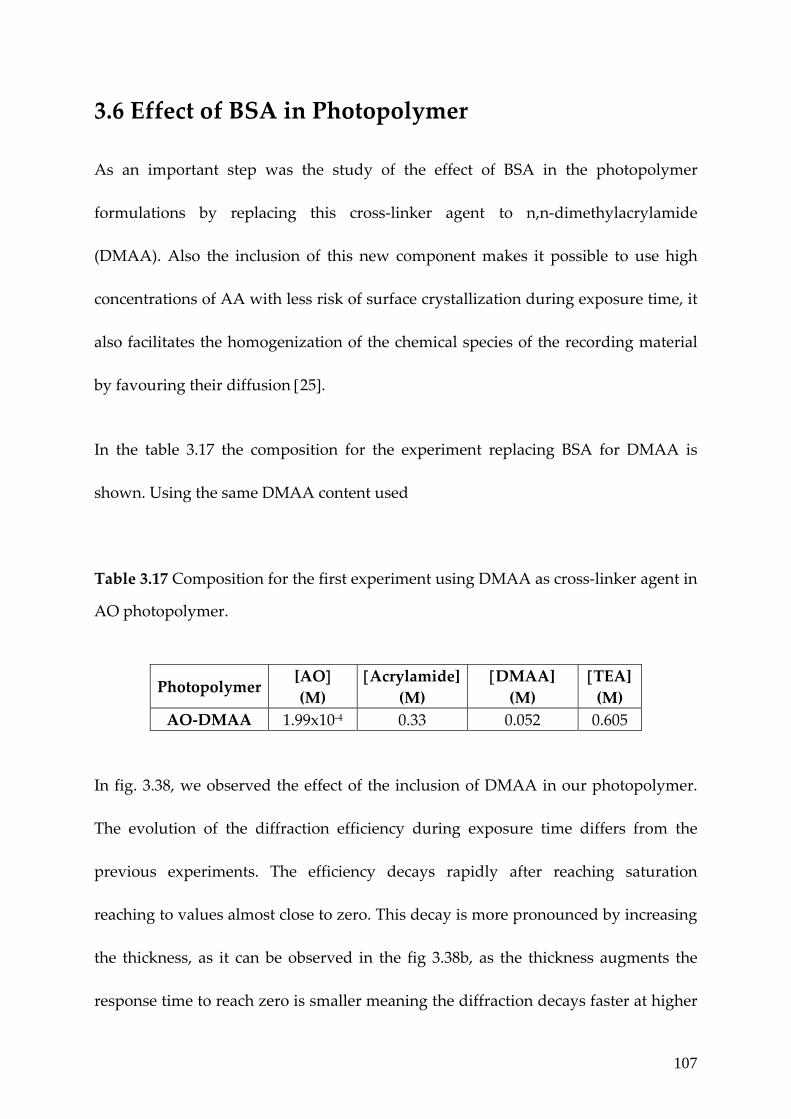
107
3.6 Effect of BSA in Photopolymer
As an important step was the study of the effect of BSA in the photopolymer
formulations by replacing this cross‐linker agent to n,n‐dimethylacrylamide
(DMAA). Also the inclusion of this new component makes it possible to use high
concentrations of AA with less risk of surface crystallization during exposure time, it
also facilitates the homogenization of the chemical species of the recording material
by favouring their diffusion [25].
In the table 3.17 the composition for the experiment replacing BSA for DMAA is
shown. Using the same DMAA content used
Table 3.17 Composition for the first experiment using DMAA as cross‐linker agent in
AO photopolymer.
Photopolymer [AO] (M)
[Acrylamide](M)
[DMAA] (M)
[TEA] (M)
AO‐DMAA 1.99x10‐4 0.33 0.052 0.605
In fig. 3.38, we observed the effect of the inclusion of DMAA in our photopolymer.
The evolution of the diffraction efficiency during exposure time differs from the
previous experiments. The efficiency decays rapidly after reaching saturation
reaching to values almost close to zero. This decay is more pronounced by increasing
the thickness, as it can be observed in the fig 3.38b, as the thickness augments the
response time to reach zero is smaller meaning the diffraction decays faster at higher

108
thickness of the material. As can be seen the diffraction efficiency increases until it
reaches a maximum of nearly 10%. However when the exposure continues to
increase the diffraction efficiency drops almost to zero. The shape of this curve
suggests that the photopolymer exhibits over‐modulation effects [26]. A higher DE is
reached outside the Bragg condition and when this condition is fulfilled the DE
drops to zero. This is due to the fact that when the Bragg condition is fulfilled the DE
is determined by the function sin2(ν) [26]. At this point, over‐modulation of the index
occurs and this can be explained mainly by polymerization of the monomer in the
bright zones. In other words, the refractive index modulation is so great that ν takes
on a value greater than π/2. This means that the diffraction efficiency at the Bragg
condition begins to decrease (since the diffraction efficiency at the Bragg angle is
governed by the function sin2ν, whereas it increases at the lateral lobes [26].
Very high values of RIM are necessary to obtain a value of ν=π however such values
were obtained in our materials due to the fact that a high acrylamide concentration
(∼0.43 M) was used thereby enabling us to observe the non‐linear behaviour of this
material which is known as the overmodulation effect [27]. In this way we were able
to observe that the DE decreases rapidly after reaching its maximum value not
because of the grating is destroyed but the index modulation is too high.

109
0 2 4 6 8 1 0 1 2 1 4 1 60
2
4
6
8
1 0
1 2
1 4
DE
(%)
E x p o s u re tim e (s e c )
DE ≈ 9% for a 15 μm thick film.
0 2 4 6 8 10 12 14 160
2
4
6
8
10
12
14
DE
(%)
Exposure time (sec)
0 2 4 6 8 10 12 14 160
2
4
6
8
10
12
14
DE
(%)
Exposure time (sec)
DE ≈ 10.3 % for a 77 μm thick film. DE ≈ 4.3% for a 96 μm thick film. Fig. 3.38 Diffraction efficiency as a function of time for photopolymer AO‐4 using DMAA as cross‐linking monomer for different thickness.
In an attempt to improve our previous results using DMAA as second monomer, an
optimization of the composition of the components of the pre‐polymer syrup was
carried out. For these experiments, photopolymers using BSA and DMAA as cross‐
linker agents were fabricated. The DMMA concentration used here was increased in

110
300 400 500 600 7000.0
0.1
0.2
0.3
0.4
Abso
rban
ce
Wavelength (nm)
AO4-BSA AO4-DMAA
50% compare to the first experiments. In table 3.18, the composition of the
components used in this section is shown.
Table 3.18 Composition of the photopolymer comparing different cross‐linker
agents.
AO photopolymer
[AO] (M)
[Acrylamide](M)
[Cross‐linker] (M)
[TEA] (M)
4‐B 1.99x10‐4 0.452 0.052 (BSA) 0.605 4‐D 1.99x10‐4 0.452 0.078
(DMAA) 0.605
In fig. 3.39, the UV‐Vis absorption spectrum for photopolymer using different cross‐
linker monomers is presented. It could be observed not significant differences
between their spectra. Both photopolymers exhibits the same absorption peak at 500
nm of wavelength.
Fig. 3.39 UV/Vis spectra of acridine orange photopolymer AO‐4 using different cross‐linking monomers (BSA and DMAA).

111
Fig. 3.40 shows the performance of diffraction efficiency as a function of exposure
time when a holographic diffraction grating is stored on the AO4 photopolymer
layers using two types of cross‐linker agents. The formation of gratings was recorded
using the set up and parameters used in the first experiments using AO as
photosensitizer. It can be seen that the diffraction efficiency increases and reaches
their maximum values of 60 and 50% for AO4‐B and AO4‐D respectively and then
stabilizes. We observed the grating formation for the case of AO4‐D was stable for
the whole time period of 90 sec compared to the previous results. The diffraction
efficiency for material with DMAA remains stable longer by increasing the DMAA
content.
0 15 30 45 60 75 900
10
20
30
40
50
60
70
80
90
100
DE
(%)
Exposure time (sec)
AO4_BSA AO4_DMAA
Fig. 3.40 Diffraction efficiency as a function of time for photopolymer AO‐4 using different cross‐linking monomers (BSA and DMAA).
In table 3.19, the optical parameters obtained by the comparison between BSA and
DMAA as alternative monomer is exhibited. We observed, by replacing the BSA for

112
DMAA the diffraction efficiency, sensitivity and RIM decreases by almost 50%
despite the concentration of DMAA over BSA was higher.
Table 3.19 Optical parameters of photopolymers AO4 using different cross‐linking
agents after 2.273 sec.
AO photopolymer
DE (%)
Sensitivity(cm/J)
RIM Thickness (μm)
4 (BSA) 68.1 607.7 4.01x10‐3 36 4 (DMAA) 28.0 389.6 2.31x10‐3 36
The temporal evolution of the angular response as a function of diffraction efficiency
is shown in fig. 3.41. The angular response for both materials was after recorded 3
min after exposure was finished. At this point, the over‐modulation effect occurs, we
observed that the diffraction efficiency for AO4‐D decays by 50% of its original value
obtained during exposure while the efficiency for A04‐B remains the same. No
difference was observed in the angular width by replacing BSA for DMAA but the
satellite peaks (first diffracted order) for AO4‐D are almost vanished compare to
AO4‐B are clearly resolved. It was foreseen that if we continued to measure the
angular response for AO4‐D at different times the efficiency was going to reach zero
due to this effect [25].

113
0
10
20
30
40
50
60
70
80
90
100
-3 -2 -1 3210
DE
(%)
Angular response (sec)
AO4_BSA AO4_DMAA
Fig. 3.41 Angular Selectivity of diffraction efficiency for photopolymer AO‐4 using different cross‐linking monomers (BSA and DMAA).
In fig. 3.42 the DSC thermogram and FT‐IR measurements were carried out to try to
understand this effect. The Tg value for the AO4‐D is higher than the one obtained
with AO4‐B representing that the material becomes more rigid and the diffusion is
more difficult leading to a decrease in DE and RIM as well [16].
Specific interactions between AA and TEA can reduce the mobility of AA affecting
the diffusion process that an important effect on the photopolymerization of AA
resulting in a decrease of both diffraction efficiency and energetic sensitivity for
AO4‐D [16]. It was illustrated from the results of FT‐IR measurements that the
characteristic peak intensity of acrylamide (1670cm‐1 C=O stretch) are smaller in the
of AO4‐B compare to AO4‐D meaning more acrylamide was converted to polymer
in the case of the photopolymer using BSA as cross‐linker agent.

114
400 600 800 1000 1200 1400 1600 1800 20000,3
0,4
0,5
0,6
0,7
0,8
0,9
Abso
rban
ce
Wavenumber (cm-1)
AO4-DMAA AO4-BSA
Fig. 3.42a DSC thermogram of the acridine orange photopolymer AO‐4 using BSA and DMAA as cross‐linking monomers.
Fig. 3.42b FTIR spectra of the polymerized acridine orange photopolymer (AO‐4‐BSA‐DMAA).
We successfully explored here that by adding DMAA to the composition of the
photopolymer, it is possible to control certain properties of the polymer film, once it
is dry. As a consequence, it can be said that if DMAA is not used it is possible to
preserve the stored gratings for longer time by replacing it for BSA, but this is not the
case if DMAA is used [26,27].

115
II. Reflection volume phase gratings By comparing the results presented in the last section to those obtained in chapter 2
with a red sensitizing dye, we experienced that the volume phase transmission
gratings can be stocked in photopolymers with even higher spatial frequency as the
wavelength of the recording beam is lowered from red to green. We also observed
by rotating the sample, parallel to the intersecting beams, the recording process can
lead to achieve an improved grating quality with even higher spatial frequency.
Although this process has limitations (Reflection holograms are formed of fringes,
the plane of which is parallel to the plane of the emulsion. These fringes are more
difficult to record than those of the transmission gratings: they are more sensitive to
shrinkage effects due to the polymerization reaction) [28] and its not always possible
to use this step as an advantage, we can still expect a further increase in the capacity
of the same recording media just by changing the recording geometry from
transmission to reflection. Reflection recording geometry allows the stockage of the
optical information through bulk of the recording material hence the whole thickness
of the recording media can be used to store the induced gratings. In coming sections
we study the photopolymer compositions already explored in section I with an aim
to further enhance the storage capacity of the green sensitized photopolymers.

116
3.7 Reflection experimental set‐up
We first describe the experimental set‐up used for the grating formation using this
new geometry. Figure 3.43 depicts the set‐up used for recording the reflection phase
gratings in our studies. Distinct from the previously used transmission set‐up, the
two Ar‐ion laser beams, B1 and B2, here propagate from the opposite directions and
overlap in the film, S [29]. The beams propagate at 3° and 6° with respect to the film
normal, so that beam 1R, reflected by the film surface and does not coincide with
transmitted beam 2. Two quarter‐wave plates convert the linear polarization of the
beams to right circular polarization. As the beams are counter propagating, however,
their electric vectors rotate in opposite senses, and the intensity of the resulting light
that induces the holographic modulation in the sample is constant.
The gratings were written using a line from an Argon ion laser that was split into
two beams using a 50:50 cubic beam splitter and recombined at an angle of 30° with
respect to the normal from the opposite sides of the sample. Sample was mounted on
a rotary stage. For writing, the samples were tilted 3° in order to separate the
reflection of the glass and distinguish the diffracted and transmitted beams. The
gratings were probed with the same laser at the same writing wavelength at low
intensity for further unwanted polymerization.
The sample plane is set slightly tilted from the equal‐phase plane of the interference
field of the two beams, such that the part of beam 2 that is reflected by the sample
surface does not hit detector D, which is used to record only the light in the direction

117
of beam 1. The two beams make a crossing angle of 30° and therefore form an
interference modulation of particular period with its equal‐phase plane
approximately parallel to the surface of the sample. Thus the two beams split from an
Argon‐ion laser interfere at the sample area. Beam 2 is vertically polarized
(perpendicular to the plane of the optical table). The beams hit the film at almost the
same angle, so the planes with equal phase differences are almost parallel to the film
surface.
The Diffraction efficiency in reflection geometry is defined [30‐32] as :
22 ⎟
⎠⎞
⎜⎝⎛ Δ
≈⎟⎠⎞
⎜⎝⎛ Δ
=λ
πλ
πη LnLnr
tanh (1)
and λπ 22 wL = (2)
Fig. 3.43 Experimental set up used in reflection experiments. M: mirrors, BS: beam splitter, D: detector.
Ar Laser 6° 3°
1R
B1 B2
D
BSM
M
D
Rotary stage
….
S

118
where L is the length of the grating, Δn is the refractive index modulation, λ is the
wavelength and w is the radius of the laser beam.
The fringe spacing for reflection geometry is defined [33,34] as:
intcosθλ
n2=Λ (3)
Where θint is the internal angle of the beam with respect to the surface normal and n
is the refractive index of the composite. The θc‐int is the complementary angle inside
the sample and is calculated according to Snell’s Law. It is defined as:
n x sin θc‐int = n x sin (θc‐ext )
intint n
ext x θextn=θ
In the following figures we observed that the first increase of the signal when the
laser is switch on should correspond to the transmitted beam (no grating formation),
then the signal is raised, this could be due to the development of holographic
reflection, meaning the grating formation [29].
3.8 RB photopolymer for reflection holographic gratings
recording
A set of three photopolymers using Rose Bengal as a dye were fabricated in order to
record reflection holographic gratings. The mixture of the components early
described (AA, BSA, TEA, RB) were dissolved in a PVA solution at 10% wt/v. An
optimization of the acrylamide (by increasing its content) was carried out in this kind

119
of photopolymer. In table 3.20, the composition of the component is shown. The
thickness of this photopolymer was 27 μm.
Table 3.20 Composition of the components for reflection gratings in different
photopolymers.
Photopolymer AA
(M) TEA (M)
BSA (M)
Dye (M)
3a 3.7 x 10‐1 0.602 0.052 2.88x10‐5 (RB) 3b 4.05 x 10‐1 0.602 0.052 2.88x10‐5 (RB) 3c 4.33 x 10‐1 0.602 0.052 2.88x10‐5 (RB) 3d 4.05 x 10‐1 0.602 0.052 0.064%v/v Eosin)
To illustrate the performance of RB photopolymers for recording holographic
reflection data storage, gratings were recorded using the reflection geometry set up
described. In the table 3.21 the parameters used for the characterization are
illustrated.
Table 3.21 Reflection geometry parameters used to evaluate the RB and Eosin photopolymers. Photo‐ polymer
θ/2 (°)
WB (nm)
RB (nm)
Λ (μm)
SF (lines/mm)
I (mW/cm2)
θ (°)
RB 30 514.5 514.5 0.210 4761 153.3 33.0 (I) 35.5 (F)
Eosin 30 514.5 514.5 0.210 4761 153.3 33.0 (I) 37.0 (F)

120
0 15 30 45 60 75 900
2
4
6
8
DE (%)
Exposure time (sec)
0
20
40
60
80
100
-3 -2 -1 3210
DE (%)
Angular response (°)
where θ/2 is the half‐angle between the interference beams; WB and RB are the
writing and reading beam respectively; Λ is the fringe period; θ (I,F) are the initial
and final reading angle (before and after shrinkage) and I is the recording intensity.
Figure 3.44 represents the angular selectivity of transmission gratings with
previously obtained 95% diffraction efficiency. It was recorded in our photopolymer
RB‐3 under 1943 lines/mm spatial frequency, written at 514.5 nm and probed at 633
nm using a He‐Neon laser. Using the same material, reflection gratings were
recorded with a diffraction efficiency of 6.8% under 4800 lines/mm spatial frequency
(see fig 3.44b).
In order to increase the diffraction efficiency, the monomer concentration must be
optimized, due to the fact that the index modulation is proportional to the
concentration of the reacted monomer [11]. Table 3.21 shows the effect of increasing
(a)
(b)
Fig. 3.44 (a) Evolution of diffraction efficiency during time for RB_3a film. (b)Angular selectivity as a function of diffraction efficiency for RB‐3b film.

121
the AA concentration by 14 % compared to the first concentration used. This lead us
to increase the efficiency from around 6.8 % to 12.33 %, showing that a higher AA
concentration can significantly raise the efficiency. Although still avoiding
precipitation in the film. Figures 3.45 (a, b) represent the influence of AA
concentration on diffraction efficiencies as a function of exposure time. We limited
further increase in AA concentration in order to avoid precipitation.
0 15 30 45 60 75 900
2
4
6
8
10
12
14
DE (%)
Exposure time (sec)
Fig. 3.45a Evolution of diffraction efficiency during time for RB‐3b film.
Fig. 3.45b Evolution of diffraction efficiency during time for RB‐3c film.
3.8.1 Effect of photosensitizer.
Photosensitizer has a crucial role in photopolymeric systems. As discussed in chapter
1, after the photoreduction of the sensitizing dye, the radicals are produced. We
studied the effect of photosensitizers on DE by changing Rose Bengal to Eosin at
0 15 30 45 60 75 900
2
4
6
8
10
12
14
DE (%)
Exposure time (sec)

122
unchanged concentration for all other components. Figure 3.46 represents the
diffraction efficiency obtained when eosin was the dye. We notice that diffraction
efficiency raised to 17% at similar concentration (2.88 x10‐5 M) just by changing the
dye. An enhancement of almost 5% comes due to the fact that Eosin has a higher
yield of triplet excited states than Rose Bengal [34]. This provides ample time for AA
to react with the dye which further increasing the amount of polymerization and
thus finally increasing DE because of the increased sensitivity.
0 15 30 45 60 75 900
2
4
6
8
10
12
14
16
18
20
DE (%)
Exposure time (sec)
Fig. 3.46 Diffraction efficiency as function of exposure time recorded in reflection geometry using Eosin film.
3.8.2 Sensitivity The sensitivity of the recording material is defined as:
dxτxIηS1/2
= (6)

123
where τ: exposure time to reach maximum efficiency, d: thickness of the material, P:
power of each beam and A: illuminated area in the recording material.
We observed that AA concentration has a direct effect on the overall sensitivity of the
recording material. As discussed in section 3.1, by increasing the concentration of AA
one can observe further increase in sensitivity. Sensitivity calculated using equation
(6) shows that increasing the AA concentration by 20% remarkably increases the
sensitivity of our films. Figure 3.47 presents the evolution of sensitivity and DE as a
function of AA concentration. Table 3.22 summarizes the change in sensitivity and
DE with respect to the concentration of AA with Rose Bengal as the sensitizer.
3.7 x 10⁻¹ 4.05 x 10⁻¹ 4.33 x 10⁻¹⁶
8
10
12 DE S
Concentration of AA (M)
DE (%)
3600
3800
4000
4200
4400
4600
4800
5000
5200
5400
Sensitivity (cm/J)
Fig. 3.47 Effect of AA concentration on sensitivity (filled circles) and diffraction efficiency (open squares) using Rose Bengal .

124
Table 3.22 Sensitivity as a function of AA concentration using Rose Bengal as sensitizer. DE values are also given.
Photopolymer DE (%)
Sensitivity (cm/J)
3a 6.8 3760.11 3b 10.66 4658.79 3c 12.33 5342.51
Sensitivity remains high even after changing the photosensitizing dye. Although
significant increase in DE was observed using Eosin based films, almost similar
sensitivity of Eosin based films (see table 3.23) suggests that AA basically defines the
sensitivity in such films.
Table 3.23 Effect of photosensitizing dye on recording parameters for RB and Eosin film.
Dye Diffraction Efficiency (%)
Sensitivity(cm/J)
RB 10.66 4658.79 Eosin 16.98 ≅ 17 5785.83
3.8.3 Shrinkage
High sensitivity of our recording media caused relatively high shrinkage observed
while re‐reading the stored gratings. Shrinkage is a common phenomenon in
photopolymer based recording media which comes into picture because of the
swelling or shrinking of the film under illumination of the writing beam [35]. This
means that the read‐out angle varies from the recording one (Bragg’s angle).This

125
variation results in a change of the grating period due to the partial change in the
thickness or index of the film causing an alteration of the readout beam angle to
achieve maximum diffraction efficiency. The fractional change in the thickness was
calculated by relation defined in chapter 1. Irrespective of the AA concentration we
observed the same shrinkage of ~ 10% for films containing Rose Bengal. Whereas the
films with Eosin suffered relatively higher shrinkage of ~ 16% compare to the Rose
Bengal one at constant film thickness of 27 μm. Our immediate interest is to avoid
shrinkage in our films by controlling undesired diffusion of the monomer.
3.8.4 Effect of the exposure time on the diffraction efficiency
In order to study the effect of recording exposure in the diffraction efficiency, a
experiment varying the exposure time was performed using RB photopolymers.
Decreasing the cross‐linker monomer (BSA) content in photopolymer RB‐3 by 12.5 %
and varying the exposure time the efficiency reflection gratings were recorded. The
reactant composition used in the photopolymer is illustrated in the table 3.24.
Table 3.24 Composition of the pre‐polymer solution (5 ml).
Photopolymer
AA (M)
BSA (M)
TEA (M)
RB (M)
3a 3.7 x 10‐1 4.5x10‐2 0.605 2.8x10‐5

126
In fig 3.48 the evolution of the diffraction efficiency varying the recording exposure
time is shown. An improvement in the efficiency was observed by increasing the
exposure time, from 8.94% to 12.23% for 20 and 40 seconds respectively. An
explanation of this trend is at longer exposure time the density of the photons in a
given period of time will be lesser so the concentration of radicals will be smaller
resulting in a termination rate so a single radical can live longer causing the
polymerization of more acrylamide molecules.
0 5 10 15 20 25 30 35 400
3
6
9
12
15
DE (%)
Exposure time (sec)
20 sec 30 sec 40 sec
Fig. 3.48 Diffraction efficiency as function of exposure time using Rose bengal film varying the exposure time.

127
3.9. Acridine orange for reflection geometry
We obtained diffraction efficiency as high 7.3% when AO was used as a sensitizing
dye in AA based photopolymer. Table 3.26 represents the value obtained for
diffraction efficiency and sensitivity of the acridine orange photopolymer
experimental conditions are summarized in table 3.27.
Table 3.26 Acridine orange photopolymer in reflection geometry
Photopolymer
AA (M)
BSA (M)
TEA (M)
AO (M)
DE (%)
AO 4.05 x 10‐1 4.5x10‐2 0.605 7.99x10‐5 7.30
To characterize the AO photopolymers as material to create reflection holographic
gratings we recorded using the same set up used previously. In table 3.27 the
parameters used for the recording are illustrated.
Table 3.27 Reflection geometry parameters used to evaluate the AO photopolymers. Photo‐ polymer
θ/2 (°)
WB (nm)
RB (nm)
Λ (μm)
SF (lines/mm)
I (mW/cm2)
θ (°)
AO 30 514.5 514.5 0.210 4761 153.3 33.0 (I) 37.0 (F)
where θ/2 is the half‐angle between the interference beams; WB and RB are the
writing and reading beam respectively; Λ is the fringe period; θ (I,F) are the initial
and final reading angle (before and after shrinkage) and I is the recording intensity.

128
Figure 3.49 represents the efficiency using the pre‐established formulation shown in
table 3.26 using acridine orange as photosensitizer at a particular concentration in
order to achieve absorbance of 0.15. We observed the diffraction efficiency value
achieved with acridine orange as photosenstizer was lower compare to the one
obtained with Rose Bengal and eosin.
0 15 30 45 60 75 900
2
4
6
8
10
DE(%)
Exposure time (sec)
Fig. 3.49 Diffraction efficiency as function of exposure time recorded in reflection geometry using Acridine orange.

129
3.10 Conclusion
In this chapter we first explored the suitability of green wavelength recording for
holographic data storage through formation of volume phase gratings using both the
transmission and reflection recording geometries. Effect of material purity, at
optimum conditions shows a significant effect on the recording parameters.
Dynamics of grating formation and characterization are probed using various
supporting characterizations which helped us to optimize the whole photopolymer
system. Grating periods thus stored in photopolymers with Rose bengal, Eosin,
Acridine orange etc dyes have shown high diffraction efficiencies close to 95%. We
demonstrated the increase of spatial frequency from 1900 lines/mm to close to 2200
lines/mm just by slight change in recording parameters. The effect of shrinkage after
recording is probed through recording the angular response of the stored gratings in
the thick polymer films.
A further capacity enhancement was achieved using the reflection geometry
recording. Volume grating formation in the photopolymer studied in reflection
geometry is demonstrated with the same sensitizing dyes where spatial frequency
exceeds 4760 lines/mm.

130
References [1]. F. del Monte, O. Martinez, J. A. Rodrigo, M.L. Calvo and P. Cheben, “A volume
holographic sol‐gel material with large enhancement of dynamic range by
incorporation of high refractive index species. Adv. Mat. 18, 2014‐2017, (2006).
[2]. H.J. Caufield, Handbook of optical holography, (1979).
[3]. H. Sherif, I. Naydenova, S. Martin, C. McGinn and V. Toal, “Characterization of
an acrylamide‐based photopolymer for data storage utilizing holographic angular
multiplexing”, J. Opt. A: Pure Appl. Opt. 7, 255‐260 (2005).
[4]. http://en.wikipedia.org/wiki/Recrystallization
[5]. M. Ortuño, S. Gallego, C. García, C. Neipp, A. Belendez and I. Pascual,
“Optimization of a 1 mm thick PVA/acrylamide recording material to obtain
holographic memories: method of preparation and holographic properties”. Appl.
Phys. B. 76, 851‐857. (2003).
[6]. Y‐Ch. Jeong, S. Lee, and J‐K. Park,” Holographic diffraction gratings with
enhanced sensitivity based on epoxy‐resin photopolymers”, Opt. Express. 15, No.4,
1497‐1504, (2007).
[7]. J.R. Lawrence, F.T. O’Neill and J.T. Sheridan. “Photopolymer holographic
recording material”. Optic 112. No. 10 449‐463, (2001).
[8]. C. Sanchez, M.J. Escuti, C Van Heesch, C. W.M. Bastiaansen, D. J. Broer, J. loos
and R. Nussbaumer, “TiO2 nanoparticle‐Photopolymer composites for volume
holographic Recording”, Adv.Funct. Mater 15, 1623‐1629 (2005).
[9]. H. Yao, M. Huang, Z. Chen, L. Hu and F. Gan, “Optimization of two‐monomer‐
based photopolymer used for holographic recording”. Mat. Lett. 56, 3–8. (2002).
[10]. I. Naydenova, E. Mihaylova, S. Martin, V. Toal,” Holographic patterning of
acrylamide–based photopolymer surface”, Opt. Express. 13, No. 13, 4878‐4889,
(2005).

131
[11]. M.A. khodorkovskii, S.V. Murashov, T.O. Artamonova, A.L. Shakmin, A.A.
Belaeva and D.Yu. Davydov, “ Fullerene films highly reistant to laser ablation”,
Technical Phys. 49, No. 2, 258‐262 (2004).
[12]. S. Blaya, L. Carretero, R. Mallavia, A. Fimia, R.F. Madrigal, M. Ulibarrena and
D. Levy, “Optimization of an acrylamide‐based dry film used for holographic
recording”, Appl. Opt. 37, No. 32, 7604‐7609, (1998).
[13]. S. Gallego, M. Ortuño, C. Neipp, A. Marquez and A. Beléndez and I. Pascual,
“Maximum effective optical thickness of the gratings recorded in photopolymers”,
Proc. of SPIE Vol. 5827 (SPIE, Bellingham, WA), 107‐117. (2005).
[14]. S. Piazzolla and B. K. Jenkins, “First‐Harmonic diffusion model for holographic
grating formation in photopolymers”, J. Opt. Soc. Am. B. 17, No. 7, 1147‐1157, (2000).
[15]. V. Moreau, Y. Renotte and Y. lion, “Characteriztion of Dupont photopolymer:
determination of kinetic parameters in a diffusion model”, Appl. Opt. 10, 3427‐3435
(2002).
[16]. W. S. Kim, Y‐Ch. Jeong, J‐K Park, Ch‐W. Shin, N‐K, “Diffraction efficiency
behavior of photopolymer based on P(MMA‐co‐MAA) copolymer matrix”, Opt. Mat.
29, 1736–1740, (2007).
[17]. W.S. kim, H‐S. Chang, Y‐C. Jeong,Y‐M Lee, J‐K. Park, C‐W Shin, N. Kim and H‐
J. Tak, “A new phase‐stable photopolymer with high diffraction efficiency based on
modified PMMA”, Opt. Comm. 249, 65‐71 (2005).
[18]. S. Blaya, L. Carretero, R.F. Madrigal, M. Ulibarrena, P. Acebal and A. Fimia,
“Photopolerization model for holographic gratings formation in photopolymers”,
Appl. Phys. B. 77, 639‐662, (2003).
[19]. C. Garcia, I. Pascual, A. Costela, I. Garcia‐Moreno, C. Gomez, A. Fimia, and R.
Sastre, “Hologram recording in polyvinyl alcohol_acrylamide photopolymers by
means of pulsed laser exposure” Appl. Opt. 41, No. 14, 2613‐2610, (2002).
[20]. S. Gallego, M. Ortuño, C. Neipp, A. Márquez and A. Beléndez, I. Pascual, J. V.
Kelly and J. T. Sheridan, “Physical and effective optical thickness of holographic

132
diffraction gratings recorded in photopolymers”, Opt. Express. 13, No. 6 1939‐1947,
(2005).
[21]. S. Gallego, M. Ortuño, C. Neipp, A. Márquez, A. Beléndez and I. Pascual,
“Characterization of polyvinyl alcohol_acrylamide holographic memories with a
first‐harmonic diffusion model”, App. Opt. 44, No.29, 6205‐ 6210, (2005).
[22]. E. Fernández, C. García, I. Pascual, M. Ortuño, S. Gallego, and A. Beléndez,
“Optimization of a thick polyvinyl alcohol–acrylamide photopolymer for data
storage using a combination of angular and peristrophic holographic multiplexing”,
Appl. Opt. 45, No.29, 7661‐7666, (2005).
[23]. M.K. Dutt, “Aqueous solution of acridine orange in the staining of DNA‐
aldehyde”, Cellular and Mol Life Sci. 30, No. 6, 667‐669, (1974).
[24]. S. Blaya, L. Carretero, R.F. Madrigal, A. Fimia, “Optimization of a
photopolymerizable holographic recording material on polyvinylalcohol using
angular responses”, Opt. Mat. 23, 529‐538, (2003).
[25]. S. Gallego, M. Ortuño, C. Neipp, C. García, A. Beléndez and I. Pascual,
“Temporal evolution of the angular response of a holographic diffraction grating in
PVA/acrylamide photopolymer”, Opt. Express. 11, No. 2, 181‐190, (2003).
[26]. S. Gallego, M. Ortuño, C. Neipp, C; Garcia, A. Beléndez and I. Pascual, “
Overmodulation effects in volume holograms recorded on photopolymers”, Opt.
Comm. 215, 263‐269, (2003).
[27]. N. Suzuki and Y. Tomita, “Diffraction properties of volume holograms recorded
in SiO2 nanoparticle‐dispersed methacrylate photopolymer films”, Jpn. J. Appl. Phys.
42, L927‐L929 Part 2 No. 8A, (2003).
[28]. Y. Defosse, C. Cam and D. J. Lougnot, ″Use of a self‐developing polymer
material for volume reflection hologram recording”, Pure Appl. Opt. 2 437‐40. (1993).
[29]. L. Nikolova, T. Todorov, V. Dragostinova, T. Petrova, and N. Tomova,
“Polarization reflection holographic gratings in azobenzene‐containing gelatine
films”, Opt. Lett. 27, No. 2, 92‐94, (2002).
[30]. H. Kogelnik, Bell Syst. Tech. J. 48, 2909 (1969).

133
[31]. R. Jallapuran, I. Naydenova, S. Martin, R. Howard, V. Toal, S. Frohmann, S.
Orlic and H. J. Eichler, “Acrylamide‐based photopolymer for microholographic data
storage”, Opt. Mat. 28, 1329‐1333, (2006).
[32]. P. Hariharan, Optical holography – Principles, Techniques and Applications, 2 nd ed.
(Cambridge University Press, Cambridge, UK). (1996).
[33]. O‐P. Kwon, S‐L. Kwon, M. Jazbinsek, P. Gunter and S‐H. Lee, “High
performance reflections gratings in nematiclike photorefractive polymers”, Appl.
Phys. Lett. 87, 121910‐12, (2005).
[34]. F.T. O’Neill, J.R. Lawrence and J.T. Sheridan, “Thickness variation of self‐
preocessing acrylamide‐based photopolymer and reflection holography”, Opt. Eng.
40 (4), 533‐539, (2001).

134
4 Chapter 4 Novel dyes for blue wavelength volume transmission grating recording
4.1. Introduction
One of the major challenges in the area of holographic data storage is the
development of suitable storage materials. Holographic media must satisfy stringent
criteria, including high dynamic range, high photosensitivity, dimensional stability,
optical clarity and flatness, nondestructive readout. Next generation recording media
requires an optimal recording material with higher storage capacity and low volume
shrinkage. For achieving higher storage capacity, the use of photosensitizer that
absorbs in the blue wavelength region is required thus minimizing the fringe spacing
and increasing the spatial frequency. Sony and Inphase has developed a new
generation of blue laser DVD with a storage capacity up to 50 GB using 400 to 410
nm lasers. The replacement from red to blue laser DVD provides an improvement in
storage capacity from 5 GB to 50 GB [1,2].
In this section we introduce series of blue sensitive dyes suitable for high capacity
holographic data storage. First we used acridine yellow (AY), then Perylene diimide
and finally we synthesize a natural colorant Curcumin and demonstrate their

135
capability in a photopolymer composition using transmission and reflection volume
phase recording.
4.2 Acridine Yellow: a blue photosensitizer.
Acridine yellow is used in microbiology as a mutagen damaging the DNA, as
photosensitizer for blue holographic grating recordings at 457,9 nm wavelength in
acrylamide based photopolymers. Our approach allows us flexibility in tailoring the
media to the particular needs of high density holographic data storage. In order to
enhance the RIM up to 4x10‐3, three photopolymers were prepared and the effect of
acridine yellow concentration on this parameter is investigated. The influence of
exposure intensity on the diffraction efficiency and refractive index modulation is
also studied. An angular selectivity study as function of the diffraction efficiency is
performed which determines the shrinkage suffered by the recording media.
Photopolymers were prepared by combining AY (λmax=464.2 nm) as radical generator
(KODAK) at different proportions. Fig 4.1 shows the molecular structure and UV‐Vis
spectrum of the acridine yellow that exhibits a maximum absorption peak at 433.6
nm which makes it suitable for blue region recording. Acrylamide was used as
monomer, N,N’‐methylenebisacrylamide was employed as cross‐linking monomer,
triethanolamine used as initiator and all dissolved in a poly(vinyl alcohol) solution
at 10 and 13% w/v. All the reagents were used without further purification. The
average layer thickness measured using a DEK‐TAK 6M profilometer was 34 μm.

136
300 400 500 600 7000,0
0,2
0,4
0,6
0,8
1,0
N
CH3
NH2 NH2
CH3
HCL
Abs
orba
nce
W avelength (nm )
Fig. 4.1 Chemical structure and UV/Vis spectrum of AY solution (water) at 2.629x10‐4 M of concentration.
In fig. 4.2 the cyclic voltammograms of acridine yellow are shown as were recorded
in DMF. This measurement allows understanding the limitations of AY as acceptor of
electron from TEA (electron donor) during the photoreduction involved in the
photopolymerization process. The reduction peak of AY occurs at ‐1.6 V which
enables this dye to be used in the final photopolymer composition.

137
Fig. 4.2 Cyclyc Voltammograms of Acridine Yellow
Two sets of three photopolymers were fabricated varying the acridine yellow
concentration mixed with the rest of the components (AA, BSA, TEA) at different
PVA solution, 10 % wt/v (Case I) and 13% wt/v (Case II) in order to study the effect
of dye concentration and thickness.
4.2.1 Optimization of the acridine yellow concentration.
We demonstrated in last chapter that the quantity of photosensitizer used in the
recording material is an important factor, which also controls the diffraction
efficiency of the recorded data. As photosensitizer can accelerate the initiation of the
polymerization process, light sensitivity, diffraction efficiency as well as RIM can be
enhanced by increasing this component in the photopolymer. Also it was observed
CV of AY

138
that the excess of the photosensitizer has a negative influence on the diffraction
efficiency due to additional energy required to bleach this component during the
photopolymerization [3,4]. To verify the effect of the acridine yellow on the
diffraction efficiency, a series of photopolymers were prepared containing different
AY proportions. The photopolymer compositions are summarised in table 4.1.
Table 4.1 Composition of photopolymers using acridine yellow at different AY
concentration.
Dye AA (M) BSA (M) TEA (M) AY (M) AY1 0.43 0.05 0.57 5.28x10‐5 AY2 0.43 0.05 0.57 8.812x10‐5 AY3 0.43 0.05 0.57 1.376x10‐4
The characterization of holographic gratings storage capacity of this media was
carried out using our previously described setup in transmission interference
geometry. The parameters used for the recordings are presented in the table 4.2.
Table 4.2 Parameters for gratings recording using AY
Photo‐ polymer
θ/2 (°)
WB (nm)
RB (nm)
Λ (μm)
SF (lines/mm)
I (mW/cm2)
θB (°)
AY 33.5 457.9 633 0.418 2392 60 49.72 where θ/2 is the half‐angle between the interference beams; WB and RB are the
writing and reading beam respectively; Λ is the fringe period; θB(T,E) are the
theoretical and experimental Bragg angle and I is the recording intensity.

139
Fig.4.2 illustrates the UV‐Vis spectra of the photopolymers films prepared using
different AY concentration. The square, full circle and triangle curves are attributed
to photopolymer AY1‐3 varying the AY concentration from 1.376x10‐4 to 5.28x10‐5 M.
By blending the AY with the rest of reactants the maximum absorption peak shifts
from 433.6 nm to 463.26 nm in all the photopolymers with a thickness of 34 μm. It is
observed that by increasing the AY concentration the overall absorption increases.
300 400 500 600 7000,00
0,05
0,10
0,15
0,20
0,25
0,30
Abs
orba
nce
Wavelength (nm)
AY1 AY2 AY3
Fig. 4.2 UV‐Vis spectra of acridine yellow photopolymer films at different concentrations using PVA at 10%wt/v.
In Fig 4.3 the typical growth of the diffraction efficiency with respect to exposure
time and as function of the AY content for the three photopolymers in matrix of PVA
solution at 10%wt/v are shown. The exposure intensity was 60mW/cm2.

140
0 10 20 30 40 50 60 700
10
20
30
40
50
60
70
80
90
100
Laser on
AY1-PVA:10% AY2-PVA:10% AY3-PVA:10%
DE
(%)
Exposure time (sec)
Fig. 4.3 Diffraction efficiency during exposure time of photopolymers AY1‐3 using PVA solution at 10%wt/v at different concentration of acridine yellow.
We observed that there was no induction time required for formation of the gratings
as the diffraction efficiency started to grow instantly after the exposure. This is in
contrast to the acrylate‐containing system which requires initial oxygen
consummation for the diffraction efficiency to grow, resulting in a delay [5]. We can
see that the diffraction efficiency increased by augmenting the dye concentration.
This tendency can be explained as the lower diffraction efficiency obtained at the
lower concentration of acridine yellow due to existence of lesser molecules capable of
starting the polymer chain leading to low level of polymerisation and consequently
decay in the diffraction efficiency. After 1.376x10‐4 M of AY concentration,
agglomeration of AY was observed in the samples.
Increasing the concentration of the matrix to 13% wt/v we observed the same
performance as the photopolymers with less PVA concentration. The diffraction

141
efficiency as presented in figure 4.4 increased when higher AY concentration was
used.
0 10 20 30 40 50 60 700
10
20
30
40
50
60
70
80
90
100
Laser on
DE
(%)
Exposure time (sec)
AY1-PVA:13% AY2-PVA:13% AY3-PVA:13%
Fig. 4.4 Diffraction efficiency as a function of exposure time for photopolymer AY1‐3 using PVA solution at 13%wt/v.
The difference between these two cases (I and II) is the exposure time required to
achieve maximum diffraction efficiency was longer for the case of II compared to
case I. The efficiency grew up to reach its highest value before stabilizing. The
diffraction efficiencies obtained for case II were slightly lesser compare to case I.
For the case I, it was noticeable that the evolution growth of the diffraction efficiency
followed different pattern. For photopolymer with higher AY content (AY3), the
diffraction efficiency increased and reached a saturation value and then stabilized
after approximately 6 sec of exposure. For the other two photopolymers (AY1‐2) the
behaviour is similar, the diffraction increased until 8 and 15 sec, suffering little decay
and then stabilize. Maximum diffraction efficiency of 76.6 % was obtained for the

142
photopolymer AY3 with the highest amount of AY compare to 45% for the
photopolymer with the lowest AY content. Highest sensitivity of 670 cm/J for the
photopolymer AY3 was achieved defined earlier. For the cases of AY1‐2, lower
sensitivities were obtained caused for the retardation of photopolymerization process
compare to AY3 [6]. The recording parameters of the photopolymers are shown is
table 4.3.
Table 4.3 Properties of acridine yellow photopolymers at 6.136 sec of recording for
photopolymers (AY1‐3) using PVA solution at 10%wt/v.
Case I η (%) d (μm) Δn S (cm/J)
AY1 45.6 34 2.84x10‐3 539 AY2 57.97 34 3.31x10‐3 608 AY3 70.1 34 3.80x10‐3 669
The sensitivity relies on the concentration of photosensitizer used because it has an
essential effect on photopolymerization rate for a given photopolymer at any
particular fringe spacing. As the photosensitizer amount is higher in AY3 the amount
of monomer polymerized is expected to increase.
For case II, as well as for the case I, higher diffraction efficiencies were achieved by
increasing the dye content reflecting a significant increase of the sensitivity. Higher
as 242 sensitivities for the highest AY concentration was found. An increase in the DE
represents a positive change in the RIM, reaching as high as 1.34x10‐3 for AY3 (table
4.4). The results obtained for case II concerned for these two optical parameters were

143
inferior than the one obtained for case I. This effect can be explained as the
concentration of AA diminishes when the thickness increases.
Table 4.4 Properties of acridine yellow photopolymers (AY1‐3) using PVA solution
at 13%wt/v after 6.136 sec.
CaseII
η (%) d (μm) Δn S (cm/J)
AY1 13.6 90 5.46x10‐4 111 AY2 35 90 9.16x10‐4 178 AY3 64.4 90 1.34x10‐3 242
Angular response curves have been used to study the effect of thickness on the
performance of above photopolymerizable films [4]. Fig. 4.5, illustrates the
diffraction efficiencies as function of reconstruction angle for transmission grating
recorded on the three photopolymers measured using a He‐Ne beam laser for case I.
We observed only the zero and first order diffraction efficiencies as function of the
reconstructed angle for all photopolymer layers under a spatial frequency of 2410
lines/mm. Only the first order was noticeable indicating the formation of thick
holograms [7]. By fine‐tuning the Bragg angle position the diffraction efficiency
decreases but doesn’t reach to zero. The recording films were rotated with a
resolution of 0.1° monitoring the diffracted beam along the Bragg condition. By
increasing the AY content in this material the side peaks (first diffracted order)
intensity compared to the zero order becomes more uniform and visible. For AY1
these lobes are barely visible compare to AY3 that are more clearly resolved. This can
be caused by non‐uniformity of the grating formation in good agreement with the

144
evolution of the diffraction efficiency during time. It was noticed that the FWHM
increased slightly by increasing the AY content achieving 0.58° for the case of AY3
corresponding to a high angular selectivity which is an important parameter for
quality holographic storage applications.
0
10
20
30
40
50
60
70
80
90
100
-3 -2 -1 3210
DE
(%)
Angular response (°)
AY1-PVA:10% AY2-PVA:10% AY3-PVA:10%
Fig. 4.5 Angular selectivity of diffraction efficiency of photopolymer AY1‐3 using PVA solution at 10%wt/v at different dye concentration.

145
0
10
20
30
40
50
60
70
80
90
100
-3 -2 -1 3210
DE
(%)
Angular response (sec)
AY1-PVA:13% AY2-PVA:13% AY3-PVA:13%
Fig. 4.6 Angular selectivity of diffraction efficiency of photopolymer AY1‐3 using PVA solution at 13%wt/v.
For case II, in Fig. 4.6, the corresponding angular scans for the diffraction efficiency
are represented for gratings stored in photopolymer with PVA solution at 13% wt/v.
In this figure the physical thickness controls the central lobe width.
In figure 4.7 similar compositions in two different recording layer thickness is
presented. The narrowing of the principal lobe can be observed when the physical
thickness is higher. But this effect can be observed in gratings of 35 μm and 90 μm of
thickness because their composition is very similar and the attenuation of index
profile limit the optical grating thickness.

146
0
20
40
60
80
100
-3 -2 -1 3210
DE
(%)
Angular response (°)
AY2_P:60 mw/cm2
AY2_P:33.6mW/cm2
AY2_P:24mW/cm2
AY2_P:9mW/cm2
0
20
40
60
80
100
-1-2-3 3210
DE
(%)
Angular response (°)
AY3_P:60mW/cm2
AY3_P:33,6mW/cm2
AY3_P:24mW/cm2
AY3_P:9mW/cm2
Photopolymer AY2 Photopolymer AY3 Fig. 4.7 Angular selectivity of diffraction efficiency for photopolymer AY2‐3 using PVA solution at 10%wt/v at different intensity.
Once the diffraction efficiency is measured, the refractive index modulation was
calculated using the Kogelnik’s coupled wave theory under Bragg condition. In Fig.
4.8 the refractive index modulation is plotted as function of diffraction efficiency and
exposure intensity for the photopolymer with highest AY content (AY3). A
remarkable increase of diffraction efficiency was observed by increasing the
recording intensity. The diffraction efficiency raised from 17.68% at 9mW/cm2 to 75%
at 60 mW/cm2 recording intensity. The increase on the diffraction efficiency obtained
represents an enhancement of the refractive index modulation from 1.66x10‐3 to 4x10‐3
for the lower and the maximum exposure intensity respectively, which is
considerably higher than other similar systems [8].

147
0 10 20 30 40 50 6010
20
30
40
50
60
70
80
(Exposure intensity (mW/cm2)
DE
(%)
1,5x10⁻³
2,0x10⁻³
2,5x10⁻³
3,0x10⁻³
3,5x10⁻³
4,0x10⁻³
RIM
Reffractive index
Fig. 4.8 Effect of the exposure intensity on the diffraction efficiency and refractive index for photopolymer AY3 (PVA:10 %wt/v).
Using higher irradiation the free radical concentration becomes high and the
termination is favoured leading to the creation of shorter polymer chains. As the
polymer pattern being less dense, the observed shrinkage is also less. For the case of
low irradiance, the polymerization process is not complete; the reaction sites are
numerous so the monomer molecules are forced to diffuse onto longer distances to
join a polymer chain in formation. When the polymer density is elevated the
diffusion is obstructed and some molecules will not be polymerized [9].
4.2.2 Shrinkage
A study of angular selectivity as function of the diffraction efficiency was
investigated in order to determine thus the shrinkage suffered by the films. The
existence of shrinkage in the layers results in a change of the thickness of the fringe

148
spacing, in this case it is required to modify the readout beam angle to achieve
maximum diffraction efficiency. This change in the layer thickness [10] is defined as:
⎥⎦
⎤⎢⎣
⎡−= 1
tanφtanφ d Δd
i
f0 (1)
where ϕI and ϕf are the initial and final angle of the recorded grating. Knowing this
change the percentage of the shrinkage can be calculated as follows:
100 x dΔd Shrinkage %
o
= (2)
where Δd and do are the fraction of thickness change and the initial thickness of the
layer. Considerably small shrinkage of 1% was measured on the photopolymer AY3.
For further understanding of the degree of polymerization of all photopolymers, the
photopolymeric layers were irradiated with a collimated light from a Argon ion laser
at 457,9 nm for 70 sec in order to polymerize the acrylamide in the samples. 1.2 mg of
each sample was milled with 136 mg of KBr pressed in form of disks. Fig. 4.9 shows
the FT‐IR spectra of polymerized samples containing different AY proportion;
measured using FT‐IR spectrometer. We observed the stretching vibration of
acrylamide at 1675 cm‐1. We noticed a decrease in the absorbance by increasing the
AY content. For the case of AY1‐2 the absorbance are closer compared to AY3 that is
much lower. These can explained that more acrylamide molecules are polymerizaed
in the AY3 compared to AY1‐2, which is concordance with the diffraction efficiency
obtained previously where DEAY1‐2 are 45 and 50% smaller compare to DEAY3 obtained
of 75%.

149
1400 1500 1600 1700 1800 1900 20000,4
0,5
0,6
0,7
0,8
0,9
1,0
1,1
1,2
1,3
AY1 AY2 AY3
Abso
rban
ce
Wavenumber (cm-1)
Fig. 4.9 F‐TIR spectra of the acridine yellow photopolymers varying its concentration.
Fig 4.10 shows a microscope image of the volume grating created in the acridine
yellow photopolymer (AY3) after interferometric exposition at 0.418 μm of grating
spacing. This image confirms the formation of well‐defined gratings in present
photopolymer.
Fig. 4.10 Microscope image of the grating formation of the sample containing 1.367x10‐4 M of AY concentration.

150
4.3 Perylene diimide photopolymers
Perylene is a widely studied dye where a diimide functional group attached to the
perylene enhances the electron acceptor strength. Also perylene diimide shows the
significant overlap in the blue wavelength region. In this section we report our
studies on water soluble perylene diimide dye as sensitizer via free radical
polymerization in acrylamide base photopolymer.
We fabricated three photopolymer layers (PP1‐3) under red light ambiance, by
mixing (AA, BSA, TEA) and perylene di‐imide (Peryl) as photosensitizer illustrated
in fig. 4.11 with an average molecular weight of 502 g/mol.
Fig. 4.11 Water soluble molecule of perylene diimide
This photosensitizer presents long wavelength absorption spectra (fig. 4.12) showing
two relevant peaks at 494 nm and 529 nm making it suitable to use for recoding
under blue wavelength.
NN
O
O
O
CH2COOHHOOCH2
O O
NN
O
O
O
CH2COO-Na++Na-OOCH2
2NaOH

151
400 500 600 7000,00
0,05
0,10
0,15
0,20
0,25
0,30
0,35
0,40
Abso
rban
ce
W avelength (nm)
Fig.4.12 Absorption Spectrum of the PP1 photopolymer film with a 90 μm thick layer
This new dye was introduced at our optimised concentration of 6.8 x10‐4 M in a basic
solution (NaOH) using a molar relation of 1:2 respectively. All these components
were dissolved in a 13 %wt poly(vinylalcohol) (PVA) solution. The variation for
photopolymers PP2‐3 was the acrylamide content was increased in 24 and 48%
respectively (table 4.5).
Table 4.5 Monomer concentration for the different photopolymers
Monomer conc. PP1 PP2 PP3
[AA] (M) 0.434 0.539 0.643
4.3.1 Photosensitization mechanism
Polycyclic aromatic hydrocarbons such as anthracene and perylene have been used
as sensitizer in cationic polymerization (photolysis) using onion salts as coinitiator
and THF as monomer [11]. The addition of the sensitizer in polymerization can guide
to complex mechanism that involves intermediates. There is evidence that perylene

152
diimides proceeds the polymerization through singlet and not via triplet excited state
initiation due to energy difference between them, 54 and 27 Kcal/mol respectively
[12]. This sensitizer is capable to react as electron acceptor as well electron donor.
The photopolymerization process starts when the perylene diimide (D) absorbs a
photon from the light then is promoted to an excited singlet state. The singlet state
dye reacts with the electron donor, triethanolamine (ED) to produce the radical
initiating species that will initiate the polymerization process. Then this species react
with the monomer (acrylamide) to form the growing chain. Then this growing chain
will react with another and the polymer is produced. A possible mechanism to
explain the polymerization process if radicals initiating species are formed though a
singlet excited state (exciplex) in this material is shown in the scheme 1.
D + hv → D* (absorption of photon) (a) D* → D (deactivation (singlet) (b)
D* + ED→ [D…ED]* (formation of radical initiating species) (c) ↓ Radical species
RS* + M → RSM* (formation of growing chain) (d) RSM* + RSM* → Polymer (formation of the polymer network) (e)
(scheme 1)
Due to the dye and inhibition effects, some polymers do not react instantaneously to
the exposure, negligible polymerization occurs for some period of time at the start of
the recording. To study the behaviour of the holographic gratings using a novel
perylene‐ acrylamide based photopolymer the diffraction efficiency was monitored
during exposure time. Table 4.6 summarizes the recording parameters.

153
Table 4.6 Parameters for recording the formation of the gratings using perylene.
Photo‐ polymer
θ/2 (°)
WB (nm)
RB (nm)
Λ (μm)
θB (°)
P (mW)
SF (lines/mm)
PP 33.5 488 633 0.44 45.7 40 2262
where θ/2 is the half‐angle between the intereference beams, WB and RB are the
writing and reading beam respectively, Λ is the fringe period, θB is the theoretical
Bragg angle and I is the recording intensity.
In fig.4.13 (a‐c) the temporal evolution of the DE for the photopolymer varying the
monomer content is presented. The maximum diffraction efficiency achieved was
44% for the photopolymer with smaller acrylamide content compared to 39 and 14%
for the photopolymer PP2‐3 with higher content (table 4.7).
Table 4.7 Diffraction efficiency and RIM for all the materials.
Once the diffraction efficiency (η) is obtained, we determined the refractive index
modulation of all the materials using kogelnik’s coupled wave theory (equation 3)
dπcosθ λλarcsinΔn η= (3)
where d is the sample thickness (90 μm), λ is the wavelength for the reconstructed
beam (633 nm) and θ is the reconstructed angle (45.7°). We observed the same
PP1 PP2 PP3 DE (%) 44 39 14 Δn 1.13x10‐3 1.054x10‐4 5.99x10‐4

154
behaviour as the diffraction efficiency while the acrylamide content increased the
RIM decreased.
0 15 30 45 60 75 900
10
20
30
40
50
Laser on
DE
(%)
Exposure time
0 15 30 45 60 75 900
10
20
30
40
50
Laser on
DE
(%)
Exposure time
0 15 30 45 60 75 900
10
20
30
40
50
Laser on
DE
(%)
Exposure time
Fig. 4.13. Temporal evolution of diffraction efficiency as function of time for photopolymers PP1‐3.
(a) PP1 (b) PP2 (c) PP3
We observed by increasing the concentration up to 24% for photopolymer PP2, a
different development in grating was observed, the diffraction efficiency reached to
the maximum value and then decreased by 10% and then stabilize at 52.5 sec of
exposure time. For the photopolymer PP3 with higher over the content of acrylamide
compare to PP2 the grating formation shows dynamic behaviour with exposure time.
This variation involves some issues in the grating formation that will be discussed in
next sections.
In fig 4.14, grating patterns were visible under optical microscope with a CCD
Camera. The bleached areas appear in lighter bands using a thinner layer of 20 μm.

155
Fig. 4.14 Optical microscope image of grating formation in PP1.
A refractive index modulation (RIM) takes place as a result of the compositional
changes between dark and bright regions. The diffusion of the monomer, chain
flexibility and mobility of the monomer have a great influence in the amount of
photopolymerization as consequence in the refractive index modulation [13]
meaning higher is the diffusion of monomer higher RIM will be achieved. Another
factor that influences the polymeric termination reaction is the formation of new
species such as dimmers contributing in a decrease of the polymerization rate [14].
Using perylene as sensitizer, the maximum DE achieved as high as 44% occurred
after a long induction period of 52.5 sec of illumination for the photopolymer PP1,
this can be attributed to the perylene inhibition effect [15], size of its molecule and its
higher transition glass temperature (Tg) in polymer blend superior to 100°C making
the polymer chain more rigid restricting the diffusion of monomer to be higher. In an
effort to increase the diffraction efficiency we increased the acrylamide content up to
48% more than PP1. We found by increasing the acrylamide content the diffraction
2 μm

156
decreased. Observing these results, it’s clear that acrylamide concentration has an
effect on the declining of the diffraction efficiency as well as the refractive index
modulation. This suggests that there is a limit for the acrylamide concentration due
to its compatibility and solubility in the photopolymer. This limitation on the content
can be a contribution to the presence of hydrogen bonding interactions in the blend.
Certain types of interactions such as hydrogen bonding can modulate the refractive
index of a medium through a change in its polarizability [16]. The existence of
hydrogen bonds can affect the properties of polymer blends such as mechanical
properties. FT‐IR spectra were employed to characterize the existence of hydrogen
bond on the photopolymers. Mixed samples with oven dried KBr powder then
pressed as discs were used to record the spectra using a FT‐IR spectrometer (Bio Rad
FTS‐155) within a range of 4000 and 400 cm‐1. As shown in fig. 4.15 two bands
appeared close 1738 cm‐1 attributed to the carbonyl vibration and about 1720 cm‐1
assigned to the hydrogen‐bonded carbonyl groups (shown in region R1). We
observed that the spacer length between these two bands decreased as the
acrylamide content increased. Longer spacers favour the formation of strong
hydrogen bond. Other band at 3402 cm‐1 (shown in region R2) arise from the
hydroxyl vibration region for PP1, as incrementing the monomer content this band
shifted to a higher wave number, 3383 and 3384 cm‐1 for PP2‐3 respectively. This
indicates that the % of transmission of this group becomes weak by increasing the
monomer concentration as well as the glass transition temperature increases.

157
Fig. 4.15 FT‐IR spectra for photopolymers PP1‐3
The transition glass temperature (Tg) that controls the elasticity of the chain, smaller
is its value higher is the stiffness attained by the material [12] is shown in fig. 4.16,
was measured by differential scanning calorimetry using a DSC 2010 CE (TA
instruments) at a heating rate of 10°C/min from 0 to 200°C. The Tg of the 3
photopolymers was found to be between 110 and 150°C (table 4.8) observing lower
temperature was established for PP1 and the highest for PP3 pointing out as the
acrylamide content the photopolymer less flexible with lesser thermal stability. The
latter can be confirmed by a thermogravimetric analysis; this was performed using
TGA 2050 CE (TA instruments) under nitrogen atmosphere. The samples were
heated from 0 to 1000°C within a rate of 10°C/min. In fig. 4.17, a 3‐step thermal
decomposition was observed in all 3 materials, starting the decomposition around
145°C representing a moderate thermal stability in comparison to others perylene
based polymer [18]. The first weight loss observed in the polymers was found to be
PP1
PP2
PP3
3402
3383
3384
R2 R1

158
between 145‐165°C about 45‐46 %. The maximum weight loss of 54 % (PP1‐PP2) and
56 % (PP3) occurred in the second step between 230‐240°C arriving almost to 99% of
loss in all the samples. In the third degradation step no residue was found, this
supports the existence of a decomposition mechanism of the main chain and
formation of oligomers, especially dimers and trimers [19]. According to table 4.8 the
thermal stability based on the initial temperature occurred between 145 to 165°C
pursues the following order: PP1>PP2>PP3 indicating that PP1 has a higher thermal
stability than the rest of the materials.
Table 4.8 Tgi values for the PP1‐3
Temp (°C) PP1 PP2 PP3 Tg 114.5 144.31 149.97
PP1 (green), PP2 (red) and PP3 (blue) lines Fig. 4.16 DSC thermograms of PP1‐3 Fig. 4.17 Thermogravimetric analysis of
PP1‐3
From these results, lower crystallization temperature, higher thermal stability,
smaller wavenumber of hydroxyl vibration in the spectra, higher spacers length

159
between carbonyl and hydrogen bonded groups will favour the strong formation of
hydrogen bonded between acrylamide molecules and poly (acrylamide) leading to a
higher mobility of the monomer molecules to diffuse, thus increasing the refractive
index modulation and diffraction efficiency among the photopolymer in study.
Figure 4.18 (a‐b) present once the gratings are recorded the angular response for the
diffraction efficiency as a function of the reconstructed angle using a He‐Ne laser as
a probe beam using 2 silicon detectors (Hamamatsu) for photopolymers (PP1‐2) with
the similar composition (varying the monomer content). The samples were
positioned on a computer‐controlled rotary stage with a resolution of 0.1°.
We achieved as high as 44 % of diffraction efficiency for PP1 close to the Bragg angle
position, while the sample was detuned the diffraction efficiency decreased. The
maximum diffraction obtained shows FWHM < 0.46° which represents a high
angular selectivity, important quality for holographic storage applications and
hardly found in thin films. As we can see only the first diffracted order is visible
indicating that the thick holograms were created [5]. We observed by increasing the
monomer content in 24 % the angular width of the photopolymer PP2 was found
double compare to the first one increasing the FWHM from 0.46 to 0.92° affecting the
narrowing of the principal lobe by increasing the monomer content and the
secondary lobes have vanished for the case of PP2.

160
0
10
20
30
40
50
DE
(%)
Sample rotation (°)-4 -3 -2 -1 +4+3+2+1O
45,7°
0
5
10
15
20
25
30
35
40
45
50
-4 -3 -2 -1 +4+3+2+1O
DE
(%)
Sample rotation (°)
45,7°
Fig. 4.18a Angular response as function of Diffraction efficiency for PP1.
Fig. 4.18b Angular response as function of Diffraction efficiency for PP2
Due to modification in the illuminating angle the grating period is transformed into
Λf with a value of 0.445 μm, defining it as [20,21]
o
fof θ
θcoscos
Λ=Λ (4)
where Λo, θo and Λf , θf are the period and angle of the grating before and after
shrinkage.
We observed reduced shrinkage of 1.38% which is small (∼2 – 3%) according to Gallo
et al. [20] and final grating period of which not differs significantly to the original
one, making it suitable for HDS applications.

161
4.4 Curcumin sensitized photopolymer recording
Turmeric is a yellow colour compound found in the Rhizomes of the Curcuma longa
plant. Turmeric is a typical spice that provides curry powder, flavour and the
yellowish colour in many eastern plates. The important ingredient in turmeric is the
curcumin [(E,E)‐1,7‐bis(4‐hydroxy‐3‐methoxyphenyl)‐1,6heptadiene‐3,5‐one] and is
the major yellow‐orange pigment extracted from it [22]. This compound is present in
the turmeric in a 1 ‐ 2% by weight [23]. Natural curcumin is approved as colouring
agent in food, drugs and cosmetics [24]. Curcumin is also known for its antitumor
and anticancer properties. It is widely used as household medicine. It has been used
to shield sun from the skin burns. Curcumin is found to be an efficient
photosensitizer for diaryliodonium salts in cationic polymerization and
copolymerization of styrene and methacrylate using photolysis to create
photopolymerization [23,25].
In this section we present the results using a novel inexpensive, natural and non toxic
photosensitizer: curcumin in acrylamide based photopolymer for transmission
holographic recording at 488 nm recording wavelength. The effect of dye
concentration and the tuning of the recording wavelength are studied. The angular
response as function of diffraction efficiency (DE) is also investigated.
Fig 4.19, illustrates the chemical structure of curcumin.

162
OH OH
OMeMeO
O O
H
Fig. 4.19 Chemical structure of the curcumin in its enol form.
The cyclic voltammogram (CV) of curcumin is presented in fig 4.20 was recorded in
order to try to exaplain the limitations of curcumin as a electron acceptor, role of the
dye in the Photopolymerization process. The values were obtained in 0.76mM of
curcumin CH2Cl2 at a scan rate of 200 mV/s.
Fig. 4.20 Cyclyc Voltammogram of Curcumin.
CV of Curcumin

163
4.4.1 Synthesis of curcumin
The isolation of Curcumin from turmeric powder was performed using the method
proposed by R. Mohan [22]. Turmeric was acquired from a grocery store. 40 grams of
turmeric powder was added into 100 ml of dichloromethane and then stirred
magnetically at 40°C for 1 hr. The mixture was suction‐filtered and the filtrate was
concentrated using a rota‐evaporator at 40‐50°C. The reddish yellow oily residue was
agitated magnetically overnight with 40 ml of hexane. The resulting solid was
dissolved in a minimum amount of a solvent mixture (99% dichloromethane and 1%
of methanol) loaded onto a column packed with 70 grams of silica gel. The column
was eluted with the same mixture. The least polar colored component was
concentrated using a rota‐evaporator. Curcumin can exist in at least two tautomeric
forms, keto and enol. The enol form is more energetically stable in the solid phase
and in solution. The final product was verified as curcumin in hydrogen‐bond‐
stabilized enol form by H NMR [26] using DMSO as a solvent due to this compound
is not sufficiently soluble in CDCl3 illustrated in fig 4.21. The existence of the
extended conjugation, aromatic and enol groups will impart the long‐wavelength
absorption of this dye [23]. Curcumin displays different solubility in several organic
solvents but it is insoluble in water (acidic or neutral water) but becomes soluble in a
basic solution of NaOH.
The pre‐polymer solutions were prepared by mixing the following materials: the
monomer: Acrylamide, N,N’‐methylbisacrylamide, Triethanolamine and

164
Curcuminas (sensitized by previous method) at different concentrations in a 13%
(w/v) of poly(vinyl alcohol) matrix solution. The composition of these materials is
shown in table 4.9. Curcumin has a broad UV absorption from 300 to almost 500 nm
depending on the concentration. This compound is readily soluble and pH
dependent due to the phenolic groups in different solvents. Due to the presence of
the methoxy and hydroxyl groups, a bathochromic shift is produced at the maximum
absorption wavelength of this compound [22]. In Fig. 4.21, It was confirmed by
changing the pH from acid to basic using 3 different solvents: Ethanol, DMSO and
Basic aqueous solution (NaOH at 0.5 M) where the absorption peak shifts towards
red wavelength. The maximum absorption occurs at 427.3 nm, 433,5 nm and 469.32
nm in ethanol, DMSO and aqueous solution respectively.
Table 4.9 Composition of materials inside of the curcumin pre‐polymer solutions.
Material AA (M) BSA (M) TEA (M) Cur (M) Cur1 0.45 0.05 0.6 1.54x10‐4 Cur2 0.45 0.05 0.6 4.86x10‐4 Cur3 0.45 0.05 0.6 1.01x10‐3

165
Fig. 4.21a UV spectra for photopolymers Cu1‐3 in 50 μm thick films.
Fig. 4.21b Curcumin UV/Vis spectra indifferent solvents at 2.7x10‐4 M of concentration: square, solid circle and triangle curves are assigned in Ethanolanhydrous, DMSO and aqueous basic solution (0.5 M).
Fig. 4.21c Curcumin solution (M) using different solvent. (a: DMSO, b: Ethanol anhydrous and c: NaOH aqueous solution).
A BC
350 400 450 500 550 600 6500.0
0.1
0.2
0.3
0.4
0.5
0.6
0.7
0.8
0.9
1.0
Cu3
Cu2
Cu1
Abs
orba
nce
Wavelength (nm)
300 400 500 600 7000,0
0,1
0,2
0,3
0,4
0,5
0,6
0,7
0,8
0,9
1,0
Abso
rban
ce
Wavelength (nm)

166
Fig. 4.22 RMN of curcumin using DMSO deuterated as solvent. The Raman spectrum of the curcumin is shown in figure 4.22. The gratings were
formed using the following parameter shown in table 4.10. The diffraction efficiency
was defined as the ratio between the diffracted and the sum of transmitted and
diffracted intensities.
Table 4.10 Parameter used for grating formation.
Photo‐ polymer
θ/2 (°)
WB (nm)
RB (nm)
Λ (μm)
θB (°)
I (mW/cm2)
SF (lines/mm)
Cu 34.25 488 633 0.433 46.8 133 2306
where θ/2 is the half‐angle between the interference beams, WB and RB are the
writing and reading beam respectively, Λ is the fringe period, θB is the theoretical
Bragg angle and I is the recording intensity.

167
Fig. 4.23a illustrates the evolution of diffraction efficiency during exposure time of
curcumin photopolymer (Cur3) monitored using a He‐Ne laser beam. As evident
from figure 4.23a, no induction period after illumination was observed as the
diffraction efficiency increased instantaneously and saturates after exposure of 60 sec
yielding diffraction efficiency up to 15.5%.
0 15 30 45 60 75 900
2
4
6
8
10
12
14
16
18
20
Laser on
DE(
%)
Exposure time (sec)
0.0 2.0x10⁻⁴ 4.0x10⁻⁴ 6.0x10⁻⁴ 8.0x10⁻⁴ 1.0x10⁻³⁰
2
4
6
8
10
12
14
16
18
20
DE
(%)
Curcumin concentration (M)
Fig. 4.23a Evolution of diffraction efficiency as function of exposure time for Cu3.
Fig. 4.23b Diffraction efficiency as a function of curcumin concentration.
In Fig. 4.23b, the diffraction efficiency is plotted as function of the curcumin content.
A reduction of the diffraction efficiency of the photopolymer is observed by
decreasing the proportion of curcumin. The diffraction efficiency decreased from
15.5% to 7.8% for the photopolymer with less curcumin content. This trend can be
explained as more acrylamide molecules are expected to polymerize when the
concentration is high providing enough dye molecules to initiate the polymerization

168
process hence an increase in DE [4].Due to limited solubility of curcumin, further
increase of curcumin content yielded inferior film quality and hence not presented.
An increase in the curcumin content enhanced the refractive index modulation from
7.8x10‐4 for the photopolymer with the lowest curcumin content to 1.1x10‐3 for the
photopolymer with the highest content. We also explored the influence of NaOH
content used in the curcumin photopolymers. We prepared different photopolymers
keeping the rest of the reactants constant varying the NaOH content. We recorded
holographic gratings in all the layers using identical condition for 90 sec of exposure
at 133 mW/cm2. Table 4.11 summarizes the effect of the basic media on the diffraction
efficiency using constant curcumin concentration at 2.15x10‐4 M while varying the
NaOH content.
Table 4.11 Diffraction efficiencies using different NaOH concentration at 2.18x10‐4 M
curcumin concentration.
As the NaOH content was decreased the diffraction efficiency was reduced
indicating a large impact of the basic media on the curcumin solution. This suggests
a better curcumin miscibility when the pH of the aqueous solution was increased.
Basic solution [NaOH ] (M)
DE (%)
0.29 4.67 0.2 2.89 0.083 1.2

169
Fig. 4.24 illustrates the angular response of the curcumin photopolymer as function
of diffraction efficiency with a spatial frequency of 2306 lines/mm. By rotating the
samples with a resolution of 0.1° the angular selectivity of recorded gratings on the
photopolymers was investigated. For this the diffraction efficiency at 633 nm was
monitored. The zero diffracted beam was only visible in all the materials. A broader
peak of zero diffracted beam is observed by decreasing the curcumin content in the
photopolymers. A symmetric shape is observed in all photopolymer around the
maximum diffraction efficiency. By increasing the curcumin concentration the
FWHM is reduced up to 0.5°, an important feature observed in thick volume
holograms [7].
0
2
4
6
8
10
12
14
16
-1-2-3 320 1
DE
(%)
Angular response (°)
0
2
4
6
8
10
12
14
16
3-3 2-2 -1 1
DE
(%)
0
Angular response (°)
0
2
4
6
8
10
12
14
16
-3 -2 -1 3210
DE
(%)
Angular response (°)
Fig. 4.24 Angular response as function of Diffraction efficiency for all photopolymers;C1, C2,C3.
Due to the long‐wavelength absorbance of the photosensitizer, the effect of tuning
the recording wavelength on the curcumin photopolymer (Cu3) is investigated. In
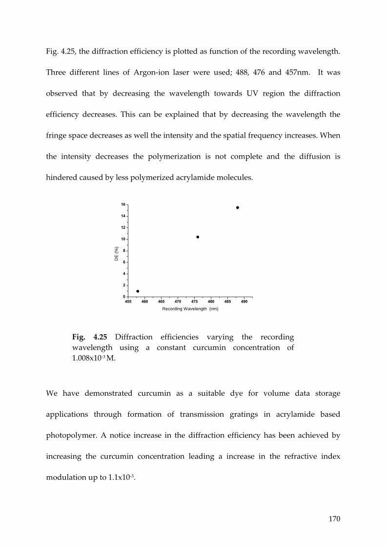
170
Fig. 4.25, the diffraction efficiency is plotted as function of the recording wavelength.
Three different lines of Argon‐ion laser were used; 488, 476 and 457nm. It was
observed that by decreasing the wavelength towards UV region the diffraction
efficiency decreases. This can be explained that by decreasing the wavelength the
fringe space decreases as well the intensity and the spatial frequency increases. When
the intensity decreases the polymerization is not complete and the diffusion is
hindered caused by less polymerized acrylamide molecules.
455 460 465 470 475 480 485 4900
2
4
6
8
10
12
14
16
DE
(%)
Recording Wavelength (nm)
Fig. 4.25 Diffraction efficiencies varying the recording wavelength using a constant curcumin concentration of 1.008x10‐3 M.
We have demonstrated curcumin as a suitable dye for volume data storage
applications through formation of transmission gratings in acrylamide based
photopolymer. A notice increase in the diffraction efficiency has been achieved by
increasing the curcumin concentration leading a increase in the refractive index
modulation up to 1.1x10‐3.

171
4.5. Conclusion
This chapter discloses the novel dyes suitable for blue wavelength recording. We
studied two newly synsthesized dyes in water soluble photo polymer systems.
Efficient grating formation is achieved in all these blue sensitized photopolymers.
We studied two newly synthesized dyes in water soluble acrylamide photopolymer
systems. Efficient grating formation is achieved in all these blue sensitised
photopolymers. We found by decreasing the recording wavelength up to blue region
we diminished the grating period and we increased the spatial frequency using the
same set up from close to 2173 nm to 2410 nm. We have demonstrated Perylene and
Curcumin as two novel photosensitizers suitable for blue recording at 488 nm of
wavelength with a reduced shrinkage yielding a better efficiency. Shifting the
wavelength further to the blue region we found acridine yellow as a appropriate dye
for recording at 457 nm of wavelength achieving a high RIM of 4x10‐3 with a
shrinkage measured by angular selectivity as lower as 1% making a proper material
for holographic data storage.

172
References [1] http://www.inphase‐technologies.com/downloads/pdf/technology/techTour.pdf
[2] http://www.tapeonline.com/blu‐ray.axpx
[3] S. Martin, P.E.L.G. Leclere, Y. L.M. Renotte, V. Toal and Y.F. Lion,
“Characterization of an acrylamide‐based dry photopolymer holographic recording
material”, Opt. Engineering 33, 3942‐3946 (1994).
[4] M. Ortuño, S. Gallego, C. Garcia, C.Neipp, A. Belendez and I. Pascual,
“Optimization of a 1 mm thick PVA/Acrylamide recording material to obtain
holographic memories: method of preparation and holographic properties”, Appl.
Phys. B 76, 851‐857 (2003).
[5] C. Sanchez, M.J. Escuti, C Van Heesch, C. W.M. Bastiaansen, D. J. Broer, J. loos
and R. Nussbaumer, “TiO2 nanoparticle‐Photopolymer composites for volume
holographic Recording”, Adv.Funct. Mater 15, 1623‐1629 (2005).
[6] W.S. kim, H‐S. Chang, Y‐C. Jeong,Y‐M Lee, J‐K. Park, C‐W Shin, N. Kim and H‐J.
Tak, “A new phase‐stable photopolymer with high diffraction efficiency based on
modified PMMA”, Opt. Comm. 249, 65‐71 (2005).
[7] H. Kogelnik, Bell Syst. Tech. J. 48, 2909 (1969).
[8] R. Jallapuram, I. Naydenova, S. Martin, R. Howard, V. Toal, S. Frohmann, S. Orlic
and H. J. Eichler, “Acrylamide‐based photopolymer for microholograpic data
storage”, Opt. Mater. 28, 1329‐1333 (2006).
[9] V. Moreau, Y. Renotte and Y. lion, “Characteriztion of Dupont photopolymer:
determination of kinetic parameters in a diffusion model”, Appl. Opt. 10, 3427‐3435
(2002).
[10] H. Sherif, I. Naydenova, S. Martin, C. McGinn and V. Toal, “Characteriztion of
an acrylamide‐based photopolymer for data storage utilizing holographic angular
multiplexing”, J. Opt. A: Pure Appl. Opt. 7, 255‐260 (2005).
[11]. M. Magini, M.R. Rodrigues, « A dynamical system to describe the cationic
photopolymerization of tetrahydrofuran initiated by systems sensitizer‐sulfonium

173
salt”, Polymer, 46, 3489–3495, (2005). J. V. Crivello* and J. L. Lee, “Photosensitized
Cationic Polymerizations Using Dialkylphenacylsulfonium and Dialkyl(4‐
hydroxyphenyl)sulfonium Salt Photoinitiators”
Macromolecules, Vol. 14, No. 5, 1141‐1147, (1981).
[12]. S. Icli , S. Demiça, B. Dindar, A. O. Doroshenko and C. Timur, “Photophysical
and photochemical properties of a water‐soluble perylene diimide derivative”, J. P. P.
A: Chem, 136, 15–24, (2000)
[13]. W.S. Kim, Y‐C Jeong, J‐K. Park, C‐W Shin and N‐Kim, “Diffraction efficiency
behaviour of photopolymer based on P5MMA‐co‐MMA) copolymer matrix”, Opt.
Mater. Doi:10.1016/j.optmat.2006.09.007, (2006).
[14]. S. Blaya, L. Carretero, R. Mallavia, A. Fimia, R. Madrigal, M. Ulibarrena, D.
Levy, “Optimization of an acrylamide‐based dry film used for holographic
recording”, Applied Optics, Vol. 37, No. 32, 7604‐7610, 1998.
[15]. D.M. Connor, S.D. Allen, D.M. Collard, C.L.Liotta and D. A. Schiraldi. “Effect of
linear comonomers on the rate of crystallization of copolyesters”. Journal of applied
polymer science. Vol.80, 2896‐2704 (2201).
[16]. K. Wu, J. G. Snijders and C. Lin, “Reinvestigation of Hydrogen Bond Effects on
the Polarizability and hyperpolarizability of Urea Molecular Clusters” J. Phys. Chem.
B 2002, 106, 8954‐8958
[17]. A. Sosnik and D. Cohn, “Poly(ethylene glycol)‐poly(epsilon‐caprolactone) block
oligomers as injectable materials”, Polymer, 44, 7033–7042, (2003).
[18]. X. He, H. Liu, Y. Li, Y. Liu, F. Lu, Y. Li and D. Zhu, “A New Copolymer
Containing Perylene Bisimide and Porphyrin Moieties: Synthesis and
Characterization”, Macromol. Chem Physic, Vol. 206, Iss. 21, 2199‐2205, (2005).
[19]. D. Kucklingl, H‐J. P. Adler, L. Ling, W. D. Habicher and K‐F Arndt,
“Temperature sensitive polymers based on 2‐(dimethylmaleinimido)‐N‐ethyl‐
acrylamide: Copolymers withN‐isopropylacrylamide”, Polymer Bulletin, 44, 269–276
(2000)

174
[20]. J.T. Gallo and M. Verber, “Model for the effects of material shrinkage on volume
holograms”, Appl. Opt. 29, 6797‐ 6804. 1994.
[21]. L. Shou‐Jun, L. Guo‐Dong, H. Qing‐Sheng, W. Min‐Xian, Jin Guo‐Fan, S. Meng‐
Quan, W. Tao and W. Fei‐Peng, “Holographic grating formation in dry
photopolymer film with shrinkage”, Chinese Physics, Vol. 13, No. 9, 1428‐1431.
[22] A. Anderson, M. Mitchell and R.S. Mohan, “Isolation of Curcumin from
Turmeric”, J. Chem. Educ. 77, 359‐360 (2000).
[23] J.V. Crivello and U.Bulut, “Curcumin: A naturally ocurring lon‐wavelength
photosensitizer for diaryliodonium salts”, J of Polym Sci Part A; Polym Chem 43,
5217‐5231, (2005).
[24] E. M. Bruzell, E. Morisbak and H. H. Tonnesen, “Studies on curcumin and
curcuminoids. XXIX. Photoinduced cytoxicity of curcumin in selected aqueous
preparations”, Photochem. Photobiol. Sci. 4, 523‐530 (2005).
[25] Mishra and S. Daswal, “Curcumin, A Novel Natural Photoinitiator for the
Copolymerization of Styrene and Methylmethacrylate”, Journal of Macromolecular
Science, Part A: Pure and Applied Chemistry 42, 1667‐1678 (2005).
[26] J. Crivello and U. Bulut, “Indian turmeric and its use in cationic
Photopolymeriztion”, Macrom. Symp. 240, 1‐11 (2006).

175
5
Chapter 5 Nano‐composite photopolymer recording media for reduced shrinkage
5.1 Introduction
In previous chapters we explored the potential of photopolymer systems as high
capacity data storage media capable of full range recording. With all the excellent
properties obtained including high diffraction efficiency at high spatial frequencies,
the photopolymer system mainly suffers from the undesired shrinkage after
recording. Shrinkage is due to the loss in volume fraction of the recording media as it
undergoes polymerization process [1]. The effective film thickness is reduced
because of such loss which leads to change in the stored grating period [1]. The basic
photopolymer component responsible for such changes during recording process is
the non‐active component called binder. Almost all polymeric binder materials suffer
from loss in volume fraction mainly because of the loss in their mechanical properties
caused by polymerization process. To improve the mechanical property of binders
like PVA, inorganic based composite materials such as CdS, Carbon nanotubes,

176
silver, and clays have been employed to prepare polymer nano‐composite [2]. Here
the crystallization behaviour of polymer material is also crucial since it governs the
thermal and stress‐strain properties [3]. The crystallisation of PVA is drastically
affected by the tactility of main chain, molecular weight and plasticizer amount.
More recently, PVA‐based nano‐composites have been produced to improve its
mechanical properties. Most PVA‐based nanocomposites are prepared with the sol–
gel method [3]. The main concern for fabricating a polymeric–inorganic
nanocomposite is the dispersion of inorganic nanoparticles in a polymer matrix
uniformly while avoiding the aggregation. To solve this problem, various processes
have been developed. Extensive methods for synthesizing polymeric–inorganic
nanocomposites included solgel, in situ polymerization, intercalation polymerization,
and blending techniques has been used.
Several Attempts have been proposed to improve some features of photopolymers
such as enhancement of the refractive index modulation and reduction of the
shrinkage by the incorporation of high or low refractive index species such as silica
and titania nanoparticles, in addition to the regular photopolymerizable monomers.
Suzuki et al. [4] demonstrated the recording of permanent holographic gratings in
the green with high diffraction efficiency in TiO2 nanoparticle‐dispersed
methacrylate photopolymer films. The photopolymer was prepapred by dispersing
the TiO2 nanoparticles combined with a solution of methyl isobutyl ketone and n‐
butyl alcohol, to methacrylate monomers [~2‐methyl‐acrylic acid 3‐[2‐methyl‐
acryloyloxymethyl)‐octahydro‐4,7‐methano‐iden‐5‐ylmethyl ester] at a wavelength

177
of 589 nm. The photosensitizer used was titanocene (Irgacure 784, Ciba) at 1 wt%
allowing the photosensitivity at wavelengths shorter than 550 nm. The difference
between the refractive index of the nanoparticles and the monomers in the solid
phase is one, which is greater than that (~0.1) between polymerized monomers and
host polymers usually in binder based photopolymers. Diffraction efficiencies near
90% were obtained after 100 seconds of the exposure time with an intensity of 100
mW/cm2 and a fringe spacing of 1.0 μm. It is shown that the diffraction efficiency as
well as the recording sensitivity significantly increased with a raise of nanoparticles
content. It is also found that shrinkage during holographic exposure is noticeably
suppressed by inclusion of the nanoparticles. The inclusion of nanoparticles
represented an enhancement of the refractive index modulation up to 5.1x10‐3.
Kim et al. [5] reported an organic‐inorganic photopolymer containing surface‐
nanoparticles, which exhibits a significant enhancement in optical properties. The
photopolymer was prepared using poly(methylmethacrylate‐co‐methacrylate acid)
as a polymer matrix, acrylamide and surface‐photoreactive silica nanoparticles as
photopolymerizable monomers, triethanolamine and methylene blue as initiator and
photosensitizer respectively. The composition of the reactants was: matrix:monomer:
TEA:MB was dissolved in the ratio 51:25:24:0.06 wt% in tetrahydrofuran. Two
different photopolymers were prepared to verify the effect of the nanoparticles. The
photopolymer containing surface‐photoreactive nanoparticles showed diffraction
efficiency near 90% after 90 sec of exposure and reduced volume shrinkage without
affecting the energetic sensitivity using a fringe spacing of about 1 μm.

178
Sanchez et al. [6] reported another new and efficient inorganic‐organic photopolymer
for the recording of volume transmission holograms. The mixture comprises a
mixture of UV‐acrylates and grafted titanium dioxide nanoparticles with an average
size of 4 nm. Diffraction efficiencies near 90 % were achieved with a space fringe of
500 nm for the maximum nanoparticles content of 30%. A dramatic increase in
efficiency was observed as the nanoparticles concentration was increased. The
inclusion of nanoparticles represented an enhancement of refractive index
modulation up to 15.5x10‐3‐ an improvement of more than a factor of four over the
material without nanoparticles while maintaining high transparency in the visible
wavelength range.
Another hybrid photopolymer by incorporating Zr(OiPr)4 nanoparticles was
investigated by F. del Monte et al. [7] Diffraction efficiencies of 97% yielding
refractive index modulation up to 1.5x10‐2 and 69% of diffraction efficiency were
achieved for photopolymers with and without Zr nanoparticles respectively in a 35
μm thick film. The components of the holographic material were 2,4‐pentadienone
and POEA as the first and second photopolymerizable monomers, Irgacure 784 as
photoinitiator and the binder of choice was a nanoporous hybrid silica glass.
In present chapter we incorporate inorganic nano‐composite materials such as SiO2
nanoparticles in our recording media in order to reduce the overall shrinkage.
Furthermore, incorporation of such nano‐composite materials often brings the much
desired modulation of effective refractive index as the inorganic materials generally
exhibit different refractive index. Formation of the transmission volume phase

179
gratings are demonstrated with excellent control over shrinkage [5,6]. Before going
into experimental details it is required to understand the additional photo‐physical
steps involved in a nano‐composite photopolymer media.
5.2 Preparation of PVA/SiO2 nano‐composite based photopolymer
recording media
SiO2 nanoparticles employed have a lower index of refraction (n∼1.46) than that of
presently used monomers (n=1.55) in our experiments [8]. Firstly the SiO2
nanoparticles aqueous dispersion is treated with an ultrasonic vibrator [3]. Its pH
value is adjusted to a basic one so as to negatively charge the SiO2 nanoparticles,
which act as templates to absorb positively charged polyallilamine hydrochloride
(PAH) surfactant chains through electrostatic adsorptive interaction. After
incorporation of this pH controlled solution into PVA solution dissolved in water,
PVA molecular chains are then assembled on the surface of SiO2 nanoparticles
through hydrogen bonding between hydroxyl groups of the PVA and the amino
group of the PAH. Finally the treated SiO2 nanoparticles are uniformly dispersed in
bulk PVA matrix, which then combines with the rest of pre‐polymer components [3].
In this experiment we used 17 mg of SiO2 nanoparticles aqueous dispersion (3.2 ml)
which was ultrasonically agitated for 30 min. The pH of the solution was adjusted
from acid to a basic with addition of 1.5 ml of 0.2M NaOH water solution. A 0.5 wt%
positively charged Poly (allylamine hydrochloride)‐(PAH:Aldrich, Mw: 70,000)
solution (1.6 ml) was dropped into the SiO2 dispersion, immediately a precipitation

180
of the solids appeared. The dispersion was centrifuged and rinsed to eliminate the
PAH not absorbed onto the surface of the SiO2. This step was repeated four times. A
0.5wt% (3.2 ml) PVA solution, Mw: 31,000‐50,000 (87‐89% hydrolyzed) was dropped
into the rinsed SiO2 dispersion which was ultrasonically stirred for 40 min to produce
a stable SiO2 dispersion. The stable SiO2 dispersion was dropped into a 10% wt
(3.2ml) of a PVA solution with the same molecular weight used previously were
ultrasonically stirred for 40 min at room temperature to produce a homogeneous
PVA/SiO2 nanocomposite.
The influence of NaOH quantity plays an important role in the dispersion of the
nanoparticles, as it helps the formation of SiO2 solids wrapped in PAH. The
formation of solids is indispensable in the functionalization process. The NaOH
amount used after nanoparticles aqueous solution in the first step of the
nanoparticles functionalization will decide the formation of solids to rinse and then
to disperse. If the NaOH amount is less than 1.5 ml in 3.2 ml of nanoparticles‐water
dispersion no solids are formed, if the quantity is higher than this value the solution
becomes transparent, in this way there are no solids formed.
5.2.1 Interaction forces in the nanocomposite
Acute aggregations exist if SiO2 nanoparticles are dispersed in pure water without
any treatment. However, there is a not obvious aggregation after SiO2 nanoparticles
are treated with PAH as the surfaces of negatively charges SiO2 nanoparticles are

181
covered by positively charged molecular chain of PAH through electrostatic
adsorptive interactions which considerably improve the dispersion of SiO2
nanoparticles [9].
The amido functional groups of PAH thus covers the surface of SiO2 nanoparticles,
which are then subsequently linked to hydroxyl‐functional groups of PVA by
hydrogen‐bonding interactions. This results in the uniform dispersion of SiO2
nanoparticles in the PVA matrix [9]. Figure 5.1 schematically demonstrates such
process.
Fig. 5.1 Scheme of the nanocomposite process.
5.2.2 Photopolymerization process incorporating nanoparticles
The polymerization process with nano‐particles inside the PVA matrix is slightly
different from the general photo polymerization process. When the interfering light
pattern reaches the film rapid cross‐linking takes place in the high intensity fringes

182
while there is almost no reaction in the dark areas. As a result of the depletion of
highly reactive monomer in the bright regions, the non consumed monomers then
start diffusing from dark to bright areas [6].
The consumption of monomers due to photopolymerization breaks the
thermodynamical equilibrium balance of monomers and nanoparticles which
requires that their volume fraction ratio between two components be constant at any
position. Such imbalance leads to migration (diffusion) of monomers from dark to
bright regions while photosensitive inorganic nanoparticles experience the counter‐
diffusion from bright to dark regions since they are not consumed. In this way a
permanent RI grating is formed owing to a spatial frequency distribution of the
effective RI with periodically distributed nanoparticles and polymerized monomers
[8]. Such a dynamic process essentially continues until monomers are consumed
completely by the monomolecular and bimolecular termination processes and/or
until the increased viscosity of a surrounding medium due to growing polymer
networks makes oligomers and nanoparticles immobilize as a result spatially
periodic distributions of nanoparticles and if any polymer concentrations are formed
in the material [8]. Because unreacted monomers in the dark regions are sooner or
later photopolymerized, a fixed RI grating maybe formed owing to a difference in
effective refractive indices of periodically distributed nanoparticles and uniformly
distributed polymerized monomers.
Such counter‐diffusion process is illustrated in Fig. 5.2. The inorganic nanoparticles
experience counterdiffusion from the bright to the dark regions, since the particles

183
are not consumed and their chemical potential increases in the bright regions as a
result of depletion of monomers in those areas [10].
Fig. 5.2 Holographic recording process in a nanoparticle‐dispersed photopolymer. (a) before and (b) during holographic exposure.
As a consequence the low reactivity species the monomer and non‐reactive
nanoparticles counter‐diffuse to the dark areas resulting in a compositional
modulation [6]. The chemical affinity of the nanoparticles with the low‐reactivity
monomer could enhance this diffusion of the nanoparticles to the dark areas. This
compositional segregation together with the significant difference in refractive index
of nanoparticles with respect to the rest of the components of the polymer is expected
to enhance the diffraction efficiency of the stored grating structures.
MonomerNanoparticles
Laser beams
a
b

184
5.2.3 Hydrogen Bond interaction theory
Another dominating effect may come from the formation of hydrogen bonds within
the PVA‐SiO2 network [11]. Figure 5.3 illustrates the interfacial structure of
photopolymer. Before photopolymerization, the acrylamide molecules ought to
induce efficient hydrogen bonding with silica nanoparticles (stability of silica/PVA
dispersion is lower than that of silica/AA dispersion, thus it could be concluded that
acryl amide molecules induce the most strong hydrogen bonding with silica
nanoparticles) and then the acrylamide become into poly(acrylamide) by
photopolymerization in the illuminates areas [11].
Fig. 5.3 Interfacial structure of nanocomposite_ photopolymer. (a) non_illuminated area (b) illuminated area.
After photopolymerization, acrylamide groups in the PAA chains may form
hydrogen bonding with silica nanoparticles in the bright region, while there may be
Illuminated area
Illuminated area
OH
OH AA
Illuminated area OH
OH
Non illuminated
area
(a)
(b)

185
weak hydrogen bonding in the dark region due to the diffusion of acryl amide
molecules into the bright region by the concentration gradient. This difference in the
hydrogen bonding strength between the bright region and the dark region may lead
to the additional refractive index modulation, in other words, the enhancement in the
diffraction efficiency [11].
5.3 Formation of transmission phase gratings in Eosin‐
nanocomposite photopolymer
We prepared different wt% SiO2 nano‐particle incorporated into PVA matrix with
previously described Eosin sensitizer. For successful comparison of the effect of
nanoparticles in this recording media It was ensured that the concentration of all the
component except SiO2 remains the same. The final concentration for all three
samples is tabulated in table 5.1.
Table 5.1 Composition of pre‐polymer solution in 2.5 ml of solution.
Eosin/SiO2
photopolymer
[Eosin]
(%)
[Acrylamide]
(M)
[BSA]
(M)
[TEA]
(M)
EOS 0.080 0.452 0.052 0.605
EOS‐Si1 0.080 0.452 0.052 0.605
EOS‐Si2 0.080 0.452 0.052 0.605

186
Fig. 5.4 shows the UV‐Vis spectra for eosin composite without and for composite
containing nanoparticles shows a maximum absorption at the same wavelength of
530 nm. The gradual increase in general absorption features with incorporation of
SiO2 nanoparticles indicates towards the amount of scattering taking place as any
increase caused by nano‐particle is not expected to increase the eosin absorption.
300 400 500 600 7000,00
0,03
0,06
0,09
0,12
0,15
Abs
orba
nce
Wavelength (nm)
EOS EOS-Si1 EOS-Si2
Fig. 5.4 UV/Vis spectra of the Eosin photopolymers and nanocomposites.
Table 5.2 shows the recording and reconstruction parameters used for
characterization.
Table 5.2 Experimental parameters for grating formation in Eosin nanocomposite.
Photo‐ polymer
θ/2 (°)
WB (nm)
RB (nm)
Λ (μm)
SF (lines/mm)
I (mW/cm2)
θB (°)
EOS+SiO2 34 514.5 633 0.46 2173 153.3 43.4 (T)

187
where θ/2 is the half‐angle between the interference beams; WB and RB are the
writing and reading beam respectively; Λ is the fringe period; θB(T,E) are the
theoretical and experimental Bragg angle and I is the recording intensity.
Figure 5.5 illustrates the response of transmission gratings stored in this recording
media with respect to increasing exposure time. A dramatic rise in the diffraction
efficiency was observed by the addition of SiO2 nanoparticles compared to the
photopolymer without nanoparticles [6]. Also by increasing the nanoparticles
loading the time to reach saturation is reduced drastically. The DE increased from
25% to 75% for the composite with higher 0.354 wt% nanoparticles. The less time
required for saturation explains the light dispersion created by nano particles which
creates a uniform illumination inside the media through out the recording film
thickness.
0 15 30 45 60 75 900
20
40
60
80
100
DE
(%)
Exposure time (sec)
EOS EOS-Si1 EOS-Si2
Figure 5.5 Diffraction efficiency with respect to exposure time.

188
There is a remarkable increase in the DE with exposure time for both cases with
nanoparticles. Similar phenomena was reported in previous studies focusing on the
nanoparticles‐dispersed binder photopolymer and the origin of increase in DE was
attributed to a counter‐diffusion in DE resulting in the spatially periodic distribution
of nanoparticles during holographic exposure. Table 5.3 summarizes the results.
Table 5.3 Optical parameters of photopolymer that contains Eosin + nanocomposites
after 2.045 sec using a 25 μm thick layer.
Eosin/SiO2 photopolymer
SiO2
content (%)
DE (%)
Sensitivity (cm/J)
RIM Thickness(μm)
EOS 0 3.3 232 1.07x10‐3 25
EOS‐Si1 0.208 23.8 623 3.0x10‐3 25
EOS‐Si2 0.354 74.1 1099 6.1x10‐3 25
We observed that for higher nanoparticles loads the pre‐polymer syrup becomes
viscous and the dispersion turbidity increases leading to non‐uniform grating
formation caused by higher surface scattering.
For comparing the amount of film shrinkage, the grating angular response half‐width
(θ) was obtained from the measurement of the diffraction efficiency as a function of
angular deviation from the Bragg condition [5,12]. The angular response of the two
photopolymers was measured by rotating the photopolymer from the position
satisfying the Bragg condition. Figure 5.6 compares the angular response of the
photopolymer with and without nanoparticles: it should be noticed that the

189
uniformity of grating throughout the depth of the photopolymer is also enhanced by
the introduction of small amount of surface‐photo reactive nanoparticles.
0
10
20
30
40
50
60
70
80
90
100
-2 -1 210
DE
(%)
Angular response (°)
EOS EOS-Si1 EOS-Si2
Fig. 5.6 Angular Selectivity of diffraction efficiency for composite without and nanoparticles.
As evident from the above figure, the increase in nano‐particle presence inside the
recording media narrows the angular selection of the gratings. This is a clear
indication of less shrinkage suffered by the films over those without nano‐particles as
the presence of rigid nano‐particles compensates the loss of volume fraction caused
by the binder. The Bragg condition in the case of higher amount of nanoparticles was
closer to the theoretically calculated Bragg angle while with no to little nanoparticles
the Bragg condition varies by angle of 2°. This means the composition with
nanoparticles has an ideal response and doesn’t suffer from shrinkage as the case of

190
others. Table 5.4 illustrates the amount of shrinkage taking place in the films with
and without nanoparticles.
Table 5.4 Shrinkage calculated using eosin nanocomposite.
Photopolymer Δd (μm)
Shrinkage(%)
EOS 1.48 5.9 EOS‐Si1 1.48 5.9 EOS‐Si2 0.47 1.9
5.3.1 Effect of intensity on the EOS‐Si2 nanocomposite recording
media
In fig. 5.7, the growth of diffraction efficiency is plotted as a function of intensity for
the nanocomposite with higher amount of nanoparticles. A clear rise in the
diffraction efficiency was observed by increasing the intensity This evolution is
different for 40 mW/cm2 and 100mW/cm2 where by raising the intensity the
diffraction efficiency reached the maximum value and then decays and stabilizes but
for the case of maximum intensity at 153 mW/cm2 the diffraction reaches fast
saturation and stabilizes.. Also higher sensitivities were found by increasing the
intensity. Table 5.5 summarizes the results obtained by varying the intensity of the
recording beams.

191
Table 5.5 Optical parameters of photopolymer that contains EOS‐Si2 nanocomposite
varying the recording intensity after 2.045 sec.
Intensity (mW/cm2)
DE (%)
Sensitivity(cm/J)
RIM Thickness (μm)
153.3 74.1 1099 6.1x10‐3 25
100 18 830 2.5x10‐3 25
40 5.4 1136 1.3x10‐3 25
0 15 30 45 60 75 900
10
20
30
40
50
60
70
80
90
100EOS-Si2
DE
(%)
Exposure time (sec)
153.3 mW/cm-2
100 mW/cm-2
40 mw/cm-2
Fig. 5.7 Traces of diffraction vs exposure time for EOS‐Si6 photopolymer at different intensities.
The angular response of the recording media with varying intensity is presented in
figure 5.8. As we can clearly observe, by decreasing the recording intensity for the
photopolymer EOS‐Si2 the first diffracted orders start to vanish.

192
0
10
20
30
40
50
60
70
80
90
100
EOS-Si2
-2 -1 210
DE
(%)
Angular response (°)
153.3 m W /cm 2
100 m W /cm 2
40 m W /cm 2
Fig. 5.8 Angular Selectivity of diffraction efficiency of nanocomposite EOS‐Si2 at different intensities.
The features for photopolymer with nanoparticles are more clearly resolved. The
uplift of the first satellite peaks may result from the non‐uniformity in grating
strength. Also, by increasing the intensity, the angular width of the zero diffracted
order was considerably reduced.
5.3.2 FT‐IR and Tg analysis
To clarify the difference in the hydrogen bonding between ‘dark’ and ‘bright’ region,
we prepared samples firstly illuminated (3mg) then mild with KBR and pressed as
disc form for FT‐IR measurement photopolymerization of acrylamide molecules, as
shown in Fig.5.9. The PVA is a kind of semicrystalline polymer, quite a few
crystallites are formed on the surface of the pure PVA fracture, whereas there are no

193
crystallites on the surface of the PVA/SiO2 nanocomposite because of the well‐
dispersed SiO2 nanoparticles being able to prevent the formation of crystallites [9].
400 600 800 1000 1200 1400 1600 1800 20000,4
0,5
0,6
0,7
0,8
0,9
1,0
1,1
1,2Ab
sorb
ance
Wavenumber (cm-1)
EOS EOS-Si9 EOS-Si6
Fig. 5.9 FTIR spectra of the polymerized Eosin photopolymers with and without nanoparticles.
The characteristic absorbing peaks of SiO2 (1100 and 466 cm‐1) are not simply
overlapped by those of PVA but move to 1089 and 473 cm‐1, respectively. This may
be due to the interaction between the SiO2 nanoparticles and PVA matrix, which
obstructs the formation of intrahydrogen bonding. This further indicates that the
PVA matrix and SiO2 nanoparticles are not simply blended, and complex interactions
exist [9].
Figure 5.10 shows the DSC thermogram of photopolymer with and without
nanoparticles. The addition of the SiO2 nanoparticles to the PVA matrix results in a
significant increase in Tg, this suggest that the SiO2 nanoparticles are well dispersed
in the PVA matrix due to nanoparticles have huge relative surface areas and strong

194
absorptive power [9]. The amorphous molecular chains of PVA become stable and
interact strongly with the SiO2 nanoparticles. As a result the SiO2 nanoparticles
restrict the thermal motion of the PVA molecular segment [9]. Because Tg is the
limiting temperature for the applications of plastic polymers; an improvement is very
important for PVA [7]. The Tg (EOS‐without nanoparticles) =90°C and Tg (EOS‐Si2,
containing nanoparticles)= 94°C resulting Tg(nanocomposite) > Tg (PVA).
Fig. 5.10 DSC thermogram with Eosin photopolymers containing nanoparticles
Figure 5.11 (a,b) compares the grating surface profile after recording with and
without nanoparticels.
Fig. 5.11a Microscope image with Eosin without nanoparticles.
Fig. 5.11b Microscope image with Eosin with nanoparticles.

195
From the highly resolved features it can be concluded that the incorporation of nano‐
particles are helpful for high refractive index modulation which essentially gives the
higher diffraction efficiency.

196
Conclusion
We successfully achieved the minimum shrinkage limit of a photopolymer recording
system by incorporation of inorganic nanoparticles inside the eosin sensitized
recording media. The reason for such decrease in the shrinkage may be attributed to
an increase in rigidity of sample material resulting from dispersion of nanoparticles.
The structure of photopolymerised polymer and the mechanism of reduction of
volume shrinkage due to the rigid silica are conceptually described. Surface‐
photoreactive nanoparticles introduced in this study are expected to play dual roles
with respect to the reduction of volume shrinkage. Due to the rigid silica
nanoparticles incorporated in this grating by chemical bonds, the mobility of
photopolymerised polymer chains can be reduced and this can lead to a reduction of
free volume change and thus to the volume shrinkage of recorded gratings. In
addition to the contribution to the reduction in the free volume change during
Photopolymerization, the nanoparticles also contribute the rigidity enhancement of
the medium which also leads to the reduction of volume shrinkage. It is seen that
while shrinkage decreases with an increase of nanocomposite concentration. As the
shrinkage decreases, Sensitivity (S), Dynamic sensitivity (S*) and Δn increases.

197
References
[1] S. Martin, P. E. L. G. Leclere, Y. L.M. Renotte, V. Toal, Y. F. Lion,
“Characterization of an acrylamide‐based dry photopolymer holographic recording
material”, Optical Engineering, Vol. 33, No. 12, 3942‐3946, 1994.
[2] Z. Peng, L. X. Kong and S.D. Li, “Dynamic mechanical analysis of poly
vinylalcohol/silica nanocomposite”, Synthetic Metals 152, 25‐28, (2005).
[3] Z. Peng, L. X. Kong and S.D. Li, “Non‐isothermal crystallization kinetics of self‐
assembled polyvinylalcohol/silica nanocomposite”, Polymer 46, 1949‐1955, (2005).
[4]. N. Suzuki, Y. Tomita and T. Kojima, “Holographic recording in TiO2
nanoparticle‐dispersed methacrylate photopolymer films”, Appl. Lett. Vol. 81, No.
22, 4121‐4123, (2002).
[5] W. S. Kim, Y‐Ch. Jeong and J‐K Park, “Organic‐inorganic hybrid photopolymer
with reduced volume shrinkage”, Appl. Phys. Lett. 87, 012106‐1‐3, (2005).
[6] C. Sanchez, M.J. Escuti, C Van Heesch, C. W.M. Bastiaansen, D. J. Broer, J. loos
and R. Nussbaumer, “TiO2 nanoparticle‐Photopolymer composites for volume
holographic Recording”, Adv.Funct. Mater 15, 1623‐1629 (2005).
[7]. F. del Monte, O. Martinez, J. A. Rodrigo, M.L. Calvo and P. Cheben, “A volume
holographic sol‐gel material with large enhancement of dynamic range by
incorporation of high refractive index species. Adv. Mat. 18, 2014‐2017, (2006).
[8] N. Suzuki and Y. Tomita, “Diffraction properties of volume holograms recorded
in SiO2 nanoparticle‐dispersed methacrylate photopolymer films”, Jpn. J. Appl. Phys.
42, L927‐L929 Part 2 No. 8A, (2003).
[9] Z. Peng, L. X. Kong and S.D. Li, “Thermal properties and morphology of a
polyvinylalcohol/silica nanocomposite prepared with a self‐assembled monolayer
technique”, J. Appl. Pol. Sci. 96, 1436‐1442, (2005).
[10] Y. Tomita, N. Suzuki and K. Chikana, “ Holographic manipulation of
nanoparticles distribution morphology in nanoparticles‐dispersed photopolymers”,
Opt. Lett. 30, No.8, 839‐841, (2005).

198
[11] W. S. Kim, Y‐Ch. Jeong, and J‐K Park, “Nanoparticle‐induced refractive index
modulation of organic‐inorganic hybrid photopolymer”, Opt. Express 14, No. 20
8967‐8973, (2006).
[12] N. Suzuki, Y.Tomita and T. Kojima, “Holographic recording in TiO2
nanoparticle‐dispersed methacrylate photopolymer films”, Appl. Phys. Lett. 81, No.
22, 4121‐4123, (2002).

199
Conclusions In chapter 1, we presented the theory of holography and different photosensitive
media commonly used for recording purposes focussing in the description of
photopolymers. We described the principle of photopolymers, main components
exhibiting the materials chosen in our study, the photopolymerization process which
undergo once is illuminated by a coherent light (Laser), terminology related to this
kind of materials. The diffusion process of the monomer is examined due to its
importance in the enhancement of their refractive index modulation. We also
discussed the different geometries used for holographic grating recording such as
transmission and reflection ones.
In this study we presented the novel water soluble photopolymer systems and their
suitability as volume information storage media highlighting on increasing the
capacity of the information storage in terms of increased spatial frequency. Different
photosensitizing dyes with sensitivity from red to blue spectral region were revised
demonstrated spatial frequency as high as 2400 lines/mm arriving close to 6000
lines/mm in transmission and reflection geometry respectively.
In the chapter 2 we presented the optimization of different components of
photopolymer system suitable for red wavelength recording in transmission
geometry selecting methylene blue as photosensitizer. The diffraction grating

200
formation was monitored achieving a diffraction efficiency of 40% under a high
spatial frequency of 1767 lines/mm with high angular response of 0.5°. By optimizing
this component we demonstrated its limitations as a dye in the acrylamide based
photopolymers giving an explanation of this phenomenon.
In chapter 3 we studied different photopolymers using Rose Bengal, Erythrosin B,
Acridine Orange and Eosin as photosensitizers in transmission as well as reflection
geometry recording using green sensitizing dyes, showing a significant advantage of
higher capacity grating storage over red sensitized dye studied in chapter 2. An
optimization using the different dyes was carried out. An efficient dye is desired to
achieve maximum diffraction efficiencies in holographic grating recording in
photopolymer. The importance of role as electron acceptor in the photoreaction was
studied using CV. We found the maximum diffraction efficiency among all the dyes
used of 90% using EryB exhibiting the best acceptor electron nature with a peak
reduction of ‐1.1eV with the highest angular selectivity of 0.48° very important effect,
because this effect increases the capacity of the futures holography memories made
with photopolymers, compare to the rest of the dyes, 80%, 75% and 70% for RB, AO
and Eosin respectively. The effect of the cross‐linking agent in photopolymer is also
studied by the replacement of the N’,N‐methylenebisacrylamide for N,N‐
dimethylacrylamide observing a negative influence (overmodulation process) in the
DE evolution due to the drastic decay suffered after achieving its maximum value.
The results were analysed with help of optical techniques such as UV/Vis spectra

201
(layer absorption) DSC (Tg, diffusion of monomer), FTIR measurements (quantity of
monomer become polymer) and through diffraction grating formation in the bulk of
the recording medium. By changing from transmission to reflection geometry using
the same set up we increased more than two times the spatial frequency but lower
diffraction efficiencies were achieved. In reflection, we observed by increasing the
exposure time the diffraction efficiency was improved. RB was found the more
efficient dye in this geometry showing as high as 12.33% of diffraction efficiency.
In chapter 4 we develop novel photopolymer system suitable for blue wavelength
recording. Two new water soluble blue sensitizing dyes are synthesized and their
information storage capacity is demonstrated. We found between the two novel
materials Perylene as more efficient dye at 488 nm of wavelength for creation of
holographic grating recording. We demonstrated by increasing the acrylamide
content the width of the first lobe (zero diffracted order beam) is increased. By
reducing to recording wavelength to 457.9 nm closer UV spectral region we achieved
75% of diffraction efficiency using acridine yellow as dye with reduced shrinkage of
1% making ideal material for data storage. We also discussed by increasing the layer
thickness the sensitivity and diffraction efficiency as well as RIM decreased.
Finally; in chapter 5 we studied the response of the refractive index modulation by
incorporating low index in‐organic nanoparticles in the photopolymer composition.
Large modulation of index is thus created in recording system. A better

202
understanding of the monomer diffusion process involved in the
photopolymerization process was achieved. We observed a drastic increase of
diffraction efficiency from 25% to 75% as well as the sensitivity and the RIM with the
higher load of SiO2 nanoparticles in the photopolymer. An important diminution of
the angular width leading to a reduction of the shrinkage also was found.
For further enhancement of the spatial frequency we propose to find a dye sensible in
region close to UV (410 nm). By reducing even more the recording wavelength the
spatial frequency will be increased thus increasing the store capacity of the material.