Hollow boron nitride nanospheres as boron reservoir for ... · High global incidence of prostate...
12
ARTICLE Received 23 Jul 2014 | Accepted 15 Nov 2016 | Published 6 Jan 2017 Hollow boron nitride nanospheres as boron reservoir for prostate cancer treatment Xia Li 1 , Xiupeng Wang 2 , Jun Zhang 1,3 , Nobutaka Hanagata 4 , Xuebin Wang 1 , Qunhong Weng 1 , Atsuo Ito 2 , Yoshio Bando 1 & Dmitri Golberg 1 High global incidence of prostate cancer has led to a focus on prevention and treatment strategies to reduce the impact of this disease in public health. Boron compounds are increasingly recognized as preventative and chemotherapeutic agents. However, systemic administration of soluble boron compounds is hampered by their short half-life and low effectiveness. Here we report on hollow boron nitride (BN) spheres with controlled crystal- linity and boron release that decrease cell viability and increase prostate cancer cell apop- tosis. In vivo experiments on subcutaneous tumour mouse models treated with BN spheres demonstrated significant suppression of tumour growth. An orthotopic tumour growth model was also utilized and further confirmed the in vivo anti-cancer efficacy of BN spheres. Moreover, the administration of hollow BN spheres with paclitaxel leads to synergetic effects in the suppression of tumour growth. The work demonstrates that hollow BN spheres may function as a new agent for prostate cancer treatment. DOI: 10.1038/ncomms13936 OPEN 1 World Premier International Center for Materials Nanoarchitectonics (WPI-MANA), National Institute for Materials Science (NIMS), Namiki 1-1, Tsukuba, Ibaraki 305-0044, Japan. 2 Health Research Institute, Department of Life Science and Biotechnology, National Institute of Advanced Industrial Science and Technology (AIST), Central 6, 1-1-1 Higashi, Tsukuba, Ibaraki 305-8566, Japan. 3 School of Materials Science and Engineering, Hebei Key Laboratory of Boron Nitride Micro and Nano Materials, Hebei University of Technology, Tianjin 300130, China. 4 Nanotechnology Innovation Station, National Institute for Materials Science (NIMS), 1-2-1 Sengen, Tsukuba, 305-0047, Japan. Correspondence and requests for materials should be addressed to X.L. (email: [email protected]) or to J.Z. (email: [email protected]) or to D.G. (email: [email protected]). NATURE COMMUNICATIONS | 8:13936 | DOI: 10.1038/ncomms13936 | www.nature.com/naturecommunications 1
Transcript of Hollow boron nitride nanospheres as boron reservoir for ... · High global incidence of prostate...

ARTICLE
Received 23 Jul 2014 | Accepted 15 Nov 2016 | Published 6 Jan 2017
Hollow boron nitride nanospheres as boronreservoir for prostate cancer treatmentXia Li1, Xiupeng Wang2, Jun Zhang1,3, Nobutaka Hanagata4, Xuebin Wang1, Qunhong Weng1, Atsuo Ito2,
Yoshio Bando1 & Dmitri Golberg1
High global incidence of prostate cancer has led to a focus on prevention and treatment
strategies to reduce the impact of this disease in public health. Boron compounds are
increasingly recognized as preventative and chemotherapeutic agents. However, systemic
administration of soluble boron compounds is hampered by their short half-life and low
effectiveness. Here we report on hollow boron nitride (BN) spheres with controlled crystal-
linity and boron release that decrease cell viability and increase prostate cancer cell apop-
tosis. In vivo experiments on subcutaneous tumour mouse models treated with BN spheres
demonstrated significant suppression of tumour growth. An orthotopic tumour growth model
was also utilized and further confirmed the in vivo anti-cancer efficacy of BN spheres.
Moreover, the administration of hollow BN spheres with paclitaxel leads to synergetic effects
in the suppression of tumour growth. The work demonstrates that hollow BN spheres may
function as a new agent for prostate cancer treatment.
DOI: 10.1038/ncomms13936 OPEN
1 World Premier International Center for Materials Nanoarchitectonics (WPI-MANA), National Institute for Materials Science (NIMS), Namiki 1-1, Tsukuba,Ibaraki 305-0044, Japan. 2 Health Research Institute, Department of Life Science and Biotechnology, National Institute of Advanced Industrial Science andTechnology (AIST), Central 6, 1-1-1 Higashi, Tsukuba, Ibaraki 305-8566, Japan. 3 School of Materials Science and Engineering, Hebei Key Laboratory of BoronNitride Micro and Nano Materials, Hebei University of Technology, Tianjin 300130, China. 4 Nanotechnology Innovation Station, National Institute forMaterials Science (NIMS), 1-2-1 Sengen, Tsukuba, 305-0047, Japan. Correspondence and requests for materials should be addressed to X.L.(email: [email protected]) or to J.Z. (email: [email protected]) or to D.G. (email: [email protected]).
NATURE COMMUNICATIONS | 8:13936 | DOI: 10.1038/ncomms13936 | www.nature.com/naturecommunications 1

Prostate cancer is one of the most common cancers formales, particularly in the developed Western countries1.The high global incidence of prostate cancer has led to a
focus on prevention and treatment strategies to reduce thepublic health impact of the disease2–6. For prostate cancer, themain clinical treatments include active surveillance, surgery7,radiation therapy8, hormone therapy9 and chemotherapy10
with drugs, such as paclitaxel (PTX) and docetaxel. Anepidemiological study shows that boron (B) intake can reducethe risk of prostate cancer11,12 for human by up to 54%(ref. 13). Boric acid (BA), the dominant form of B in plasma,has been tested as a preventative and therapeutic agent againstprostate cancer. For example, in vitro BA inhibits theproliferation of prostate cancer cells in a dose-dependentmanner14. In the animal model, BA decreases serum prostate-specific antigen (PSA) levels by 88%, inhibits the LNCaptumour growth by 38% and reduces insulin-like growth factor-1in nude mice injected with human LNCap prostate cancercells15. Mechanisms of B-mediated anticancer action in prostatecancer include the reduction of intracellular calcium signals andstorage, the decrease in enzymatic activities (serine protease,NAD-dehydrogenases and so on) and the inhibition of thecancer cell proliferation14,16–19.
Recently, B compounds have attracted attention as preventa-tive and therapeutic agents for prostate cancer and othercancers11–17,19–27. However, systemic administration of solubleB compounds, such as BA, associates with the drawbacks of itsshort half-life period, low bioavailability, requirement of frequentadministration, low fraction arrived in the tumour site andlimited effectiveness for prostate cancer treatments. Moreover,the therapeutic window of BA for prostate cancer cells, which isB100 times higher than its average serum level in human,suggests difficulty in systemic administration of soluble Bcompounds without toxicity14,21,23. Local delivery of anticancerdrug or agents in a sustained manner either to the regionthat contains a tumour or directly within the tumour hasthe advantage of increasing tumour exposure to drug whilelimiting systemic toxicity28,29. Therefore, local delivery of asparsely soluble B-containing compound, as an alternative tosoluble B compounds, might hold promise as a preventative andtherapeutic agent for prostate cancer treatment. In addition, thesystematic administration of a sparsely soluble B-containingcompound in a nanoparticle format can facilitate the passivetargeting of drugs into the tumour sites, decrease the side effectsand enhance the antitumour efficacy with the aid of enhancedpermeability and retention effects30. Typically, the sparselysoluble B-containing compound is boron nitride (BN) that isstructurally analogous to carbon. To date, BN has been usedas a delivery vehicle31–33 for anticancer drugs such asdoxorubicin, similarly to other nano delivery vehicles, such ascarbon nanotubes34, graphene35, mesoporous silica36, calciumphosphate37 and polymers38. However, there have been noreports regarding the effectiveness of BN itself in cancertreatments.
Herein we fabricated hollow BN spheres with controlledcrystallinity and solubility to guide B release by adjusting theposttreatment temperature. Androgen-sensitive LNCap andandrogen-independent DU-145 prostate cancer cell lines wereused to evaluate the effects of hollow BN spheres on theapoptosis, necrosis and proliferation of the prostate cancer cellsin vitro. The death mechanism of LNCap and DU145 prostatecancer cells treated with BA or BN spheres were evaluated andcompared by a spectral cell analyzer. Then male BALB/c-nu/numice were used to confirm the effects of hollow BN spheres on thesuppression of prostate cancer occurrence and inhibition oftumour growth in vivo.
ResultsPhysicochemical characterization of hollow BN spheres.Hollow BN spheres were synthesized via the chemical vapourdeposition (CVD) reaction of trimethoxyborane (B(OMe)3) usingmodified method developed previously39, in which the second-stage annealing process was conducted in Ar rather than in NH3
atmosphere. Transmission electron microscopic images prove thesuccessful synthesis of BN nanospheres (Fig. 1a). The BNs-asample shows a solid sphere structure with an approximatediameter of 200 nm and low crystallinity characterized by a long-range disorder. The BNs-b shows a hollow sphere structure withthe same diameter and wall thickness of 50–60 nm. For BNs-c, ahollow nanostructure with a wall thickness of about 20 nm andhigh crystallinity characterized by the state of long-range order isobserved.
The BN spheres are adjustable with respect to crystallinity bymeans of posttreatment temperature variations (Fig. 1b). As awhole, with an increase in posttreatment temperature, thecrystallinity increased and the full width at half-maximum forthe peaks in the wide-angle X-ray diffraction patterns becomesnarrower. The BNs-a spheres exhibit an amorphous naturewith a broad peak at around 26�. The BNs-b spheres are higherin crystallinity than BNs-a, exhibiting two crystalline peaks ataround 26.4� and 42� that correspond to (002) and (100),respectively. The BNs-c spheres are the highest with regard tocrystallinity, exhibiting a much narrower peak width for (002)and (100) reflections than those for BNs-b.
All the BN spheres show typical fourier transform infraredspectroscopy (FTIR) absorption bands of B-N stretching at1,382 cm� 1 and B-N-B bending at 795 cm� 1 (Fig. 1c). Thepresence of hydroxyl groups is confirmed by the O-H stretchingband at 3,421 cm� 1 for BNs-a. A shoulder at 3,227 cm� 1
indicates the asymmetric stretching of the N-H group. A weakband at around 1,200–1,250 cm� 1 is attributed to B-N-Ostretching. With an increase in calcination temperature from900 to 1,025 �C for BNs-b, the O-H stretching band becomesweaker while the N-H stretching band becomes stronger. ForBNs-c, calcined at 1,400 �C, both the O-H and N-H bandsbecome very weak. X-ray photoelectron spectroscopy reveals thehigh content of oxygen (O) in BN spheres (Supplementary Fig. 1).As shown in Fig. 1d, the BNs-a, -b and -c nanospheresexhibit hydrodynamic diameters of 263.5±66.5, 334.1±87.8and 351.8±56.3, respectively, which are slightly higher thanthose obtained from the transmission electron microscopicobservations. In addition, unlike other BN materials such as BNnanotubes, all the BN spheres show good dispersibility in cellculture medium (Fig. 1e). The presence of large amount ofhydroxyl groups (Fig. 1c) and a high content of oxygen(Supplementary Fig. 1) result in their high hydrophilicity andgood dispersibility in an aqueous solution. At pH¼ 7.4, theBNs-a, -b and -c spheres all show zeta potentials centred ataround � 21 to � 25 mV (Fig. 1f).
B release from hollow BN spheres. Release of B from BN sphereswith controlled crystallinity kept in dialysis membrane bags wasanalysed in an acetate buffer at pH 4.6. BN spheres with highcrystallinity show slow B release (Fig. 1g). BNs-a spheres with thelowest crystallinity show the highest B release speed, which isabout 20 times that of BNs-c spheres and 2 times that of BNs-bspheres. Figure 1h displays the BN nanospheres remain in thedialysis membrane bags after immersing the same amount ofBNs-a, -b and -c samples in the acetate buffer for 1 month. It canbe seen that the BNs-a sample almost totally degrades, while theBNs-c sample remains in a large amount. Supplementary Fig. 2shows the B release rate for different BN spheres under various
ARTICLE NATURE COMMUNICATIONS | DOI: 10.1038/ncomms13936
2 NATURE COMMUNICATIONS | 8:13936 | DOI: 10.1038/ncomms13936 | www.nature.com/naturecommunications

conditions of pH, temperature and concentration. For all the BNspheres, the B release rate increases with the increase in tem-perature. In addition, the B release rate nearly linearly increaseswith the increase in the initial BN materials concentration. ForBNs-b, pH value has a negligible effect on B release at a lowtemperature, whereas, at a high temperature, high pH valueresults in the increase in the B release. The dynamic studiesof structural evolution for BN spheres are presented inSupplementary Fig. 3. For BNs-a sample, the spherical particle,around 200 nm in diameter, gradually degrades from the edgearea at the initial stage and then transforms into smaller clusters,about 5–20 nm in diameter, 3 days later. For BNs-b sample, thehollow spherical particle degrades in a different way comparedwith the BNs-a sample. The defect sites in the wall graduallydegrade to form a porous structure and the hollow spherical wallstructures are still preserved after 3 days. After 10 days, the partialdegradation in the edge area and a marginal amount of smallclusters are observed. Supplementary Fig. 4 reveals FTIR spectraof the hydrolysed products of BN nanospheres. New absorptionbands at 1,228 and 1,185 cm� 1 are attributed to the B-O groupfor the BA and B-N-O groups, respectively. In addition, theobtained products possess new absorption bands at 1,096, 1,023,916 and 689 cm� 1, which are identified as ammonium boratehydrates. In contrast with Fig. 1b, wide-angle X-ray diffractionpatterns of the hydrolysed products of BN spheres suggest thepresence of BA and ammonium borate hydrates (SupplementaryFig. 5), which is consistent with the FTIR results.
Cytotoxicity assay. Inhibitory effects of hollow BN spheres onproliferation and viability of androgen-sensitive LNCap andandrogen-independent DU145 prostate cancer cell lines were
assessed by the WST-8 method (Cell Counting Kit-8). Both BAand the BN spheres reduce cell viability in a dose-dependentmanner (Supplementary Fig. 6). For LNCap prostate cancer cells,all the BN spheres decrease cell viability greater than BA. Amongthem, BNs-b with a moderate crystallinity and B release bestinhibits LNCap cell viability. For example, after 3 days’ exposureat 5 mg ml� 1, BNs-b induces much lower cell viability than BA,BNs-a and BNs-c. In addition, at a higher concentration or longerexposure time, BNs-b as well as BNs-c induces much higherinhibition for LNCap cells than BA and BNs-a. There is also atendency for BNs-b to inhibit best DU145 cell viability after 3-dayexposure. Moreover, after 6 days’ exposure at a concentration ofup to 5mg ml� 1, BNs-b still induces the lowest DU145 cell via-bility. After long incubation for 6 days at a high concentration of25 mg ml� 1, BA, BNs-a and BNs-b decrease DU145 cell viabilitygreatly, compared with BNs-c. As a whole, LNCap prostate cancercells are more sensitive to BNs-b and -c with relatively highercrystallinity and slower B release, while DU145 prostate cancercells are more sensitive to BA, BNs-a and BNs-b with the lowercrystallinity and faster B release.
Light microscopic observation revealed significant morpholo-gical alterations between the cells treated with BN spheres and BAas shown in Fig. 2. After 6-day exposure of LNCap cancer cells toBA and BNs-a, there are no obvious changes in morphology.However, the presence of BNs-b results in a significant decreasein cell number and obvious aggregation. The presence of BNs-calso results in a decrease in cell number, although the trend is notobvious as for BNs-b. DU145 cells treated with BA, BNs-a andBNs-b for 6 days overall shrink and become smaller, showing atypical apoptosis process. Especially for BNs-b, the obviousaggregation of particles in the cytoplasm can be observed.However, for BNs-c, a large number of DU145 cells remain
4010
10
a b c
d e f
h
g
20 30 40
2� (degree)
50 60 70 4,000
3,421 3,227
1,382 795
3,000
Wavenumbers (cm–1)
2,000 1,000
7060504030
B r
elea
se (
µg m
l–1)
20100
0 5 10
Time (days)
15 20
8
6
4
2
0–50 –40 –30 –20
Zeta potential (mV)
–10 0 10
BNs-aBNs-bBNs-c
BNs-aBNs-bBNs-c
BNs-aBNs-bBNs-c
BNs-aBNs-bBNs-c
BNs-aBNs-bBNs-c
30
20
Inte
nsity
(a.
u.)
Inte
nsity
(a.
u.)
Inte
nsity
(a.
u.)
Tra
nsm
ittan
ce (
a.u.
)
10
00 200 400
Diameter (nm)
600 800
Figure 1 | Physicochemical characterization of BN spheres. (a) Transmission electron microscopic images of BN nanospheres: BNs-a (a,b), BNs-b (c,d),
and BNs-c (e,f). Scale bar: a,c,e, 200 nm; b,d,f, 5 nm. (b) Wide-angle X-ray diffraction patterns of the BN nanospheres. (c) FTIR patterns of the BN
nanospheres. (d) Particle size distribution of the BN spheres in water. (e) Suspension of BN spheres in culture medium at 100mg ml� 1. (f) Zeta potential of
BNs-a, -b and -c in PBS buffer. (g) Boron release for BNs-a, -b and -c in acetate buffer pH¼4.6 at different time. (h) The residual particles after immersing
same amount of BNs-a, -b and -c samples in acetate buffer for 1 month.
NATURE COMMUNICATIONS | DOI: 10.1038/ncomms13936 ARTICLE
NATURE COMMUNICATIONS | 8:13936 | DOI: 10.1038/ncomms13936 | www.nature.com/naturecommunications 3

attached to the plate and only a small number of cells becomesmaller.
Cell death mechanism induced by hollow BN spheres. LNCapand DU145 prostate cancer cells treated with BA or BN sphereswere evaluated and compared by a spectral cell analyser (Figs 3and 4). Cells stained annexin-V-FITCþ /PI� are considered asearly apoptotic; cells stained annexin-V-FITCþ /PIþ are con-sidered as late apoptotic; cells stained annexin-V-FITC� /PIþ areconsidered as necrotic. Compared with blank control, all BNspheres exhibit obvious cytotoxicity against LNCap cells in adose-dependent manner (Fig. 3c,d). In contrast, BA shows onlyweak cytotoxicity to LNCap cells regardless of dose. BNs-aenhances the fraction of early apoptosis, late apoptosis andnecrosis, compared with BA (Fig. 3a,b). BNs-b best enhances thefraction of early apoptosis, late apoptosis and necrosis regardlessof incubation time. For example, 3 days’ exposure to 5 mg ml� 1 ofBNs-b is obviously cytotoxic, being characterized by 11.82% inearly apoptosis, 29.04% in late apoptosis and 26.80% in cellularnecrosis fractions (Fig. 3a(f)). When BNs-b dose is increased to25 mg ml� 1, the cytotoxicity increases to a level of 6.42% in earlyapoptosis, 47.01% in late apoptosis and 37.47% in necrosis(Fig. 3a(g)). However, exposure to BNs-c with high crystallinityand a very low rate of B release results in lower level of earlyapoptosis and late apoptosis than exposure to BNs-b. Forexample, 3 days’ exposure to 25 mg ml� 1 of BNs-c shows 0.69%in early apoptosis and 9.68% in late apoptosis (Fig. 3a(i)), which is
much lower than exposure to BNs-b under the same conditions(6.42% and 47.01%, respectively). In addition, a decrease insolubility, thus an increase in crystallinity, clearly correlates withan increase in necrosis of LNCap cells: for example, the necrosisratios for BA, BNs-a, -b and -c are about 14.48, 18.71, 37.47 and43.76%, respectively, at 25 mg ml� 1 for 3 days.
Similarly, BA and BN spheres exhibit obvious cytotoxicityagainst DU145 cells in a dose-dependent manner (Fig. 4c,d). ForBA at a low concentration of 5mg ml� 1 or after a short period of3 days, cytotoxicity against DU145 cells is weak. However, 6-daysexposure to 25 mg ml� 1 of BA is obviously cytotoxic, beingcharacterized by 9.60% in cellular necrosis, 22.57% in lateapoptosis and 5.48% in early apoptosis fractions (Fig. 4b(c)),which suggests that the cytotoxicity of BA is related to both theapoptosis and necrosis. Three days’ exposure to 25mg ml� 1 ofBNs-a enhances fractions of early (16.96%) and late (14.42%)apoptosis (Fig. 4a(e)), compared with that of BA (3.57% and4.43%, respectively) (Fig. 4a(c)). Three days’ exposure to BNs-bconsiderably enhances fractions of early apoptosis (40.05% at5 mg ml� 1; 32.68% at 25mg ml� 1) and late apoptosis (8.62% at5 mg ml� 1; 52.41% at 25 mg ml� 1) (Fig. 4a(f,g)). After 6 days’incubation, BNs-b demonstrates similar results to those after 3days. However, BNs-c shows much weaker effects on DU145 cellscompared with BA and other two BN spheres regardless ofincubation time.
Furthermore, the levels of two key damage-associated mole-cular pattern protein biomarkers, capase-3/7 and lactate dehy-drogenase (LDH) release, were examined to evaluate apoptosis
a b
Figure 2 | BN spheres alter cell morphology. (a,b) Optical microscopy images of (a) LNCap and (b) DU145 prostate cancer cells after exposure to
original culture mediums (a) and culture medium containing different samples at 5 mg ml� 1 (b: BA; d: BNs-a; f: BNs-b; h: BNs-c) and 25mg ml� 1
(c: BA; e: BNs-a; g: BNs-b; i: BNs-c) for 6 days. BA at the equivalent B concentration was used as control.
ARTICLE NATURE COMMUNICATIONS | DOI: 10.1038/ncomms13936
4 NATURE COMMUNICATIONS | 8:13936 | DOI: 10.1038/ncomms13936 | www.nature.com/naturecommunications

Annexin V-FITC
PI
106
105
104
103
102
101
100
100 101 102 103 104 105 106
Annexin V-FITC
PI
106
105
104
103
102
101
100
100 101 102 103 104 105 106
Annexin V-FITC
PI
106
105
104
103
102
101
100
100 101 102 103 104 105 106
Annexin V-FITC
PI
106
105
104
103
102
101
100
100 101 102 103 104 105 106
Annexin V-FITC
PI
106
105
104
103
102
101
100
100 101 102 103 104 105 106
Annexin V-FITC
PI
106
105
104
103
102
101
100
100 101 102 103 104 105 106
Annexin V-FITC
PI
106
105
104
103
102
101
100
100 101 102 103 104 105 106
Annexin V-FITC
PI
106
105
104
103
102
101
100
100 101 102 103 104 105 106
Annexin V-FITC
PI
106
105
104
103
102
101
100
100 101 102 103 104 105 106
Annexin V-FITC
PI
106
105
104
103
102
101
100
100 101 102 103 104 105 106
Annexin V-FITC
PI
106
105
104
103
102
101
100
100 101 102 103 104 105 106
Annexin V-FITC
PI
106
105
104
103
102
101
100
100 101 102 103 104 105 106
Annexin V-FITC
PI
106
105
104
103
102
101
100
100 101 102 103 104 105 106
0Control BA BNs-a BNs-b BNs-c
50
100
Per
cent
age
of a
popt
osis
and
necr
osis
(%
)
**** **
Control BA BNs-a BNs-b BNs-c
Per
cent
age
of a
popt
osis
and
necr
osis
(%
)
0
50
100
**** **
**
Q1: 15.16% Q2: 6.05% Q1: 14.48% Q2: 1.86% Q1: 9.13% Q2: 12.04%
Q3: 78.20%
Annexin V-FITCP
I
106
105
104
103
102
101
100
100 101 102 103 104 105 106
Q1: 9.98% Q2: 3.08%
Q3: 86.09% Q4: 0.85%
Q4: 0.59% Q3: 83.37% Q4: 0.29% Q3: 72.49% Q4: 6.35%
Q1: 6.40% Q2: 12.65%
Q3: 75.34% Q4: 5.61%
Q1: 32.23% Q2: 22.56%
Q3: 40.02% Q4: 5.19%
Q1: 19.24% Q2: 7.47%
Q3: 68.87% Q4: 4.41%
Q1: 18.71% Q2: 9.22%
Q3: 69.88% Q4: 2.19%
Q1: 16.27% Q2: 6.86%
Q3: 74.90% Q4: 1.97%
Q1: 26.80% Q2: 29.04%
Q3: 32.35% Q4: 11.82%
Q1: 37.47% Q2: 47.01%
Q3: 9.10% Q4: 6.42%
Q1: 30.23% Q2: 28.93%
Q3: 28.20% Q4: 12.64%
Q1: 43.62% Q2: 44.09%
Q3: 7.08% Q4: 5.21%
Annexin V-FITC
PI
106
105
104
103
102
101
100
100 101 102 103 104 105 106
Annexin V-FITC
PI
106
105
104
103
102
101
100
100 101 102 103 104 105 106
Q1: 55.99% Q2: 11.22%
Q3: 30.93% Q4: 1.87%
Q1: 17.58% Q2: 18.74%
Q3: 57.76% Q4: 5.93%
Annexin V-FITC
PI
106
105
104
103
102
101
100
100 101 102 103 104 105 106
Annexin V-FITC
PI
106
105
104
103
102
101
100
100 101 102 103 104 105 106
Q1: 43.76% Q2: 9.68%
Q3: 45.86% Q4: 0.69%
Q1: 22.32% Q2: 5.55%
Q3: 71.47% Q4: 0.67%
a
c d
ba a
b c
e
g
i
d
f
h
b c
e
g
i
d
f
h
Figure 3 | BN spheres induce apoptosis and necrosis in LNCap prostate cancer cells. (a,b) Evaluation of the death pathways of LNCap cells treated with
BA or BN nanoparticles supplemented culture medium at BN concentration of 5mg ml� 1 (b: BA; d: BNs-a; f: BNs-b; h: BNs-c) and 25 mg ml� 1 (c: BA; e:
BNs-a; g: BNs-b; i: BNs-c) for 3 days (a) or 6 days (b). BA containing equivalent B was used for comparison. Culture medium was used as control (a). Q1,
Q2, Q3 and Q4 zones represent necrosis, late apoptosis, normality and early apoptosis, respectively. (c,d) Statistical analysis of the percentage of apoptosis
and necrosis in LNCap cells treated with BA or BN nanoparticles for 3 days (c) and 6 days (d). Blank: 5mg ml� 1; Slash: 25 mg ml� 1. Data in c,d are shown as
mean±s.d., ANOVA, *Po0.05, n¼ 3.
NATURE COMMUNICATIONS | DOI: 10.1038/ncomms13936 ARTICLE
NATURE COMMUNICATIONS | 8:13936 | DOI: 10.1038/ncomms13936 | www.nature.com/naturecommunications 5

PI
106
105
104
103
102
101
100
PI
106
105
104
103
102
101
100
PI
106
105
104
103
102
101
100
PI
106
105
104
103
102
101
100
Annexin V-FITC106105104103102101100
PI
106
105
104
103
102
101
100
Annexin V-FITC106105104103102101100
PI
106
105
104
103
102
101
100
Annexin V-FITC106105104103102101100
PI
106
105
104
103
102
101
100
Annexin V-FITC106105104103102101100
PI
106
105
104
103
102
101
100
Annexin V-FITC106105104103102101100
PI
106
105
104
103
102
101
100
Annexin V-FITC106105104103102101100
PI
106
105
104
103
102
101
100
Annexin V-FITC106105104103102101100
PI
106
105
104
103
102
101
100
Annexin V-FITC106105104103102101100
PI
106
105
104
103
102
101
100
Annexin V-FITC106105104103102101100
Annexin V-FITC106105104103102101100
Annexin V-FITC106105104103102101100
Annexin V-FITC106105104103102101100
a b
c d
bc c
de e
f fg g
b
d
***
*
**
*
0
50
100
Per
cent
age
of a
popt
osis
and
necr
osis
(%
)
Control BA BNs-a BNs-b BNs-c0
50
100
Per
cent
age
of a
popt
osis
and
necr
osis
(%
)
Control BA BNs-a BNs-b BNs-c
*****
*
**
Annexin V-FITCP
I
106
105
104
103
102
101
100
106105104103102101100
aQ1: 0.75% Q2: 1.17%
Q4: 0.86%Q3: 97.22%
Q1: 2.29% Q2: 2.17%
Q4: 1.41%Q3: 94.13%
Q1: 0.69% Q2: 2.21%
Q4: 3.31%Q3: 93.79%
Q1: 2.28% Q1: 2.27%Q2: 4.43%
Q4: 3.57%Q3: 89.73%
Q1: 2.36% Q2: 14.42%
Q4: 16.96%Q3: 66.26%
Q2: 1.66%
Q4: 0.72%Q3: 95.35%
Q1: 1.94% Q2: 1.95%
Q4: 1.01%Q3: 95.09%
Q1: 9.60% Q2: 22.57%
Q4: 5.48%Q3: 62.35%
Q1: 8.19% Q2: 18.13%
Q4: 7.23%Q3: 66.45%
Q1: 0.68% Q2: 8.62%
Q4: 40.05%Q3: 50.65%
Q1: 1.09% Q2: 52.41%
Q4: 32.68%Q3: 13.82%
Q1: 1.12% Q2: 7.36%
Q4: 30.33%Q3: 61.19%
Q1: 0.50% Q2: 40.78%
Q4: 40.63%Q3: 18.09%
PI
106
105
104
103
102
101
100
Annexin V-FITC106105104103102101100
PI
106
105
104
103
102
101
100
Annexin V-FITC106105104103102101100
h iQ1: 1.52% Q2: 2.66%
Q4: 2.15%Q3: 93.67%
Q1: 2.86% Q2: 4.59%
Q4: 6.10%Q3: 86.45%
PI
106
105
104
103
102
101
100
Annexin V-FITC106105104103102101100
PI
106
105
104
103
102
101
100
Annexin V-FITC106105104103102101100
h iQ1: 1.44% Q2: 1.64%
Q4: 1.08%Q3: 95.84%
Q1: 3.50% Q2: 3.50%
Q4: 3.25%Q3: 89.75%
PI
106
105
104
103
102
101
100
Annexin V-FITC106105104103102101100
aQ1: 0.70% Q2: 0.70%
Q3: 98.18% Q4: 0.43%
Figure 4 | BN spheres induce apoptosis and necrosis in DU145 prostate cancer cells. (a,b) Evaluation of the death pathways of DU145 cells treated with
BA or BN nanoparticles supplemented culture medium at BN concentration of 5 mg ml� 1 (b: BA; d: BNs-a; f: BNs-b; h: BNs-c) and 25mg ml� 1 (c: BA;
e: BNs-a; g: BNs-b; i: BNs-c) for 3 days (a) or 6 days (b). BA containing equivalent B was used for comparison. Culture medium was used as control (a). Q1,
Q2, Q3 and Q4 zones represent necrosis, late apoptosis, normality and early apoptosis, respectively. (c,d) Statistical analysis of the percentage of apoptosis
and necrosis in DU145 cells treated with BA or BN nanoparticles for 3 days (c) and 6 days (d). Blank: 5 mg ml� 1; Slash: 25mg ml� 1. Data in c,d are shown as
mean±s.d., ANOVA, *Po0.05, n¼ 3.
ARTICLE NATURE COMMUNICATIONS | DOI: 10.1038/ncomms13936
6 NATURE COMMUNICATIONS | 8:13936 | DOI: 10.1038/ncomms13936 | www.nature.com/naturecommunications

and necrosis, respectively (Fig. 5; Supplementary Figs 7 and 8).The results indicate that the apoptosis (capase-3/7) and the necrosis (LDH) are enhanced remarkably byBN spheres, compared with BA, further confirming the flowcytometric results. Both LNCap and DU145 prostate cancer cellsshow an increase in LDH release with the increase in crystallinityand decrease in solubility. It can be seen that the LDHrelease shows the following sequence: BNs-c4BNs-b4BNs-a4BA4control. As a whole, LNCap cells exhibit much higherLDH release than DU145 cells. With the addition of BA or BNspheres, caspase-3/7 contents increase. Among all the samples,BNs-b shows the highest caspase-3/7 activity both for LNCap andDU145 cells. Moreover, totally, DU145 cells exhibit highercaspase-3/7 activity than LNCap cells.
In vivo anticancer effects of hollow BN spheres. We investigatedin vivo anticancer efficacy of BA and hollow BN spheres tosuppress the tumour growth in mice with prostate cancersinduced by the injection of LNCap prostate cancer cells throughsubcutaneously injected models first (Fig. 6). Saline group wasused as the control. BN spheres and BA significantly suppress theprostate tumour growth compared with the control (Fig. 6c).Among all the groups, BNs-b is the most effective for controllingtumour progression. Thirty-three days after inoculation ofLNCap cells, 50% of mice in the saline groups are free fromtumour formation by visual inspection, compared with 75% inthe BA, BNs-a and BNs-c groups and 100% in the BNs-b group.After 61 days, ratios of tumour-free mice for the saline, BA,BNs-a, BNs-b and BNs-c groups are 0, 50, 75, 100 and 25%,respectively. Finally, after 96 days, the ratios of tumour-free micefor the saline, BA, BNs-a, BNs-b and BNs-c groups are 0, 25, 50,75 and 25%, respectively. Over the 3-month period, the averagetumour volume increases to 827 mm3 in the saline group,compared with 2 mm3 in the BNs-b group and 287 mm3 in theBA group (Fig. 6d). It can be seen that BNs-b can inhibit tumour
volume by about 99.75% compared with the control. The fact thatthe tumour growth is significantly suppressed in the BA groupover the control group indicates that B can inhibit prostatecancer. The inhibitory effects on prostate tumour growth arefurther enhanced by the alternative use of BNs-a or -b sphere as anovel B carrier to realize the sustained release of B. However,BNs-c spheres exhibit much weaker inhibitory effects on prostatetumour growth owing to too high crystallinity associated with toolow B release.
Then orthotopical tumour growth models in mouse injectedwith low dose of LNCap cells were used to further confirm in vivoanticancer efficacy of BA and hollow BN spheres to suppress theLNCap tumour occurrence and growth (Fig. 7). At the end point,12 weeks later, the average mouse weight for the saline, BA andBNs-b groups is 17, 24 and 27 g, respectively, while the averagetumour volume is in the following sequence: control group(494 mm3)4BA group (224 mm3)4BNs-b group (39 mm3). Theorthotopical tumour growth models exhibit the same trends withsubcutaneously injected models.
Cancer therapy efficacies using hollow BN spheres, chemother-apy drug paclitaxel (PTX) and the PTX–hollow BN spherescomplex were further evaluated in mice orthotopically injectedwith high dose of LNCap cells (Fig. 8). Both hollow BNs-bspheres and PTX drugs significantly inhibit the tumour growthcompared with the control of saline group. Most importantly, thecombination of hollow BNs-b spheres with PTX drugs demon-strates the cooperative and synergetic effects on the tumoursuppression, as the PTX–hollow BN spheres complex groupexhibits the minimum tumour volume among all the groups.
In vivo safety and systemic biodistribution. Healthy C57/BL6mice were intravenously administered via tail vein with 50mg ofBNs-b and then the blood biochemistry, haematology and bio-distribution analysis were carried out. Various biochemistryparameters, such as blood urea nitrogen, creatinine, alanine
3.0a b
c d5,000
5,500
5,000
4,500
4,000
3,500
3,000
4,500
4,000
3,500
3,000
* ** *
* **
**
** *
* ** *
* * * *
2.5
2.0
1.5
1.5
1.0
0.5
0.0
Abs
orba
nce
(a.u
.)Lu
min
esce
nce
(RLU
)
Lum
ines
cenc
e (R
LU)
Abs
orba
nce
(a.u
.)
1.0
0.5
0.00 BA BNs-a BNs-b BNs-c Triton-X
0 BA BNs-a BNs-b BNs-c 0 BA BNs-a BNs-b BNs-c
0 BA BNs-a BNs-b BNs-c Triton-X
Figure 5 | The levels of two key damage-associated molecular pattern protein biomarkers. LDH cytotoxicity for (a) LNCap and (b) DU145 prostate
cancer cells after 16 h (n¼4); Caspase-3/7 activity for (c) LNCap and (d) DU145 prostate cancer cells after 16 h (n¼ 2). Data in a–d are shown as
mean±s.d., ANOVA, *Po0.05.
NATURE COMMUNICATIONS | DOI: 10.1038/ncomms13936 ARTICLE
NATURE COMMUNICATIONS | 8:13936 | DOI: 10.1038/ncomms13936 | www.nature.com/naturecommunications 7

aminotransferase, aspartate aminotransferase, alkaline phospha-tase were tested (Supplementary Fig. 9A–E). The liver functionmarkers (alanine aminotransferase, aspartate aminotransferase,alkaline phosphatase) and kidney function markers (creatinine,blood urea nitrogen) are only slightly varied and are still withinthe normal range, compared with the control. No obvious hepaticor renal toxicity is observed in treated mice. For the haematolo-gical analysis, the following important parameters, such as whiteblood cells, red blood cells, haemoglobin, haematocrit, meancell volume, mean corpuscular hemoglobin, mean corpuscularhemoglobin concentration and platelet, were tested (Supple-mentary Fig. 10). All of the above parameters in the BNs-b treatedgroups appear to be normal compared with the control groups.As a whole, no obvious toxicity of BNs-b is noted from the bloodbiochemistry and haematological data.
Moreover, the biodistribution of BNs-b after intravenousinjection into C57/BL6 mice was monitored after 1 h, 1 day, 3
BA
a b
c d
LDH release
B release
Caspase-3/7
Apoptosis
LNCap
Necrosis
BNs-a
BNs-b
BNs-c
BNs-cBNs-bBNs-aBASaline BNs-c
BNs-bBNs-aBASaline
100 1,200
Tum
our
volu
me
(mm
3 )
1,000
800
600
400
200
0Mic
e w
ithou
t tum
ours
(%
)
80
60
40
20
00 20 40 60
Days after cells injection (d)
80 100 0 20 40 60
Days after cells injection (d)
80 100
*
* ** ** ** **** *
*
Saline BA BNs-b
Figure 6 | Effects of BN spheres on cellular and in vivo subcutaneously injected prostate cancer models. (a) BA or hollow BN spheres with controlled B
release resulting in different LDH release and caspase-3/7 activity in LNCap prostate cancer, which is responsible for necrosis and apoptosis, respectively;
(b) Effects of saline, BA and hollow BNs-b spheres on mice preinjected with LNCap prostate cancer cells, respectively. (c) Percentage of mice without
development of tumour over time after LNCap cancer cell injection (data are shown as mean±s.d., Kaplan–Meier log rank test, *Po0.05 vs saline group,
n¼4); (d) Quantitative analysis of the effects of different samples on tumour size (data are shown as mean±s.d., t-test, *Po0.05, n¼4).
30
a b 800
600
400
200
Tum
our
volu
me
(mm
3 )
0
* *
*
*
20
Mou
se w
eigh
t (g)
10
0Control BA BNs-b Control BA BNs-b
Figure 7 | In vivo anticancer effects of BN spheres by using orthotopically injected models. (a) Mouse weight for different groups 12 weeks after LNCap
cancer cell injection at the dose of 2� 106 cells per mouse (n¼4); (b) quantitative analysis of the effects of different samples on tumour size at the end
point (data are shown as mean±s.d., t-test, *Po0.05, n¼4).
800
**
*
600
400
Tum
our
volu
me
(mm
3 )
200
0Control BNs-b PTX PTX-BNs-b
Figure 8 | Comparison of cancer therapy efficacies using different
combinations of BN spheres and PTX. Quantitative analysis of the tumour
size for different groups 8 weeks after LNCap cancer cell injection at the
dose of 5� 106 cells per mouse (data are shown as mean±s.d., t-test,
*Po0.05, n¼4).
ARTICLE NATURE COMMUNICATIONS | DOI: 10.1038/ncomms13936
8 NATURE COMMUNICATIONS | 8:13936 | DOI: 10.1038/ncomms13936 | www.nature.com/naturecommunications

days and 15 days (Supplementary Fig. 9F). During the wholeperiod of time, no obvious signs of abnormal behaviour in eating,drinking and activity were documented. At a certain time, themouse was killed, various organs and tissues were collected andthe boron contents were measured by ICP. The results show thatBNs-b distributes in many different organs and mainlyaccumulates in the reticuloendothelial system such as the liverand spleen. The distributed amounts in the different organs andtissues decrease with time owing to gradual degradation andclearance.
DiscussionChallenges in applying nanomaterials in medicine are rapidlyincreasing and offer excellent prospects for the development ofnew non-invasive or minimally invasive strategies for thetreatment of cancer34,38,40,41. Several kinds of nanoparticlesystems for prostate cancer diagnosis and therapy42–44 havepreviously been developed to improve the efficacy of specificdelivery and to decrease the incidence of serious side effects.However, in all cases, nanoparticles are used to act as the carrierfor other chemotherapy drugs. Here we designed BN as asubstantial anticancer nanomedicine that delivers and slowlyreleases B to play the important anticancer roles.
BN nanomaterials have recently attracted attentions in thebiomedical field, for example, with respect to bone tissueengineering45, drug delivery32,46, boron neutron capture cancertherapy47, irreversible lethal electroporation cancer treatment48
and so on. In the previous study, surface-modified BN nano-tubes32 and highly water-soluble porous BN nanomaterials33
were effectively loaded with doxorubicin and enhanced intra-cellular drug delivery into LNCap prostate cancer cells. Moreover,boron neutron capture cancer therapy is a targeted radiationtherapy for cancer that significantly increases the therapeuticratio relative to conventional radiotherapeutic modalities49.Boron-containing nanoparticles, such as BN nanotubes47, boroncarbide50 and C2B10 carborane cage-attached carbon nanotubes51
have shown high concentration of boron atoms in tumour cellsthan in blood and other organs, which provided the targeteddelivery of boron to tumour cells for an effective boron neutroncapture under cancer therapy. However, compared with othernanomedicines, the exploration of BN nanomaterials for cancerdiagonosis and therapy is just emerging. In this study, we havefirst demonstrated a cancer-therapeutic function in hollow BNnanomaterials.
Hollow BN spheres with controlled crystallinity were success-fully fabricated using conventional CVD system and subsequentAr treatment. The second-stage annealing process conducted inAr rather than in NH3 atmosphere is the key point to form thehollow structures, which is quite different from the solid sphere inthe previous report39. The CVD reaction of B(OMe)3 withammonia initially results in the formation of the sphericalprecursor B(OMe)3-xH3-xN, which is a metastable intermediatephase. Moreover, the remnant oxygen impurity also has a crucialrole in the resultant spherical morphology and accumulation ofB-O in the core rather than in the sphere surface39. Thus, in thesecond-stage annealing process conducted in Ar, evaporation ofB-O species causes voids in the obtained BN spheres. With theincrease in temperature, the void size increases and shellthickness decreases.
Thus the control of B release was realized through adjusting thetreatment temperature and the crystallinity of BN nanospheres.In this study, BNs nanospheres with different crystallinity andmorphology were synthesized to act as a reservoir of B. B has ahigh affinity to oxygen and is present in aqueous solution,depending on pH, as either BA (B(OH)3) or borate (B(OH)4)� .
As the pKa of the equilibrium between B(OH)3 and (B(OH)4)� is9.2, at the intracellular pH¼ 7.4 free boron exists as the weakLewis acid BA22. BN can be hydrolysed into BA and ammonia,which then transformed into ammonium borate hydrates52. Anamorphous form is more soluble as compared with a crystallineform owing to the random configuration of atoms in anamorphous matter as compared with the ordered configurationin a crystalline matter. BNs-a treated at 900 �C has an amorphousnature and high surface energy, which facilitated dissolution ofBN and the release of B. With the increase in treatmenttemperature for BNs-b to 1,025 �C, stability of the crystal latticeis gradually increased and the solubility of BN is graduallydecreased. For BNs-c treated at 1,400 �C, the crystal structure ishighly ordered having superb stability, which results in moreenergy required to break down a crystal lattice, low solubility ofBN and a slow release of B.
Since recently, a mainstream medical path of boron has beenfocussed on drugs and surgical procedures as a means of therapyfor prostate cancer14,15. The prostate cancer cell lines includeLNCap, DU-145, PC-3 and so on. BA, the dominant form ofboron in plasma, has a number of distinctive features that make ita promising pharmaceutical agent for cancer treatments. BA is amild organic Lewis acid with structural features similar to carbon,allowing it to act as a competitive inhibitor for many carbon-containing substrates, such as kinds of enzymes21. This chara-cteristic of BA makes it an effective inhibitor of enzymes, such aspeptidases, proteases, proteasomes, arginase, nitric oxide synthaseand transpeptidases21. BA in the blood lowers the risk of prostatecancer by inhibiting the NAD metabolite cADPR and reducingintracellular calcium signaling and storage19,23. At an appropriateconcentration, BA selectively inhibits prostate cancer cellproliferation while allowing non-tumorigenic cells to grow14.BA decreases serum PSA levels by 88%, inhibits the LNCaptumour growth by 38% and reduces insulin-like growth factor-1in nude mice injected with human LNCap prostate cancer cells15.Boron compounds inhibit the activity of serine protease, inclu-ding PSA16, presumably by binding to its active site53. BA of250 mM or 1 mM causes a reduction in cell volume, F-actin-stained filopodia extending around the periphery of the cells andthus cell migration and invasion of human DU-145 prostatecancer cells in a dose-dependent manner22.
In vitro and in vivo experimental results confirm the advantageof the BN nanospheres as a delivery vehicle of B over BA toprevent prostate cancer and inhibit tumour growth. Although BAinhibits the prostate cancer cell activity in a dose-dependentmanner, the effective dose (400 mM or 25mg ml� 1 B1,000 mM)in vitro is 40–100 times higher than average serum levels of BA inhuman21. The free B form, such as BA, will quickly excrete viaurine with a half-life time of 24 h (ref. 54) and cannot takeefficacy for a long time, which needs frequent and high-dose Badministration that may result in drug resistance and side effectsto healthy tissues. For instance, BA (1.7 mg B kg� 1 day� 1, equalto 42.5 mg B per mouse per day) was needed to administer everyday by oral gavage to inhibit LNCap tumour growth in the miceby 38% (ref. 15). In contrast, in our study, the sustained release ofB from BNs-b nanospheres obviously prevented LNCap tumouroccurrence by 75% and inhibited LNCap tumour growth by99.75%, when administrating them around the tumour sites at thedose of 30mg BN per mouse once in 4 days for the initial 3 timesand once in 7 days afterwards. Thus it is possible to achieve a highconcentration of B in the tumour while avoiding accumulation ofB in the normal tissues. Unexpectedly, hollow BN spheres withmoderate crystallinity and B release speed effectively preventprostate cancer and inhibit tumour growth.
The death mechanism study demonstrates that hollow BNspheres result in apoptosis and necrosis in prostate cancer cells,
NATURE COMMUNICATIONS | DOI: 10.1038/ncomms13936 ARTICLE
NATURE COMMUNICATIONS | 8:13936 | DOI: 10.1038/ncomms13936 | www.nature.com/naturecommunications 9
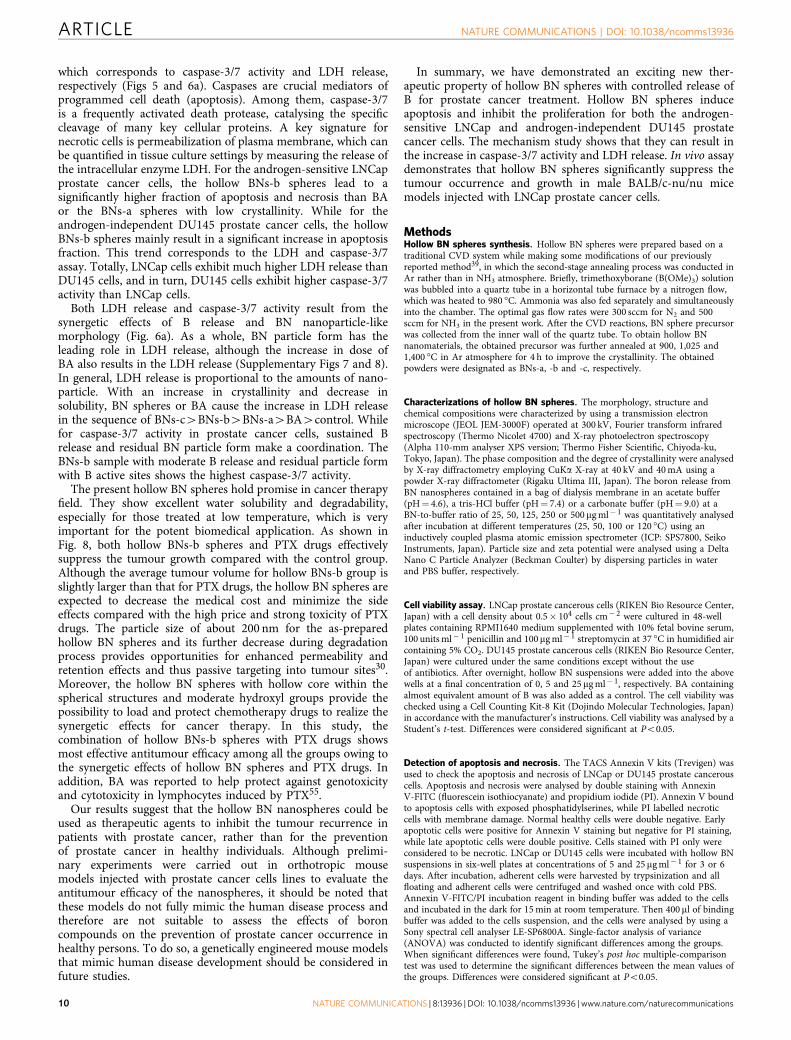
which corresponds to caspase-3/7 activity and LDH release,respectively (Figs 5 and 6a). Caspases are crucial mediators ofprogrammed cell death (apoptosis). Among them, caspase-3/7is a frequently activated death protease, catalysing the specificcleavage of many key cellular proteins. A key signature fornecrotic cells is permeabilization of plasma membrane, which canbe quantified in tissue culture settings by measuring the release ofthe intracellular enzyme LDH. For the androgen-sensitive LNCapprostate cancer cells, the hollow BNs-b spheres lead to asignificantly higher fraction of apoptosis and necrosis than BAor the BNs-a spheres with low crystallinity. While for theandrogen-independent DU145 prostate cancer cells, the hollowBNs-b spheres mainly result in a significant increase in apoptosisfraction. This trend corresponds to the LDH and caspase-3/7assay. Totally, LNCap cells exhibit much higher LDH release thanDU145 cells, and in turn, DU145 cells exhibit higher caspase-3/7activity than LNCap cells.
Both LDH release and caspase-3/7 activity result from thesynergetic effects of B release and BN nanoparticle-likemorphology (Fig. 6a). As a whole, BN particle form has theleading role in LDH release, although the increase in dose ofBA also results in the LDH release (Supplementary Figs 7 and 8).In general, LDH release is proportional to the amounts of nano-particle. With an increase in crystallinity and decrease insolubility, BN spheres or BA cause the increase in LDH releasein the sequence of BNs-c4BNs-b4BNs-a4BA4control. Whilefor caspase-3/7 activity in prostate cancer cells, sustained Brelease and residual BN particle form make a coordination. TheBNs-b sample with moderate B release and residual particle formwith B active sites shows the highest caspase-3/7 activity.
The present hollow BN spheres hold promise in cancer therapyfield. They show excellent water solubility and degradability,especially for those treated at low temperature, which is veryimportant for the potent biomedical application. As shown inFig. 8, both hollow BNs-b spheres and PTX drugs effectivelysuppress the tumour growth compared with the control group.Although the average tumour volume for hollow BNs-b group isslightly larger than that for PTX drugs, the hollow BN spheres areexpected to decrease the medical cost and minimize the sideeffects compared with the high price and strong toxicity of PTXdrugs. The particle size of about 200 nm for the as-preparedhollow BN spheres and its further decrease during degradationprocess provides opportunities for enhanced permeability andretention effects and thus passive targeting into tumour sites30.Moreover, the hollow BN spheres with hollow core within thespherical structures and moderate hydroxyl groups provide thepossibility to load and protect chemotherapy drugs to realize thesynergetic effects for cancer therapy. In this study, thecombination of hollow BNs-b spheres with PTX drugs showsmost effective antitumour efficacy among all the groups owing tothe synergetic effects of hollow BN spheres and PTX drugs. Inaddition, BA was reported to help protect against genotoxicityand cytotoxicity in lymphocytes induced by PTX55.
Our results suggest that the hollow BN nanospheres could beused as therapeutic agents to inhibit the tumour recurrence inpatients with prostate cancer, rather than for the preventionof prostate cancer in healthy individuals. Although prelimi-nary experiments were carried out in orthotropic mousemodels injected with prostate cancer cells lines to evaluate theantitumour efficacy of the nanospheres, it should be noted thatthese models do not fully mimic the human disease process andtherefore are not suitable to assess the effects of boroncompounds on the prevention of prostate cancer occurrence inhealthy persons. To do so, a genetically engineered mouse modelsthat mimic human disease development should be considered infuture studies.
In summary, we have demonstrated an exciting new ther-apeutic property of hollow BN spheres with controlled release ofB for prostate cancer treatment. Hollow BN spheres induceapoptosis and inhibit the proliferation for both the androgen-sensitive LNCap and androgen-independent DU145 prostatecancer cells. The mechanism study shows that they can result inthe increase in caspase-3/7 activity and LDH release. In vivo assaydemonstrates that hollow BN spheres significantly suppress thetumour occurrence and growth in male BALB/c-nu/nu micemodels injected with LNCap prostate cancer cells.
MethodsHollow BN spheres synthesis. Hollow BN spheres were prepared based on atraditional CVD system while making some modifications of our previouslyreported method39, in which the second-stage annealing process was conducted inAr rather than in NH3 atmosphere. Briefly, trimethoxyborane (B(OMe)3) solutionwas bubbled into a quartz tube in a horizontal tube furnace by a nitrogen flow,which was heated to 980 �C. Ammonia was also fed separately and simultaneouslyinto the chamber. The optimal gas flow rates were 300 sccm for N2 and 500sccm for NH3 in the present work. After the CVD reactions, BN sphere precursorwas collected from the inner wall of the quartz tube. To obtain hollow BNnanomaterials, the obtained precursor was further annealed at 900, 1,025 and1,400 �C in Ar atmosphere for 4 h to improve the crystallinity. The obtainedpowders were designated as BNs-a, -b and -c, respectively.
Characterizations of hollow BN spheres. The morphology, structure andchemical compositions were characterized by using a transmission electronmicroscope (JEOL JEM-3000F) operated at 300 kV, Fourier transform infraredspectroscopy (Thermo Nicolet 4700) and X-ray photoelectron spectroscopy(Alpha 110-mm analyser XPS version; Thermo Fisher Scientific, Chiyoda-ku,Tokyo, Japan). The phase composition and the degree of crystallinity were analysedby X-ray diffractometry employing CuKa X-ray at 40 kV and 40 mA using apowder X-ray diffractometer (Rigaku Ultima III, Japan). The boron release fromBN nanospheres contained in a bag of dialysis membrane in an acetate buffer(pH¼ 4.6), a tris-HCl buffer (pH¼ 7.4) or a carbonate buffer (pH¼ 9.0) at aBN-to-buffer ratio of 25, 50, 125, 250 or 500mg ml� 1 was quantitatively analysedafter incubation at different temperatures (25, 50, 100 or 120 �C) using aninductively coupled plasma atomic emission spectrometer (ICP: SPS7800, SeikoInstruments, Japan). Particle size and zeta potential were analysed using a DeltaNano C Particle Analyzer (Beckman Coulter) by dispersing particles in waterand PBS buffer, respectively.
Cell viability assay. LNCap prostate cancerous cells (RIKEN Bio Resource Center,Japan) with a cell density about 0.5� 104 cells cm� 2 were cultured in 48-wellplates containing RPMI1640 medium supplemented with 10% fetal bovine serum,100 units ml� 1 penicillin and 100mg ml� 1 streptomycin at 37 �C in humidified aircontaining 5% CO2. DU145 prostate cancerous cells (RIKEN Bio Resource Center,Japan) were cultured under the same conditions except without the useof antibiotics. After overnight, hollow BN suspensions were added into the abovewells at a final concentration of 0, 5 and 25 mg ml� 1, respectively. BA containingalmost equivalent amount of B was also added as a control. The cell viability waschecked using a Cell Counting Kit-8 Kit (Dojindo Molecular Technologies, Japan)in accordance with the manufacturer’s instructions. Cell viability was analysed by aStudent’s t-test. Differences were considered significant at Po0.05.
Detection of apoptosis and necrosis. The TACS Annexin V kits (Trevigen) wasused to check the apoptosis and necrosis of LNCap or DU145 prostate cancerouscells. Apoptosis and necrosis were analysed by double staining with AnnexinV-FITC (fluorescein isothiocyanate) and propidium iodide (PI). Annexin V boundto apoptosis cells with exposed phosphatidylserines, while PI labelled necroticcells with membrane damage. Normal healthy cells were double negative. Earlyapoptotic cells were positive for Annexin V staining but negative for PI staining,while late apoptotic cells were double positive. Cells stained with PI only wereconsidered to be necrotic. LNCap or DU145 cells were incubated with hollow BNsuspensions in six-well plates at concentrations of 5 and 25 mg ml� 1 for 3 or 6days. After incubation, adherent cells were harvested by trypsinization and allfloating and adherent cells were centrifuged and washed once with cold PBS.Annexin V-FITC/PI incubation reagent in binding buffer was added to the cellsand incubated in the dark for 15 min at room temperature. Then 400 ml of bindingbuffer was added to the cells suspension, and the cells were analysed by using aSony spectral cell analyser LE-SP6800A. Single-factor analysis of variance(ANOVA) was conducted to identify significant differences among the groups.When significant differences were found, Tukey’s post hoc multiple-comparisontest was used to determine the significant differences between the mean values ofthe groups. Differences were considered significant at Po0.05.
ARTICLE NATURE COMMUNICATIONS | DOI: 10.1038/ncomms13936
10 NATURE COMMUNICATIONS | 8:13936 | DOI: 10.1038/ncomms13936 | www.nature.com/naturecommunications

Caspase-Glo 3/7 assay. Caspase-3/7 activities were measured in cells treated withhollow BN nanospheres using the Caspase-Glo 3/7 Assay Kits (Promega) accordingto the manufacturer’s protocol. Briefly, 100 ml LNCap cells (5� 104 cells cm� 2)were seeded into each well of a 96-well culture plate and incubated at 37 �Covernight. Then 100ml of hollow BN suspensions were added to the well to get thefinal concentration of 5 mg ml� 1 of BN or BA containing almost equivalent Bfollowed by incubation for 16 h. Single-factor ANOVA was conducted to identifysignificant differences among the groups. When significant differences were found,Tukey’s post hoc multiple-comparison test was used to determine the significantdifferences between the mean values of the groups. Differences were consideredsignificant at Po0.05.
LDH assay. LDH cytotoxicity caused by hollow BN spheres was measuredby a detection kit (Takara Bio Inc. Japan). Briefly, 100 ml LNCap cells(5� 104 cells cm� 2) were seeded into each well of a 96-well culture plate andincubated at 37 �C overnight. Then 100 ml of hollow BN suspensions at variousconcentrations were added to the well to get the final concentrations of 1, 5 and25mg ml� 1 of BN or BA containing almost equivalent B followed by incubation for16 h. The microplate was centrifuged to get the supernatant. LDH cytotoxicity waschecked by using the above supernatant with a reaction time about 12 min inaccordance with the manufacturer’s instructions. The Triton X-100 (1%) was usedas a positive control in the assay. Single-factor ANOVA was conducted to identifysignificant differences among the groups. When significant differences were found,Tukey’s post hoc multiple-comparison test was used to determine the significantdifferences between the mean values of the groups. Differences were consideredsignificant at Po0.05.
In vivo antitumour experiment. Male BALB/c-nu/nu mice (Charles river, Japan)were used in this study. Human LNCap prostate cancer cells were collected fromexponentially growing cultures with 90% confluent.
For subcutaneous tumour growth models, 20 mice were divided randomly into5 groups. Collected LNCap cells were mixed with Matrigel 1:1 by volume and kepton ice until use. A total amount of 8� 106 LNCap cells were injectedsubcutaneously into the left flank of the mice. Four days later, mice wereadministered subcutaneously approximately 0.5–1 cm apart from the tumour sitewith saline, hollow BN suspension in saline (BNs-a, BNs-b, BNs-c, 30 mg,respectively) or BA containing almost equivalent B in saline, once in 4 days for theinitial 3 times and once in 7 days afterwards before killing. Tumour diameter wasmeasured using calipers at a certain time interval. Once the tumour diameterreached 2 cm, the experiment was discontinued to limit the tumour burden inmouse. Percentage of mice without development of tumours was analysed by aKaplan–Meier log-rank test. Tumour volume was analysed by Student’s t-test.Differences were considered significant at Po0.05.
For orthotopical tumour growth models, 12 mice were divided randomly into 3groups. A transverse incision was made in the lower abdomen. The bladder andseminal vesicles were delivered through the incision to expose the prostate. Whenthe prostate was identified, a total amount of 2� 106 cells suspended in 20ml ofHEPES buffer were carefully injected under the prostatic capsule. The incision wasclosed using a suture of 4-0 silk. Mice were administered intravenously with PEG-saline (1 mg ml� 1), hollow BNs-b (30 mg) suspension in PEG-saline or BAcontaining almost equivalent B in PEG-saline, once in 1 day for the initial 3 timesand once in 4 days for the subsequent 6 times. Then, 12 weeks later, body weightswere measured and a transverse incision was made in the lower abdomen tomonitor the tumour size. Tumour volume and body weights were analysed by aStudent’s t-test. Differences were considered significant at Po0.05.
In addition, orthotopical tumour growth models were also used to check andcompare the treatment efficacy of hollow BN spheres, clinically used chemotherapydrug (PTX) and the PTX–hollow BN spheres complex. The saline group was usedas a control. Sixteen mice were divided randomly into four groups. A total amountof 5� 106 cells suspended in 30ml of HEPES buffer were carefully injected underthe prostatic capsule. Mice were administered intravenously with saline, hollowBNs-b (30 mg) suspension in PEG-saline, PTX (15mg) in saline or the PTX–hollowBN spheres complex in PEG-saline, once in 1 day for the initial 3 times and once in3 days for the subsequent 3 times. Then, 8 weeks later, a transverse incision wasmade in the lower abdomen to monitor the tumour size. Tumour volume wasanalysed by a Student’s t-test. Differences were considered significant at Po0.05.
In vivo safety and biodistribution. To examine the in vivo safety, six C57/BL6mice were divided randomly into two groups and administered BNs-b (50 mg inPEG-saline) or saline intravenously via tail-vein injection. The acute toxicology,such as blood biochemistry and haematology analysis, was measured 3 days later.The results were analysed by a Student’s t-test. Differences were considered sig-nificant at Po0.05. In addition, 12 C57/BL6 mice were divided randomly into 4groups and administered BNs-b (50 mg in PEG-saline) intravenously via tail-veininjection. At several fixed time points (1 h, 1 day, 3 days, 15 days), the tissues, suchas heart, liver, spleen, lung, kidney and stomach, were discretized. The tissuedistribution of BNs-b particles was semiquantified by hydrothermal digestion in1 M NaOH solution. The double-blind experiments were carried out.
All the animal experiments were permitted by the Ethical Committee of theNational Institute for Materials Science (NIMS), Japan. All the animal experimentsand feeding were carried out in accordance with the guidelines of the EthicalCommittee of NIMS, Japan.
Data availability. The data that support the findings of this study are availablefrom the authors upon reasonable request.
References1. Ito, K. Prostate cancer in Asian men. Nat. Rev. Urol. 11, 197–212 (2014).2. Thompson, I. M., Cabang, A. B. & Wargovich, M. J. Future directions in the
prevention of prostate cancer. Nat. Rev. Clin. Oncol. 11, 49–60 (2014).3. Klein, E. A. Can prostate cancer be prevented? Nat. Clin. Pract. Urol. 2, 24–31
(2005).4. Guns, E. S. & Cowell, S. P. Drug insight: lycopene in the prevention and
treatment of prostate cancer. Nat. Clin. Pract. Urol. 2, 38–43 (2005).5. Tanaka, H. et al. Monoclonal antibody targeting of N-cadherin inhibits prostate
cancer growth, metastasis and castration resistance. Nat. Med. 16, 1414–1420(2010).
6. Ravindranathan, P. et al. Peptidomimetic targeting of critical androgenreceptor-coregulator interactions in prostate cancer. Nat. Commun. 4, 1923(2013).
7. Klotz, L. & Emberton, M. Management of low risk prostate cancer-activesurveillance and focal therapy. Nat. Rev. Clin. Oncol. 11, 324–334 (2014).
8. Mohiuddin, J. J., Baker, B. R. & Chen, R. C. Radiotherapy for high-risk prostatecancer. Nat. Rev. Urol. 12, 145–154 (2015).
9. Wong, Y. N., Ferraldeschi, R., Attard, G. & de Bono, J. Evolution of androgenreceptor targeted therapy for advanced prostate cancer. Nat. Rev. Clin. Oncol.11, 365–376 (2014).
10. Yasufuku, T., Shigemura, K., Matsumoto, O., Arakawa, S. & Fujisawa, M.Combination chemotherapy with weekly paclitaxel or docetaxel, carboplatin,and estramustine for hormone-refractory prostate cancer. J. Infect. Chemother.16, 200–205 (2010).
11. Zhang, Z. F., Winton, M. I., Rainey, C. & Eckhert, C. D. Boron is associatedwith decreased risk of human prostate cancer. FASEB. J. 15, A1089 (2001).
12. Barranco, W. T., Hudak, P. F. & Eckhert, C. D. Evaluation of ecological andin vitro effects of boron on prostate cancer risk. Cancer Causes Control 18,71–77 (2007).
13. Cui, Y. et al. Dietary boron intake and prostate cancer risk. Oncol. Rep. 11,887–892 (2004).
14. Barranco, W. T. & Eckhert, C. D. Boric acid inhibits human prostate cancer cellproliferation. Cancer Lett. 216, 21–29 (2004).
15. Gallardo-Williams, M. T. et al. Boron supplementation inhibits the growth andlocal expression of IGF-1 in human prostate adenocarcinoma (LNCaP) tumorsin nude mice. Toxicol. Pathol. 32, 73–78 (2004).
16. Gallardo-Williams, M. T., Maronpot, R. R., Wine, R. N., Brunssen, S. H. &Chapin, R. E. Inhibition of the enzymatic activity of prostate-specific antigen byboric acid and 3-nitrophenyl boronic acid. Prostate 54, 44–49 (2003).
17. Scorei, R. I. & Popa, R. Boron-containing compounds as preventive andchemotherapeutic agents for cancer. Anticancer Agents Med. Chem. 10,346–351 (2010).
18. Kim, D. H., Marbois, B. N., Faull, K. F. & Eckhert, C. D. Esterification of boratewith NADþ and NADH as studied by electrospray ionization massspectrometry and 11B NMR spectroscopy. J. Mass. Spectrom. 38, 632–640(2003).
19. Henderson, K., Stella, S. L., Kobylewski, S. & Eckhert, C. D. Receptor activatedCa2þ release is inhibited by boric acid in prostate cancer cells. PLoS ONE 4,e6009 (2009).
20. Cui, Y. et al. Dietary boron intake and reduced risk of prostate cancer. Am.J. Epidemiol. 155, s56–s56 (2002).
21. Bradke, T. M., Hall, C., Carper, S. W. & Plopper, G. E. Phenylboronic acidselectively inhibits human prostate and breast cancer cell migration anddecreases viability. Cell Adh. Migr. 2, 153–160 (2008).
22. Barranco, W. T. & Eckhert, C. D. Cellular changes in boric acid-treated DU-145prostate cancer cells. Br. J. Cancer 94, 884–890 (2006).
23. Barranco, W. T., Kim, D. H., Stella, Jr S. L. & Eckhert, C. D. Boric acid inhibitsstored Ca2þ release in DU-145 prostate cancer cells. Cell Biol. Toxicol. 25,309–320 (2009).
24. Korkmaz, M., Uzgoren, E., Bakirdere, S., Aydin, F. & Ataman, O. Y. Effects ofdietary boron on cervical cytopathology and on micronucleus frequency inexfoliated buccal cells. Environ. Toxicol. 22, 17–25 (2007).
25. Scorei, R. I. & Popa, R. Sugar-borate esters--potential chemical agents inprostate cancer chemoprevention. Anticancer Agents Med. Chem. 13, 901–909(2013).
26. Das, B. C. et al. Boron chemicals in diagnosis and therapeutics. Future Med.Chem. 5, 653–676 (2013).
27. Baker, S. J. et al. Therapeutic potential of boron-containing compounds. FutureMed. Chem. 1, 1275–1288 (2009).
NATURE COMMUNICATIONS | DOI: 10.1038/ncomms13936 ARTICLE
NATURE COMMUNICATIONS | 8:13936 | DOI: 10.1038/ncomms13936 | www.nature.com/naturecommunications 11

28. Goldberg, E. P., Hadba, A. R., Almond, B. A. & Marotta, J. S. Intratumoralcancer chemotherapy and immunotherapy: opportunities for nonsystemicpreoperative drug delivery. J. Pharm. Pharmacol. 54, 159–180 (2002).
29. Seto, T. et al. Intrapleural hypotonic cisplatin treatment for malignant pleuraleffusion in 80 patients with non-small-cell lung cancer: a multi-institutionalphase II trial. Br. J. Cancer 95, 717–721 (2006).
30. Barreto, J. A. et al. Nanomaterials: applications in cancer imaging and therapy.Adv. Mater. 23, H18–H40 (2011).
31. Li, X. et al. Multimodal luminescent-magnetic boron nitride nanotubes@NaGdF4:Eustructures for cancer therapy. Chem. Commun. 50, 4371–4374 (2014).
32. Li, X. et al. Boron nitride nanotubes functionalized with mesoporous silica forintracellular delivery of chemotherapy drugs. Chem. Commun. 49, 7337–7339(2013).
33. Weng, Q. et al. Highly water-soluble, porous, and biocompatible boron nitridesfor anticancer drug delivery. ACS Nano 8, 6123–6130 (2014).
34. Liu, Z. et al. In vivo biodistribution and highly efficient tumour targeting ofcarbon nanotubes in mice. Nat. Nanotechnol. 2, 47–52 (2007).
35. Yang, K. et al. Multimodal imaging guided photothermal therapy usingfunctionalized graphene nanosheets anchored with magnetic nanoparticles.Adv. Mater. 24, 1868–1872 (2012).
36. Li, X. et al. Mesoporous silica-calcium phosphate-tuberculin purified proteinderivative composites as an effective adjuvant for cancer immunotherapy. Adv.Healthc. Mater. 2, 863–871 (2013).
37. Wang, X. P., Ito, A., Li, X., Sogo, Y. & Oyane, A. Signal molecules-calciumphosphate coprecipitation and its biomedical application as a functionalcoating. Biofabrication 3, 022001 (2011).
38. Park, J. et al. Combination delivery of TGF-beta inhibitor and IL-2 bynanoscale liposomal polymeric gels enhances tumour immunotherapy. Nat.Mater. 11, 895–905 (2012).
39. Tang, C. C., Bando, Y., Huang, Y., Zhi, C. Y. & Golberg, D. Synthetic routes andformation mechanisms of spherical boron nitride nanoparticles. Adv. Funct.Mater. 18, 3653–3661 (2008).
40. Chen, Y., Chen, H. R. & Shi, J. L. In vivo bio-safety evaluations and diagnostic/therapeutic applications of chemically designed mesoporous silicananoparticles. Adv. Mater. 25, 3144–3176 (2013).
41. Wang, X. P. et al. Zn- and Mg- containing tricalcium phosphates-basedadjuvants for cancer immunotherapy. Sci. Rep. 3, 2203 (2013).
42. Ghosh, D. et al. M13-templated magnetic nanoparticles for targeted in vivoimaging of prostate cancer. Nat. Nanotechnol. 7, 677–682 (2012).
43. Cho, H. S. et al. Fluorescent, superparamagnetic nanospheres for drug storage,targeting, and imaging: a multifunctional nanocarrier system for cancerdiagnosis and treatment. ACS Nano 4, 5398–5404 (2010).
44. Yang, H. W. et al. Cooperative dual-activity targeted nanomedicine for specificand effective prostate cancer therapy. ACS Nano 6, 1795–1805 (2012).
45. Lahiri, D. et al. Boron nitride nanotube reinforced polylactide-polycaprolactonecopolymer composite: mechanical properties and cytocompatibility withosteoblasts and macrophages in vitro. Acta Biomater. 6, 3524–3533 (2010).
46. Ciofani, G. et al. Boron nitride nanotubes: a novel vector for targeted magneticdrug delivery. Curr. Nanosci. 5, 33–38 (2009).
47. Buzatu, D. A. et al. Nanotubes for cancer therapy and diagnostics, US patent7608240 (2009).
48. Raffa, V. et al. BNNT-mediated irreversible electroporation: Its potential oncancer cells. Technol. Cancer Res. Treat. 11, 459–465 (2012).
49. Coderre, J. A. & Morris, G. M. The radiation biology of boron neutron capturetherapy. Radiat. Res. 151, 1–18 (1999).
50. Petersen, M. S. et al. Boron nanoparticles inhibit tumour growth by boronneutron capture therapy in the murine B16-OVA model. Anticancer Res. 28,571–576 (2008).
51. Yinghuai, Z. et al. Substituted carborane-appended water-soluble single-wallcarbon nanotubes: new approach to boron neutron capture therapy drugdelivery. J. Am. Chem. Soc. 127, 9875–9880 (2005).
52. Cao, F., Liu, K., Fang, Z. Y. & Wang, S. Q. Hydrolysis mechanism of borazine-derived boron nitride pyrolysized below 1,200 oC. J. Mater. Sci. Technol. 28,956–960 (2012).
53. Bone, R., Shenvi, A. B., Kettner, C. A. & Agard, D. A. Serine proteasemechanism: structure of an inhibitory complex of a-lytic protease and a tightlybound peptide boronic acid. Biochemistry 26, 7609–7614 (1987).
54. Moseman, R. F. Chemical disposition of boron in animals and humans.Environ. Health Perspect. 102, 113–117 (1994).
55. Turkez, H., Tatar, A., Hacimuftuoglu, A. & Ozdemir, E. Boric acid as aprotector against paclitaxel genotoxicity. Acta Biochim. Pol. 57, 95–97 (2010).
AcknowledgementsWe thank Dr X.L. Li, Dr H. Morita, Dr Y. Shirai, Dr X.D. Wang, Dr R. Nagano,Dr K. Iiyama, Dr M. Mitome and Dr N. Kawamoto for their generous technicalassistance. J.Z. thanks the support from the National Natural Science Foundationof China (21103056) and the Natural Science Foundation of Hebei Province(B2016202213). This work was funded by the World Premier International Center forMaterials Nanoarchitectonics (WPI-MANA) of the National Institute for MaterialsScience (NIMS), Tsukuba, Japan.
Author contributionsX.L. designed the concept of BN with controlled B release for prostate cancer and carriedout most of the experiments. J.Z. carried out BN synthesis. X.P.W. and A.I. supported theanimal model and ICP measurements. N.H. supported in vitro experiments. X.B.Wcarried out the XPS measurements. All the authors discussed the results. X.L. analysed allthe data and wrote the manuscript. D.G. and A.I revised the manuscript. D.G. and Y.B.oversaw the whole project.
Additional informationSupplementary Information accompanies this paper at http://www.nature.com/naturecommunications
Competing financial interests: The authors declare no competing financial interests.
Reprints and permission information is available online at http://npg.nature.com/reprintsandpermissions/
How to cite this article: Li, X. et al. Hollow boron nitride nanospheres as boron reservoirfor prostate cancer treatment. Nat. Commun. 8:13936 doi: 10.1038/ncomms13936(2017).
Publisher’s note: Springer Nature remains neutral with regard to jurisdictional claims inpublished maps and institutional affiliations.
This work is licensed under a Creative Commons Attribution 4.0International License. The images or other third party material in this
article are included in the article’s Creative Commons license, unless indicated otherwisein the credit line; if the material is not included under the Creative Commons license,users will need to obtain permission from the license holder to reproduce the material.To view a copy of this license, visit http://creativecommons.org/licenses/by/4.0/
r The Author(s) 2017
ARTICLE NATURE COMMUNICATIONS | DOI: 10.1038/ncomms13936
12 NATURE COMMUNICATIONS | 8:13936 | DOI: 10.1038/ncomms13936 | www.nature.com/naturecommunications