HNHA (N-hydroxy-7-(2-naphthylthio) heptanomide), … · N-hydroxy-7-(2-naphthylthio) heptanomide...
40
N-hydroxy-7-(2-naphthylthio) heptanomide (HNHA), Histone Deacetylase Inhibitors, Suppresses COX-2 expression in colon cancer Ji Hyun Park Department of Medical Science The Graduate School, Yonsei University
-
Upload
nguyendien -
Category
Documents
-
view
219 -
download
0
Transcript of HNHA (N-hydroxy-7-(2-naphthylthio) heptanomide), … · N-hydroxy-7-(2-naphthylthio) heptanomide...

N-hydroxy-7-(2-naphthylthio)
heptanomide (HNHA), Histone
Deacetylase Inhibitors, Suppresses
COX-2 expression in colon cancer
Ji Hyun Park
Department of Medical Science
The Graduate School, Yonsei University
N-hydroxy-7-(2-naphthylthio)
heptanomide (HNHA), Histone
Deacetylase Inhibitors, Suppresses
COX-2 expression in colon cancer
Directed by Professor Jin Suck Suh
The Master’s Thesis
Submitted to the Department of Medical Science,
the Graduate School of Yonsei University
in partial fulfillment of the requirements for the
degree of Master of Medical Science
Ji Hyun Park
June 2009
This certifies that the Master’s Thesis
of Ji Hyun Park is approved.
________________________________________
Thesis Supervisor : Jin Suck Suh
________________________________________
Thesis Committee Member : Seung Hoon Choi
________________________________________
Thesis Committee Member : Ho Guen Kim
The Graduate School
Yonsei University
June 2009

Acknowledgments
First of all, I am deeply grateful to Professor Jin Suk Suh,
Seung Hun Choi, Ho Guen Kim for their supports to complete
this thesis.
And I want to thank Ki Chong Park and Jeong Won Yang
for their supports during my Master’s course. Also I am really
appreciate that Eun Hye Song, So Jung Yoon, Hyo Jin Kang, and
Mei Hua Jin, who gave me great helps even though not belong to
same lab each other.
Also, I want to give special thanks to my parents and my
little brother (Eun Suk Park), whose constant love can make me
always happy and keep going my works. I always think and
remember that their huge supports for me.
Finally, I would like to express my gratitude to all who
gave me the possibility to complete this thesis.
JUNE 2009

Table of contents
Abstract ················································································1
I. INTRODUCTION ·································································3
II. MATERIALS AND METHODS
1. Cell Culture···································································7
2. Tumor Animal Model and Experimental Designs························7
3. Immunohistochemistry for COX-2, HIF-1α······························8
4. Western Blot analysis for COX-2 and TNF- α···························8
5. Cell cycle analysis by flow cytometry·····································9
III. RESULTS
1. Effect of drug HNHA on C1300··········································12
2. Effect of drug HNHA on HT-29··········································14
3. Cell cycle analysis··························································16
4. Expression of COX-2······················································17
5. Expression of HIF-1α······················································19
6. Expression of TNF-α·······················································21
IV. DISCUSSION ···································································22

V. CONCLUSION ··································································25
REFERENCES ······································································26
ABSTRACT (IN KOREAN) ······················································30

List of Figures
Figure 1. Chemical structure of HDAC inhibitors····························6
Figure 2. Size of HT-29 tumor and Survival rate····························11
Figure 3. Effect of drug HNHA on C1300·······································12
Figure 4. Effect of drug HNHA on HT-29········································14
Figure 5. Cell cycle analysis·······························································16
Figure 6. Expression of COX-2··························································17
Figure 7. Expression of HIF-1α··························································19
Figure 8. Detection of TNF-α·····························································21
Figure 9. Proposed mechanism of cancer suppression by
HNHA···················································································23

List of Tables
Table 1. IC50 concentration of HNHA and SAHA·······················11
Abstract
N-hydroxy-7-(2-naphthylthio) heptanomide (HNHA), Histone
Deacetylase Inhibitors, Suppresses COX-2 expression in colon cancer
Ji Hyun Park
Department of Medical Science
The Graduate School, Yonsei University
(Directed by Professor Jin Suck Suh)
Cyclooxygenase-2 (COX-2) plays important roles in cell adhesion,
apoptosis, and angiogenesis, and is essential for tumor angiogenesis. Levels of
COX-2 expression are low in normal cells, but very high level in human and animal
colorectal tumors.
Histone deacetylase (HDAC) plays a crucial role in carcinogenesis1 and is
over-expressed in several tumor cells2. The histone deacetylase inhibitor N-
hydroxy-7-(2-naphthylthio) heptanomide (HNHA) is a new anti-angiogenesis drug,
and has been shown to inhibit COX-2 expression in colon cancer. Here, we
confirm that HNHA decreases COX-2 expression in colon cancer, similar to COX-2
inhibitors such as Celecoxib and Rofecoxib. Moreover, hypoxia inducible factor-
1α (HIF-1α), and tumor necrosis factor-α (TNF-α) are involved in COX-2
expression and their expression is also decreased by HNHA in colon cancer. The
effect of HNHA on tumor growth was examined in a human colon cancer tumor
model and investigated by western blot, immunohistochemistry, and FACS analysis.
Key Words: Histone deacetylase (HDAC), N-hydroxy-7-(2-naphthylthio)
heptanomide (HNHA), (Hypoxia-inducible factor-1α (HIF-1α), Cyclooxygenase-2
(COX-2)

N-hydroxy-7-(2-naphthylthio) heptanomide (HNHA), Histone
Deacetylase Inhibitors, Suppresses COX-2 expression in colon cancer
Ji Hyun Park
Department of Medical Science
The Graduate School, Yonsei University
(Directed by Professor Jin Suck Suh)
I. INTRODUCTION
Histone acetylation is regulated by acetylase and deacetylase and plays a
key role in gene expression in eukaryotes. Recent studies have shown that histone
deacetylase (HDAC) plays a crucial role in carcinogenesis1 and is over-expressed in
several tumor cells2. A new HDAC inhibitor N-hydroxy-7-(2-naphthylthio)
heptanomide (HNHA) has anti-angiogenic activity3. HNHA suppresses the hypo-
acetylation of histones, down-regulating the target gene and inhibiting invasion of
the cancer cells and tumor growth by repressing enzyme activity3. Thus, if HDAC
inhibitors strongly repress cancer cells, they could be used to repress cancer
metastasis or growth. Cyclooxygenase (COX) is an enzyme that catalyzes the
formation of prostaglandins from arachidonic acid4. There are two COX enzyme
isoforms: cyclooxygenase-1 (COX-1) is constitutively expressed in normal tissues
as a ―housekeeper‖ of mucosal integrity5, whereas cyclooxygenase-2 (COX-2) is not
normally expressed in most tissues, but is induced by a wide spectrum of growth
factors and pro-inflammatory cytokines in specific pathophysiological conditions6.
COX-2 has important functions in cell adhesion and apoptosis, and is essential for
tumor angiogenesis. If angiogenesis is blocked, the tumor cannot survive. The
level of COX-2 expression is low in normal cells, but is very high in human and
animal colorectal tumors.
COX-2 is a highly inducible by pro-inflammatory cytokines such as TNF-
α18
, which in turn is regulated by NF-κB. HDAC inhibitor blocks TNF-α
activation of COX-2 protein and mRNA synthesis, and suppresses COX-2 activity7.
TNF-α is involved in promotion and progression of experimental and human cancer
through intracellular signaling pathways leading to activation of the NF-κB and AP-
1 transcription factor complexes17
. Moreover, COX-2 up-regulation is associated
with induction of hypoxia-inducible factor-1α (HIF-1α)8 through binding of HIF-1α
to a hypoxia-responsive element (HRE) on the COX-2 promoter. COX-2 up-
regulation during hypoxia is accompanied by increased levels of prostaglandin E2
(PGE2), which promote tumor cell survival under hypoxic conditions8.
Hypoxia, oxygen deficiency in tissues, is a universal characteristic of solid
tumors and represents a key regulatory factor in tumor growth and survival9. The
coordinated homeostatic response to hypoxia is largely transcriptional and is
mediated primarily through activation of the heterodimeric transcription factor
hypoxia-inducible factor (HIF)-110
. HIF-1 is composed of two subunits: the
oxygen sensitive HIF-1α subunit and the constitutively expressed HIF-1β subunit10
.
Under normoxic conditions, HIF-1α is hydroxylated at key proline residues
facilitating von Hippel-Lindau protein binding, which in turn allows ubiquitination
and subsequent proteasome-targeted degradation11
. Under hypoxic conditions,
proline hydroxylation is inhibited, thereby stabilizing HIF-1α, which can translocate
into the nucleus and bind to constitutively expressed HIF-1β, forming the active
HIF-1 complex12
. HIF-1α is over-expressed in various types of cancer, including
colorectal cancer13
, and compelling evidence supports a role for HIF-1 in
tumorigenesis14, 15
. HIF-1α levels are elevated in the cytoplasm and the nucleus of
cells in various solid tumors16
. Moreover, under hypoxic conditions COX-2
protein levels increase in colorectal adenoma and carcinoma cells, indicating that
COX-2 up-regulation is associated with HIF-1α induction. Therefore, if HNHA
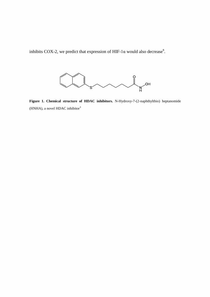
inhibits COX-2, we predict that expression of HIF-1α would also decrease8.
Figure 1. Chemical structure of HDAC inhibitors. N-Hydroxy-7-(2-naphthylthio) heptanomide
(HNHA), a novel HDAC inhibitor3
II. MATERIALS AND METHODS
1. Cell Culture
HT-29 is a human colorectal carcinoma cell line with a doubling time of
20-24 hours. C1300 cell is a mouse neuroblastoma cell line with a doubling time
of 30-33 hours. These two cell lines were cultured in RPMI 1640 medium
supplemented with 10% fetal bovine serum (FBS), 2 mg/mL sodium bicarbonate,
10% penicillin/streptomycin and 5% CO2 incubator at 37ºC. Drug treatment was
started the day after seeding using an Inhibiting Concentration 50 (IC50) dose.
2. Tumor Animal Model and Experimental Designs
BALB/C nude mice are used as a general-purpose strain in a variety of
research areas including cancer, immunology and inflammation, and cardiovascular
biology research. HT-29 colorectal cancer cells were cultured in vitro, harvested,
and injected (1.0 x 10⁷/mouse) into the dorsal sub-membrane of a 6-week-old mouse.
Drug injection was performed once every 2 days, 10-12 times in total, when the
tumor size reached 0.6 cm x 0.6 cm. Two different drugs were injected at Lethal
Dose 50 (LD50): N-Hydroxy-7-(2-naphthylthio) heptanomide (HNHA, 206 µg/mL),
and suberoylanilide hydroxamic acid (SAHA, 138 µg/mL).
3. Immunohistochemistry for COX-2, HIF-1α
Immunostaining was performed on 5 µm sections after deparaffinization in
xylene and re-hydration in descending alcohols. In the primary antibody reaction,
slides were incubated with monoclonal mouse anti-COX-2 (Santa Cruz
Biotechnology) and monoclonal mouse anti-HIF-1α (Novus) for 1 hour. The
sections were incubated with biotinylated secondary antibodies (1:500) for 10
minutes, washed with PBS, and an avidin–biotin complex (Strept ABComplex;
DAKO, CA) was applied. Finally, the sections were rinsed in PBS, developed
with diaminobenzidine tetrahydrochloride substrate for 3 minutes and
counterstained with hematoxylin.
4. Western blot analysis for COX-2, HIF-1α and TNF- α
The homogenate was sonicated three times for 20 seconds at RT and
centrifuged at 12,000 rpm and -4ºC for 10 minutes. The supernatant was diluted
with electrophoretic sample buffer to a final protein concentration of 5.0µg/µL and
heated at 100ºC for 7 minutes prior to electrophoresis under denaturing conditions
by SDS-PAGE using a discontinuous procedure with 4.5% polyacrylamide stacking
gels and 12% polyacrylamide separating gels. Paired mini-gels (Mini-protein II
cell, Bio-Rad Laboratories, U.S.A) were loaded with 35.0 µg protein per well.
After electrophoresis, one mini-gel was routinely stained by the Coomassie blue
staining method and the other was equilibrated in a transfer buffer (25 mM Tris, 192
mM glycine, 20% methanol (v/v), pH 7.3). The proteins were electrotransferred in
transfer buffer to a PVDF transfer membrane (0.2 µm immunoblot TM PVDF
membrane for protein blot analysis, Bio-Rad) for 1 hour at RT and 90 volts. To
visualize the transferred proteins, the PVDF membrane was stained with ponceau S
(Amresco, Solon, Ohio) for 10 minutes and subsequently incubated in TBS-T (TBS
with 0.05% Tween-20) with 1% skim milk for 1 hour at RT to block non-specific
sites. The blot was rinsed with TBS-T and incubated with primary antibody
overnight at 4ºC, then with horseradish peroxidase conjugated secondary antibody
to IgG for 2 hours at RT. The peroxidase reaction was developed with Amersham
ECL reagents (Amersham Biosciences, USA).
5. Cell cycle analysis by flow cytometry
HT-29 cells were seeded in plates (5 x 101 cells/plate) and incubated for 24
hours prior to treatment with the indicated drugs and further incubation for 24 hours
in RPMI 1640 with 10% FBS. Cells were harvested, centrifuged, and resuspended
in phosphate buffered saline (PBS, pH7.4). RNase (80µg/mL) was added to
reduce background staining and DNA was labeled using propidium iodide
(50µg/mL). DNA histograms were determined using a Beckton-Dickenson FACS
Vantage flow cytometer system (Beckton-Dickenson, San Jose, CA) and the cell
cycle distribution was analyzed using Cell Quest software version 3.2 (Beckton-
Dickenson).

III. RESULT
Table 1. IC50 concentration of HNHA and SAHA. Using CCK-81
1 CCK-8 : Cell counting kit-8
Figure 2. Size of HT-29 tumor and survival rate. A. Size of HT-29 tumor. B. Survival rate.
A, tumor size was calculated by (length*width2)/2. B. Control group was treated with saline ( ),
the drug treatment groups were treated with SAHA or HNHA, and the negative control received no
treatment ( ).
Cell line Tissue derived HNHA IC50 (μM) SAHA IC50 (μM)
HT-29 Human colorectal 16.98 24.91
C1300 Mouse neuroblastoma 55.63 78.22
MCF-7 Human breast 14.32 19.95
A B

Figure 3. Effect of drug treatment on growth of C1300 neuroblastoma cell line. A. Control, B.
SAHA, C. HNHA. C1300 cells were treated with SAHA and HNHA after preculture in vitro. The
same number of cells (5 x 101 cells) was seeded on each plate.
C1300 (mouse neuroblastoma cell line) was treated with HNHA and
SAHA to compare the effects of the new drug and the existing drug. The initial
cell population was the same because the plates were seeded at the same density, but
B
C
A
the cells treated with the new drug, HNHA, grew slowly compared with the control
and SAHA treated-plate; while the cell population increased on control plates
treated with saline, the number of cells on HNHA-treated plates was significantly
decreased. In addition, the morphology of drug-treated cells was also different.
In the control plate, although the cell number increased the cell morphology did not
change between the first and last day (Figure 3. A). In contrast, the morphology of
cells treated with SAHA and HNHA changed during this time; the shape of cells
treated with SAHA differed between the first day and the last day (Figure 3. B), and
HNHA-treated cells showed both a decrease in population and altered morphology
(Figure 3. C).
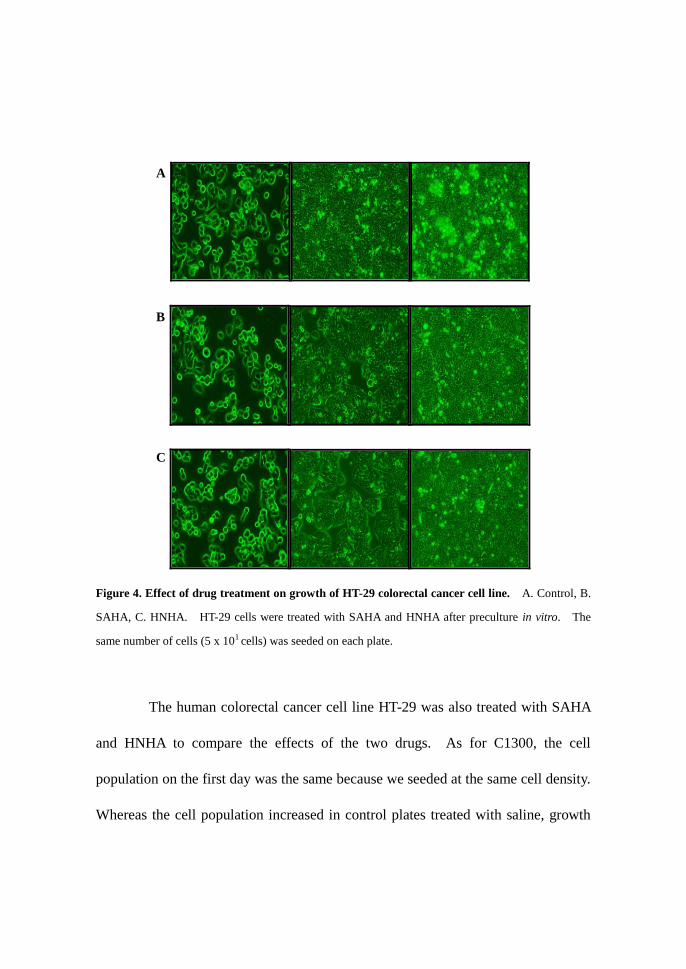
Figure 4. Effect of drug treatment on growth of HT-29 colorectal cancer cell line. A. Control, B.
SAHA, C. HNHA. HT-29 cells were treated with SAHA and HNHA after preculture in vitro. The
same number of cells (5 x 101 cells) was seeded on each plate.
The human colorectal cancer cell line HT-29 was also treated with SAHA
and HNHA to compare the effects of the two drugs. As for C1300, the cell
population on the first day was the same because we seeded at the same cell density.
Whereas the cell population increased in control plates treated with saline, growth
A
B
C

of the cells treated with HNHA was obviously decreased compared with the control
and SAHA treatment. Therefore the new drug, HNHA, exhibited a stronger effect
than SAHA.
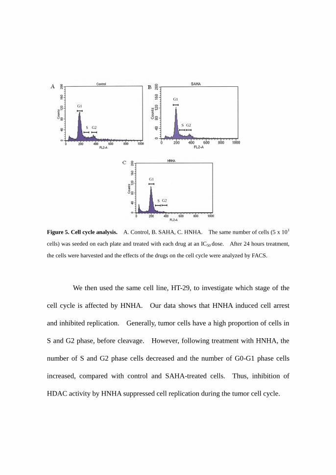
Figure 5. Cell cycle analysis. A. Control, B. SAHA, C. HNHA. The same number of cells (5 x 101
cells) was seeded on each plate and treated with each drug at an IC50 dose. After 24 hours treatment,
the cells were harvested and the effects of the drugs on the cell cycle were analyzed by FACS.
We then used the same cell line, HT-29, to investigate which stage of the
cell cycle is affected by HNHA. Our data shows that HNHA induced cell arrest
and inhibited replication. Generally, tumor cells have a high proportion of cells in
S and G2 phase, before cleavage. However, following treatment with HNHA, the
number of S and G2 phase cells decreased and the number of G0-G1 phase cells
increased, compared with control and SAHA-treated cells. Thus, inhibition of
HDAC activity by HNHA suppressed cell replication during the tumor cell cycle.
I
S
I
G1
I
G2
I
S
I G1
I
G2
I
S
I
G1
I
G2

Figure 6. Expression of COX-2. A. Control, B. SAHA, C. HNHA. Cultured cells (1x10⁷) were
injected into the sub-membrane of BALB/C nude mouse. When the tumor reached an appropriate
size, drugs were injected at the LD50 dose once every 2 days, for a total of 10-12 injections. After
drug treatment, the cancer tissue was removed and the level of COX-2 expression was detected by
immunohistochemistry and western blot.
We next measured the level of COX-2 expression in tumor tissue of nude
mice by immunohistochemistry. COX-2 expression was lower in HNHA and
SAHA treated tissue than in controls, and HNHA showed more effective down-
regulation of COX-2 expression than SAHA. COX-2 was over-expressed in
control cancer tissue (Figure 6. A). COX-2 expression was lower in tissue treated
with SAHA than in control tissue (Figure 6. B), and very low in cancer tissue
A B
`
C D
D
Control
HNHA
SAHA
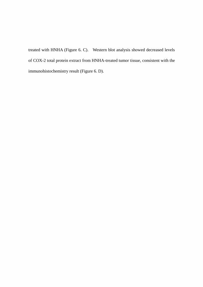
treated with HNHA (Figure 6. C). Western blot analysis showed decreased levels
of COX-2 total protein extract from HNHA-treated tumor tissue, consistent with the
immunohistochemistry result (Figure 6. D).
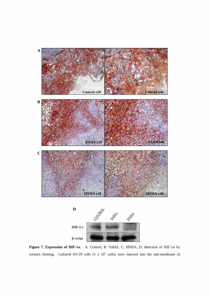
Figure 7. Expression of HIF-1α. A. Control, B. SAHA, C. HNHA, D. detection of HIF-1α by
western blotting. Cultured HT-29 cells (1 x 10⁷ cells) were injected into the sub-membrane of
A
B
C
C
Control x20
SAHA x20
HNHA x20
Control x40
SAHA x40
HNHA x40
D

BALB/C nude mice. When the tumor size was approximately 0.6 cm x 0.6 cm, the LD50 dose of each
drug was injected once every 2 days for a total of seven injections. After treatment, the animals were
sacrificed and the cancer tissue removed for measurement of HIF-1α expression by
immunohistochemistry (A, B, C). Total protein was extracted from the same tissue, and expression of
HIF-1α was measured by western blot analysis (D).
HIF-1α was widely expressed in control cells and it was hard to distinguish
morphologically between cells that do or do not express HIF-1α (Figure 7. A).
HIF-1α was also over-expressed in tissue that was treated with SAHA, but it was
easy to identify which cells expressed HIF-1α (Figure 7. B). In HNHA-treated
cancer tissue, the level of HIF-1α expression was decreased and cell morphology
was almost normal (Figure 7. C). Total protein was extracted from the tissue used
in the prior experiment and expression of HIF-1α was measured by western blot.
HIF-1α expression was decreased in HNHA-treated tissues compared with control
and SAHA-treated tissue (Figure 7. D).
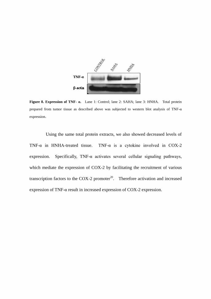
Figure 8. Expression of TNF- α. Lane 1: Control; lane 2: SAHA; lane 3: HNHA. Total protein
prepared from tumor tissue as described above was subjected to western blot analysis of TNF-α
expression.
Using the same total protein extracts, we also showed decreased levels of
TNF-α in HNHA-treated tissue. TNF-α is a cytokine involved in COX-2
expression. Specifically, TNF-α activates several cellular signaling pathways,
which mediate the expression of COX-2 by facilitating the recruitment of various
transcription factors to the COX-2 promoter20
. Therefore activation and increased
expression of TNF-α result in increased expression of COX-2 expression.

IV. DISCUSSION
Histone acetylation is regulated by acetylases and deacetylases, and plays a
key role in eukayotic gene expression. Recent studies have shown that histone
deacetylase (HDAC) plays a crucial role in carcinogenesis1 and is over-expressed in
several tumor cells2. COX-1 is constitutively expressed but COX-2 is inducible as
an early response gene controlled by growth factors, tumor promoters, oncogenes,
and carcinogens19
. More specifically, COX-2 plays an important role in colorectal
cancer carcinogenesis5. Moreover, through the production of prostaglandins,
COX-2 suppresses apoptosis and promotes angiogenesis and tumor invasion19
.
In this study, we investigated the effect of a new HDAC inhibitor, HNHA,
on the expression of COX-2 compared with the existing HDAC inhibitor SAHA.
The IC50 of SAHA is higher than that of HNHA by CCK-8 (Table 1), indicating that
HNHA is a more efficient inhibitor than SAHA. Indeed, treatment with HNHA
inhibited survival of C1300 (mouse neuroblastoma cell line) and HT-29 (human
colorectal cancer cell line) cells more effectively than treatment with SAHA (Figure
3, 4). More specifically, in vitro data showed that HNHA reduced the proportion
of colorectal cancer cells in S phase of the cell cycle but increased the number of
cells in Go-G1 phase (Figure 5). Thus, HNHA arrested cells prior to S phase,
inhibiting tumor cell replication. COX-2 expression was decreased in colon cancer
tissue treated with HNHA (Figure 6) because histone acetylation normally relaxes
the chromatin, allowing transcription factor binding and RNA polymerase II
recruitment. The addition of HDAC inhibitors has a similar net effect to
increasing the amount of histone acetylase activity, resulting in enhanced
transcription levels21
. Expression of HIF-1α was decreased in HNHA-treated
mouse tissue compared with controls (Figure 7), as was expression of TNF-α
(Figure 8).
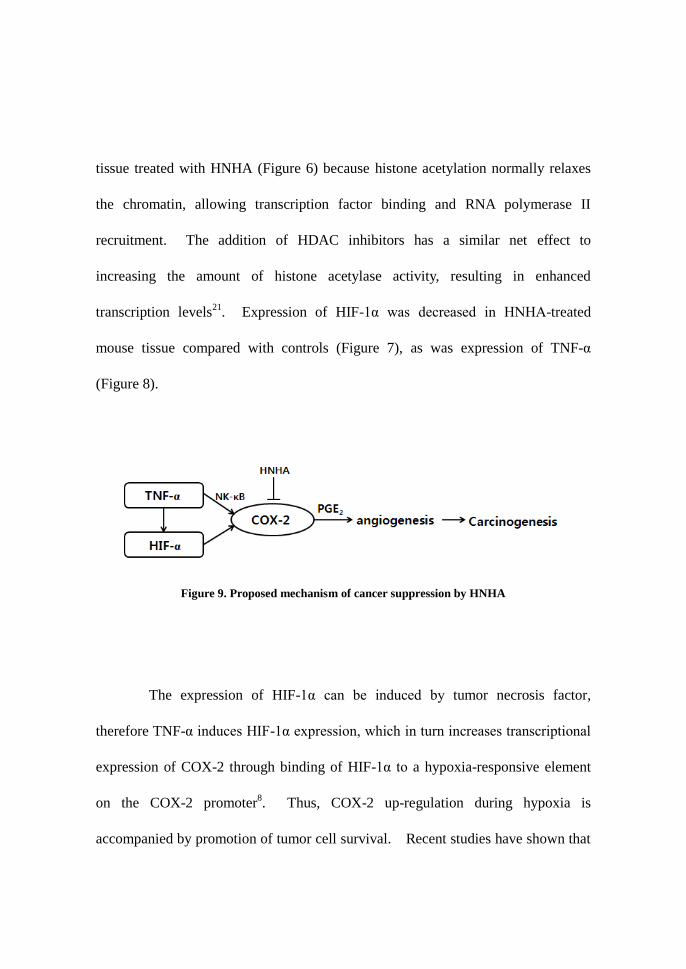
Figure 9. Proposed mechanism of cancer suppression by HNHA
The expression of HIF-1α can be induced by tumor necrosis factor,
therefore TNF-α induces HIF-1α expression, which in turn increases transcriptional
expression of COX-2 through binding of HIF-1α to a hypoxia-responsive element
on the COX-2 promoter8. Thus, COX-2 up-regulation during hypoxia is
accompanied by promotion of tumor cell survival. Recent studies have shown that

inhibition of COX-2 represses various cancers including colon cancer and cervical
cancer 5, 6, 19, 22
. Moreover, prostaglandin formation significantly correlates with
COX-2 expression. Our data indicate that the new HDAC inhibitor, HNHA, also
functions as a COX-2 inhibitor. A number of signaling pathways are involved in
the regulation of COX-2 expression in colorectal carcinoma cells and other cancer
cells. In future studies we plan to link the regulation of COX-2 expression with
key oncogenic signaling pathways.

V. CONCLUSION
The present study clarifies the effects of HNHA on colon cancer. The results yield
the following conclusions:
1. HNHA induces cell cycle arrest of cancer cells, and acts like a HDAC
inhibitor and a COX-2 inhibitor.
2. Decreased expression of COX-2 is related to reduced expression of HIF-1α
and TNF-α.
In conclusion, these data prove that the new drug HNHA inhibits COX-2 expression
in cancer cells and tumor tissues. Further studies are needed to confirm that this
effect is at the RNA level and not the protein level.
REFERENCES
1. Dhordain P, Lin RJ, Quief S, Lantoine D, Kerckaert JP, Evans RM, and et al. The
LAZ3 (BCL06) oncoprotein recruits a SMRT/Msin3A/histone deacetylase
containing complex to mediate transcriptional repression. Nucleic Acids Res.
1998; 26: 4645–4651.
2. Pandolfi PP. Transcription therapy for cancer. Oncogene 2001; 20: 3116–3127.
3. Kim DH, Lee JY, Kim KN, Kim HJ, Jeung HC, Chung HC, and et al. Anti-tumor
activity of N-hydroxy-7-(2-naphthylthio) heptanomide, a novel histone
deacetylase inhibitor. Biochemical and Biophysical Research Communications
2007; 356: 233-238.
4. Dubois RN, Abramson SB, Crofford L, Gupta RA, Simon LS, Leo BA, and et al.
Cyclooxygenase in biology and disease. The FASEB Journal 1998; 12: 1063-
1073.
5. Arber N. Cyclooxygenase-2 Inhibitors in Colorectal Cancer Prevention : Point.
Cancer Epidemiol Biomarkers Prev 2008; 17(8) : 1852-1857.
6. Gupta RA, and DuBois RN. Colorectal Cancer Prevention and Treatment by
Inhibition of Cyclooxygenase-2. Nature 2001; 1:11-21.
7. Marks PA, Richon VM, Breslow R, and Rifkind RA. Histone deacetylase
inhibitors as new cancer drugs. Current Opinion in Oncology 2001; 13:477–483

8. Kaidi A, Qualtrough D, Williams AC, and Paraskeva C. Direct Transcriptional
Up-regulation of Cyclooxygenase-2 by Hypoxia-Inducible Factor (HIF)-1
Promotes Colorectal Tumor Cell Survival and Enhances HIF-1 Transcriptional
Activity during Hypoxia. Cancer Res. 2006; 66: (13) : 6683-6691.
9. Harris AL. Hypoxia—a key regulatory factor in tumour growth. Nat Rev. Cancer
2002; 2:38–47.
10. Wang GL, Jiang BH, Rue EA, Semenza GL. Hypoxiainducible factor-1 is a
basic-helix-loop-helix-pas heterodimer regulated by cellular O2 tension. Proc
Natl Acad Sci U S A 1995; 92:5510–5514.
11. Jaakkola P, Mole DR, Tian YM, et al. Targeting of HIF-α to the von Hippel-
Lindau ubiquitylation complex by O2-regulated prolyl hydroxylation. Science
2001; 292:468–472.
12. Jiang BH, Zheng JZ, Leung SW, Roe R, Semenza GL. Transactivation and
inhibitory domains of hypoxia inducible factor 1α. Modulation of
transcriptional activity by oxygen tension. J Biol Chem 1997; 272: 19253–
19260.
13. Zhong H, De Marzo AM, Laughner E, et al. Over-expression of hypoxia-
inducible factor 1α in common human cancers and their metastases. Cancer
Res 1999; 59:5830–5835.
14. Carmeliet P, Dor Y, Herbert JM, et al. Role of HIF-1α or in hypoxia-mediated
apoptosis, cell proliferation, and tumour angiogenesis. Nature 1998; 394:485–
490.
15. Maxwell PH, Dachs GU, Gleadle JM, et al. Hypoxiainducible factor-1
modulates gene expression in solid tumors and influences both angiogenesis
and tumor growth. Proc Natl Acad Sci U S A 1997; 94:8104–8109.
16. Talks, K. L. et al. Expression and distribution of the hypoxia inducible factors
HIF-1α and HIF-2α in normal human tissues, cancers, and tumor-associated
macrophages. Am. J. Pathol. 2000; 157: 411–421.
17. Balkwill F. TNF-α in promotion and progression of cancer. Cancer Metastasis
Rev. 2006; 25:409-416.
18. Nakao S, Ogtata Y, Shimizu E, Yamazaki M, Furuyama S, and Sugiya H. Tumor
necrosis factor α (TNF-α)-induced prostaglandin E2 release is mediated by the
activation of cyclooxygenase-2 (COX-2) transcription via NFκB in human
gingival fibroblasts. Molecular and Cellular Biochemistry 2002; 238: 11–18,.
19. Saldivar JS, Lopez D, Feldman RA, Reena TJ, Antonio de la Rosa, Terreros D,
and et al. COX-2 overexpression as a biomarker of early cervical
carcinogenesis: A pilot study. Gynecologic Oncology 2007; 107: S155–S162.
20. Yang WL, Roland IH, Godwin AK and Xu XX. Loss of TNF-α-regulated COX-
2 expression in ovarian cancer cells. Oncogene 2005; 24: 7991–8002.
21. Glozak MA and Seto E. Histone deacetylases and cancer. Oncogene 2007; 26:
5420–5432.
22. Shao J, Sheng H, Inoue H, Morrow JD, and DuBois RN. Regulation of
Constitutive Cyclooxygenase-2 Expression in Colon Carcinoma Cells. The
Journal of Biological Chemistry 2000; 275 : 43 : 33951–33956.
ABSTRACT ( IN KOREAN )
대장암에서 신약 HNHA(N-hydroxy-7-(2-naphthylthio)
heptanomide)의 COX-2 발현억제
박 지 현
연세대학교 대학원 의과학과
(지도교수 서 진 석)
최근의 연구를 통하여 nucleosome 의 구조가 변형하는데
전사인자와 DNA 와의 접근을 촉진시키는 SWI/SNF, RSC, NURF,
NRD 등과 같은 염색질 재형성 인자 (chromatin remodeling
factor), histone 의 아세틸화 상태를 조절하는 Histone
acetyltransferases (HATs)와 Histone deacetylase (HDACs)가
중요한 조절인자로 작용함이 밝혀졌다. 또한, HDAC 는 저산소증,
저포도당, 세포암화 등 열악한 환경조건에서 높이 발현되어
세포증식 억제인자의 발현을 저해함으로써 세포증식을 촉진시키는
역할을 하는 것이 밝혀지면서 세포의 암화 및 분화를 조절하는데
있어 중요한 인자로 인식되고 있다.
COX-2 는 cell adhesion, apoptosis, angiogenesis 에서 중요한
역할을 하여, 정상세포에서는 낮은 레벨로 발현하지만
tumor 에서는 높은 레벨로 발현되는 것으로 알려졌다. 앞서
언급한대로 COX-2 는 angiogenesis 에서 중요한 역할을 하는
것으로도 밝혀져 암 조직에서 HDAC inhibitor 를 이용한 COX-
2 의 억제는 암세포의 증식과 생존을 억제한다.
HT-29 라는 human colorectal cancer cell line 과 mouse
neuroblastoma cell line 인 C1300 을 in vitro 에서 배양하여 기존
상용화 된 유사약물과 비교하여 약을 처리하였다. In vivo
실험에서는 HT-29 를 배양하여 실험동물 모델을 만들어
일정한 tumor 크기가 되었을 때 각 종류별 drug 을 피하 주사한다.
약물처리가 완료되면 cancer tissue 를 획득한 후 total protein 을

추출하여 western blot analysis, immunohistochemistry 를
수행하여 약물이 cancer 에 미친 영향을 관찰하였다.
HDAC inhibitor 는 COX-2 의 발현을 억제하며 COX-
2 발현 전단계인 TNF-α 발현 또한 억제한다. 또한
정상세포에서는 거의 발현하지 않지만 암세포에서는 많이 발현되는
HIF-1α 의 발현도 HNHA 를 처리한 조직에서는 그 발현이
억제되었다. 이와 같이 COX-2 는 HNHA inhibitor 에 의해서도
억제됨을 확인하였으며, 따라서 암 조직에서 COX-2 의 억제는
암세포의 증식과 생존을 억제할 것이며, 또한 신생혈관 형성작용을
억제함으로써 암 성장을 억제할 수 있을 것이다.
핵심되는 말 : Histone deacetylase (HDAC), N-hydroxy-7-
(2-naphthylthio)heptanomide (HNHA), Cyclooxygenase-2
(COX-2), Hypoxia-inducible factor-1α (HIF-1α), Tumor
necrosis factor-α (TNF-α)