
The role of valproic acid, a histone deacetylase inhibitor ...

IMMUNOBIOLOGY
Histone deacetylase inhibitors impair innate immune responses to Toll-likereceptor agonists and to infectionThierry Roger,1 Jerome Lugrin,1 Didier Le Roy,1 Genevieve Goy,1 Matteo Mombelli,1 Thibaud Koessler,2 Xavier C. Ding,1
Anne-Laure Chanson,1 Marlies Knaup Reymond,1 Isabelle Miconnet,3 Jacques Schrenzel,2 Patrice Francois,2 andThierry Calandra1
1Infectious Diseases Service, Department of Medicine, Centre Hospitalier Universitaire Vaudois and University of Lausanne, Lausanne, Switzerland; 2Service ofInfectious Diseases, Genomic Research Laboratory, Geneva University Hospitals, Geneva, Switzerland; and 3Division of Immunology and Allergy, Departmentof Medicine, Centre Hospitalier Universitaire Vaudois and University of Lausanne, Lausanne, Switzerland
Regulated by histone acetyltransferasesand deacetylases (HDACs), histone acety-lation is a key epigenetic mechanism con-trolling chromatin structure, DNA accessi-bility, and gene expression. HDACinhibitors induce growth arrest, differen-tiation, and apoptosis of tumor cells andare used as anticancer agents. Here wedescribe the effects of HDAC inhibitorson microbial sensing by macrophagesand dendritic cells in vitro and host de-fenses against infection in vivo. HDAC
inhibitors down-regulated the expressionof numerous host defense genes, includ-ing pattern recognition receptors, ki-nases, transcription regulators, cyto-kines, chemokines, growth factors, andcostimulatory molecules as assessedby genome-wide microarray analyses orinnate immune responses of macro-phages and dendritic cells stimulated withToll-like receptor agonists. HDAC inhibi-tors induced the expression of Mi-2� andenhanced the DNA-binding activity of the
Mi-2/NuRD complex that acts as a tran-scriptional repressor of macrophage cyto-kine production. In vivo, HDAC inhibitorsincreased the susceptibility to bacterialand fungal infections but conferred pro-tection against toxic and septic shock.Thus, these data identify an essential rolefor HDAC inhibitors in the regulation ofthe expression of innate immune genesand host defenses against microbialpathogens. (Blood. 2011;117(4):1205-1217)
Introduction
The innate immune system plays an essential role in antimicrobialdefenses. Detection of microbial pathogens is carried out bysentinel cells of the innate immune system that are located intissues (macrophages and dendritic cells [DCs]) in close contactwith the host’s natural environment or that are rapidly recruited tothe site of infection (neutrophils). Recognition of invasive patho-gens by immune cells relies on their capacity to detect microbial- orpathogen-associated molecular patterns, such as endotoxin, pepti-doglycan, lipopeptides, glucans or mannans, flagellin, and nucleicacids. This process involves the coordinated actions of soluble andcellular molecules composing components of the complementsystem, acute phase proteins, and membrane-associated or intracel-lular pattern-recognition molecules, including Toll-like receptors(TLRs), nucleotide-binding oligomerization domain-like receptors,retinoic acid-inducible gene I like receptors, C-type lectin recep-tors, and scavenger receptors. Ligand-activated receptors triggerthe mitogen-activated protein kinase, nuclear factor-�B (NF-�B),and interferon-related factor (IRF) signal transduction pathwaysthat induce the transcription and production of immune genes,including cytokines that are critical for the activation of innate andadaptive immunity.1,2
Chromatin structure plays a central role in regulating geneexpression. Acetylation of histones is an essential epigeneticmechanism controlling chromatin structure, DNA accessibility fortranscription factors, and gene expression. The net state of acetyla-
tion of the � amino groups of lysine residues of histones is regulatedby the opposing actions of histone acetyltransferases and histonedeacetylases (HDACs). Acetylation of histones relaxes the chroma-tin structure promoting gene transcription, whereas deacetylationof histones compacts the chromatin structure favoring gene silenc-ing. HDACs have been classified into 4 subclasses based on theirhomology with yeast HDACs, their subcellular localization, andtheir enzymatic activity.3 Beside histones, nonhistones proteins(such as �-tubulin, heat shock protein 90, steroid receptors, andregulators of nuclear import) are also modified by reversibleacetylation.4,5 Therefore, histone acetyltransferases and HDACsaffect diverse biologic functions, principally cell differentiation,growth, and survival.6-8
HDACs are at the center of great interest for 2 major reasons.First, dysregulated HDAC expression or activity has been linked tothe pathogenesis of cancer and inflammatory and autoimmunediseases. Second, small-molecule inhibitors of class I, II, and IVHDACs have been shown to exhibit anticancer activity with goodsafety profiles notably in patients with hematologic malignancies.Thus, these drugs are among the most promising anticanceragents under development.6,8-10 Here we analyzed the impact ofHDAC inhibitors on gene expression profiles in macrophagesand DCs in vitro and on the host response to bacteria and fungiin vivo. We found that HDAC inhibitors exerted profound inhibi-tory effects on the host innate immune antimicrobial defense
Submitted May 10, 2010; accepted October 7, 2010. Prepublished online asBlood First Edition paper, October 18, 2010; DOI 10.1182/blood-2010-05-284711.
An Inside Blood analysis of this article appears at the front of this issue.
The online version of this article contains a data supplement.
The publication costs of this article were defrayed in part by page chargepayment. Therefore, and solely to indicate this fact, this article is herebymarked ‘‘advertisement’’ in accordance with 18 USC section 1734.
© 2011 by The American Society of Hematology
1205BLOOD, 27 JANUARY 2011 � VOLUME 117, NUMBER 4
For personal use only.on November 24, 2018. by guest www.bloodjournal.orgFrom
response, down-regulating the expression of innate immune recep-tors, interfering with transcriptome remodeling after stimulationwith TLR agonists, and inhibiting the expression of key antimicro-bial cytokines and accessory molecules in whole blood, macro-phages, DCs, and splenocytes. Consistent with these immunosup-pressive effects, HDAC inhibitors enhanced the susceptibility ofmice to bacterial and fungal infections. Conversely, HDAC inhibi-tors protected mice from septic shock.
Methods
Ethics statement
All animal procedures were approved by the Office Veterinaire du Cantonde Vaud (authorizations 876.5, 876.6, and 877.5) and performed accordingto the institution guidelines for animal experiments.
Mice, cells, and reagents
Eight- to 12-week-old female BALB/c mice (Charles River Laboratories)were housed under specific pathogen–free conditions. Bone marrowderived macrophages (BMDMs), thioglycollate-elicited peritoneal macro-phages, and RAW 264.7 macrophages (ATCC TIB-71) were cultured aspreviously described.11 Bone marrow–derived DCs (BMDCs) were ob-tained by culturing bone marrow cells in Iscove modified Dulbecco mediumcontaining 10% fetal calf serum (FCS; Sigma-Aldrich), 50�M 2-mercapto-ethanol, and granulocyte-macrophage colony-stimulating factor. Spleno-cytes were cultured in RPMI medium containing 2mM L-glutamine and10% FCS and 50�M 2-mercaptoethanol. Human myeloid DCs (moDCs)were produced as described previously.12 Human whole blood assay wasperformed as described previously.13
Cells were exposed to Salmonella minnesota ultra pure lipopolysaccha-ride (LPS; List Biologicals Laboratories), Pam3CSK4 (EMC Microcollec-tions), CpG oligonucleotide (CpG ODN; Invivogen), toxic shock syndrometoxin-1 (Toxin Technology), staphylococcal enterotoxin B, concanavalin A(Sigma-Aldrich), or heat-inactivated Escherichia coli O18 (E coli), Staphy-lococcus aureus, and Candida albicans. Trichostatin A (TSA), suberoylani-lide hydroxamic acid (SAHA), and valproic acid (VPA) were fromSigma-Aldrich. The concentrations of TSA and VPA used in vitro did notaffect the viability of BMDMs (� 85% cell recovery after 18 hours). Orfiril(Desitin Pharmaceuticals), a commercial injectable solution of sodiumvalproate, was used for in vivo experiments.
Microarray analysis and quantitative real-time PCR
For each experimental condition, 2 independent samples were processed inparallel. Low RNA input fluorescent linear amplification kit (AgilentTechnologies) was used for cDNA synthesis and cRNA amplification.Experimental samples were labeled with cyanine 5-CTP, whereas a controlUniversal mouse RNA mixture (Stratagene) was labeled using cyanine3-CTP (PerkinElmer). Labeled cRNA was hybridized onto high-densityoligonucleotide microarrays containing approximately 20 000 60-mer(Mouse Development Oligo Microarray Kit, reference G4120A, and MouseOligo Microarray Kit, V2, reference G4121B, Agilent Technologies). Slideswere scanned using a Microarray Scanner G2565AA system (AgilentTechnologies) at a resolution of 5 �m. For data analysis, local background-subtracted signals were calculated using Feature Extraction software(Agilent Technologies, Version A6.1.1). To ensure spot quality, features andtheir respective background, which were not uniform in pixel fluorescenceintensity distribution in both channels, were flagged (nonuniformity outlierflagging algorithm). Data were imported in GeneSpring, Version 7.0(Agilent Technologies) and then normalized using both per spot (signalchannel divided by the corresponding control channel and generation oflog10 ratio) and per chip (to the 50th percentile). The microarray dataset hasbeen deposited in the Gene Expression Omnibus database (GEO; NationalCenter for Biotechnology Information; accession numbers GPL7291,GSE22409).
Real-time polymerase chain reaction (RT-PCR) was performed with a7500 Fast Real-Time PCR System using the Power SYBR Green PCRMaster Mix (Applied Biosystems) and primer pairs (supplemental Table4, available on the Blood Web site; see the Supplemental Materials link atthe top of the online article).14 Samples were tested in triplicates. A standardmade of successive dilutions of a reference cDNA was processed in parallel.Gene-specific expression levels were assessed relative to the expression ofglyceraldehyde-3-phosphate dehydrogenase (GAPDH) and Hprt and re-ported in arbitrary units. In selected experiments, standards consisted ofserial dilutions of a plasmid containing the target gene.
Cytokine measurements
Tumor necrosis factor (TNF) and interleukin-6 (IL-6) concentrations weremeasured by bioassay,15 IL-12p40, and interferon-� (IFN-�) by enzyme-linked immunosorbent assay (ELISA; BD Biosciences), and a broadscreening of cytokines and chemokines production was performed using theLuminex technology (Luminex Corporation).14
Proliferation
Splenocytes (1.5 � 105) were cultured for 48 hours in 96-well cultureplates. Proliferation was monitored by measuring 3H-thymidine incorpora-tion over 18 hours.
Flow cytometry
BMDCs were incubated with 2.4G2 monoclonal antibody (mAb) andmAbs (BD Biosciences) specific for mouse major histocompatibility classII (14-4-4S-fluorescein isothiocyanate), CD11c (HL3-phycoerythrin [PE]),and CD40 (3/23-biotin revealed with CyChrome-conjugated streptavidin;BD Biosciences). moDCs were incubated with the lineage Cocktail1 (fluorescein isothiocyanate–conjugated mAbs specific for CD3, CD14,CD16, CD19, CD20, and CD56) and mAbs specific for human CD11c(B-ly6-PE-Cy5), HLA-DR (L243-APC) and CD40 (5C3-PE), CD80 (L307.4-PE), CD86 (FUN-1-PE), or CC-chemokine receptor 7 (CCR7; 3D12-PE).12 Datawere analyzed using FlowJo Version 8.5.3 software (TreeStar).
Electrophoretic mobility shift assay
A total of 2 �g of nuclear extracts was incubated for 15 minutes at roomtemperature with a radiolabeled consensus NF-�B probe (Santa CruzBiotechnology) and analyzed by electrophoretic mobility shift assay.16
Western blot analyses
Cell lysates were electrophoresed through polyacrylamide gels and trans-ferred onto nitrocellulose membranes.14 Membranes were incubated withantibodies specific for phosphorylated (phospho)-extracellular signal-regulated kinase (ERK)1/2, total-ERK1/2, phospho-p38, total-p38, phospho-IRF3, (Cell Signaling Technology), c-jun, Mi2b (Santa Cruz Biotechnol-ogy), SNF2�/BRG1 (Millipore), IRF7 (Invitrogen), phospho-signal-transducer and activator of transcription protein (STAT1; BD Biosciences),and �-tubulin (Sigma-Aldrich), and then revealed with secondary horse-radish peroxidase-conjugated goat antirabbit IgG and the ECL Westernblotting analyses system (GE Healthcare). Acid-soluble proteins wereextracted and analyzed by Western blotting using antiacetylated histoneH3 and H4 antibodies (Cell Signaling Technology).13
Chromatin immunoprecipitation
Chromatin immunoprecipitation analysis was performed using the ChIPassay kit (Millipore) using antiacetylated histone H4 (06-866, UpstateBiotechnology) or Mi2b (sc-11378X, Santa Cruz Biotechnology) rabbitpolyclonal antisera and normal rabbit IgG (sc-2027, Santa Cruz Biotechnol-ogy) as described previously.13 Immunoprecipitated DNA was amplified byPCR using primers described in supplemental Table 4.
siRNA silencing in RAW 264.7 mouse macrophages
RAW 264.7 macrophages (6 � 104 cells per well) were seeded in 24-wellplates and transfected the next day with 187.5 ng of Mi-2b or Brg1 siRNA
1206 ROGER et al BLOOD, 27 JANUARY 2011 � VOLUME 117, NUMBER 4
For personal use only.on November 24, 2018. by guest www.bloodjournal.orgFrom

duplexes or negative control (sequences available in supplemental Table 4)and HiPerFect transfection reagent (QIAGEN) according to manufacturer’sinstruction. After 3 days, cells were stimulated for 4 hours with 0 to10 ng/mL LPS. Gene expression was analyzed by real-time PCR. Mi-2band Brg1 mRNA levels were decreased by 60% in cells transfected with thespecific siRNAs.
In vivo models
Klebsiella pneumonia sepsis. A total of 10 CFU of a clinical isolate ofK pneumoniae was injected intranasally into mice treated 15 minutes earlierwith a single dose of valproate (Orfiril, 200 mg/kg intraperitoneally).17
Systemic candidiasis. Mice were challenged through the tail vein with1.2 � 105 CFU of a clinical isolate of C albicans. Valproate treatment(200 mg/kg intraperitoneally) was administrated 15 minutes beforeC albicans challenge and repeated daily during 24 days.
Pam3CSK4-induced shock. Mice were injected with D-galactosamine(2 g/kg intraperitoneally) followed immediately after by Pam3CSK4
(1.6 mg/kg intraperitoneally).11 Valproate injections (200 mg/kg intraperito-neally every 12 hours) were started 2 days before D-galactosamine injectionand discontinued 48 hours after Pam3CSK4 challenge.
CLP. Animals were pretreated intraperitoneally 1 hour before cecalligation and puncture (CLP) with valproate (200 mg/kg) and injectedsubcutaneously every 12 hours with gentamicin (10 mg/kg), clindamycin(30 mg/kg), and buprenorphine (0.1 mg/kg).
Doses of valproate were selected based on previous publications andadjusted to the specific conditions of the sepsis models.
Statistical analysis
Comparisons among treatment groups were performed using the Fisherexact test for categorical data and the Mann-Whitney tests for continuousvariables. The Kaplan-Meier method was used for survival, and differenceswere analyzed by the log-rank sum test. The analyses were performed usingPrism software Version 5.03 (GraphPad). All reported P values are 2-sided,and values less than .05 were considered to indicate statistical significance.For microarray analyses, statistical significance of differentially expressedgenes (2-fold changes) was evaluated by analysis of variance using theBenjamini and Hochberg false discovery rate correction (5%).
Results
Inhibition of HDACs down-regulates the expression of innateimmune genes in macrophages
Transcriptome analyses. The transcriptome of BMDMs wasexamined using the Agilent high-density Mouse DevelopmentOligoarrays, which contained 20 280 unique 60-mer correspondingpredominantly to expressed sequence tags and with the MouseOligo Microarray Kit (V2) containing 20 156 indexed gene probes.At baseline, TSA (t 4 hours of incubation), a prototypicalbroad-spectrum inhibitor of class I, II, and IV HDACs, modifiedthe expression of 1594 macrophage genes (7.7% of the transcrip-tome), of which 772 (3.6%) were found to be down-regulated and822 (4.1%) up-regulated. HDAC inhibitors significantly reducedthe expression of numerous pattern recognition molecules andimmune receptors involved in the sensing of a broad range ofmicrobial products, including bacterial lipopeptides, lipoteichoicacid, peptidoglycan, endotoxin, flagellin, viral nucleic acids, andfungal �-glucan (supplemental Table 1). We then performedtime-course analyses of gene expression profiles of BMDMspreincubated for 1 hour with TSA before stimulation for 1, 2, 4, and20 hours with TLR1/TLR2 (Pam3CSK4 lipopeptide) or with TLR4(LPS) agonists (Figure 1A-B). TSA inhibited the up-regulation of32% to 58% and 33% to 60% of the genes induced by LPS orPam3CSK4, respectively. Conversely, TSA counter-regulated the
down-regulation of 60% to 73% and 51% to 75% of the genesrepressed by LPS or Pam3CSK4. In contrast, TSA potentiated theeffects of LPS and Pam3CSK4 on only a small proportion of genes(2%-16%). The genes modulated by TSA included signal transduc-tion, immunoregulation, cytoskeleton and cell structure, metabo-lism and cell cycle, growth, and apoptosis (complete listing of thegenes: Figure 1C-D, supplemental Tables 2-3).
We then focused our analyses on innate immune gene familiesknown to play a critical role in the host antimicrobial defenseresponse. TSA exerted prominent inhibitory effects on LPS- orPam3CSK4-induced genes (Figure 1C-D). Indeed, TSA inhibitedLPS- and/or Pam3CSK4-induced up-regulation of genes encodingfor molecules involved in the sensing of microbial compounds,such as Tlrs, Cd14, Md-2, scavenger receptors (Scarb2), cytosolicmicrobial sensors (Aim1, Mda-5, Nlrp3, Nod1, Nod2, Eif2ak2/Pkr,Pycard/Asc, Mevf/Pyrin, and RIG-I), c-type lectins (Clecs andMsr1), formyl peptide receptors (Fprs), IgE and IgG Fc receptors,complement and complement receptors (C1qa, C1r, and Cfb/H2-Bf), and adhesion molecules (Icam1, integrins, and Vcam1; Table1). The effect of TSA on the expression of Tlrs, Cd14, Cd36, andMd-2 was confirmed by real-time PCR (Figure 1E). TSA alsoinhibited the expression of adaptor molecules (MyD88 and Ti-cam2), kinases (Iraks, Jaks, Lck, Map3ks, Ripk2, Syk, Tank, Tbk1,and Traf1), phosphatases, and transcription modulators (Atfs,Cebps, Irfs, Junb, Nfkbs, Spic, Stats, and Socs1; Table 1). Inaddition, TSA down-regulated a wide range of LPS- and/orPam3CSK4-stimulated mediators involved in chemotaxis, inflamma-tion, tissue repair, and antigen processing and presentation. This listof genes included cytokines (Il1a, Il1rn, Il6, Il12a, Il12b, Il15, Il18,Il23a, Il27, Ltb, Tnf, Tnfaip3, Tnfsf4, and Tnfsf9), chemokines(Ccl4, Ccl7-9, Ccl12, Ccl17, Ccl22, Ccl24, Cklfsf3, Cklfsf6,Cklfsf7, Cxcl2, Cxcl5, Cxcl12, Cxcl16, and Cx3cl1), growthfactors (Csf2, Edn1, and Tmpo), and their receptors (Il1rl1, Il2rg,Il4ra, Il10rb, Il13ra1, Crlf3, Cxcl12, Cxcl16, Pdfrl, Tnfrsf1a,Tnfrsf14, Ifnar1, Ifnar2, Ifngr2, Csf3r, and Ednrb), cathelicidinantimicrobial peptide (Calmp), matrix metalloproteinases, ubiquit-ins, proteasome subunits and molecules involved in autophagy(Atg16l), antigen transport (Tap1 and Tap2), and peptide presenta-tion (H-2D, H-2E, and H-2Q; Table 1). Therefore, TSA stronglyaffected transcriptome remodeling of BMDMs stimulated withmicrobial products exerting predominantly inhibitory effects, indi-cating that acetylation of histones or nonhistone proteins isrequired for optimal transcription of a large number of macrophagegenes involved in microbial sensing and host defenses.
Cytokine production. To validate the observations generatedfrom microarray profiling, we quantified the production of cyto-kines (ie, TNF, IL-6, and IL-12p40) in BMDMs exposed to LPS,Pam3CSK4, E coli, or S aureus. Real-time PCR analyses (Figure2A), bioassay, and ELISA measurements (Figure 2B-C) confirmedthat TSA strongly inhibited TNF, IL-6, and IL-12p40 production ina time- and dose-dependent manner. Yet, TSA did not inhibitLPS-induced Tnf mRNA and protein (Figure 2A-B). Similar resultswere obtained in thioglycolate-elicited peritoneal macrophages(data not shown).
To confirm the findings obtained with TSA, we tested the effectsof 2 other HDAC inhibitors: SAHA, a hydroxamate, and VPA, ashort chain fatty acid. Like TSA, SAHA and VPA used at clinicallyrelevant concentrations (4-100nM and 4-100�M) dose-dependently inhibited TNF, IL-6, and IL-12p40 production inBMDMs stimulated with Pam3CSK4 (Figure 2D). VPA also markedlyreduced (up to 50-fold) the production of 13 of 15 mediatorsinduced by LPS or Pam3CSK4 in whole blood (Figure 2 E-F).
HDAC INHIBITORS IMPAIR INNATE IMMUNE RESPONSES 1207BLOOD, 27 JANUARY 2011 � VOLUME 117, NUMBER 4
For personal use only.on November 24, 2018. by guest www.bloodjournal.orgFrom

Figure 1. Trichostatin A inhibits the expression of innate immune genes in macrophages. BMDMs were preincubated for 1 hour with or without TSA (100nM) beforeexposure (C-D, 4 hours) to LPS (100 ng/mL) or Pam3CSK4 (100 ng/mL). Transcriptome was analyzed with Agilent Mouse Development Oligoarrays (A-B) or Mouse OligoMicroarray Kit V2 (C-D). (A) Number of genes either up-regulated or down-regulated by LPS and Pam3CSK4 without preincubation with TSA (fold change � 2 vs medium).(B) Effect of 1-hour preincubation with TSA on genes (expressed in percentage) either up-regulated or down-regulated by LPS or Pam3CSK4 (fold change � 2 vs medium).White represents no change; red, inhibition; and green, increase by TSA compared with stimulation with LPS or Pam3CSK4 alone. (C) Heat map of selected pattern recognitionmolecules (medium, TSA, LPS and Pam3CSK4: 4 hours of incubation, LPS TSA and Pam3CSK4 TSA; 1-hour preincubation with TSA followed by 4-hour incubation withLPS and Pam3CSK4). (D) Effect of 1-hour preincubation with TSA (fold changes were calculated vs LPS or Pam3CSK4 alone) on a selection of genes up-regulated by LPS orPam3CSK4 and grouped into various categories based on their biologic functions (microbial sensing and killing; adhesion; cytokine, growth factor and receptors; signaling;antigen processing and presentation). White represents no change; red, inhibition; and green, increase by TSA compared with stimulation with LPS or Pam3CSK4 alone.(E) Tlr1-9, Md-2, Cd14, and Cd36 mRNA copy number was determined by RT-PCR and expressed relative to that of GAPDH. Data are representative of 2 independentexperiments.
1208 ROGER et al BLOOD, 27 JANUARY 2011 � VOLUME 117, NUMBER 4
For personal use only.on November 24, 2018. by guest www.bloodjournal.orgFrom

HDAC inhibitors down-regulated the production of IL-1ra andIL-10, suggesting that the reduced expression of proinflammatorycytokines was not the result of an increased expression ofanti-inflammatory cytokines.
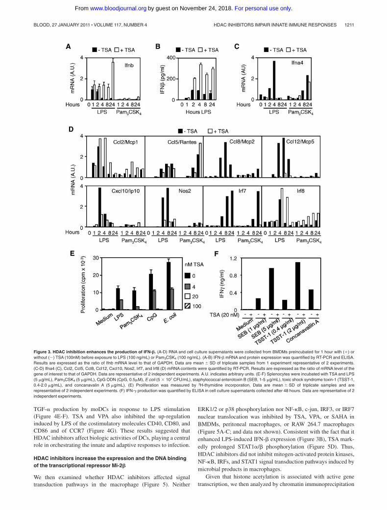
Expression of IFNs and IFN-dependent genes. Together withcytokines and chemokines, type I interferons (IFN-� and IFN-�)are central mediators of innate and adaptive immune responsesagainst viral and bacterial infections.18,19 In response to LPSstimulation, macrophages produce copious amounts of IFN-� thatstimulates the transcription of Ifna, Ccl2, Ccl8, Ccl12, Cxcl10, andNos2 (encoding for iNOS), Irf7, and Irf8.20,21 LPS, but notPam3CSK4, induced rapid (1 hour) Ifnb mRNA expression andsustained IFN-� secretion by BMDMs, whereas LPS or Pam3CSK4
induced early (2 hours and 4 hours, LPS) or late (8 hours and24 hours, Pam3CSK4) Ifna4 mRNA up-regulation (Figure3A-C). Interestingly, TSA up-regulated and markedly prolonged(up to 24 hours) IFN-� mRNA and protein expression after LPSexposure (Figure 3A-B), confirming microarray data (supplementalTable 2). In contrast, it completely inhibited the up-regulation ofIfna4 mRNA (Figure 3C). TSA also inhibited the expression ofLPS-induced Ccl8, Ccl12, Cxcl10, Nos2, and Irf7 mRNA (Figure3D) and of numerous other LPS-induced IFN-�-dependent genes
(supplemental Figure 1). Yet, TSA did not reduce LPS-inducedCcl2 and Irf8 mRNA and late (24 hours) IFN-�-independent Ifna4mRNA (Pam3CSK4) and Ccl5 mRNA (LPS and Pam3CSK4). Thus,the massive accumulation of IFN-� induced by TSA in BMDMsdid not overcome its inhibition of IFN-�-dependent gene expres-sion. Of interest, TSA dose-dependently suppressed the prolifera-tion of splenocytes induced by LPS, Pam3CSK4, CpG ODN, orE coli (Figure 3E). It also inhibited the release of IFN-� bysplenocytes exposed to staphylococcal enterotoxin B, toxic shocksyndrome toxin-1, or concanavalin A (Figure 3F).
HDAC inhibitors impair the response of DCs
We then examined the effects of HDAC inhibitors on innateimmune responses of mouse BMDCs and human moDCs. TSAinhibited the production of cytokines (IL-6 and IL-12p40) and theup-regulation of CD40 by BMDCs stimulated with LPS, Pam3CSK4,CpG ODN, E coli, S aureus, or C albicans (Figure 4A-D).Similarly, TSA and VPA inhibited proinflammatory and anti-inflammatory cytokines (TNF, IL-1�, IL-1�, IL-1ra, IL-6, IL-10,IL-12p40, and IL-12p70), chemokines (IP-10/CXCL10, MIP-1�/CCL4, MCP-1/CCL2, and RANTES/CCL5), IFN-�, G-CSF, and
Table 1. Selection of genes whose up-regulation by LPS or Pam3CSK4 is inhibited by TSA in BMDMs
Adhesionmolecule
Innatereceptor
Signaltransduction Phosphatase
Transcriptionregulator
Cytokine,chemokine,and growth
factor
Cytokine,chemokine,and growth
factor receptor
Antigenprocessing
andpresentation
Ubiquiti-nation Others
Alcam Aim1 Csnk1a1 Dusp1 (Mkp-1) Atf3 Ccl4 Ccrl2 Atg16l Ubc Calmp
Cd47 Birc (cIAP2) Irak2 Dusp4 Atf4 Ccl7 Crlf3 Ctsz Ube1l C1qa
Icam1 Card4 (Nod1) Irak3 Dusp16 Atf5 Ccl8 Csf3r H2-DMb1 Ube2c C1r
Itga4 Carc15 (Nod2) Jak1 Ptpn6 Batf Ccl9 Ednrb H2-Ea Ube2d3 H2-Bf
Itga5 Cd14 Jak2 Ptpn9 Bcl3 Ccl12 Ifnar1 H2-Eb1 Ube2e2 F10
Itgal Cias1 (Nlrp3) Lck Ptpn12 Bcl7c Ccl17 Ifnar2 H2-M3 Ube2j1 Mmp9
Itgax Clec2d Map3k7ip2 Bcl10 Ccl22 Ifngr2 H2-Q10 Ube2j2 Mmp14
Itgb3 Clec4a2 Map3k8 (Tpl2) Cebpb Ccl24 Il1rl1 H2-Q7 Ube2l6 Mmp25
Vcam1 Clec4d Mapkapk2 Cebpd Ccrl2 Il2rg Psma3 Ube2m Nos2
Clec4e Myd88 Cited2 Csf2 Il4ra Psma4 Ube2r2 Pde4b
Eif2ak2 (Pkr) Pias1 Hmgb2 Cklfsf3 Il10rb Psma5 Ube2v1 Pld2
Fcer1g Prkr Ikbke Cksfs6 Il13ra1 Psma6 Ubl3 Ptger4
Fcgr2b Ptk2 Irf1 Cklfsf7 Pdfrl Psmb7 Ubtd1 Ptges
Frp1 Ripk2 Irf2 Cxcl2 Ptafr Psmb8 Ufm1 Ptgir
Fpr-rs2 Socs1 Irf5 Cxcl5 Tnfrsf1a Psmb9 Usp12 Ptgs2
Ifih1 (Mda5) Stk19 Irf7 Cxcl12 Tnfrsf14 Psmb10 Usp18 Sod2
Ly96 (Md-2) Syk Junb Cxcl16 Psme1 Usp24 Txn1
Mefv (Pyrin) Tank Klf6 Cx3cl1 Psme2b Usp42 Tnfrsf5 (Cd40)
Msr1 Tbk1 Nfkb1 Edn1 Tap1
Pycard Ticam2 Nfkb2 Il1a Tap2
Scarb2 Traf1 Nfkbib Il1rn Tapbp
Ddx58 (RIG-l) Trim30 Nfkbie Il6 Tapbpl
Tlr1 Nfkbiz Il12a
Tlr2 Pias1 Il12b
Tlr3 Rel Il13ra1
Tlr6 Rela Il15
Tlr7 Relb Il18
Tlr9 Spic Il23a
Stat3 Il27
Stat5a Inhba
Ltb
Pbef1
Tnf
Tmpo
Tnfaip3 (A20)
Tnfsf4 (OX-40L)
Tnfsf9 (4-1BBL)
HDAC INHIBITORS IMPAIR INNATE IMMUNE RESPONSES 1209BLOOD, 27 JANUARY 2011 � VOLUME 117, NUMBER 4
For personal use only.on November 24, 2018. by guest www.bloodjournal.orgFrom

Figure 2. HDAC inhibitors inhibit cytokine release by macrophages exposed to microbial products and bacteria. BMDMs were preincubated for 1 hour with orwithout TSA (100nM unless specified) before exposure to LPS (100 ng/mL), Pam3CSK4 (100 ng/mL), and heat-killed E coli or S aureus (107 CFU/mL). (A-C) TNF, IL-6, andIL-12p40 mRNA (A) and protein (B-C) production by BMDMs. TNF, IL-6, and IL-12p40 mRNA levels were analyzed by RT-PCR, and results are expressed as the ratio ofcytokines to GAPDH mRNA levels. Data are representative of 3 independent experiments. Cytokine were quantified in cell culture supernatants collected after 8 hours (TNF)and 18 hours (IL-6 and IL-12p40). Data are mean � SD of triplicate samples from one experiment representative of 3 independent experiments. A.U. indicates arbitrary units.(D) BMDMs were preincubated for 1 hour with or without SAHA (4, 20, and 100nM) or VPA (4, 20, and 100�M) before exposure to Pam3CSK4 (100 ng/mL). TNF, IL-6, andIL-12p40 were quantified in cell culture supernatants collected after 8 hours (TNF) and 18 hours (IL-6 and IL-12p40). Data are mean � SD of triplicate samples from1 experiment representative of 3 independent experiments. (E-F) Human whole blood was incubated for 18 hours with VPA (E, 100�M) or TSA together with either LPS(10 ng/mL) or Pam3CSK4 (100 ng/mL). Cytokine and chemokine production was assessed by the Luminex technology (“Cytokine measurements”), and LPS/LPS VPA ratioswere calculated (E). TNF was quantified by bioassay. Data are mean � SD of triplicate samples from one donor and are representative of 2 independent experiments (F).
1210 ROGER et al BLOOD, 27 JANUARY 2011 � VOLUME 117, NUMBER 4
For personal use only.on November 24, 2018. by guest www.bloodjournal.orgFrom
TGF-� production by moDCs in response to LPS stimulation(Figure 4E-F). TSA and VPA also inhibited the up-regulationinduced by LPS of the costimulatory molecules CD40, CD80, andCD86 and of CCR7 (Figure 4G). These results suggested thatHDAC inhibitors affect biologic activities of DCs, playing a centralrole in orchestrating the innate and adaptive responses to infection.
HDAC inhibitors increase the expression and the DNA bindingof the transcriptional repressor Mi-2�
We then examined whether HDAC inhibitors affected signaltransduction pathways in the macrophage (Figure 5). Neither
ERK1/2 or p38 phosphorylation nor NF-�B, c-jun, IRF3, or IRF7nuclear translocation was inhibited by TSA, VPA, or SAHA inBMDMs, peritoneal macrophages, or RAW 264.7 macrophages(Figure 5A-C; and data not shown). Consistent with the fact that itenhanced LPS-induced IFN-� expression (Figure 3B), TSA mark-edly prolonged STAT1�/� phosphorylation (Figure 5D). Thus,HDAC inhibitors did not inhibit mitogen-activated protein kinases,NF-�B, IRFs, and STAT1 signal transduction pathways induced bymicrobial products in macrophages.
Given that histone acetylation is associated with active genetranscription, we then analyzed by chromatin immunoprecipitation
Figure 3. HDAC inhibition enhances the production of IFN-�. (A-D) RNA and cell culture supernatants were collected from BMDMs preincubated for 1 hour with () orwithout (�) TSA (100nM) before exposure to LPS (100 ng/mL) or Pam3CSK4 (100 ng/mL). (A-B) IFN-� mRNA and protein expression was quantified by RT-PCR and ELISA.Results are expressed as the ratio of Ifnb mRNA level to that of GAPDH. Data are mean � SD of triplicate samples from 1 experiment representative of 2 experiments.(C-D) Ifna4 (C), Ccl2, Ccl5, Ccl8, Ccl12, Cxcl10, Nos2, Irf7, and Irf8 (D) mRNA contents were quantified by RT-PCR. Results are expressed as the ratio of mRNA level of thegene of interest to that of GAPDH. Data are representative of 2 independent experiments. A.U. indicates arbitrary units. (E-F) Splenocytes were incubated with TSA and LPS(5 �g/mL), Pam3CSK4 (5 �g/mL), CpG ODN (CpG, 0.5�M), E coli (5 � 107 CFU/mL), staphylococcal enterotoxin B (SEB, 1-5 �g/mL), toxic shock syndrome toxin-1 (TSST-1,0.4-2.0 �g/mL), and concanavalin A (5 �g/mL). (E) Proliferation was measured by 3H-thymidine incorporation. Data are mean � SD of triplicate samples and arerepresentative of 2 independent experiments. (F) IFN-� production was quantified by ELISA in cell culture supernatants collected after 48 hours. Data are representative of 2independent experiments.
HDAC INHIBITORS IMPAIR INNATE IMMUNE RESPONSES 1211BLOOD, 27 JANUARY 2011 � VOLUME 117, NUMBER 4
For personal use only.on November 24, 2018. by guest www.bloodjournal.orgFrom

the extent of histone H4 acetylation of the Tnf and Il6 promoters inBMDMs stimulated with LPS or Pam3CSK4 (Figure 5E). TSAincreased histone H4 acetylation of both promoters, suggesting that
there was no correlation between the status of histone acetylationand the observed down-regulatory effect of TSA on gene transcrip-tion (Figure 2A).
Figure 4. HDAC inhibitors inhibit the response of DCs to microbial stimulation. (A-C) BMDCs were preincubated for 1 hour with TSA before stimulation for 18 hours or theindicated time with LPS (100 ng/mL), Pam3CSK4 (100 ng/mL), CpG oligonucleotide (CpG, 0.7�M), and heat-killed C albicans, E coli, or S aureus (107 CFU/mL). (A-B) IL-6 andIL-12p40 production. Data are mean � SD of triplicate samples and are representative of 4 independent experiments. (C) Cd40 mRNA expression quantified by RT-PCR.Results are expressed as the ratio of Cd40 mRNA level to that of GAPDH. Data are mean � SD of 1 experiment representative of 3 independent experiments. AU indicatesarbitrary units. (D) CD40 mean fluorescence intensity (MFI) determined by flow cytometry. Data are representative of 3 independent experiments. (E-G) Human moDCs werepreincubated for 1 hour with TSA (E-F, 100nM) or VPA (E-F, 100�M) before exposure to LPS (30 ng/mL) for 18 hours. (E) TNF and IL-6 production. Data are mean � SD oftriplicate samples and are representative of 2 independent experiments. (F) Effect of TSA and VPA on cytokine and chemokine production by 2 independent preparations ofmoDCs. Mediators were analyzed using the Luminex technology (“Cytokine measurements”). indicates inhibition (fold change � 2); and �, no effect. (G) CD40, CD80,CD86, and CCR7 expression analyzed by flow cytometry. Data are representative of 2 independent experiments.
1212 ROGER et al BLOOD, 27 JANUARY 2011 � VOLUME 117, NUMBER 4
For personal use only.on November 24, 2018. by guest www.bloodjournal.orgFrom

The Mi-2/NuRD and SWI/SNF (also called BAF in mammals)ATP-dependent remodeling complexes play a central role inregulating gene expression.22 Mi-2� (CHD4) acts as a transcrip-tional repressor, whereas BRG1 (SMARCA4, the catalytic subunitof the BAF complex) acts as a transcriptional activator ofsecondary LPS-induced genes in J77.4 macrophages.23 Given thatHDAC inhibitors impaired the expression of secondary (ie, Il6) butnot primary (ie, Tnf) LPS-induced genes in BMDMs, we hypoth-esized that TSA mediated its effects by affecting the expression ofthe Mi-2� and BRG1 dyad. Interestingly, TSA markedly up-regulated the expression of Mi-2� mRNA in BMDMs exposed toLPS and Pam3CSK4 (Figure 5F). In constrast, TSA had a modest
inhibitory effect on the expression of BRG1. In line with these findings,TSA increased basal and LPS-induced Mi-2� protein expression(Figure 5G-H). Chromatin immunoprecipitation studies revealed thatTSA strongly increased Mi-2� recruitment to the Il6 promoter inBMDMs exposed to LPS, whereas it reduced the binding of Mi-2� tothe Tnf promoter (Figure 5I). siRNA-mediated silencing of Mi-2� inRAW 264.7 mouse macrophages greatly enhanced LPS-induced Il6mRNA expression without a significant effect on Tnf mRNA levels(Figure 5J). Conversely, BRG1 silencing only had a very modest effecton both Il6 and Tnf mRNA expression. Altogether, these data suggestthat TSAmay inhibit cytokine production via an increased expression ofthe transcriptional repressor Mi-2� in macrophages.
Figure 5. HDAC inhibitors increase Mi-2� expression and recruitment to TSA-sensitive promoter. (A-I) BMDMs were preincubated for 1 hour with () or without (�) TSA(100nM unless specified) and exposed to LPS (100 ng/mL) or Pam3CSK4 (100 ng/mL) for the indicated time or 1 hour (E). NF-�B DNA binding activity and NF-�B p65, c-jun,phosphorylated (P-), and total ERK1/2 and p38 and P-STAT1 expression were analyzed by electrophoretic mobility shift assay (A) and Western blot (B-D) using nuclear (nuc)and cytosolic (cyto) extracts. The retarded complex detected by electrophoretic mobility shift assay was dose-dependently inhibited by cold wild-type but not mutant NF-�Boligonucleotide, and supershifted using anti-p65 antibody (data not shown). Acetylation of histone H4 (E) and Mi-2� recruitment (I) to Tnf and Il6 promoters were analyzed bychromatin immunoprecipitation. Mi-2� and BRG1 mRNA and protein levels were quantified by real-time PCR (F) and Western blot (G) with densitometric analyses (H). Data arerepresentative of 2 to 5 independent experiments. (J) RAW 264.7 macrophages transfected with control (Ctl), Mi-2� (Mi), or BRG1 (Br) siRNAs. After 3 days, cells wereincubated for 4 hours with () or without (�) 10 ng/mL of LPS. Il6 and Tnf mRNA levels were analyzed by RT-PCR and results expressed as the ratio of cytokine to GAPDHmRNA levels. Data are representative of triplicate determinations from 1 experiment.
HDAC INHIBITORS IMPAIR INNATE IMMUNE RESPONSES 1213BLOOD, 27 JANUARY 2011 � VOLUME 117, NUMBER 4
For personal use only.on November 24, 2018. by guest www.bloodjournal.orgFrom

HDAC inhibitors impair innate immune responses in vivo
We next investigated the effects of HDAC inhibitors in experimen-tal models of bacterial and fungal sepsis or toxic shock titrated tocause either mild or severe infections or shock. We first verified thatvalproate enhanced the acetylation of histone H4 in vivo inperitoneal exudate cells and splenocytes (Figure 6A). In anotherwise nonsevere, acute K pneumoniae pneumonia model,valproate increased the proportion (53% vs 80%) and magnitude ofbloodstream infections (Figure 6B; P .04) and mortality (from6% to 60%, P .0004; Figure 6C). Consistent with valproate-induced impaired cytokine production by C albicans–infectedBMDMs and BMDCs (Figure 4; and data not shown), valproatetreatment was associated with accelerated (mean time to death:21.5 days vs � 40 days) and increased mortality (44% vs 75%,P .02) in a model of chronic Candida infection (Figure 6D).Thus, inhibition of HDACs impairs host defenses in vivo, increas-ing the susceptibility to and mortality of bacterial and fungal sepsis.
Severe sepsis and septic shock are characterized by an earlyoverwhelming inflammatory response to microbial invasion, andinhibition of proinflammatory mediators confers protection insepsis models.24,25 We therefore tested whether valproate mightexert protective effects in models of fulminant toxic shock inducedby Pam3CSK4 or CLP. Administration of valproate caused a 2- to3-fold reduction of IL-6 and IL-12p40 circulating levels (Figure7A) and a notable increase in survival (0%-64%, P .001) in thePam3CSK4 toxic shock model (Figure 7B). Similarly, valproatetreatment increased survival from 17% to 42% (P .04) in theCLP model (Figure 7C).
Discussion
These studies identify an essential role for acetylation of histonesand nonhistone proteins in the regulation of inflammatory andinnate immune gene expression, and in host defensive responsesagainst microbes. Genome-wide microarray analyses revealed acritical role for HDACs in the expression of host defense genes,including pattern-recognition receptors, adaptor molecules, ki-nases, transcription regulators, complement factors, cytokines,chemokines, and growth factors. HDAC inhibitors exert both
immunosuppressive (a predominant effect) and immunostimula-tory activities. Located at the forefront of the host defenses againstmicrobial invasion, macrophages and DCs are an important source
Figure 6. HDAC inhibition increases mortality to nonsevere infectionwith K pneumonia and C albicans. BALB/c mice were injected intraperi-toneally with valproate (Orfiril, 200 mg/kg) or phosphate-buffered saline(PBS). (A) Peritoneal exudate cells and splenocytes were collected after1 hour. Histone H4 acetylation (AcH4) was analyzed by Western blotting.(B-C) Mice were infected intranasally with 10 CFU of K pneumoniae15 minutes after valproate. (B) Circulating bacterial counts 3 days afterinfection and (C) survival (n 15 mice per treatment groups; P .04 and.0004 for bacterial counts and survival, respectively). The dashed linerepresents the lower limit of detection. (D) Survival of BALB/c mice injectedwith 1.2 � 105 CFU of C albicans and treated with valproate or PBS daily(n 18 mice per treatment group; P .02).
Figure 7. HDAC inhibition protects from lethal toxic shock and severe sepsis.(A-B) BALB/c mice were injected intraperitoneally with valproate (Orfiril, 200 mg/kgstarted 2 days before and discontinued 2 days after induction of shock) or PBS.Animals were sensitized with D-galactosamine and injected intraperitoneally withPam3CSK4. (A) Plasma levels of IL-6 and IL-12p40 were determined 1 hour and3 hours after challenge with Pam3CSK4. Data are mean � SD of 8 mice per treatmentgroup. *P .001 for valproate versus PBS. (B) Survival of BALB/c mice subjected toPam3CSK4-induced shock (n 11 mice per treatment group; P .001). (C) Survivalof BALB/c mice subjected to CLP and treated with either valproate or PBS (every12 hours, starting 30 minutes after surgery; n 16-18 mice per treatment group).P .04.
1214 ROGER et al BLOOD, 27 JANUARY 2011 � VOLUME 117, NUMBER 4
For personal use only.on November 24, 2018. by guest www.bloodjournal.orgFrom

of cytokines. Inhibition of HDACs caused a marked reduction ofcytokine and chemokine production induced by both cell typesafter exposure to TLR agonists. Of note, HDAC inhibitors alsosuppressed the release of IFN-� and IFN-� by macrophages andsplenocytes. Likewise, TSA inhibited IFN-�–stimulated gene acti-vation and late IFN-� induction by Sendai virus and doubled-stranded RNA, and prevented IFN-�–mediated inhibition of thecytopathic effects of vesicular stomatitis virus.26,27 Unexpectedly,we observed that HDAC inhibitors sustained LPS-induced IFN-�production in BMDMs. Nevertheless, the expression of numerousIFN-�/STAT1-dependent genes was strongly inhibited by TSA andVPA, indicating that the increased production of IFN-� did notovercome the potent inhibitory effects of HDAC inhibitors.
Phagocytes and professional antigen-presenting cells help bridgeinnate and adaptive immunity. Macrophages and DCs are keyproducers of IL-12 and IL-23 promoting the generation of protec-tive Th1 and Th17 responses against intracellular and extracellularpathogens.28,29 HDAC inhibitors were found to be potent inhibitorsof IL-12 and IL-23 production by macrophages and DCs as well asCCR7, which is critical for the migration of DCs to secondarylymphoid organs (present data).30-32 TSA inhibited the expressionof several genes involved in antigen processing and presentation.Moreover, HDAC inhibitors have been reported to inhibitDC-stimulated allogeneic T-cell proliferation and Th1-cell activa-tion32-34 and to block the differentiation of IL-17–producingT cells.35 Thus, inhibition of HDACs impacts on several keymacrophages and DCs functions likely to affect the production ofcritical cytokines and the mounting of protective Th1 and Th17immune responses. Of note, TSA did not inhibit TNF production byBMDMs and peritoneal macrophages stimulated with LPS. Incontrast, TSA exerted potent inhibitory effects on whole blood,DCs, and BMDMs stimulated with microbial products other thanLPS (Pam3CSK4, E coli, and S aureus). Similar results wereobtained using SAHA and VPA. These observations suggest thatHDAC inhibitors differentially affect gene expression according tothe cell type or the stimulus studied. This could account for thediscrepant effects of HDAC inhibitors on TNF production reportedin the literature.30-33,36 Indeed, delaying TSA treatment until 1 hourafter the addition of Pam3CSK4 to BMDMs, or inhibiting proteinsynthesis at the time of preincubation with TSA, abrogatedTSA-mediated inhibition of Pam3CSK4-induced TNF production.Thus, inhibition of stimulus-induced Tnf mRNA expression by TSArequires de novo protein synthesis, which cannot overcome a rapidinduction of gene expression in BMDMs stimulated with LPS.
HDAC inhibitors have been reported to interfere with theactivation of the mitogen-activated protein kinases, IRFs, STAT1,AP-1, or NF-�B signal transduction pathways. However, thesefindings have been inconsistent and controversial.26,30,31,36,37 InBMDMs, we did not observe any impact of 3 HDAC inhibitors(TSA, VPA, and SAHA) on ERK1/2 or p38 phosphorylation or onNF-�B, c-jun, IRF3, or IRF7 nuclear translocation induced byLPS or Pam3CSK4. Yet, we found that TSA markedly inhibited therecruitment of NF-�B p65 and of RNA polymerase to the Il6promoter (data not shown). This finding is consistent with thenotion that acetylation of the NF-�B subunits themselves or ofmolecules involved in the NF-�B signal transduction pathwaycontrols the extent, potency, and duration of NF-�B–mediatedtranscriptional activity.38
Transcriptional repression mediated by HDAC inhibitors mayalso rely on acetylation-dependent recruitment of transcriptionalcorepressors or changes in chromatin architecture.38 In line withthis hypothesis, we have previously shown that TSA inhibits the
expression of the proinflammatory cytokine macrophage migrationinhibitory factor via a local deacetylation of chromatin impairingthe recruitment of the basal transcriptional machinery to themacrophage migration inhibitory factor promoter.13,39 In the presentstudy, we provide data suggesting that HDAC inhibitors induce theexpression of Mi-2� and the activity of the Mi-2/NuRD complex,which acts as a transcriptional repressor of secondary LPS-inducedcytokines, such as IL-6. Although little is known about the in vivofunction of Mi-2�, mice deficient in metastasis-associated protein2, a component of the Mi-2/NuRD complex, develop a lupus-likesyndrome characterized by the hypersecretion of cytokines.40
A main finding of our study is the fact that HDAC inhibitorsimpair the host natural defenses against microbial pathogens.Administration of valproate increased the susceptibility of mice tobacterial and fungal infections converting a nonsevere bacterialpneumonia into a highly lethal infection and markedly increasingthe mortality of invasive candidiasis. Moreover, we have alsoobserved that HDAC inhibitors reduced the expression of phago-cytic receptors and phagocytosis and killing of bacteria by macro-phages (M.M., J.L., T.C., T.R., manuscript in preparation). Theseresults clearly demonstrate that the inhibitory effects of HDACinhibitors on innate immune cells in vitro translate into robustimmunosuppressive effects in vivo that negatively impact on thesusceptibility to and outcome of infections. Several arguments ledus to believe that these observations may have clinical implica-tions. A vast amount of data indicate that interfering with criticalmediators of innate or adaptive immunity (eg, TNF or IL-1)increases the risk of infections.41 Indeed, treatment of patients withTNF antagonists has been associated with an overall increased riskof bacterial (tuberculosis, nontuberculous mycobacteriosis, listerio-sis, and salmonellosis) and fungal (candidiasis) infections.42,43
HDAC inhibitors are currently used for the treatment of hemato-logic malignancies and solid tumors often in combination withother cancer or immune suppressive therapies increasing the risk ofinfection. In phase 1 and 2 clinical trials, patients treated withvalproate, SAHA, MS-275, and ITF2357 have developed severeinfections, even without neutropenia, that could be the result of theimmune-suppressive activities of these HDAC inhibitors.44-49 Thesedata suggest that monitoring of infections may be warranted inclinical trials of HDAC inhibitors.
Taken advantage of their broad anti-inflammatory and immuno-modulatory properties targeting several key mediators implicatedin the pathogenesis of septic shock (such as TLRs, MyD88, signaltransducing molecules, and proinflammatory cytokines), we rea-soned that HDAC inhibitors may prove to be beneficial asadjunctive therapy for septic shock. Consistent with this assump-tion, valproate exhibited remarkable protective effects in a toxicshock model induced by Pam3CSK4 and in the CLP peritonealsepsis model. In a recent fascinating article, Xu et al detectedhistones in the circulation of septic patients and baboons andshowed that these proteins played a pathogenic role in sepsis.50
Purified histones H3 and H4 were found to be toxic for endothelialcells, to cause microvascular thrombosis, and to be lethal wheninjected intravenously into mice. Notably, antihistone H4 antibod-ies rescued mice from lethal shock induced by LPS, TNF, or CLP.Given that histones appear to act as noxious danger-associatedendogenous molecular patterns, one may wonder whether theacetylation status of extracellular histones correlates with toxicity.If so, deacetylation of histones released by necrotic and apopticcells could exert detoxifying and cytoprotective effects.
Taken together, the present data identify protein acetylation as akey mechanism regulating the expression of important innate
HDAC INHIBITORS IMPAIR INNATE IMMUNE RESPONSES 1215BLOOD, 27 JANUARY 2011 � VOLUME 117, NUMBER 4
For personal use only.on November 24, 2018. by guest www.bloodjournal.orgFrom

immune genes critically implicated in the sensing of and hostresponses to microbial pathogens. Inhibition of HDACs impairsessential biologic functions of innate immune cells, reducing theircapacity to induce a proinflammatory response, to engulf and killpathogens, and to mount an adaptive response increasing thesusceptibility to infection. On the other hand, the broad anti-inflammatory and immune-suppressive properties of HDAC inhibi-tors were found to be beneficial as adjunctive therapy for septicshock. Thus, these results suggest that the use of HDAC inhibitorsas anticancer agents may increase the risk of infection and sepsis,whereas they may offer new therapeutic options for the manage-ment of patients with septic shock.
Acknowledgments
This work was supported by research funding from the SwissNational Science Foundation (310000-114073/1 and 310030-132744/1, T.R.; 310030-118266, T.C.; 3100A0-112370/1, J.S.; and
3100A0-116075, P.F.), by the Swiss Society for Infectiology(Merck Sharp & Dohme–Chibret AG Award; T.R.), and by theLeenaards Foundation (T.R., J.S.).
Authorship
Contribution: T.R. conceived and supervised the studies, performedexperiments, and wrote the paper; J.L., G.G., X.C.D., M.M.,A.-L.C., M.K.R., and I.M. carried out in vitro experiments; D.L.R.performed in vivo experiments; T.K., P.F., and J.S. performedmicroarray analyses; and T.C. discussed the study results and wrotethe paper.
Conflict-of-interest disclosure: The authors declare no compet-ing financial interests.
Correspondence: Thierry Roger, Infectious Diseases Service,Department of Medicine, Centre Hospitalier Universitaire Vaudois andUniversity of Lausanne, BH 19-111, rue du Bugnon 46, CH-1011Lausanne, Switzerland; e-mail: [email protected].
References
1. Ishii KJ, Koyama S, Nakagawa A, Coban C, Akira S.Host innate immune receptors and beyond: mak-ing sense of microbial infections. Cell HostMicrobe. 2008;3(6):352-363.
2. Medzhitov R. Recognition of microorganisms andactivation of the immune response. Nature. 2007;449(7164):819-826.
3. Yang XJ, Seto E. The Rpd3/Hda1 family of lysinedeacetylases: from bacteria and yeast to miceand men. Nat Rev Mol Cell Biol. 2008;9(3):206-218.
4. Glozak MA, Seto E. Histone deacetylases andcancer. Oncogene. 2007;26(37):5420-5432.
5. Xu WS, Parmigiani RB, Marks PA. Histonedeacetylase inhibitors: molecular mechanisms ofaction. Oncogene. 2007;26(37):5541-5552.
6. Bolden JE, Peart MJ, Johnstone RW. Anticanceractivities of histone deacetylase inhibitors. NatRev Drug Discov. 2006;5(9):769-784.
7. Haberland M, Montgomery RL, Olson EN. Themany roles of histone deacetylases in develop-ment and physiology: implications for diseaseand therapy. Nat Rev Genet. 2009;10(1):32-42.
8. Minucci S, Pelicci PG. Histone deacetylase inhibi-tors and the promise of epigenetic (and more)treatments for cancer. Nat Rev Cancer. 2006;6(1):38-51.
9. Marks PA, Breslow R. Dimethyl sulfoxide to vori-nostat: development of this histone deacetylaseinhibitor as an anticancer drug. Nat Biotechnol.2007;25(1):84-90.
10. Prince HM, Bishton MJ, Harrison SJ. Clinicalstudies of histone deacetylase inhibitors. ClinCancer Res. 2009;15(12):3958-3969.
11. Roger T, Froidevaux C, Le Roy D, et al. Protec-tion from lethal gram-negative bacterial sepsis bytargeting Toll-like receptor 4. Proc Natl Acad SciU S A. 2009;106(7):2348-2352.
12. Miconnet I, Pantaleo G. A soluble hexameric formof CD40 ligand activates human dendritic cellsand augments memory T cell response. Vaccine.2008;26(32):4006-4014.
13. Lugrin J, Ding XC, Le Roy D, et al. Histonedeacetylase inhibitors repress macrophage mi-gration inhibitory factor (MIF) expression by tar-geting MIF gene transcription through a localchromatin deacetylation. Biochim Biophys Acta.2009;1793(11):1749-1758.
14. Delaloye J, Roger T, Steiner-Tardivel QG, et al.Innate immune sensing of modified vaccinia virusAnkara (MVA) is mediated by TLR2-TLR6,MDA-5 and the NALP3 inflammasome. PLoSPathog. 2009;5(6):e1000480.
15. Roger T, David J, Glauser MP, Calandra T. MIFregulates innate immune responses throughmodulation of Toll-like receptor 4. Nature. 2001;414(6866):920-924.
16. Roger T, Chanson AL, Knaup-Reymond M,Calandra T. Macrophage migration inhibitory factorpromotes innate immune responses by suppressingglucocorticoid-induced expression of mitogen-activated protein kinase phosphatase-1. Eur JImmunol. 2005;35(12):3405-3413.
17. Le Roy D, Di Padova F, Adachi Y, et al. Criticalrole of lipopolysaccharide-binding protein andCD14 in immune responses against gram-negative bacteria. J Immunol. 2001;167(5):2759-2765.
18. Bogdan C, Mattner J, Schleicher U. The role oftype I interferons in nonviral infections. ImmunolRev. 2004;202:33-48.
19. Borden EC, Sen GC, Uze G, et al. Interferons atage 50: past, current and future impact on bio-medicine. Nat Rev Drug Discov. 2007;6(12):975-990.
20. Theofilopoulos AN, Baccala R, Beutler B, KonoDH. Type I interferons (alpha/beta) in immunityand autoimmunity. Annu Rev Immunol. 2005;23:307-336.
21. Thomas KE, Galligan CL, Newman RD, Fish EN,Vogel SN. Contribution of interferon-beta to themurine macrophage response to the toll-like re-ceptor 4 agonist, lipopolysaccharide. J BiolChem. 2006;281(41):31119-31130.
22. Chi T. A BAF-centered view of the immune sys-tem. Nat Rev Immunol. 2004;4(12):965-977.
23. Ramirez-Carrozzi VR, Nazarian AA, Li CC, et al.Selective and antagonistic functions of SWI/SNFand Mi-2beta nucleosome remodeling complexesduring an inflammatory response. Genes Dev.2006;20(3):282-296.
24. van der Poll T, Opal SM. Host-pathogen interac-tions in sepsis. Lancet Infect Dis. 2008;8(1):32-43.
25. Rittirsch D, Flierl MA, Ward PA. Harmful molecu-lar mechanisms in sepsis. Nat Rev Immunol.2008;8(10):776-787.
26. Nusinzon I, Horvath CM. Interferon-stimulatedtranscription and innate antiviral immunity requiredeacetylase activity and histone deacetylase1. Proc Natl Acad Sci U S A. 2003;100(25):14742-14747.
27. Nusinzon I, Horvath CM. Positive and negativeregulation of the innate antiviral response andbeta interferon gene expression by deacetylation.Mol Cell Biol. 2006;26(8):3106-3113.
28. Korn T, Bettelli E, Oukka M, Kuchroo VK. IL-17 andTh17 cells. Annu Rev Immunol. 2009;27:485-517.
29. Trinchieri G. Interleukin-12 and the regulation ofinnate resistance and adaptive immunity. Nat RevImmunol. 2003;3(2):133-146.
30. Bode KA, Schroder K, Hume DA, et al. Histonedeacetylase inhibitors decrease Toll-like receptor-mediated activation of proinflammatory gene ex-pression by impairing transcription factor recruit-ment. Immunology. 2007;122(4):596-606.
31. Bosisio D, Vulcano M, Del PA, et al. BlockingTH17-polarizing cytokines by histone deacetylaseinhibitors in vitro and in vivo. J Leukoc Biol. 2008;84(6):1540-1548.
32. Brogdon JL, Xu Y, Szabo SJ, et al. Histonedeacetylase activities are required for innate im-mune cell control of Th1 but not Th2 effector cellfunction. Blood. 2007;109(3):1123-1130.
33. Reddy P, Sun Y, Toubai T, et al. Histone deacety-lase inhibition modulates indoleamine 2,3-dioxygenase-dependent DC functions and regu-lates experimental graft-versus-host disease inmice. J Clin Invest. 2008;118(7):2562-2573.
34. Reilly CM, Thomas M, Gogal R Jr, et al. The his-tone deacetylase inhibitor trichostatin A upregu-lates regulatory T cells and modulates autoimmu-nity in NZB/W F1 mice. J Autoimmun. 2008;31(2):123-130.
35. Koenen HJ, Smeets RL, Vink PM, et al. HumanCD25highFoxp3pos regulatory T cells differenti-ate into IL-17-producing cells. Blood. 2008;112(6):2340-2352.
36. Cao W, Bao C, Padalko E, Lowenstein CJ.Acetylation of mitogen-activated protein kinasephosphatase-1 inhibits Toll-like receptor signal-ing. J Exp Med. 2008;205(6):1491-1503.
37. Aung HT, Schroder K, Himes SR, et al. LPS regu-lates proinflammatory gene expression in macro-phages by altering histone deacetylase expres-sion. FASEB J. 2006;20(9):1315-1327.
38. Calao M, Burny A, Quivy V, Dekoninck A, Van LC.A pervasive role of histone acetyltransferasesand deacetylases in an NF-kappaB-signalingcode. Trends Biochem Sci. 2008;33(7):339-349.
39. Calandra T, Roger T. Macrophage migration in-hibitory factor: a regulator of innate immunity. NatRev Immunol. 2003;3(10):791-800.
40. Lu X, Kovalev GI, Chang H, et al. Inactivation ofNuRD component Mta2 causes abnormal T cellactivation and lupus-like autoimmune disease inmice. J Biol Chem. 2008;283(20):13825-13833.
41. Scheinecker C, Redlich K, Smolen JS. Cytokines
1216 ROGER et al BLOOD, 27 JANUARY 2011 � VOLUME 117, NUMBER 4
For personal use only.on November 24, 2018. by guest www.bloodjournal.orgFrom

as therapeutic targets: advances and limitations.Immunity. 2008;28(4):440-444.
42. Wallis RS. Tumour necrosis factor antagonists:structure, function, and tuberculosis risks. LancetInfect Dis. 2008;8(10):601-611.
43. Dixon WG, Watson K, Lunt M, et al. Rates of seri-ous infection, including site-specific and bacterialintracellular infection, in rheumatoid arthritis pa-tients receiving anti-tumor necrosis factortherapy: results from the British Society for Rheu-matology Biologics Register. Arthritis Rheum.2006;54(8):2368-2376.
44. Kelly WK, O’Connor OA, Krug LM, et al. Phase Istudy of an oral histone deacetylase inhibitor,suberoylanilide hydroxamic acid, in patients with
advanced cancer. J Clin Oncol. 2005;23(17):3923-3931.
45. Ryan QC, Headlee D, Acharya M, et al. Phase Iand pharmacokinetic study of MS-275, a histonedeacetylase inhibitor, in patients with advancedand refractory solid tumors or lymphoma. J ClinOncol. 2005;23(17):3912-3922.
46. Candelaria M, Gallardo-Rincon D, Arce C, et al.A phase II study of epigenetic therapy with hydral-azine and magnesium valproate to overcomechemotherapy resistance in refractory solid tu-mors. Ann Oncol. 2007;18(9):1529-1538.
47. Gojo I, Jiemjit A, Trepel JB, et al. Phase 1 andpharmacologic study of MS-275, a histonedeacetylase inhibitor, in adults with refractory and
relapsed acute leukemias. Blood. 2007;109(7):2781-2790.
48. Rocca A, Minucci S, Tosti G, et al. A phase I-IIstudy of the histone deacetylase inhibitor valproicacid plus chemoimmunotherapy in patients withadvanced melanoma. Br J Cancer. 2009;100(1):28-36.
49. Galli M, Salmoiraghi S, Golay J, et al. A phaseII multiple dose clinical trial of histone deacety-lase inhibitor ITF2357 in patients with relapsed orprogressive multiple myeloma. Ann Hematol.2010;89(2):185-190.
50. Xu J, Zhang X, Pelayo R, et al. Extracellular his-tones are major mediators of death in sepsis. NatMed. 2009;15(11):1318-1321.
HDAC INHIBITORS IMPAIR INNATE IMMUNE RESPONSES 1217BLOOD, 27 JANUARY 2011 � VOLUME 117, NUMBER 4
For personal use only.on November 24, 2018. by guest www.bloodjournal.orgFrom

online October 18, 2010 originally publisheddoi:10.1182/blood-2010-05-284711
2011 117: 1205-1217
Schrenzel, Patrice François and Thierry CalandraXavier C. Ding, Anne-Laure Chanson, Marlies Knaup Reymond, Isabelle Miconnet, Jacques Thierry Roger, Jérôme Lugrin, Didier Le Roy, Geneviève Goy, Matteo Mombelli, Thibaud Koessler, Toll-like receptor agonists and to infectionHistone deacetylase inhibitors impair innate immune responses to
http://www.bloodjournal.org/content/117/4/1205.full.htmlUpdated information and services can be found at:
(5640 articles)Immunobiology and Immunotherapy Articles on similar topics can be found in the following Blood collections
http://www.bloodjournal.org/site/misc/rights.xhtml#repub_requestsInformation about reproducing this article in parts or in its entirety may be found online at:
http://www.bloodjournal.org/site/misc/rights.xhtml#reprintsInformation about ordering reprints may be found online at:
http://www.bloodjournal.org/site/subscriptions/index.xhtmlInformation about subscriptions and ASH membership may be found online at:
Copyright 2011 by The American Society of Hematology; all rights reserved.of Hematology, 2021 L St, NW, Suite 900, Washington DC 20036.Blood (print ISSN 0006-4971, online ISSN 1528-0020), is published weekly by the American Society
For personal use only.on November 24, 2018. by guest www.bloodjournal.orgFrom


![Genome-wide Target Mapping Shows Histone Deacetylase ... · Genome-wide Target Mapping Shows Histone Deacetylase Complex1 Regulates Cell Proliferation in Cucumber Fruit1[OPEN] Zhen](https://static.fdocuments.net/doc/165x107/6016d3d27496f8274240bb84/genome-wide-target-mapping-shows-histone-deacetylase-genome-wide-target-mapping.jpg)