THE BREAST Dr.JAMIL SAWAKED. LATISSIMUS DORSI TERES MAJOR SERRATUS ANTER ANATOMY.
Histomorphometric study of the anterior latissimus dorsi muscle andevaluation of enzymatic markers of broilers affected with dorsal cranial
myopathy
R. Sesterhenn,∗ F. M. Siqueira,†,1 A. C. Hamerski,∗ D. Driemeier,† S. F. Valle,† S. L. Vieira,‡L. Kindlein,∗ and V. P. Nascimento∗
∗Department of Preventive Veterinary Medicine; †Department of Clinical Pathology; and ‡Department of AnimalScience, Universidade Federal do Rio Grande do Sul, Porto Alegre, 91540-000 Brazil
ABSTRACT Dorsal cranial myopathy (DCM), whichaffects the anterior latissimus dorsi (ALD) musclesof commercial broilers, is of unknown etiology, and itrepresents up to 6% of the partial condemnations inBrazilian slaughterhouses. This study was performedto achieve histomorphometric characterizations of theALD muscles from male Cobb 500 broilers slaughteredat either 35 d or 42 d and to evaluate the effects ofDCM on the enzymatic markers aspartate aminotrans-ferase (AST), alanine aminotransferase (ALT), creatinekinase (CK), and lactate dehydrogenase (LDH) andon uric acid and creatinine metabolites. Blood sam-ples (1.5 to 3 mL) and ALD muscle fragments werecollected from each carcass, all of which were pro-cessed in a commercial inline processing system. Foreach age, twelve macroscopically normal animals andtwelve animals found to exhibit DCM were randomly
selected for histomorphometric evaluation and analy-sis of serologic profiles. Microscopic evaluations demon-strated that the muscle fibers of those with DCM ex-hibited a strong presence of multifocal regenerativemyodegeneration as well as a substitution of muscletissue with connective tissue (P < 0.001) through fi-brosis, thus characterizing the chronicity and hardnessof the affected muscle. It is suggested that DCM isa localized muscle lesion because the detected serumlevels of CK (P < 0.001), AST (P < 0.001), ALT(P = 0.01), and LDH (P < 0.001) enzymes werestrongly associated with the group affected by DCM.Additional studies are needed to gain an understand-ing of this myopathy because it is an emerging prob-lem in the poultry industry. In addition, it is related toDCM lesions in fast-growing broilers with the greatestslaughter weights.
Key words: broiler, morphometry, muscular fiber, myopathy, serum enzymes2017 Poultry Science 0:1–7
http://dx.doi.org/10.3382/ps/pex252
INTRODUCTION
In poultry production, rising economic pressures onthe slaughter of broilers with the fastest weight gainin the shortest amount of time has allowed genetics toevolve very rapidly in the last decades with a specialfocus on improving muscular performance (Olivo andShimokomaki, 2002; Havenstein et al., 2003; Vellemanand Nestor, 2003; Vieira, 2008). Nevertheless, these in-creases in growth rates and muscle size, as the con-sequences of production intensification to meet mar-ket demands, have contributed to metabolic alterationsthat resulted in damage to the cell structure of skeletalmuscle, both morphologically and biochemically, withconsequential meaningful losses in the poultry chain(Dransfield and Sosnicki, 1999; MacRae et al., 2006;
C© 2017 Poultry Science Association Inc.Received February 23, 2017.Accepted August 17, 2017.1Corresponding author: [email protected]
MacRae et al., 2007). Among muscle changes presentin chickens, dorsal cranial myopathy (DCM) standsout, evidenced in the anterior latissimus dorsi (ALD)muscle. The etiology and other factors that cause thismuscular disorder have not yet been identified. How-ever, this lesion affects male broilers of high perfor-mance lineages that are in good body condition withthe greatest slaughter weights and no other apparentproblems or infection (Zimermann et al., 2012). Thispathology has been reported in several slaughterhousesin Brazil, initially detected in the southern region inthe 2000s, reaching 6% of the partial condemnations ofthe country’s slaughterhouses (Giacomin et al., 2011;Roso and Dickel, 2011; Ferreira et al., 2012; Zimermannet al., 2012; Sesterhenn et al., 2014; Amaral et al.,2015). Our objective was to analyze the histomorpho-metric characteristics of the ALD muscle from maleCobb 500 broilers slaughtered at 35 and 42 d andto evaluate the effects of DCM on the serum enzy-matic markers alanine aminotransferase (ALT), aspar-tate aminotransferase (AST), creatine kinase (CK),
1
Downloaded from https://academic.oup.com/ps/article-abstract/doi/10.3382/ps/pex252/4201683/Histomorphometric-study-of-the-anterior-latissimusby gueston 03 October 2017

2 SESTERHENN ET AL.
γ-glutamyltransferase (GGT), and lactate dehydroge-nase (LDH) and the metabolites of uric acid and cre-atinine.
MATERIALS AND METHODS
Poultry Management
Male Cobb 500 commercial broiler chickens aged35 and 42 d were obtained during two sequentialslaughterings in a processing plant located in southernBrazil. Average body weights were 2.8 ± 0.2 kg and3.2 ± 0.2 kg, respectively. Of 1,224 carcasses analyzed,48 (3.52%) were selected and collected for further eval-uation. Twenty-four birds of each age were randomlyselected and classified as normal (NORM; n = 12) orDCM (n = 12) based on the degree of severity of the cri-teria established by Zimermann et al. (2012). The ALDmuscles were classified based on the visual appearanceof the skin covering this muscle, its coloration, shape,and consistency, as well as the presence and type ofexudate and/or hemorrhages.
Morphometry Measurements
All muscle fragments collected were fixed in 10%buffered neutral formalin, dehydrated with ethanol inincreasing concentrations, diaphanized in xylol, andembedded in paraffin. From each sample, 10 semi-serial cross-sections and longitudinal sections (3 μmthick) were cut (Microtome Leica DM500; Leica Biosys-tems Nussloch GmbH, Germany) and stained withhematoxylin-eosin for analysis the myopathic lesionin the tissue. In addition, 4 semi-serial cross-sectionsand longitudinal were stained with Masson’s Trichromestains to confirm the presence collagen. Microscopy wasperformed using a light microscope (Leica DM500; Le-ica Biosystems Nussloch GmbH, Germany) with an im-age capture system (ICC50HD) and LAS EZ softwarefor image capture and storage. Morphometric analy-sis of the density and partial volume (Vv, %) of themuscular and connective tissue of each sample was per-formed. In total, 672 images (480 of muscle tissue and192 of connective tissue) were captured in cross-sectionand submitted to the multifunctional M42 test systemusing the superposition method (Weibel, 1979; Wilsonet al., 1990; An et al., 2010).
Serum Analysis
Blood collection for serology (between 1.5 and 3 mLfrom each bird) was performed upon bleeding af-ter electronarcosis. Samples were stored in disposablecapped vacuum tubes (Vacutainer) without anticoag-ulant. Samples were kept at room temperature for 30to 60 min then centrifuged (3,000 × g for 10 min at4◦C), and the serum was stored at −20◦C until an-alyzed. Biochemical tests were performed on thawedsamples in duplicate. A high performance reading spec-
trophotometer with automatic calibration (CM 200;Wiener lab, Rosario, Argentina) was used at a wave-length of 340 nm (Konelab 20i; ThermoElectro Corpo-ration, Espoo, Finland) by means of commercially diag-nostic kits (Wiener lab, Rosario, Argentina) to estimatethe serum enzyme activities of ALT, AST, CK, GGT,LDH, and the metabolites of uric acid and creatinine.The methodology used by UV-Kinetic (IFCC) method(Autopack, Bayer Diagnostics).
Statistical Analysis
Data were analyzed through the analysis ofcovariance considering the effects of myopathy(NORM/DCM) and age at slaughter (35 d and 42 d)on muscle tissue, connective tissue, enzyme activitiesof ALT, AST, CK, GGT, LDH, and the metabolites ofuric acid and creatinine. The statistical analyses wereperformed using IBM SPSS 18.0, with the significancelevel set at 95%. In the presence of meaningful differ-ences (P < 0.05), multiple comparisons were performedusing the Bonferroni test. In addition, carcass weightwas applied as a co-variable because previous stud-ies showed it influenced DCM prevalence (Zimermannet al., 2012).
RESULTS
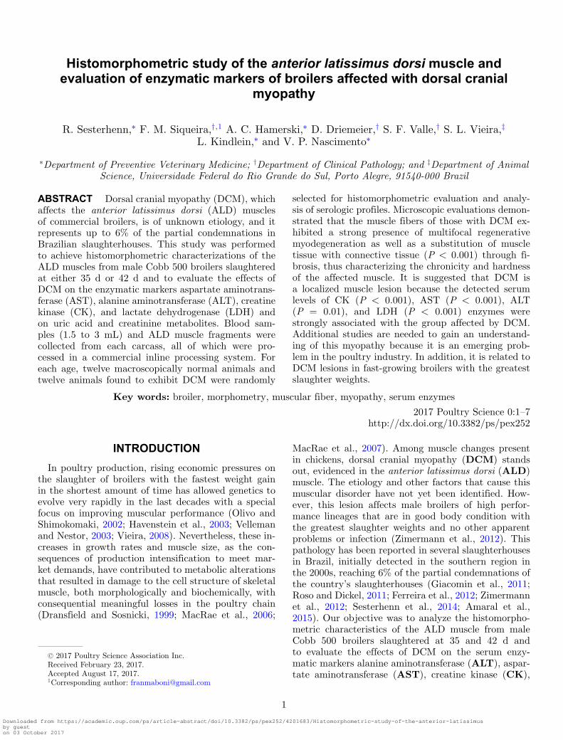
Of 1,224 carcasses analyzed, 90 (7.35%) broilersshowed macroscopic DCM signs. This frequency wasgreater among those aged 35 d (n = 58, 4.73%) com-pared to those aged 42 d (n = 32, 2.61%). Macroscop-ically, the NORM group did not present lesions in theskin and muscle (Figures 1a and b). However, the DCMgroup presented yellowish coloration on the skin andgreater subcutaneous volume (Figure 1c). After cuttingthe skin, one or both portions of the ALD muscle wereremarkably hardened, pale, and thick. The surface wasoften covered with a thin layer of clear or slightly tur-bid viscous material as well as scattered petechiae orhemorrhages (Figure 1d). Upon histopathologic evalu-ation of the ALD muscle, important alterations wereobserved in both groups. Normal ALD muscle eventu-ally presented hypereosinophilic and hypercontractedfibers, flocculus sarcoplasm, and macrophage and lym-phocyte infiltrate in addition to a remarkable presenceof connective and adipose tissue (Figure 2a). Nonethe-less, the lesions were evidently more intense in mus-cle fragments of carcasses affected by DCM (degenera-tion, necrosis, fibrosis, and regeneration; Figures 2b and2c). Staining with Masson’s trichrome showed a prolif-eration of connective tissue (Figure 2d). Altered mus-cle fibers were characterized by the presence of hyper-contraction, hypereosinophilia, rounded edges (loss ofpolyhedral pattern), and sarcoplasmic homogenization,characterizing a hyaline degeneration. An abundanceof interspersed necrotic cells in connective and adiposetissue was seen in addition to inflammatory infiltrate,which was predominantly mononuclear. Variations in
Downloaded from https://academic.oup.com/ps/article-abstract/doi/10.3382/ps/pex252/4201683/Histomorphometric-study-of-the-anterior-latissimusby gueston 03 October 2017

MACROSCOPIC ANALYSIS ON BROILER DORSAL CRANIAL MYOPATHY 3
Figure 1. Broiler carcasses and anterior latissimus dorsi (ALD) muscles. (a) Normal carcass showing no dorsal cranial myopathy (DCM);(b) normal ALD muscle showing no DCM; (c) carcass with DCM evidencing volume increase and yellow coloration (circles); (d) ALD muscleexhibiting DCM with scattered petechiae and hemorrhage on the surface and a greater consistency (arrows).
the sizes of myofibers with cytoplasmic fragmentation,flocculus sarcoplasm (floccular necrosis), and regenera-tion were also observed.
According to results obtained in the morphomet-ric analysis of the relationship between the partialvolume of muscle tissue (Vvm, %) and that of con-nective tissue (Vvc, %; Figure 3), meaningful differ-ences were found (P < 0.001) with a strong influ-ence of carcass weight (P = 0.001) on Vvc. Also, Vvmshowed that broilers with DCM had less muscle tis-sue (34.63% ± 2.67%) when compared to those withnormal muscles (47.28% ± 4.78%). Additionally, Vvcwas higher in those with DCM (29.98% ± 2.79% vs.15.61% ± 5.13%). However, the relationships betweenVvm and Vvc with age at slaughter (35 d or 42 d) werenot significantly different between groups (P > 0.05).
Enzyme and metabolite levels were estimated to as-sess tissue damage associated with DCM. In this way,serum levels for ALT (P = 0.01), AST, CK, and LDH(P < 0.001) were found to be greater in broilers affectedby DCM (Table 1). Carcass weight strongly influencedALT, AST (P ≤ 0.001), and LDH (P = 0.05) enzymes.A difference (P < 0.001) was found in GGT enzymelevels, which were lower in the DCM group. Moreover,no differences (P > 0.05) in uric acid and creatininemetabolites were found between the groups even thoughserum levels were lower in the DCM group.
Greater serum levels of ALT, AST, CK, LDH, andcreatinine were seen in broilers aged 42 d when com-pared to those aged 35 d (Table 2); however, differenceswere significant only in ALT, GGT, LDH (P < 0.001),and CK (P = 0.02) enzymes. Broilers aged 42 d showed
Downloaded from https://academic.oup.com/ps/article-abstract/doi/10.3382/ps/pex252/4201683/Histomorphometric-study-of-the-anterior-latissimusby gueston 03 October 2017
4 SESTERHENN ET AL.
Figure 2. Photomicrographs of the anterior latissimus dorsi (ALD) muscles in broilers. Normal muscle with the presence of hypereosinophiliaand primarily mononuclear inflammatory infiltrate (a); muscle with DCM presenting loss of the polyhedral pattern in the fibers and size variationat 42 d (b); and 35 d (c); multifocal areas of muscle fibers in hyaline degeneration (asterisk), floccular necrosis (arrow), and infiltration byinflammatory cells, mainly heterophils and macrophages (arrowhead tip; hematoxylin and eosin). Proliferation of connective tissue (fibrosis)loosely organized, colored in blue (Masson’s trichrome) (d).
lower serum levels of GGT (P < 0.001) and uric acid(P < 0.03) compared with those aged 35 d.
DISCUSSION
This study indicates that DCM is histopathologi-cally characterized by chronic myopathic lesions with anotable presence of muscular polyphasic degeneration,regeneration, necrosis with a variable amount of inter-stitial connective tissue, fibrosis, and adipocytes. Simi-lar results were reported in muscle myopathies of DCM(Zimermann et al., 2012), white striping (Kuttappanet al., 2012, 2013b), and wooden breast (Sihvo et al.,2013; Velleman and Clark, 2015). In addition, lesionsfound in these samples are very similar to those foundwith wooden breast: pallor, increased hardness andmuscle thickness, and exudate on the muscular surface(Sihvo et al., 2013). According to Dubowitz (1985) themyopathic lesions are nonspecific and might be the re-sult of any of several neuromuscular disturbances. Fur-thermore, genetic selection has led to greater musclemass, tripling muscle size and rendering it vulnerable
to oxidative damage and antioxidant deficiencies as aresult of metabolic residue accumulation, leading to ox-idative stress and tissue lesions (MacRae et al., 2006).In addition, under stress, animals’ antioxidant mecha-nisms could define the quality of the final food product.Indeed, stress has been associated with losses in the ox-idative stability of tissues (Young et al., 2003). Untilnow, the precise etiology of DCM could not be con-firmed. Previous studies suggest that high-performancelineages and the heaviest broilers could be associatedwith DCM (Zimermann et al., 2012) and white strip-ing myopathy (Kuttappan et al., 2012, 2013a,b). Bauer-meister et al. (2009) reported that the severity of whitestriping can increase between the ages of six and eightweeks, suggesting it correlates with age. In addition, lit-erature data (Mudalal et al., 2014) show that woodenbreast myopathy is also generally seen with greaterweight and thickness.
These results corroborate with studies suggestingthat greater growth rates could predispose broilers tothese myopathies (Dransfield and Sosnicki, 1999). Thewhite fibers of breast muscle are more sensitive to
Downloaded from https://academic.oup.com/ps/article-abstract/doi/10.3382/ps/pex252/4201683/Histomorphometric-study-of-the-anterior-latissimusby gueston 03 October 2017

MACROSCOPIC ANALYSIS ON BROILER DORSAL CRANIAL MYOPATHY 5
Figure 3. Distribution of the partial volumes of muscle (Vvm, %)and connective tissue (Vvc, %) from samples of the anterior latissimusdorsi muscle of broilers macroscopically normal (NORM) and those af-fected by dorsal cranial myopathy (DCM). a, bMeans within each mor-phometric analysis are significantly different (P < 0.001). NORM =normal (no dorsal cranial myopathy); DCM (dorsal cranial myopathy).
Table 1. Serological profile of normal (NORM) broilers andthose affected by dorsal cranial myopathy (DCM).
Attribute NORM DCM P value
Enzyme (U/L)ALT 1.82 3.58 ∗AST 533.91 910.76 ∗∗CK 15,312.95 24,332.95 ∗∗GGT 3.72 1,60 ∗∗LDH 4,863.30 7,042.42 ∗∗
Metabolite (mg/dL)Uric acid 0.51 0.50 NSCreatinine 1.39 1.36 NS
NS = P > 0.05; ∗P ≤ 0.01; and ∗∗P ≤ 0.001; ∗ or ∗∗indicates a signif-icant difference between NORM and DCM groups within each row.
Table 2. Serological profile of broilers relative to age atslaughter.
Attribute 35 d 42 d P value
Enzyme (U/L)ALT 1.42 6.83 ∗∗AST 651.35 793.32 NSCK 17,792.60 21,852.87 ∗GGT 4.04 1.92 ∗∗LDH 5,272.71 6,632.61 ∗∗
Metabolite (mg/dL)Uric acid 0.56 0.46 ∗Creatinine 1.35 1.40 NS
NS = P > 0.05; ∗P ≤ 0.05; and ∗∗P ≤ 0.001. ∗ or ∗∗indicates a signif-icant difference between NORM and DCM groups within each row.
oxidation, as well as vitamin deficiencies, than redfibers, and they have a greater tendency to suffer in-juries due to exercise (Page, 1995). On the other hand,red fibers, as in the ALD muscle, are more sensitiveto changes in oxygen levels followed by reperfusion(Carmo-Araujo et al., 2007). Also, research has shownthat fast-growing broilers have up to 20% more fibers inthe ALD muscle, and the fiber size is larger comparedto slow-growing broilers (Remignon et al., 1994, 1995).However, fiber size is of more importance in determin-
ing muscle size than the number of fibers (Prentis et al.,1984). Differences in growth rates among various musclefiber types could contribute to differences in myopathicchanges in these muscles (Ono et al., 1993).
These results agree with those found by Gross et al.(2016), who verified that broilers affected by woodenbreast myopathy exhibited lower Vvm and greater VVccompared to normal broilers. Similarly, Kuttappan etal. (2012) verified that with white striping, fat was in-creased and protein decreased relative to normal broil-ers. Thus, fibrosis could explain the characteristic hard-ness of the affected tissue (Sihvo et al., 2013).
Increased levels of serum enzymes in broilers of great-est body weight indicate muscle damage along withmyopathic changes (MacRae et al., 2006). Increasedaverage concentrations of ALT, AST, CK, and LDH en-zymes were observed. According to previous research,Kuttappan et al. (2013a) found that serum levels ofALT, AST, CK, and LDH were much greater in broilersaffected by white striping compared to normal broilers.Research suggests that changes in plasmatic enzymesindicate hepatic damage or damage in the skeletal mus-cles of broilers (Mitchell and Sandercock, 1995; Yanet al., 2009).
The enzyme CK is considered the primary organ-specific serum enzyme indicative of muscle lesions(Kramer, 1989). Acute elevations in CK enzyme ac-tivity in the plasma were reported in several muscu-lar pathological conditions in broilers, including othermyopathies (Mitchell et al., 1992; Mitchell and Sander-cock, 1994, 1995) and congenital muscular dystrophyin the skeletal muscle (Stewart et al., 1981). Compar-ing fast growing genetic lines with those exhibitinglower growth rates, higher levels of CK (Sandercockand Mitchell, 2003) were observed. High levels of AST,ALT, and LDH enzymes along with the muscle-specificenzyme CK confirms muscular lesions (Gonzales andSilva, 2006; Schmidt et al., 2007; Hoffman and Solter,2008). However, increases in AST, ALT, and LDH en-zyme activities are associated not only with muscledamage, but also with hepatic damage (Hochleithner,1994; Lumeij, 2008; Grunkemeyer, 2010; Capitelli andCrosta, 2013).
According to Kramer and Hofmann (1997), muscleand hepatic lesions can occur concomitantly throughthe same pathological process or a distinct process.Nonetheless, the plasma activity of LDH increases con-siderably with hepatocellular disease or muscle injuryand, in comparison to AST or ALT, its increase anddecrease are faster (Capitelli and Crosta, 2013) andcan provide information on the chronicity of the disease(Grunkemeyer, 2010). Normal AST activity in broilersis less than 275 IU/L (Thrall et al., 2004). In general,moderate AST activity (350 IU/L) and AST activity(from 800 IU/L) are highly suggestive of severe hepaticdamage (Campbell, 2004; Capitelli and Crosta, 2013).
Nevertheless, the enzymes are age dependent, vary-ing between species and generating distinct interpre-tations (Lumeij, 1997). Mitchell (1999) reported that
Downloaded from https://academic.oup.com/ps/article-abstract/doi/10.3382/ps/pex252/4201683/Histomorphometric-study-of-the-anterior-latissimusby gueston 03 October 2017

6 SESTERHENN ET AL.
increased muscle growth compromises the integrity ofskeletal muscle membranes, thus causing alterations atthe enzymatic level, for example, quantitative changesin CK, AST, and LDH enzyme activities. Similarly,ALT activities in broilers can increase as a result ofdamage to various tissues, making interpretation dif-ficult (Grunkemeyer, 2010). Kuttappan et al. (2013a)suggest that when serological findings such as this arediscovered, severe myopathy is associated with muscledamage and not with liver abnormalities.
Although differences (P < 0.001) between the NORMand DCM groups were found in serum GGT levels, theenzyme GGT remained normal in the DCM group. Thisenzyme is an indicator of hepatocellular and renal dam-age, which is manifested in increased levels in serumor plasma and urine (Hochleithner, 1994). Kuttappanet al. (2013a) similarly showed that serum GGT levelswere higher in normal broilers even though no differencewas found (P > 0.05).
Uric acid metabolites were lower in broilers aged 42d compared with those aged 35 d (P < 0.03) eventhough these levels are generally lower in younger broil-ers (Hochleithner, 1994; Capitelli and Crosta, 2013).However, in a study of seagulls, it is generally lower inadults (Alonso-Alverez, 2005). Birds may also presentgreater uric acid after ingesting high-protein diets, pro-longed fasting, or severe muscle degeneration (Gregory,2003; Lierz, 2003; Campbell, 2004).
No differences (P > 0.05) were found in creatininelevels, suggesting that renal function was similar be-tween the NORM and DCM groups. Creatinine levelsacross bird species are normally constant (between 0.1and 0.4 mg/dL) independent of muscle mass (Hochlei-thner, 1994). However, Rajman et al. (2006) proposedthat elevations are related to increased muscle activityand food restrictions in some species.
CONCLUSIONS
Based on these results, we confirmed that DCMcaused alterations in the histomorphometric character-istics of the muscle fibers in the ALD muscle in broilers,with a striking presence of multifocal regenerative my-odegeneration and of fibrosis. A decrease in Vvm andan increase in Vvc (P < 0.001) associated with DCMindicates that the hardness found in the ALD musclesin broilers with DCM can be explained by this fibro-sis. It is suggested that DCM is signified by a local-ized muscle lesion because serum levels of CK, AST,ALT, and LDH enzymes were strongly associated withthe DCM group. Also, CK, the primary organ-specificserum enzyme linked to muscle lesions, was found atgreater levels in the DCM group. This, accompaniedby much greater AST levels, suggests not only mus-cle damage, but also hepatic damage. High serum ac-tivities of LDH and ALT highlight that even thoughthey are considered less specific indicators, they arenevertheless good indicators of muscle damage in broil-ers. Additional studies are needed to enhance our un-
derstanding of this myopathy because it is an emerg-ing problem in the poultry industry, and DCM lesionsare associated with fast-growing broiler strains with thegreatest slaughter weights.
ACKNOWLEDGMENTS
The authors are appreciative for the support of Co-ordenacao de Aperfeicoamento de Pessoal de NıvelSuperior (CAPES) and the Conselho Nacional deDesenvolvimento Cientıfico e Tecnologico (CNPq)throughout this Project.
REFERENCES
Alonso-Alvarez, C. 2005. Age-dependent changes in plasma biochem-istry of yellow-legged gulls (Larus cachinnans). Comp. Biochem.Physiol. A Mol. Integr. Physiol. 140:512–518.
Amaral, P. C., L. R. dos Santos, M. L. R. Provin, L. F. Pedrotti,and E. L. Dickel. 2015. Dorsal cranial myopathy as a cause ofsentencing in broiler slaughterhouses. Hig. aliment. 29:93–97.
An, J. Y., J. X. Zheng, J. Y. Li, D. Zeng, L. J. Qu, G. Y. Xu, andN. Yang. 2010. Effect of myofiber characteristics and thicknessof perymisium and endomysium on meat tenderness of chickens.Poult. Sci. 89:1750–1754.
Bauermeister, L. J., A. U. Morey, E. T. Moran, M. Singh, C. M.Owens, and S. R. Mckee. 2009. Occurence of white striping inchicken breast fillets in relation to broiler size. Poult. Sci. 88(Supl.1):33. (Abstr.).
Campbell, T. W. 2004. Clinical Chemistry of Birds. Pages 479–492in Veterinary Hematology and Clinical Chemistry. M. A. Thrall,ed. Philadelphia: Williams and Wilkins.
Capitelli, R., and L. Crosta. 2013. Overview of psittacine blood anal-ysis and comparative retrospective study of clinical diagnosis,hematology and blood chemistry in selected psittacine species.Vet. Clin. North Am. Exot. Anim. Pract. 16:71–120.
Carmo-Araujo, E. M., M. Dal-Pai-Silva, V. Dal-Pai, R. Checchini,and A. L. A. Ferreira. 2007. Ischaemia and reperfusion effects onskeletal muscle tissue: morphological and histochemical studies.Int. J. Exp. Pathol. 88:147–154.
Dransfield, E., and A. A. Sosnicki. 1999. Relationship between mus-cle growth and poultry meat quality. Poult. Sci. 78:743–746.
Dubowitz, V. 1985. Definition of pathological changes seen in musclebiopsies. Pages 82–128 in Muscle Biopsy. A Practical Approach.2nd ed. V. Dubowitz, ed. Balliere Tindall, London, UK.
Ferreira, T. Z., L Kindlein, R. Sesterhenn, and G. P. Bergmann.2012. Prevalence of cranial dorsal myopathy in broiler slaughteredin RS under federal inspection. III Brazilian South Congress onPoultry, Swine and Dairy Products. In Press.
Giacomin, L., C. Tochetto, L. I. Roman, G. Testolin, M. H. Barreta,and F. C. Zimermann. 2011. The role of exercise in the inductionof dorsal cranial myopathy (MDC) in broilers. The First ScientificInitiation. In Press.
Gonzalez, F. H. D., and S. C. Silva. 2006. Introduction to VeterinaryClinical Biochemistry. 2th rev. ed. Univ. Federal do Rio Grandedo Sul, Brazil.
Gregory, C. R. 2003. Urinary System. Pages 231–259. In: VeterinaryLaboratory Medicine – Clinical Pathology. K. S. Latimer, E. A.Mahaffey, and K. W. Prasse 4th rev.ed. Blackwell Publishing.
Gross, L., R. Sesterhenn, T. Z. Ferreira, S. Vieira, G. P. Bergmann,and L. Kindlein. 2016. Morphometric changes in the breast muscleof broilers with the wooden breast condition. Poult. Sci. 95:206.(Abstr.).
Grunkemeyer, V. L. 2010. Advanced diagnostic approaches and cur-rent management of avian hepatic disorders. Vet. Clin. North Am.Exot. Anim. Pract. 13:413–427.
Havenstein, G. B., P. R. Ferket, and M. A. Qureshi. 2003. Growth,livability, and feed conversion of 1957 versus 2001 broilers whenfed representative 1957 and 2001 broiler diets. Poult. Sci. 82:1500–1508.
Downloaded from https://academic.oup.com/ps/article-abstract/doi/10.3382/ps/pex252/4201683/Histomorphometric-study-of-the-anterior-latissimusby gueston 03 October 2017

MACROSCOPIC ANALYSIS ON BROILER DORSAL CRANIAL MYOPATHY 7
Hoffman, W. E., and P. F. Solter. 2008. Diagnostic enzymologyof domestic animals. Pages 351–378 in Clinical Biochemistry ofDomestic Animals. J. J Kaneko, J. W. Harvey, and M. L Brusseds. Burlington, MA: Academic Press.
Hochleithner, M. 1994. Biochemistries. Pages 223–245 in AvianMedicine: Principles and Application. B. W Ritchie, G. J Harri-son, and L. R. Harrison ed. Lake Worth, FL: Wingers PublishingInc.
Kuttappan, V. A., V. B. Brewer, P. W. Waldroup, and C. M. Owens.2012. Influence of growth rate on the occurrence of white stripingin broiler breast fillets. Poult. Sci. 91:2677–2685.
Kuttappan, V. A., G. R. Huff, W. E. Huff, B. M. Hargis, J. K.Apple, C. Coon, and C. M. Owens. 2013a. Comparison of hema-tologic and serologic profiles of broiler birds with normaland se-vere degrees of white striping in breast fillets. Poult. Sci. 92:339–345.
Kuttappan, V. A., H. L. Shivaprasad, D. P. Shaw, B. A. Valentine,B. M. Hargis, F. D. Clark, S. R. McKee, and C. M. Owens. 2013b.Pathological changes associated with white striping in broilerbreast muscles. Poult. Sci. 92:331–338.
Kramer, J. W. 1989. Clinical enzymology. Pages 338–363 in Clinicalbiochemistry of domestic animals. J. J. Kaneko ed. San Diego:Academic Press.
Kramer, J. W., and W. E. Hoffmann. 1997. Clinical enzymology.Pages 303–325 in Clinical biochemistry of domestic animals. J. J.Kaneko, J. W. Harvey, and M. L. Bruss ed. San Diego: AcademicPress.
Lierz, M. 2003. Avian renal disease: pathogenesis, diagnosis and ther-apy. Vet Clin North Am Exot Anim Pract. 6:29–55.
Lumeij, J. T. 1997. Avian Clinical Biochemistry. Pages 857–883 inClinical Biochemistry of Domestic Animals. J. J. Kaneko, J. W.Harvey, and M. L. Bruss ed. San Diego: Academic Press.
Lumeij, J. T. 2008. Avian clinical biochemistry. Pages 839–872 inClinical Biochemistry of Domestic Animals. Kaneko J. J., J. W.Harvey, and M. L. Bruss ed. Burlington, MA: Academic Press.
MacRae, V. E., M. Mahon, S. Gilpin, D. A. Sandercock, and M.A. Mitchell. 2006. Skeletal muscle fibre growth and growth as-sociated myopathy in the domestic chicken (Gallus domesticus).Poult. Sci. 47:264–272.
Macrae, V. E., M. Mahon, S. Gilpin, D. A. Sandercock, R. R. Hunter,and M. A. Mitchell. 2007. A comparison of breast muscle char-acteristics in three broiler great-grandparent lines. Poult. Sci.86:382–385.
Mitchell, M. A., P. J. Kettlewell, and M. H. Maxwell. 1992. Indicatorsof physiological stress in broiler chickens during road transporta-tion. Anim. Welf. 1:91–103.
Mitchell, M. A., and D. A. Sandercock. 1994. Age dependent changesin plasma creatine kinase activity in broiler chickens. 9th Euro-pean Poultry Conference of the World’s Poultry Science Associ-ation. In Press.
Mitchell, M. A., and D. A. Sandercock. 1995. Creatine kinase isoen-zyme profiles in the plasma of the domestic fowl (Gallus domes-ticus): effects of acute heat stress. Res. Vet. Sci. 59:30–34.
Mitchell, M. A. 1999. Muscle abnormalities – pathophysiologicalmechanisms. Pages 65–98 in Poultry meat science – Poultry sci-ence symposium series. R. I. Richardson, and G. C. Mead ed.CABI International, Wallingford, UK.
Mudalal, R., M. Lorenzi, F. Soglia, C. Cavani, and M. Petracci. 2014.Implications of white striping and wooden breast abnormalitieson quality traits of raw and marinated chicken meat. Anim. 9:728–734.
Olivo, R., and M. Shimokomaki. 2002. Meats: on the way to theresearch. 2th rev. ed. Cocal do Sul, Brazil.
Ono, Y., H. Iwamoto, and H. Takahara. 1993. The relationship be-tween muscle growth and the growth of different fiber types inthe chicken. Poult. Sci. 72:568–576.
Page, P. 1995. Pathophysiology of acute exercise – induced muscularinjury: clinical implications. J. Athl. Train. 30:29–34.
Prentis, P. F., R. K. Penney, and G. Goldspink. 1984. Possible useof an indicator muscle in future breeding experiments in domesticfowl. Poult. Sci. 25:33–41.
Rajman, M., M. Jurani, D. Lamosova, M. Macajova, M. Sedlackova,L. Kostal, D. Jezova, and P. Vyboh. 2006. The effects of feedrestriction on plasma biochemistry in growing meat type chickens(Gallus gallus). Comp. Biochem. Physiol., Part A Mol. Integr.Physiol. 145:363–371.
Remignon, H., L. Lefaucheur, J. C. Blum, and F. H. Ricard.1994. Effects of divergent selection for body weight on threeskeletal muscle characteristics in the chicken. Poult. Sci. 35:65–67.
Remignon, H., M. F. Gardahaut, G. Marche, and F. H. Ricard. 1995.Selection for rapid growth increases the number and the size ofmuscle fibers without changing their typing in chickens. J. MuscleRes. Cell Motil. 16:95–102.
Roso, K., and E. Dickel. 2011. Study of the prevalence of cranial dor-sal myopathy and economic losses occurred in a poultry slaugh-terhouse located in the north of the state of Rio Grande do Sul inthe months of April to November 2010. 38th Brazilian Congressof Veterinary Medicine. In Press.
Sandercock, D. A., and M. A Mitchell. 2003. Myopathy in broilerchickens: a role for ca (2+)-activated phospholipase a2? Poult.Sci. 82:1307–1312.
Sesterhenn, R., T. Z. Ferreira, L. Gross, D. Driemeier, and L.Kindlein L. 2014. Prevalence of cranial dorsal myopathy in broilerslaughtered under federal inspection in RS and its economic im-pact. IV Brazilian South Congress on Poultry, Swine and DairyProducts, In Press.
Sihvo, H. K., K. Immonen, and E. Puolanne. 2013. Myodegenerationwith !brosis and regeneration in the pectoralis major muscle ofbroilers. Vet. Pathol. 51:619–623.
Schmidt, G. A., A. N. LeGrande, and G. Hoffmann. 2007. Waterisotope expressions of intrinsic and forced variability in a coupledocean-atmosphere model. J. Geophys. Res. 112:1–18.
Stewart, P. A., M. E. Percy, L. S. Chang, and M. W. Thompson.1981. Creatine kinase isozyme transition in chicks with hereditarymuscular dystrophy. Muscle Nerve. 4:165–173.
Thrall, M. A., D. C. Baker, T. W. Campbell, D. B. DeNicola, M. J.Fettman, E. D. Lassen, A. Rebar, and G. Weiser. 2004. Veteri-nary Hematology and Clinical Chemistry. Pages 618. Lippincott:Williams & Wilkins.
Velleman, S. G., and K. E. Nestor. 2003. Effect of selection forgrowth rate on myosin heavy chain temporal and spatial local-ization during turkey breast muscle development. Poult. Sci. 82:1373–1377.
Velleman, S. G., and D. L. Clark. 2015. Histopathologic and myo-genic gene expression changes associated with wooden breast inbroiler breast muscles. Avian Dis. 59:410–418.
Vieira, S. L. 2008. Visual Quality of Carcass of Broiler. E-ColorEditora e Grafica Ltda, Sao Paulo, Brazil.
Weibel, E. R. 1979. Sterological Methods. Pages 375–376 in Practi-cal Methods For Biological Morphometry. New York: AcademicPress.
Wilson, B. W., P. S. Nieberg, and R. J. Buhr. 1990. Turkey musclegrowth and focal myopathy. Poult. Sci. 69:1553–1562.
Yan, J. Y., E. D. Bao, and J. M. Yu. 2009. Heat shock protein 60expression in heart, liver and kidney of broilers exposed to hightemperature. Res. Vet. Sci. 86:533–538.
Young, J. F., J. Stagsted, S. K. Jensen, A. H. Karlsson, and P.Henckel. 2003. Ascorbic acid, α-tocopherol, and oregano supple-ments reduce stress-induced deterioration of chicken meat quality.Poult. Sci. 82:1343–1351.
Zimermann, F. C., L. C. B. Fallavena, C. T. P. Salle, H. L. S. Moraes,R. A. Soncini, M. H. Barreta, and V. P. Nascimento. 2012. Down-grading of Heavy Broiler Chicken Carcasses Due to Myodegener-ation of the Anterior Latissimus Dorsi: Pathologic and Epidemi-ologic Studies. Avian Dis. 56:418–421.
Downloaded from https://academic.oup.com/ps/article-abstract/doi/10.3382/ps/pex252/4201683/Histomorphometric-study-of-the-anterior-latissimusby gueston 03 October 2017