Posterior Cadaver Torso Muscles 1. Trapezius 2. Latissimus dorsi 3. Deltoid 4. Triceps brachii Head.

Case ReportLatissimus Dorsi Tendon Transfer with GraftJacket„Augmentation to Increase Tendon Length for an IrreparableRotator Cuff Tear
John G. Skedros1,2,3 and Tanner R. Henrie2
1Department of Orthopaedic Surgery, The University of Utah, Salt Lake City, UT, USA2Utah Orthopaedic Specialists, Salt Lake City, UT, USA3Intermountain Medical Center, Salt Lake City, UT, USA
Correspondence should be addressed to John G. Skedros; [email protected]
Received 4 August 2016; Revised 20 December 2016; Accepted 25 December 2016; Published 17 January 2017
Academic Editor: Dimitrios S. Karataglis
Copyright © 2017 John G. Skedros and Tanner R. Henrie. This is an open access article distributed under the Creative CommonsAttribution License, which permits unrestricted use, distribution, and reproduction in any medium, provided the original work isproperly cited.
Massive irreparable rotator cuff tears can be reconstructed with latissimus dorsi tendon transfers (LDTT). Although uncommon,the natural length of the latissimus dorsi tendon (LDT) could be insufficient for transfer even after adequate soft tissue releases.Descriptions of cases where grafts were needed to lengthen the LDT are therefore rare. We located only two reports of the use ofan acellular dermal matrix to increase effective tendon length in tendon transfers about the shoulder: (1) GraftJacket patch for apectoralis major tendon reconstruction and (2) ArthroFlex� patch for LDTT. Both of these brands of allograft patches are obtainedfrom human cadavers. These products are usually used to cover soft tissue repairs and offer supplemental support rather thanfor increasing tendon length. Extending the LDTT with GraftJacket to achieve adequate length, to our knowledge, has not beenreported in the literature.We report the case of a 50-year-oldmale who had amassive, irreparable left shoulder rotator cuff tear thatwas reconstructed with a LDTT.The natural length of his LDT was insufficient for transfer. This unexpected situation was rectifiedby sewing two patches of GraftJacket to the LDT. The patient had greatly improved shoulder function at two-year follow-up.
1. Introduction
Massive irreparable rotator cuff tears can be reconstructedwith latissimus dorsi tendon transfers (LDTT) (Figures1(a) and 1(b)). LDTT for irreparable rotator cuff tears wasdescribed nearly three decades ago and has since become aviable treatment option in these cases [1, 2]. LDTT is indi-cated in younger patients that lack significant glenohumeralarthritis [3]. Sometimes the teres major tendon is transferredin addition to the latissimus dorsi tendon (LDT) in orderto obtain better external rotation of the shoulder [4, 5].Additionally, removal of some humeral bone along withthe LDT enables direct transosseous fixation of the LDTT,achieving higher integrity of the transfer [6, 7].
Preoperative active range of motion and sex are impor-tant predictors of outcome in LDTT (females have worseoutcomes) [8]. Additionally, inadequate subscapularis and
deltoid function and fatty infiltration of the teres minorcan adversely affect the results of LDTT [8–10]. Additionalchallenges in LDTT include obtaining an adequate view torelease the LDT and achieving sufficient length to reach theeventual attachment point [11, 12]. Adequate tendon lengthcan be reliably achieved by releasing soft tissue attachmentsalong the LDT at its muscle belly [11]. But it is known thatthe natural length of the LDT, even after adequate soft tissuereleases, could be insufficient [11, 12]. A similar problemcan occur during pectoralis major tendon reconstruction. Inthese cases, hamstring and patellar tendon-bone autograftsand fascia lata and Achilles tendon allografts have been usedto bridge the defect between the damaged muscle and itsinsertion point on the humerus [13]. Joseph et al. [14] describethe case of a pectoralis tendon rupture that was lengthenedwith an Achilles tendon allograft to provide additional lengthin order to repair the defect. The use of acellular dermal
HindawiCase Reports in OrthopedicsVolume 2017, Article ID 8086065, 6 pageshttps://doi.org/10.1155/2017/8086065

2 Case Reports in Orthopedics
matrix allograft patches to extend rotator cuff tears that hadinsufficient length for repair is well described [15–17].
Descriptions of cases where grafts were needed to extend(i.e., lengthen) the LDT are rare, showing that this situation isvery uncommon. For example, we only found one case wherean acellular dermal matrix patch was used to increase thenatural length of the LDT [12]. The patch used in that casewas a 3mm ArthroFlex patch (a brand of human acellulardermal matrix). This product is usually used to cover softtissue repairs and offer supplemental support rather than forincreasing their length [16, 18, 19].
Extending the LDTT with an alternative common acellu-lar dermal matrix (GraftJacket) to achieve adequate length,to our knowledge, has not been reported in the literature.We report the case of a 50-year-old male who had amassive, irreparable left shoulder rotator cuff tear that wasreconstructed with a LDTT. The main novel aspect of ourcase is that the patient’s LDT was found to be inadequateduring surgery. This unexpected situation was rectified bysewing two patches of GraftJacket to the free end of his LDT.This yielded greatly improved shoulder function at two-yearfollow-up.
2. Case Report
This left-hand-dominant 50-year-old male (weight: 112 kg;height: 185 cm; BMI: 33 kg/m2) fell off of his porch on31 August 2014 and sustained a massive rotator cuff tear(both the supraspinatus and infraspinatus were torn). Thesubscapularis and teres minor were deemed to be of goodquality. Three months later, he had an attempt at repair, butthe surgeon found the tendon tear irreparable. It was likelythat the patient had a previous but smaller chronic rotatorcuff tear from a sports-related injury many years previously.Thepatient understood that nothing other than a reverse totalshoulder arthroplasty could be done to adequately restoreshoulder function.
The patient came to our clinic one month after thisunsuccessful attempt at repair. Physical examination at thattime showed pseudoparalysis as exhibited by active forwardflexion and abduction at 60–65∘ and superior-posteriorshoulder subluxations (Table 1). We recommended a LDTT.If active range of motion was not achieved to his satisfactionbut the graft healed, then he would likely achieve a tenodesiseffect. This in turn would help reduce subluxations of hisglenohumeral joint and thereby reduce pain while likelyincreasing active motion to a moderate amount [21].
The patient then had an arthroscopic evaluation followedby an open acromioplasty with partial repair of the torninfraspinatus and an open LDTT.The surgery was performedby JGS in accordance with the technique described by Dr.Iannotti and colleagues [8]. A superior approach to therotator cuff was made by detaching the deltoid origin fromthe anterior aspect of the acromion and with splitting ofthe middle deltoid fibers for 3.5 cm. The coracoacromialligament was released with the deltoid and reattached atthe conclusion of the operation. The bursa was excised,and the rotator cuff was inspected. A second incision wasmade along the lateral border of the latissimus dorsi muscle,
extending to the posterior axillary crease. The LDT inser-tion was identified with the arm abducted and internallyrotated, and it was detached sharply from the humerus. Theneurovascular pedicle was identified and protected, and themuscle was released from its deep fascia attachments. Anumber 2 nonabsorbable suture was passed with use of aKrakow suture technique along each side of the tendon fromthe musculotendinous junction to the end of the tendon(Figure 1(a)). Blunt dissection was performed to construct atunnel deep to the deltoid and superficial to the posteriorrotator cuff musculature.
In the conventional surgical technique, the latissimusdorsi muscle and tendon are routinely brought over the topof the humeral head and repaired anterior to the subscap-ularis, lateral to the greater tuberosity, and medial to the tornedges of the rotator cuff (Figure 1(b)). However, during sur-gery, our patient’s LDT was found to be only 5 cm long,less than the mean LDT length reported by Goldberg etal. [22] (mean: 7.3 cm; range: 6.6–7.8 cm; SD: 0.38 cm) andnearly 5.5 cm shorter than what was needed for an ade-quate reconstruction. The patient was consented for use ofan acellular dermal matrix graft (GraftJacket) but not forother allograft or autograft tissues. The anticipated use ofGraftJacket was for augmentation at the repair site. However,GraftJacket has beenmechanically tested and was found to besuperior in strength to comparable xenografts and allografts(CuffPatch�, Restore, Permacol�, and TissueMend�) [23].Additionally, an added layer of thickness from folding theGraftJacket allograft was deemed sufficiently strong to extendthe length of the LDT.
Two 4 × 7 cm patches of GraftJacket were used to extendthe LDT.The thickness of each patchwas 2.0mm (GraftJacketMaximum Force Extreme; Wright Medical Technologies,Inc.; http://documents.wright.com/Document/Get/010660).Each patch was folded in half along its short dimension,which resulted in 4 × 3.5 cm patches that were 4.0mmthick each. The folded margins of the patches were sewntogether with number 2 nonabsorbable sutures (Figure 1(c)).The “biologically resorbable” surface of the GraftJacket wasexposed so that it would be in direct contact to the bone atthe insertion site for the tendon reconstruction. This surfacerepresents the anatomically deeper part of the graft and it isplaced facing the bone in order to allow incorporation of thegraft [24].
The first patch was overlapped on the free end of the LDTby 12.5mm and was then sutured with two rows of nonab-sorbable number 2 sutures (Figure 1(c)). The second Graft-Jacket patch was overlapped by 12.5mm to the first patch andwas sewn with two rows of nonabsorbable number 2 sutures.The total extension of the LDT was then 5.5 cm. A Krakowsuture technique with number 2 suture was then passed alongthe sides of the latissimus tendon to the free end of thegraft extension as shown in Figure 1(a). As described below,the extended LDT was then attached directly to the “foot-print” that was created for the supraspinatus, upper infra-spinatus, and upper subscapularis (Figure 1(b)).
Before attaching the extended LDT to the footprint, resid-ual infraspinatus tendon at the lower portion of the naturalinsertion site was mobilized by sharp dissection and moved

Case Reports in Orthopedics 3
Table 1: Shoulder motion and function of left shoulder.
(a)
Date Action Range of motion Strength
1 month preop∗
Forward flexion 65 2/5Abduction 60
External rotation 50 3/5Internal rotation 30
Extension 35Adduction 35
18 months postop†
Forward flexion 180 4/5Abduction 170
External rotation 60 4/5Internal rotation 70
Extension 45Adduction 45
∗Preop, preoperatively.†Postop, postoperatively.
(b) Shoulder survey scores
Preop∗ Postop†
10 cm VAS score for pain 6.5 2ASES score 26.6 68.3 (Best is 100)WORC score 923 (56.1%) (Best is 0 (100%))Simple shoulder test 10 out of 12 (12 is best)DASH score (Best is 0; worst is 100)Total 14.17Work module 0Sports/performing arts module 37.5
Short-Form 36 (SF-36) (Best is 100 for all)Physical functioning 95Physical role 100Bodily pain 61General health 82Vitality 60Social functioning 75Emotional role 100Mental health 68
∗Preop, preoperatively.†Postop, postoperatively.
15mm upward and sutured to the upper natural insertionsite for this muscle. This was done with number 2 FiberWiresutures through drill holes posteriorly and also with thesutures from a Mitek Healix� anchor that was placed at thelocation where the supraspinatus and upper infraspinatusnaturallymerge.This anchorwas double-loadedwith numberOrthocord� sutures.
The LDT with the GraftJacket extension was then pulledupward beneath the posterior deltoid and anteriorly acrossthe defect in the rotator cuff. Along the lateral portionof the footprint, four pairs of horizontal mattress sutures
(number 2 FiberWire) were passed through drill holes. TheGraftJacket extension was then fastened in place with thesenumber 2 FiberWire sutures and with the two sutures fromthe aforementionedMitek Healix anchor. Additional fixationincluded a series of separate number 2 FiberWire suturespassing through the upper subscapularis and also a seriesof separate sutures passing medially through the residualsupraspinatus tendon near the glenoid. The shoulder wasthen passively tested for stability and motion; elimination ofthe posterior-superior subluxations showed that a tenodesiseffect was clearly achieved.

4 Case Reports in Orthopedics
Deltoid m.Circumflexscapular a.
Teres major m.Teres minor m. Latissimus dorsi m.
(a)
tendonSupraspinatus
tendonSubscapularis
Latissimusdorsi tendon
(b)
4 cm
4 cm
3.5 cm
(c)
Figure 1: Drawings (a) and (b) show how a LDT is typically transferred and sewn to the defect where the rotator cuff would normally insert.Drawing (c) shows how we extended the tendon with two allograft patches. The running stitches along the margins of the LDT shown in (a)were also done, but this is not shown in (c) (images (a) and (b) are reproduced from [20] with permission of the Journal of Bone and JointSurgery, Inc.).
Figure 2: Photograph showing the patient’s left shoulder motionat 18-month follow-up (active forward flexion is 165∘). Preoperativephotographs were not available, but active forward flexion was only65∘.
After wound closure, the patient was placed in a rigidshoulder orthosis in neutral rotation and 45∘ abductionfor two months, following Gerber et al.’s [9] postoperativetreatment. Gentle passive shoulder motion was allowed onlyafter 6 weeks. Active overhead motion was not allowed untilfour months after surgery. Weight bearing exercise at theshoulder was allowed at nine months after surgery but nolifting over 20 lbs (9 kg) overhead until one year after surgery.
At follow-up 18 months later, the patient had regainedsignificant strength and range of motion in his left shoulder
(Table 1 and Figure 2). He reported being very satisfied withthe overall results achieved from his surgery. However, heavoided sleeping on his left side because his shoulder “achesreally bad” and he reported shoulder fatigue with repetitiveoverhead reach. He was not taking regular pain medicationbesides occasional over-the-counter NSAIDs.
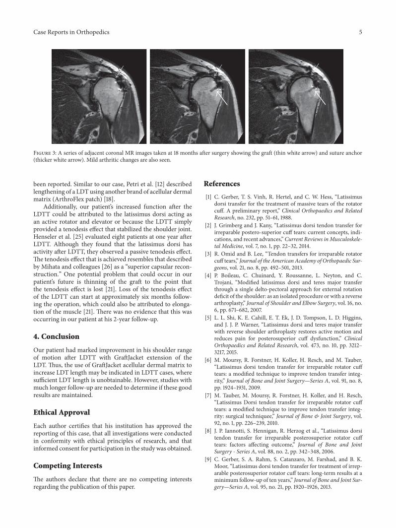
At 18 months after his LDTT, magnetic resonance (MR)scanning was done solely for the purpose of examiningintegrity of the graft. The graft was found to be intact andwithout evidence of thinning (Figure 3). Mild glenohumeralarthritis was noted in the MR images, which likely con-tributed to some of his continuing low-level pain. At finalfollow-up at two years after surgery, the pain had improvedto only trace and his function was maintained.
3. Discussion
At the time of our patient’s tendon reconstruction surgery,there was no anticipation that the length of the LDT wouldbe inadequate after releasing it sharply from its insertion andmobilizing the latissimus dorsimuscle belly of the various softtissue attachments that could restrict the transfer. Althoughremoving some humeral bone along with the LDT wouldhave increased LDT length, it would not have provided theadditional 5.5 cm needed for the LDTT. For this reason, weused GraftJacket allograft acellular dermal matrix to extendthe LDT.This material has been mechanically tested and wasfound to have superior resistance in tension loading com-pared to comparable allografts and xenografts like CuffPatch,Restore, Permacol, and TissueMend [23]. GraftJacket hasbeen used in augmentation of rotator cuff repairs and hasbeen shown to have superior results to nonaugmented repairs[19].
When GraftJacket is used in the context of LDTT forirreparable rotator cuff tear, it would be expected that itwould be for augmenting the new attachment site. To ourknowledge, the use of GraftJacket to extend the LDT has not
Case Reports in Orthopedics 5
Figure 3: A series of adjacent coronal MR images taken at 18 months after surgery showing the graft (thin white arrow) and suture anchor(thicker white arrow). Mild arthritic changes are also seen.
been reported. Similar to our case, Petri et al. [12] describedlengthening of a LDT using another brand of acellular dermalmatrix (ArthroFlex patch) [18].
Additionally, our patient’s increased function after theLDTT could be attributed to the latissimus dorsi acting asan active rotator and elevator or because the LDTT simplyprovided a tenodesis effect that stabilized the shoulder joint.Henseler et al. [25] evaluated eight patients at one year afterLDTT. Although they found that the latissimus dorsi hasactivity after LDTT, they observed a passive tenodesis effect.The tenodesis effect that is achieved resembles that describedby Mihata and colleagues [26] as a “superior capsular recon-struction.” One potential problem that could occur in ourpatient’s future is thinning of the graft to the point thatthe tenodesis effect is lost [21]. Loss of the tenodesis effectof the LDTT can start at approximately six months follow-ing the operation, which could also be attributed to elonga-tion of the muscle [21]. There was no evidence that this wasoccurring in our patient at his 2-year follow-up.
4. Conclusion
Our patient had marked improvement in his shoulder rangeof motion after LDTT with GraftJacket extension of theLDT. Thus, the use of GraftJacket acellular dermal matrix toincrease LDT length may be indicated in LDTT cases, wheresufficient LDT length is unobtainable. However, studies withmuch longer follow-up are needed to determine if these goodresults are maintained.
Ethical Approval
Each author certifies that his institution has approved thereporting of this case, that all investigations were conductedin conformity with ethical principles of research, and thatinformed consent for participation in the study was obtained.
Competing Interests
The authors declare that there are no competing interestsregarding the publication of this paper.
References
[1] C. Gerber, T. S. Vinh, R. Hertel, and C. W. Hess, “Latissimusdorsi transfer for the treatment of massive tears of the rotatorcuff. A preliminary report,” Clinical Orthopaedics and RelatedResearch, no. 232, pp. 51–61, 1988.
[2] J. Grimberg and J. Kany, “Latissimus dorsi tendon transfer forirreparable postero-superior cuff tears: current concepts, indi-cations, and recent advances,” Current Reviews inMusculoskele-tal Medicine, vol. 7, no. 1, pp. 22–32, 2014.
[3] R. Omid and B. Lee, “Tendon transfers for irreparable rotatorcuff tears,” Journal of the American Academy of Orthopaedic Sur-geons, vol. 21, no. 8, pp. 492–501, 2013.
[4] P. Boileau, C. Chuinard, Y. Roussanne, L. Neyton, and C.Trojani, “Modified latissimus dorsi and teres major transferthrough a single delto-pectoral approach for external rotationdeficit of the shoulder: as an isolated procedure or with a reversearthroplasty,” Journal of Shoulder and Elbow Surgery, vol. 16, no.6, pp. 671–682, 2007.
[5] L. L. Shi, K. E. Cahill, E. T. Ek, J. D. Tompson, L. D. Higgins,and J. J. P. Warner, “Latissimus dorsi and teres major transferwith reverse shoulder arthroplasty restores active motion andreduces pain for posterosuperior cuff dysfunction,” ClinicalOrthopaedics and Related Research, vol. 473, no. 10, pp. 3212–3217, 2015.
[6] M. Moursy, R. Forstner, H. Koller, H. Resch, and M. Tauber,“Latissimus dorsi tendon transfer for irreparable rotator cufftears: a modified technique to improve tendon transfer integ-rity,” Journal of Bone and Joint Surgery—Series A, vol. 91, no. 8,pp. 1924–1931, 2009.
[7] M. Tauber, M. Moursy, R. Forstner, H. Koller, and H. Resch,“Latissimus Dorsi tendon transfer for irreparable rotator cufftears: a modified technique to improve tendon transfer integ-rity: surgical techniquee,” Journal of Bone & Joint Surgery, vol.92, no. 1, pp. 226–239, 2010.
[8] J. P. Iannotti, S. Hennigan, R. Herzog et al., “Latissimus dorsitendon transfer for irreparable posterosuperior rotator cufftears: factors affecting outcome,” Journal of Bone and JointSurgery - Series A, vol. 88, no. 2, pp. 342–348, 2006.
[9] C. Gerber, S. A. Rahm, S. Catanzaro, M. Farshad, and B. K.Moor, “Latissimus dorsi tendon transfer for treatment of irrep-arable posterosuperior rotator cuff tears: long-term results at aminimum follow-up of ten years,” Journal of Bone and Joint Sur-gery—Series A, vol. 95, no. 21, pp. 1920–1926, 2013.

6 Case Reports in Orthopedics
[10] J. J. P. Warner and I. M. Parsons IV, “Latissimus dorsi tendontransfer: a comparative analysis of primary and salvage recon-struction of massive, irreparable rotator cuff tears,” Journal ofShoulder and Elbow Surgery, vol. 10, no. 6, pp. 514–521, 2001.
[11] P. D. G. Henry, T. Dwyer, M. D. McKee, and E. H. Schemitsch,“Latissimus dorsi tendon transfer for irreparable tears of therotator cuff: an anatomical study to assess the neurovascularhazards and ways of improving tendon excursion,”The Bone &Joint Journal, vol. 95, no. 4, pp. 517–522, 2013.
[12] M. Petri, J. A. Greenspoon, S. Bhatia, and P. J. Millett, “Patch-augmented latissimus dorsi transfer and open reduction-internal fixation of unstable os acromiale for irreparablemassiveposterosuperior rotator cuff tear,” Arthroscopy Techniques, vol.4, no. 5, pp. e487–e492, 2015.
[13] T. Dehler, A. L. Pennings, and A. W. ElMaraghy, “Dermal allo-graft reconstruction of a chronic pectoralis major tear,” Journalof Shoulder and Elbow Surgery, vol. 22, no. 10, pp. e18–e22, 2013.
[14] T. A. Joseph, M. J. Defranco, and G. G. Weiker, “Delayed repairof a pectoralis major tendon rupture with allograft: a casereport,” Journal of Shoulder and Elbow Surgery, vol. 12, no. 1, pp.101–104, 2003.
[15] D. P. Ferguson, M. R. Lewington, T. D. Smith, and I. H. Wong,“Graft utilization in the augmentation of large-to-massiverotator cuff repairs: a systematic review,”The American Journalof Sports Medicine, vol. 44, no. 11, pp. 2984–2992, 2016.
[16] C. R. Jones and S. J. Snyder, “Massive irreparable rotator cufftears: a solution that bridges the gap,” Sports Medicine andArthroscopy Review, vol. 23, no. 3, pp. 130–138, 2015.
[17] M. Petri, R. J. Warth, M. P. Horan, J. A. Greenspoon, and P. J.Millett, “Outcomes after open revision repair of massive rotatorcuff tears with biologic patch augmentation,” Arthroscopy, vol.32, no. 9, pp. 1752–1760, 2016.
[18] D. C. Acevedo, B. Shore, and R. Mirzayan, “Orthopedic appli-cations of acellular human dermal allograft for shoulder andelbow surgery,”Orthopedic Clinics of North America, vol. 46, no.3, pp. 377–388, 2015.
[19] F. A. Barber, J. P. Burns, A. Deutsch, M. R. Labbe, and R. B.Litchfield, “A prospective, randomized evaluation of acellularhuman dermal matrix augmentation for arthroscopic rotatorcuff repair,” Arthroscopy, vol. 28, no. 1, pp. 8–15, 2012.
[20] M. J. Codsi, S. Hennigan, R. Herzog et al., “Latissimus dorsitendon transfer for irreparable posterosuperior rotator cufftears. Surgical technique,”The Journal of Bone and Joint Surgery,vol. 89, supplement 2, part 1, pp. 1–9, 2007.
[21] A. Ersen, H. Ozben, M. Demirhan, A. C. Atalar, and M.Kapıcıoglu, “Time-dependent changes after latissimus dorsitransfer: tenodesis or tendon transfer?” Clinical Orthopaedicsand Related Research, vol. 472, no. 12, pp. 3880–3888, 2014.
[22] B. A. Goldberg, B. Elhassan, S.Marciniak, and J. H. Dunn, “Sur-gical anatomy of latissimus dorsi muscle in transfers about theshoulder,” American Journal of Orthopedics, vol. 38, no. 3, pp.E64–E67, 2009.
[23] F. A. Barber,M. A.Herbert, andD. A. Coons, “Tendon augmen-tation grafts: biomechanical failure loads and failure patterns,”Arthroscopy—Journal of Arthroscopic and Related Surgery, vol.22, no. 5, pp. 534–538, 2006.
[24] S. J. Snyder, S. P. Arnoczky, J. L. Bond, and R. Dopirak, “Histo-logic evaluation of a biopsy specimen obtained 3 months afterrotator cuff augmentation with GraftJacket Matrix,” Arthro-scopy, vol. 25, no. 3, pp. 329–333, 2009.
[25] J. F. Henseler, J. Nagels, R. G. H. H. Nelissen, and J. H. de Groot,“Does the latissimus dorsi tendon transfer for massive rotator
cuff tears remain active postoperatively and restore activeexternal rotation?” Journal of Shoulder and Elbow Surgery, vol.23, no. 4, pp. 553–560, 2014.
[26] T. Mihata, T. Q. Lee, Y. Itami, A. Hasegawa, M. Ohue, and M.Neo, “Arthroscopic superior capsule reconstruction for irrep-arable rotator cuff tears: a prospective clinical study in 100 con-secutive patients with 1 to 8 years of follow-up,” OrthopaedicJournal of Sports Medicine, vol. 4, no. 3, 2016.

Submit your manuscripts athttps://www.hindawi.com
Stem CellsInternational
Hindawi Publishing Corporationhttp://www.hindawi.com Volume 2014
Hindawi Publishing Corporationhttp://www.hindawi.com Volume 2014
MEDIATORSINFLAMMATION
of
Hindawi Publishing Corporationhttp://www.hindawi.com Volume 2014
Behavioural Neurology
EndocrinologyInternational Journal of
Hindawi Publishing Corporationhttp://www.hindawi.com Volume 2014
Hindawi Publishing Corporationhttp://www.hindawi.com Volume 2014
Disease Markers
Hindawi Publishing Corporationhttp://www.hindawi.com Volume 2014
BioMed Research International
OncologyJournal of
Hindawi Publishing Corporationhttp://www.hindawi.com Volume 2014
Hindawi Publishing Corporationhttp://www.hindawi.com Volume 2014
Oxidative Medicine and Cellular Longevity
Hindawi Publishing Corporationhttp://www.hindawi.com Volume 2014
PPAR Research
The Scientific World JournalHindawi Publishing Corporation http://www.hindawi.com Volume 2014
Immunology ResearchHindawi Publishing Corporationhttp://www.hindawi.com Volume 2014
Journal of
ObesityJournal of
Hindawi Publishing Corporationhttp://www.hindawi.com Volume 2014
Hindawi Publishing Corporationhttp://www.hindawi.com Volume 2014
Computational and Mathematical Methods in Medicine
OphthalmologyJournal of
Hindawi Publishing Corporationhttp://www.hindawi.com Volume 2014
Diabetes ResearchJournal of
Hindawi Publishing Corporationhttp://www.hindawi.com Volume 2014
Hindawi Publishing Corporationhttp://www.hindawi.com Volume 2014
Research and TreatmentAIDS
Hindawi Publishing Corporationhttp://www.hindawi.com Volume 2014
Gastroenterology Research and Practice
Hindawi Publishing Corporationhttp://www.hindawi.com Volume 2014
Parkinson’s Disease
Evidence-Based Complementary and Alternative Medicine
Volume 2014Hindawi Publishing Corporationhttp://www.hindawi.com