Highly multiplexed single-cell in situ RNA and DNA ...
9
Highly multiplexed single-cell in situ RNA and DNA analysis with bioorthogonal cleavable fluorescent oligonucleotides† Manas Mondal, a Renjie Liao, a Christopher D. Nazaroff, ab Adam D. Samuel a and Jia Guo * a The ability to profile transcripts and genomic loci comprehensively in single cells in situ is essential to advance our understanding of normal physiology and disease pathogenesis. Here we report a highly multiplexed single-cell in situ RNA and DNA analysis approach using bioorthogonal cleavable fluorescent oligonucleotides. In this approach, oligonucleotides tethered to fluorophores through an azide-based cleavable linker are used to detect their nucleic acids targets by in situ hybridization. After fluorescence imaging, the fluorophores in the whole specimen are efficiently cleaved in 30 minutes without loss of RNA or DNA integrity. Through reiterative cycles of hybridization, imaging, and cleavage, this method has the potential to quantify hundreds to thousands of different RNA species or genomic loci in single cells in situ at the single-molecule sensitivity. Applying this approach, we demonstrate that different nucleic acids can be detected in each hybridization cycle by multi-color staining, and at least ten continuous hybridization cycles can be carried out in the same specimen. We also show that the integrated single- cell in situ analysis of DNA, RNA and protein can be achieved using cleavable fluorescent oligonucleotides combined with cleavable fluorescent antibodies. This highly multiplexed imaging platform will have wide applications in systems biology and biomedical research. Introduction Comprehensive analyses of the copy number and spatial orga- nization of transcripts and genomic loci in single cells promise to transform our understanding of many heterogeneous biological systems, such as brain tissues, solid tumors and developing embryos. 1 Microarray technologies 2 and high-throughput sequencing 3–6 have been widely used for transcriptome- or genome-wide nucleic acids analysis. Nonetheless, as these approaches are carried out with extracted DNA or RNA, they mask the spatial complexity of nucleic acids in a heterogeneous pop- ulation. Fluorescent hybridization probes 7–12 have emerged as a powerful tool to quantify transcripts and genomic loci in their natural spatial contexts in single cells. However, only a handful of different nucleic acids species in a biological sample can be detected by these uorescence imaging-based approaches. To enable multiplexed single-cell in situ nucleic acids anal- ysis, a number of methods have been explored. For example, in situ sequencing 13,14 has been developed to enable single-cell transcriptome analysis. However, it suffers from low detection efficiency and may miss transcripts with low copy numbers. Combinatorial labeling 15–17 offers single-molecule detection sensitivity, but it has limited multiplexing capacities. Recently, sequential hybridization, 18–20 multiplexed error-robust uores- cence in situ hybridization (MER-FISH) 21–23 and reiterative hybridization 24–26 have been developed. To allow multiple anal- ysis cycles in the same specimen, these methods apply different approaches to remove the uorescence signals. Such approaches include probe degradation by DNase, photobleaching, disulde based chemical cleavage, and probe stripping by formamide. Nevertheless, probe degradation by DNase has limited efficiency and is time-consuming. Photobleaching removes uorescence signals in individual imaging areas sequentially, and thus has long assay time and low sample throughput. The endogenous thiol groups and the thiol groups generated by cleavage can react with the disulde containing probes applied in the following cycles, generating high background and false positive signals. Probe stripping by formamide removes all the probes, including the large oligonucleotides library hybridized to their RNA and DNA targets. As a result, this expensive oligonucleotides library has to be re-hybridized in every analysis cycle, which makes this approach less cost- and time-effective. Additionally, removal of the stripped oligonucleotides probes by diffusion in thick tissue samples can be inefficient and time-consuming, limiting its applications for intact tissue analysis. a Biodesign Institute, School of Molecular Sciences, Arizona State University, Tempe, Arizona 85287, USA. E-mail: [email protected] b Division of Pulmonary Medicine, Department of Biochemistry and Molecular Biology, Mayo Clinic Arizona, Scottsdale, Arizona 85259, USA † Electronic supplementary information (ESI) available: Additional gures, schemes, probe sequences and experimental details. See DOI: 10.1039/c7sc05089e Cite this: Chem. Sci. , 2018, 9, 2909 Received 28th November 2017 Accepted 8th February 2018 DOI: 10.1039/c7sc05089e rsc.li/chemical-science This journal is © The Royal Society of Chemistry 2018 Chem. Sci. , 2018, 9, 2909–2917 | 2909 Chemical Science EDGE ARTICLE Open Access Article. Published on 13 February 2018. Downloaded on 2/17/2022 7:23:58 PM. This article is licensed under a Creative Commons Attribution 3.0 Unported Licence. View Article Online View Journal | View Issue
Transcript of Highly multiplexed single-cell in situ RNA and DNA ...

ChemicalScience
EDGE ARTICLE
Ope
n A
cces
s A
rtic
le. P
ublis
hed
on 1
3 Fe
brua
ry 2
018.
Dow
nloa
ded
on 2
/17/
2022
7:2
3:58
PM
. T
his
artic
le is
lice
nsed
und
er a
Cre
ativ
e C
omm
ons
Attr
ibut
ion
3.0
Unp
orte
d L
icen
ce.
View Article OnlineView Journal | View Issue
Highly multiplex
aBiodesign Institute, School of Molecular Sc
Arizona 85287, USA. E-mail: [email protected] of Pulmonary Medicine, Departmen
Mayo Clinic Arizona, Scottsdale, Arizona 85
† Electronic supplementary informationschemes, probe sequences and experimen
Cite this: Chem. Sci., 2018, 9, 2909
Received 28th November 2017Accepted 8th February 2018
DOI: 10.1039/c7sc05089e
rsc.li/chemical-science
This journal is © The Royal Society of C
ed single-cell in situ RNA and DNAanalysis with bioorthogonal cleavable fluorescentoligonucleotides†
Manas Mondal,a Renjie Liao,a Christopher D. Nazaroff,ab Adam D. Samuela
and Jia Guo *a
The ability to profile transcripts and genomic loci comprehensively in single cells in situ is essential to
advance our understanding of normal physiology and disease pathogenesis. Here we report a highly
multiplexed single-cell in situ RNA and DNA analysis approach using bioorthogonal cleavable fluorescent
oligonucleotides. In this approach, oligonucleotides tethered to fluorophores through an azide-based
cleavable linker are used to detect their nucleic acids targets by in situ hybridization. After fluorescence
imaging, the fluorophores in the whole specimen are efficiently cleaved in 30 minutes without loss of
RNA or DNA integrity. Through reiterative cycles of hybridization, imaging, and cleavage, this method has
the potential to quantify hundreds to thousands of different RNA species or genomic loci in single cells
in situ at the single-molecule sensitivity. Applying this approach, we demonstrate that different nucleic
acids can be detected in each hybridization cycle by multi-color staining, and at least ten continuous
hybridization cycles can be carried out in the same specimen. We also show that the integrated single-
cell in situ analysis of DNA, RNA and protein can be achieved using cleavable fluorescent
oligonucleotides combined with cleavable fluorescent antibodies. This highly multiplexed imaging
platform will have wide applications in systems biology and biomedical research.
Introduction
Comprehensive analyses of the copy number and spatial orga-nization of transcripts and genomic loci in single cells promise totransform our understanding of many heterogeneous biologicalsystems, such as brain tissues, solid tumors and developingembryos.1 Microarray technologies2 and high-throughputsequencing3–6 have been widely used for transcriptome- orgenome-wide nucleic acids analysis. Nonetheless, as theseapproaches are carried out with extracted DNA or RNA, theymaskthe spatial complexity of nucleic acids in a heterogeneous pop-ulation. Fluorescent hybridization probes7–12 have emerged asa powerful tool to quantify transcripts and genomic loci in theirnatural spatial contexts in single cells. However, only a handful ofdifferent nucleic acids species in a biological sample can bedetected by these uorescence imaging-based approaches.
To enable multiplexed single-cell in situ nucleic acids anal-ysis, a number of methods have been explored. For example, insitu sequencing13,14 has been developed to enable single-cell
iences, Arizona State University, Tempe,
u
t of Biochemistry and Molecular Biology,
259, USA
(ESI) available: Additional gures,tal details. See DOI: 10.1039/c7sc05089e
hemistry 2018
transcriptome analysis. However, it suffers from low detectionefficiency and may miss transcripts with low copy numbers.Combinatorial labeling15–17 offers single-molecule detectionsensitivity, but it has limited multiplexing capacities. Recently,sequential hybridization,18–20 multiplexed error-robust uores-cence in situ hybridization (MER-FISH)21–23 and reiterativehybridization24–26 have been developed. To allow multiple anal-ysis cycles in the same specimen, these methods apply differentapproaches to remove the uorescence signals. Such approachesinclude probe degradation by DNase, photobleaching, disuldebased chemical cleavage, and probe stripping by formamide.Nevertheless, probe degradation by DNase has limited efficiencyand is time-consuming. Photobleaching removes uorescencesignals in individual imaging areas sequentially, and thus haslong assay time and low sample throughput. The endogenousthiol groups and the thiol groups generated by cleavage can reactwith the disulde containing probes applied in the followingcycles, generating high background and false positive signals.Probe stripping by formamide removes all the probes, includingthe large oligonucleotides library hybridized to their RNA andDNA targets. As a result, this expensive oligonucleotides libraryhas to be re-hybridized in every analysis cycle, which makes thisapproach less cost- and time-effective. Additionally, removal ofthe stripped oligonucleotides probes by diffusion in thick tissuesamples can be inefficient and time-consuming, limiting itsapplications for intact tissue analysis.
Chem. Sci., 2018, 9, 2909–2917 | 2909

Fig. 1 Highly multiplexed single cell in situ RNA and DNA analysis withBoCFO. Each hybridization cycle starts with target staining, which canbe achieved with two alternative approaches. (A) In the direct stainingapproach, each nucleic acids target is hybridized with a set of BoCFO.(B) In the indirect staining approach, individual nucleic acids target isfirst hybridized by a set of non-labeled oligonucleotides, which aresubsequently hybridized by BoCFO. For both approaches, after targethybridization, the fluorescence images are captured. Finally, all thedifferent fluorophores on BoCFO are chemically cleaved simulta-neously. Through cycles of target hybridization, fluorescence imagingand fluorophore cleavage, a large number of distinct transcripts orgenomic loci can be quantified at the single-molecule sensitivity insingle cells in situ. (C) Structures of BoCFO, ON-N3-Quasar 570 andON-N3-Cy5.
Chemical Science Edge Article
Ope
n A
cces
s A
rtic
le. P
ublis
hed
on 1
3 Fe
brua
ry 2
018.
Dow
nloa
ded
on 2
/17/
2022
7:2
3:58
PM
. T
his
artic
le is
lice
nsed
und
er a
Cre
ativ
e C
omm
ons
Attr
ibut
ion
3.0
Unp
orte
d L
icen
ce.
View Article Online
Here, we report a highly multiplexed single-cell in situ RNAand DNA analysis approach using bioorthogonal cleavableuorescent oligonucleotides (BoCFO). In this method, oligo-nucleotides (ON) conjugated to uorophores through an azide-based chemically cleavable linker are applied to detect theirnucleic acids targets by in situ hybridization. Upon continuouscycles of target hybridization, uorescence imaging, and uo-rophore cleavage, this approach has the potential to quantifyhundreds to thousands of different RNA species or genomic lociin individual cells at the optical resolution. To demonstrate thefeasibility of this approach, we designed and synthesizedBoCFO by coupling oligonucleotides with different cleavableuorophores. We show that the uorophores conjugated tooligonucleotides can be efficiently cleaved within the cellularenvironment in 30 minutes at 37 �C without loss of RNA or DNAintegrity. We also demonstrate that different nucleic acidsspecies can be detected in each hybridization cycle by multi-color staining, and at least ten continuous hybridizationcycles can be carried out in the same set of cells. Additionally,we show that integrated single-cell in situ analysis of DNA, RNAand protein can be achieved by using cleavable uorescentoligonucleotides together with cleavable uorescent antibodies.Applying this approach, we studied RNA expression heteroge-neity in a population of genetically identical cells, and per-formed the expression correlation analysis between differentRNA species and also between RNA and protein.
Results and discussionPlatform design
As shown in Fig. 1A and B, each hybridization cycle of thisBoCFO-based RNA and DNA proling technology consists ofthree steps. First, different RNA species or genomic loci arestained by BoCFO. This can be achieved using two alternativeapproaches. In the direct staining approach (Fig. 1A), a set ofBoCFO with varied sequences and the same uorophore ishybridized to the different regions of each nucleic acids target.In the indirect staining approach (Fig. 1B), individual nucleicacids target is rst hybridized by a set of non-labeled pre-decoding oligonucleotides with varied target binding sequences.These oligonucleotides also have one or multiple decodingoligonucleotides binding sequences, which can recruit BoCFO insubsequent hybridization. Each of these two complementaryapproaches has unique advantages. The direct staining methodhas minimized probe cross-hybridization; while the indirectstaining approach has enhanced signal to background ratio andreduced cost. In the second step, uorescence images areacquired in each uorescence channel. Under a uorescencemicroscope, each RNA molecule or genomic locus is visualizedas a single spot. Finally, all the different uorophores in thewhole specimen are simultaneously removed by chemicalcleavage of the linker. This signal removal step enables theinitiation of the next hybridization cycle. Through reiterativecycles of target hybridization, uorescence imaging and uo-rophore cleavage, highly multiplexed RNA or DNA proling canbe achieved in single cells in situ. For example, by stainingdifferent nucleic acids in sequential hybridization cycles, an
2910 | Chem. Sci., 2018, 9, 2909–2917
overall M � N nucleic acids can be quantied in individual cellsin situ, where M is the number of varied uorophores used ineach analysis cycle, and N is the number of hybridization cycles.When the same set of nucleic acids are stained in sequentialhybridization cycles (Fig. S1†), each nucleic acid is identied bya uorescence sequence barcode. In this case, with M uo-rophores applied in each cycle and N sequential cycles, a total ofMN nucleic acids can be proled in single cells in situ.
This journal is © The Royal Society of Chemistry 2018

Fig. 2 (A) GAPDH transcripts are detected with ON-N3-Quasar 570using the direct staining approach. (B) Quasar 570 is cleaved by TCEP.(C) Fluorescence intensity profiles corresponding to the yellow linespositions in (A) and (B). (D) MKI67 transcripts are detected with ON-N3-Cy5 using the indirect staining approach. (E) Cy5 is cleaved by TCEP. (F)Fluorescence intensity profiles corresponding to the yellow linespositions in (D) and (E). (G) Genomic locus 4p16.1 is stained with ON-N3-Cy5 using the indirect staining approach. (H) Cy5 is cleaved withTCEP. Cell nuclei are stained with DAPI (blue) in (G) and (H). (I) Fluo-rescence intensity profiles corresponding to the yellow lines positionsin (G) and (H). Scale bars, 5 mm.
Edge Article Chemical Science
Ope
n A
cces
s A
rtic
le. P
ublis
hed
on 1
3 Fe
brua
ry 2
018.
Dow
nloa
ded
on 2
/17/
2022
7:2
3:58
PM
. T
his
artic
le is
lice
nsed
und
er a
Cre
ativ
e C
omm
ons
Attr
ibut
ion
3.0
Unp
orte
d L
icen
ce.
View Article Online
Design and synthesis of BoCFO-based probes
To demonstrate the feasibility of this BoCFO-based RNA andDNA proling approach, we designed and synthesized ninelibraries of direct staining probes and three libraries of indirectstaining probes. The direct staining probes target mRNA topo-isomerase I (TOP1), V-akt murine thymoma viral oncogenehomolog 1 (AKT1), transferrin receptor (TFRC), breast cancer 1(BRCA1), breast cancer 2 (BRCA2), glyceraldehyde-3-phosphatedehydrogenase (GAPDH), polymerase II polypeptide A(POLR2A), actin beta (ACTB) and PR domain containing 4(PRDM4). Each library of the direct staining probes is composedof about forty 20 mer BoCFO. The indirect staining probestarget mRNA GAPDH andmarker of proliferation Ki-67 (MKI67),along with a 5 kb genomic locus at 4p16.1. Each library of theindirect mRNA and DNA staining probes is composed of �40and 100 predecoding oligonucleotides, respectively. These pre-decoding oligonucleotides include one target binding site, oneor multiple decoding oligonucleotides binding sites, and poly-Tlinkers inserted between the binding sites. Each library of thepredecoding oligonucleotides can recruit a correspondingdecoding oligonucleotide, which is conjugated with cleavableuorophores and function as BoCFO.
To prepare BoCFO, we tethered uorophores to oligonucle-otides through an azide-based cleavable linker27 in three steps.First, Quasar 570 (Scheme S1†), and Cy5 N-hydroxysuccinimide(NHS) ester (Scheme S2†) were coupled to the cleavable linker.Subsequently, the coupling products were converted to theircorresponding NHS esters. Finally, the cleavable uorophoreNHS esters were coupled with the amino groups on oligonu-cleotides to afford ON-N3-Quasar 570 and ON-N3-Cy5 (Fig. 1C).The synthesized BoCFO were puried by high-performanceliquid chromatography (HPLC) (Fig. S2†) to remove excess u-orophores and unlabeled oligonucleotides. The detailedsynthesis and characterization of BoCFO are described in ESI.†Unlike the disulde-based probes, these BoCFO probes don'tcross-react with cellular biomolecules, as the azide group isinert toward endogenous biological functionalities.28,29 Addi-tionally, aer cleavage by Staudinger reaction, the hydroxylgroup le on the oligonucleotides (Fig. S3†) will not react withthe probes applied in subsequent cycles. Therefore, the falsepositive signals generated by cross-reactions between differentprobes are also avoided.
Fluorophore cleavage efficiency
One critical requirement for the success of this BoCFO-basedRNA and DNA proling technology is that uorophores needto be cleaved very efficiently at the end of each hybridizationcycle within the cellular environment. In this way, theminimum uorescence signal leover generated in previouscycles will not result in false positive signals in the subsequentcycles. To assess the uorophore cleavage efficiency, we stainedmRNA GAPDH (Fig. 2A) with ON-N3-Quasar 570 using the directstaining approach, mRNA MKI67 (Fig. 2D) and genomic locus4p16.1 (Fig. 2G) with ON-N3-Cy5 using the indirect stainingapproach. To evaluate the signal removal efficiency at differentcleavage times, we incubated the stained cells with tris(2-
This journal is © The Royal Society of Chemistry 2018
carboxyethyl)phosphine (TCEP) for 15, 30 and 60 minutes at37 �C (Fig. S4†). Among these conditions, 30 minutes is theminimum time required to achieve the maximum cleavageefficiency. Thus the cleavage time of 30 minutes was applied toremove the uorescence signals from labeled mRNA GAPDH,MKI67 and genomic locus 4p16.1. Aer cleavage, the uores-cence signals were removed almost completely (Fig. 2B, E andH), and almost all the original FISH spots become undetectable(Fig. 2C, F and I). We also performed control experiments bystaining mRNA GAPDH and genomic locus 4p16.1 withconventional non-cleavable RNA and DNA FISH probes(Fig. S5†). Aer the TCEP treatment, the uorescence intensitiesof the Quasar 570 and Cy5 stained GAPDH and Cy5 stained4p16.1 remained largely unchanged. These results suggest thatthe uorescence signals generated by hybridization of BoCFOcan be efficiently erased using TCEP by cleavage of the uo-rophores attached to oligonucleotides.
Effects of the TCEP treatment on nucleic acids integrity
Another requirement for the success of this BoCFO-basedapproach is that the TCEP treatment should not lead to loss ofRNA or DNA integrity. It has been documented that the integrity
Chem. Sci., 2018, 9, 2909–2917 | 2911

Fig. 3 (A) After incubation with TCEP for 24 hours, ACTB transcripts are detected with ON-N3-Quasar 570 using the direct staining approach. (B)Without incubation with TCEP, ACTB transcripts are detected by conventional RNA FISH. (C) After incubation with TCEP for 24 hours, MKI67transcripts are detected with ON-N3-Cy5 using the indirect staining approach. (D) Without TCEP incubation, MKI67 transcripts are detected byconventional RNA FISH. (E) After incubation with TCEP for 24 hours, genomic locus 4p16.1 is detected with ON-N3-Cy5 using the indirectstaining approach. (F) Without incubation with TCEP, genomic locus 4p16.1 is detected by conventional DNA FISH. Cell nuclei are stained withDAPI (blue) in (E) and (F). (G) Themean copy number of ACTB andMKI67 transcripts per cell obtainedwith andwithout the TCEP treatment beforehybridization (P > 0.3; error bars, s.d.; n¼ 30 cells). The y axe in (G) is on a logarithmic scale. (H) Copy numbers of genomic locus 4p16.1 per cell (n¼ 30 cells) obtained with and without the TCEP treatment before hybridization. Scale bars, 5 mm.
Fig. 4 (A) GAPDH transcripts are detectedwithON-N3-Quasar 570 using the indirect staining approach. (B)Quasar 570 is cleaved by TCEP. (C) In thesecond cycle, GAPDH transcripts in the same cell are stained using ON-N3-Cy5. (D) Cy5 is cleaved by TCEP. (E) In the third cycle, GAPDH transcriptsin the same cell are stained usingON-N3-Quasar 570. (F) Quasar 570 is cleaved by TCEP. (G) Digital overlay of (A) and (C). (H) Digital overlay of (C) and(E). (I) Fluorescence intensity profiles corresponding to the yellow lines positions in (A) and (B). (J) Fluorescence intensity profiles corresponding to theyellow lines positions in (C) and (D). (K) Fluorescence intensity profiles corresponding to the yellow lines positions in (E) and (F). Scale bars, 5 mm.
2912 | Chem. Sci., 2018, 9, 2909–2917 This journal is © The Royal Society of Chemistry 2018
Chemical Science Edge Article
Ope
n A
cces
s A
rtic
le. P
ublis
hed
on 1
3 Fe
brua
ry 2
018.
Dow
nloa
ded
on 2
/17/
2022
7:2
3:58
PM
. T
his
artic
le is
lice
nsed
und
er a
Cre
ativ
e C
omm
ons
Attr
ibut
ion
3.0
Unp
orte
d L
icen
ce.
View Article Online

Fig. 5 (A) MKI67 and GAPDH transcripts are detected in the same cellusing the indirect staining approach with ON-N3-Quasar 570 (green)and ON-N3-Cy5 (red), respectively. (B) The mean copy numbers ofMKI67 and GAPDH transcripts per cell measured by RNA FISH withBoCFO and conventional RNA FISH (P > 0.2; error bars, s.d.; n ¼ 30cells). The y axe in (B) is on a logarithmic scale. Scale bars, 5 mm.
Fig. 6 (A) Ten different transcripts are detected with the correspondingdetected in different cells by conventional RNA FISH. Scale bars, 5 mm.
This journal is © The Royal Society of Chemistry 2018
Edge Article Chemical Science
Ope
n A
cces
s A
rtic
le. P
ublis
hed
on 1
3 Fe
brua
ry 2
018.
Dow
nloa
ded
on 2
/17/
2022
7:2
3:58
PM
. T
his
artic
le is
lice
nsed
und
er a
Cre
ativ
e C
omm
ons
Attr
ibut
ion
3.0
Unp
orte
d L
icen
ce.
View Article Online
of genome30 and transcriptome22,23 is maintained following therepeated TCEP treatment. To further assess the effects of theTCEP treatment on RNA targets, we incubated the xed cellswith TCEP for 24 hours, and then applied the direct stainingapproach to label mRNA ACTB with ON-N3-Quasar 570 (Fig. 3A)and the indirect staining approach to label mRNA MKI67 withON-N3-Cy5 (Fig. 3C). We also stained these two mRNA using theconventional RNA FISH approach without the pretreatment ofTCEP (Fig. 3B and D). The expression patterns (Fig. 3A–D) andcopy numbers (Fig. 3G) obtained by these two methods closelyresemble each other. To assess the effects of the TCEP treatmenton DNA integrity, we incubated the xed cells with TCEP for 24hours, and then applied the indirect staining approach to labelgenomic locus 4p16.1 with ON-N3-Cy5. The obtained spatialdistribution (Fig. 3E) and copy number (Fig. 3H) are similar to
ON-N3-Cy5 in the same set of cells. (B) Ten different transcripts are
Chem. Sci., 2018, 9, 2909–2917 | 2913
Chemical Science Edge Article
Ope
n A
cces
s A
rtic
le. P
ublis
hed
on 1
3 Fe
brua
ry 2
018.
Dow
nloa
ded
on 2
/17/
2022
7:2
3:58
PM
. T
his
artic
le is
lice
nsed
und
er a
Cre
ativ
e C
omm
ons
Attr
ibut
ion
3.0
Unp
orte
d L
icen
ce.
View Article Online
those generated using the conventional DNA FISH approachwithout the pretreatment of TCEP (Fig. 3F and H). These resultsindicate that the RNA and DNA integrity is maintained aer theTCEP treatment, which allows the nucleic acids in the samespecimen to be accurately proled in subsequent cycles.
To quantify hundreds of RNA species simultaneously insingle cells by sequential staining,19,21–23 an expensive oligonu-cleotide library containing thousands of predecoding probeshave to be rst hybridized to their RNA targets. Additionally, thehybridization of this predecoding oligonucleotide library(overnight to 36 hours) takes much longer than the hybridiza-tion of the subsequent decoding probes (15 to 30 minutes).Therefore, to minimize the assay cost and time, it is preferred tokeep the predecoding probes hybridized to their targetsthroughout the assay, rather than to remove them by DNase orformamide and re-hybridize them later in every analysis cycle.To demonstrate that the predecoding probes remain in thesame place aer the TCEP treatment, we stained mRNA GAPDHin three continuous hybridization cycles (Fig. 4). In each cycle,the decoding probe hybridizes to the probe used in the previouscycle, and also introduces binding sites for the probe of thefollowing cycle. With this approach, 99% of the spots colo-calized in the rst two cycles reappear in the third cycle (n ¼1036 spots). In comparison, only 78% of the spots reoccur in thethird cycle when DNase is applied to remove the all the probesin every analysis cycle.18 These results conrm that the TCEPtreatment does not damage the nucleic acids integrity, whichallows the predecoding probes to remain hybridized to theirtargets throughout the analysis cycles. In this way, the assay costand time are reduced and the analysis accuracy is enhanced.
Multiplexed single-cell in situ nucleic acids analysis
To evaluate the effectiveness of our approach to quantifydifferent nucleic acids in one hybridization cycle, we used theindirect staining method to simultaneously label mRNA MKI67and GAPDH with ON-N3-Quasar 570 and ON-N3-Cy5, respec-tively. The obtained expression patterns (Fig. 5A) and copynumbers (Fig. 5B) closely resemble those generated by the
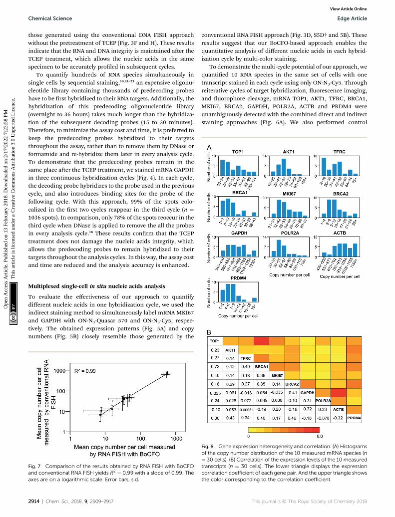
Fig. 7 Comparison of the results obtained by RNA FISH with BoCFOand conventional RNA FISH yields R2 ¼ 0.99 with a slope of 0.99. Theaxes are on a logarithmic scale. Error bars, s.d.
2914 | Chem. Sci., 2018, 9, 2909–2917
conventional RNA FISH approach (Fig. 3D, S5D† and 5B). Theseresults suggest that our BoCFO-based approach enables thequantitative analysis of different nucleic acids in each hybrid-ization cycle by multi-color staining.
To demonstrate themulti-cycle potential of our approach, wequantied 10 RNA species in the same set of cells with onetranscript stained in each cycle using only ON-N3-Cy5. Throughreiterative cycles of target hybridization, uorescence imaging,and uorophore cleavage, mRNA TOP1, AKT1, TFRC, BRCA1,MKI67, BRCA2, GAPDH, POLR2A, ACTB and PRDM4 wereunambiguously detected with the combined direct and indirectstaining approaches (Fig. 6A). We also performed control
Fig. 8 Gene expression heterogeneity and correlation. (A) Histogramsof the copy number distribution of the 10 measured mRNA species (n¼ 30 cells). (B) Correlation of the expression levels of the 10 measuredtranscripts (n ¼ 30 cells). The lower triangle displays the expressioncorrelation coefficient of each gene pair. And the upper triangle showsthe color corresponding to the correlation coefficient.
This journal is © The Royal Society of Chemistry 2018

Edge Article Chemical Science
Ope
n A
cces
s A
rtic
le. P
ublis
hed
on 1
3 Fe
brua
ry 2
018.
Dow
nloa
ded
on 2
/17/
2022
7:2
3:58
PM
. T
his
artic
le is
lice
nsed
und
er a
Cre
ativ
e C
omm
ons
Attr
ibut
ion
3.0
Unp
orte
d L
icen
ce.
View Article Online
experiments to stain these 10 RNA species in 10 different sets ofcells using the conventional RNA FISH method (Fig. 6B). Theexpression patterns obtained by these two approaches (Fig. 6Aand B) closely resemble each other. To evaluate the accuracy ofour approach, we measured the average copy numbers of tran-scripts per cell generated by our approach and conventionalRNA FISH. For all the 10 transcripts with copy numbers per cellranging from 10 to 1000, the results obtained by the twomethods (Fig. S6A†), together with those reported previouslyusing RNA-Seq,31 are consistent with each other. Comparison ofthe results obtained using our method and conventional RNAFISH yields an R2 value of 0.99 with a slope of 0.99 (Fig. 7). Theseresults conrm that the nucleic acids integrity is maintainedfollowing the repeated TCEP treatment. We also compared thesignal to noise ratios generated by our approach and conven-tional RNA FISH (Fig. S6B†). The results obtained by bothmethods are similar for all the measure transcripts. Theseresults demonstrate that the BoCFO-based approach enablesquantitative and comprehensive nucleic acids proling insingle cells in situ by multi-cycle staining.
Expression heterogeneity and correlation
As demonstrated inmany experiments, genetically identical cellscan exhibit signicant cell-to-cell variations in gene expres-sion.32–38 Our BoCFO-based single-cell nucleic acids prolingapproach allows the investigation of such cell-to-cell expressionheterogeneity. As shown in Fig. 8A, the RNA copy numbers percell are distributed in a wide range. This signicant expressionvariation leads to the relatively large error bars in Fig. 7. For allthe 10 measured transcripts, the square of the expression stan-dard deviation is much higher than the mean copy numbers.These results suggest that the 10 measured transcripts aregenerated in bursts rather than at a constant rate.39
To study expression correlation of different RNA species,bulk cell experiments usually require external stimuli to intro-duce gene expression variation. At the single-cell level,stochastic gene expression generates expression variation inindividual cells naturally. This allows us to perform single-cellexpression correlation analysis to study whether transcriptionof different genes is coordinated. Using this approach, weexamined the pairwise expression covariation of the 10
Fig. 9 (A) Ki-67 protein (yellow), mRNA MKI67 (green) and genomic locuand ON-N3-Cy5, respectively. Cell nuclei are stained with DAPI (blue). Scprotein Ki-67, each spot corresponds to one cell with transcript copy nu
This journal is © The Royal Society of Chemistry 2018
measured transcripts (Fig. S7†), and calculated the corre-sponding correlation coefficient of each transcript pair (Fig. 8B).These correlation coefficients range from �0.41 to 0.73, sug-gesting that the synthesis of these measured transcripts areheterogeneously coordinated.
Integrated DNA, RNA and protein analysis
Combined analysis of nucleic acids and proteins in the samespecimen in situ is of increasing importance in disease diag-nosis40 and studies of gene expression regulation.41 Recently,our laboratory developed cleavable uorescent antibodies formultiplexed single-cell in situ protein analysis.42,43 We demon-strated that the uorophores tethered to antibodies througha cleavable linker can be efficiently cleaved using TCEP withoutloss of protein antigenicity. We also documented that compre-hensive in situ protein proling can be achieved throughcontinuous cycles of protein binding, uorescence imaging anduorophore cleavage.
To test the hypothesis of applying BoCFO together withcleavable uorescent antibodies (CFA) for integrated DNA, RNAand protein in situ proling, we stained protein Ki67, mRNAMKI67 and genomic locus 4p16.1 in the same set of cells. Cellswere rst incubated with cleavable Cy5 conjugated antibodies tostain protein Ki67 (Fig. S8A†). Aer removing the uorescencesignals with TCEP, mRNA MKI67 (Fig. S8B†) and genomic locus4p16.1 (Fig. S8C†) were sequentially stained with ON-N3-Cy5using the indirect staining approach. The obtained spatialdistributions (Fig. S8A–C†) and abundances (Fig. S9A–C†)closely resemble those (Fig. S8D†, 3D, F and S9A–C†) generatedby conventional immunouorescence and FISH methods.These results indicate our approach enables the direct visuali-zation (Fig. 9A) and quantitative analysis of DNA, RNA andprotein molecules together in the same specimen.
To study whether the copy numbers of transcripts can be usedto predict the abundances of the corresponding proteins, weperformed the single-cell RNA-protein expression correlationanalysis. This analysis of mRNA MKI67 and protein Ki67 yieldsthe correlation coefficient value of 0.54 (Fig. 9B). These resultsare in line with the weak correlations betweenmRNA and proteinlevels reported previously,44 and suggest that post-transcriptionalregulation plays an import role on protein synthesis.
s 4p16.1 (red) are sequentially detected with Ab-N3-Cy5, ON-N3-Cy5ale bars, 5 mm. (B) Raw expression correlation data of mRNA MKI67 andmbers in the x axis and protein expression levels in the y axis.
Chem. Sci., 2018, 9, 2909–2917 | 2915

Chemical Science Edge Article
Ope
n A
cces
s A
rtic
le. P
ublis
hed
on 1
3 Fe
brua
ry 2
018.
Dow
nloa
ded
on 2
/17/
2022
7:2
3:58
PM
. T
his
artic
le is
lice
nsed
und
er a
Cre
ativ
e C
omm
ons
Attr
ibut
ion
3.0
Unp
orte
d L
icen
ce.
View Article Online
Conclusions
We have designed and synthesized BoCFO, and applied themfor multiplexed single cell in situ nucleic acids proling.Compared with the existing technologies, our approach has thefollowing advantages. (i) In this method, nucleic acids targetsare detected directly by in situ hybridization without targetsequence amplication. Therefore, transcripts and genomicloci can be visualized at the single-molecule sensitivity. (ii) Ourtechnology has high multiplexing capacity as it allows a largenumber of the same or different nucleic acids to be detected indifferent analysis cycles by sequential staining or reiterativehybridization, respectively. (iii) The TCEP treatment simulta-neously cleaves all the different uorophores in the wholespecimen within 30 minutes. Thus, our method has highsample throughput, and permits a large number of cells to beanalyzed in a short time. (iv) As BoCFO has high signal removalefficiency and avoids the cross-reactions with endogenousbiomolecules and other probes, our approach has enhancedsignal to noise ratio and analysis accuracy. (v) Rather than re-hybridizing the expensive target-binding oligonucleotidelibrary in every analysis cycle, our technology only applies thistime-consuming hybridization in the rst cycle. Therefore, ourmethod is more time- and cost-effective. (vi) As the smallcleaved uorophores diffuse out faster than the large strippedoligonucleotide probes, our technology facilitates the analysisof intact tissues. (vii) By cleaving the uorophores while main-taining the integrity of almost all the biomolecules, ourapproach can be applied for the integrated single-cell in situDNA, RNA and protein analysis.
The number of nucleic acids that can be quantied in singlecells using this BoCFO-based approach depends on two factors:the number of hybridization cycles and the number of uo-rophores applied in each cycle. As we have shown, TCEP canefficiently remove the uorophores within 30minutes, while theintegrity of RNA and DNA is preserved aer the treatment withTCEP for at least 24 hours. This suggests that the cyclingnumber can be further increased signicantly. Additionally,classical uorophores with four or ve varied colors can beapplied simultaneously to visualize different nucleic acids inone hybridization cycle. And multispectral uorophorescoupled with the hyperspectral imaging method45 will enablemore uorophores to be differentiated and applied in eachhybridization cycle. Therefore, by combining reiterativehybridization and sequential staining to quantify nucleic acidswith high and low copy numbers, respectively, we envision thatthis BoCFO-based approach has the potential to detecthundreds to thousands of nucleic acids species at the singlemolecule sensitivity in single cells in situ. Additionally, theBoCFO probes developed here integrated with cleavable uo-rescent antibodies we reported previously enable the compre-hensive and integrated DNA, RNA and protein proling at theoptical resolution in single cells. This highly multiplexedimaging platform will bring new insights into cell signalingnetwork, gene expression regulation, molecular diagnosis andcellular targeted therapy.
2916 | Chem. Sci., 2018, 9, 2909–2917
Conflicts of interest
J. G. is an inventor on a patent application led by Arizona StateUniversity that covers the method of using bioorthogonalcleavable uorescent oligonucleotides for multiplexed nucleicacids analysis.
Acknowledgements
We are grateful for support from the National Institute OfAllergy And Infectious Diseases of the National Institutes ofHealth (R21AI132840), Arizona State University startup funds,Arizona State University/Mayo Clinic seed grant (ARI-219693)and Cystic Fibrosis Foundation (FIRTH17XX0).
Notes and references
1 N. Crosetto, M. Bienko and A. Van Oudenaarden, Nat. Rev.Genet., 2014, 16, 57–66.
2 J. D. Hoheisel, Nat. Rev. Genet., 2006, 7, 200–210.3 M. L. Metzker, Nat. Rev. Genet., 2010, 11, 31–46.4 J. Guo, L. Yu, N. J. Turro and J. Ju, Acc. Chem. Res., 2010, 43,551–563.
5 S. Bose, Z. Wan, A. Carr, A. Rizvi, G. Vieira, D. Pe'er andP. Sims, Genome Biol., 2015, 16, 120.
6 J. Yuan and P. A. Sims, Sci. Rep., 2016, 33883.7 A. Raj, P. Van Den Bogaard, S. A. Riin, A. Van Oudenaardenand S. Tyagi, Nat. Methods, 2008, 5, 877–879.
8 J. Fei, D. Singh, Q. Zhang, S. Park, D. Balasubramanian,I. Golding, C. K. Vanderpool and T. Ha, Science, 2015, 347,1371–1374.
9 K. Huang and A. a. Martı, Anal. Bioanal. Chem., 2012, 402,3091–3102.
10 A. A. Martı, S. Jockusch, N. Stevens, J. Ju and N. J. Turro, Acc.Chem. Res., 2007, 40, 402–409.
11 R. M. Franzini and E. T. Kool, J. Am. Chem. Soc., 2009, 131,16021–16023.
12 B. J. Beliveau, E. F. Joyce, N. Apostolopoulos, F. Yilmaz,C. Y. Fonseka, R. B. McCole, Y. Chang, J. B. Li,T. N. Senaratne, B. R. Williams, J.-M. Rouillard and C. Wu,Proc. Natl. Acad. Sci. U. S. A., 2012, 109, 21301–21306.
13 J. H. Lee, E. R. Daugharthy, J. Scheiman, R. Kalhor,J. L. Yang, T. C. Ferrante, R. Terry, S. S. F. Jeanty, C. Li,R. Amamoto, D. T. Peters, B. M. Turczyk,A. H. Marblestone, S. a. Inverso, A. Bernard, P. Mali,X. Rios, J. Aach and G. M. Church, Science, 2014, 343,1360–1363.
14 R. Ke, M. Mignardi, A. Pacureanu, J. Svedlund, J. Botling,C. Wahlby and M. Nilsson, Nat. Methods, 2013, 10, 857–860.
15 J. M. Levsky, S. M. Shenoy, R. C. Pezo and R. H. Singer,Science, 2002, 297, 836–840.
16 E. Lubeck and L. Cai, Nat. Methods, 2012, 9, 743–748.17 M. J. Levesque and A. Raj, Nat. Methods, 2013, 10, 246–248.18 E. Lubeck, A. F. Coskun, T. Zhiyentayev, M. Ahmad and
L. Cai, Nat. Methods, 2014, 11, 360–361.19 S. Shah, E. Lubeck, W. Zhou and L. Cai, Neuron, 2016, 92,
342–357.
This journal is © The Royal Society of Chemistry 2018

Edge Article Chemical Science
Ope
n A
cces
s A
rtic
le. P
ublis
hed
on 1
3 Fe
brua
ry 2
018.
Dow
nloa
ded
on 2
/17/
2022
7:2
3:58
PM
. T
his
artic
le is
lice
nsed
und
er a
Cre
ativ
e C
omm
ons
Attr
ibut
ion
3.0
Unp
orte
d L
icen
ce.
View Article Online
20 Y. Takei, S. Shah, S. Harvey, L. S. Qi and L. Cai, Biophys. J.,2017, 112, 1773–1776.
21 K. H. Chen, A. N. Boettiger, J. R. Moffitt, S. Wang andX. Zhuang, Science, 2015, 1363, 1360–1363.
22 J. R. Moffitt, J. Hao, D. Bambah-Mukku, T. Lu, C. Dulac andX. Zhuang, Proc. Natl. Acad. Sci. U. S. A., 2016, 113, 14456–14461.
23 J. R. Moffitt, J. Hao, G. Wang, K. H. Chen, H. P. Babcock andX. Zhuang, Proc. Natl. Acad. Sci. U. S. A., 2016, 113, 11046–11051.
24 S. M. Shaffer, M. C. Dunagin, S. R. Torborg, E. A. Torre,B. Emert, C. Krepler, M. Beqiri, K. Sproesser,P. A. Brafford, M. Xiao, E. Eggan, I. N. Anastopoulos,C. A. Vargas-Garcia, A. Singh, K. L. Nathanson, M. Herlynand A. Raj, Nature, 2017, 546, 431–435.
25 L. Xiao and J. Guo, Anal. Methods, 2015, 7, 7290–7295.26 J. Guan, H. Liu, X. Shi, S. Feng and B. Huang, Biophys. J.,
2017, 112, 1077–1084.27 J. Guo, N. Xu, Z. Li, S. Zhang, J. Wu, D. H. Kim, M. S. Marma,
N. J. Turro and J. Ju, Proc. Natl. Acad. Sci. U. S. A., 2008, 105,3–8.
28 J. A. Prescher, D. H. Dube and C. R. Bertozzi, Nature, 2004,430, 873–877.
29 J. C. Jewett and C. R. Bertozzi, Chem. Soc. Rev., 2010, 39,1272–1279.
30 D. R. Bentley, S. Balasubramanian, H. P. Swerdlow,G. P. Smith, J. Milton, C. G. Brown, K. P. Hall, D. J. Evers,C. L. Barnes, H. R. Bignell, J. M. Boutell, J. Bryant,R. J. Carter, R. Keira Cheetham, A. J. Cox, D. J. Ellis,M. R. Flatbush, N. A. Gormley, S. J. Humphray, L. J. Irving,M. S. Karbelashvili, S. M. Kirk, H. Li, X. Liu,K. S. Maisinger, L. J. Murray, B. Obradovic, T. Ost,M. L. Parkinson, M. R. Pratt, I. M. J. Rasolonjatovo,M. T. Reed, R. Rigatti, C. Rodighiero, M. T. Ross, A. Sabot,S. V. Sankar, A. Scally, G. P. Schroth, M. E. Smith,V. P. Smith, A. Spiridou, P. E. Torrance, S. S. Tzonev,E. H. Vermaas, K. Walter, X. Wu, L. Zhang, M. D. Alam,C. Anastasi, I. C. Aniebo, D. M. D. Bailey, I. R. Bancarz,S. Banerjee, S. G. Barbour, P. A. Baybayan, V. A. Benoit,K. F. Benson, C. Bevis, P. J. Black, A. Boodhun,J. S. Brennan, J. A. Bridgham, R. C. Brown, A. A. Brown,D. H. Buermann, A. A. Bundu, J. C. Burrows, N. P. Carter,N. Castillo, M. C. E. Catenazzi, S. Chang, R. Neil Cooley,N. R. Crake, O. O. Dada, K. D. Diakoumakos,B. Dominguez-Fernandez, D. J. Earnshaw, U. C. Egbujor,D. W. Elmore, S. S. Etchin, M. R. Ewan, M. Fedurco,L. J. Fraser, K. V. Fuentes Fajardo, W. Scott Furey,D. George, K. J. Gietzen, C. P. Goddard, G. S. Golda,P. A. Granieri, D. E. Green, D. L. Gustafson, N. F. Hansen,K. Harnish, C. D. Haudenschild, N. I. Heyer, M. M. Hims,J. T. Ho, A. M. Horgan, K. Hoschler, S. Hurwitz,D. V. Ivanov, M. Q. Johnson, T. James, T. A. Huw Jones,G. D. Kang, T. H. Kerelska, A. D. Kersey, I. Khrebtukova,A. P. Kindwall, Z. Kingsbury, P. I. Kokko-Gonzales,A. Kumar, M. A. Laurent, C. T. Lawley, S. E. Lee, X. Lee,
This journal is © The Royal Society of Chemistry 2018
A. K. Liao, J. A. Loch, M. Lok, S. Luo, R. M. Mammen,J. W. Martin, P. G. McCauley, P. McNitt, P. Mehta,K. W. Moon, J. W. Mullens, T. Newington, Z. Ning, B. LingNg, S. M. Novo, M. J. O'Neill, M. A. Osborne, A. Osnowski,O. Ostadan, L. L. Paraschos, L. Pickering, A. C. Pike,A. C. Pike, D. Chris Pinkard, D. P. Pliskin, J. Podhasky,V. J. Quijano, C. Raczy, V. H. Rae, S. R. Rawlings, A. ChivaRodriguez, P. M. Roe, J. Rogers, M. C. Rogert Bacigalupo,N. Romanov, A. Romieu, R. K. Roth, N. J. Rourke,S. T. Ruediger, E. Rusman, R. M. Sanches-Kuiper,M. R. Schenker, J. M. Seoane, R. J. Shaw, M. K. Shiver,S. W. Short, N. L. Sizto, J. P. Sluis, M. A. Smith, J. ErnestSohna Sohna, E. J. Spence, K. Stevens, N. Sutton,L. Szajkowski, C. L. Tregidgo, G. Turcatti, S. Vandevondele,Y. Verhovsky, S. M. Virk, S. Wakelin, G. C. Walcott,J. Wang, G. J. Worsley, J. Yan, L. Yau, M. Zuerlein,J. Rogers, J. C. Mullikin, M. E. Hurles, N. J. McCooke,J. S. West, F. L. Oaks, P. L. Lundberg, D. Klenerman,R. Durbin and A. J. Smith, Nature, 2008, 456, 53–59.
31 M. Uhlen, L. Fagerberg, B. M. Hallstrom, C. Lindskog,P. Oksvold, A. Mardinoglu, A. Sivertsson, C. Kampf,E. Sjostedt, A. Asplund, I. Olsson, K. Edlund, E. Lundberg,S. Navani, C. A. Szigyarto, J. Odeberg, D. Djureinovic,J. O. Takanen, S. Hober, T. Alm, P. Edqvist, H. Berling,H. Tegel, J. Mulder, J. Rockberg, P. Nilsson, J. M. Schwenk,M. Hamsten, K. Von Feilitzen, M. Forsberg, L. Persson,F. Johansson, M. Zwahlen, G. Von Heijne, J. Nielsen andF. Ponten, Science, 2015, 347, 1260419.
32 A. Becskei, B. B. Kaufmann and A. van Oudenaarden, Nat.Genet., 2005, 37, 937–944.
33 W. J. Blake, M. KAErn, C. R. Cantor and J. J. Collins, Nature,2003, 422, 633–637.
34 M. B. Elowitz, A. J. Levine, E. D. Siggia and P. S. Swain,Science, 2002, 297, 1183–1186.
35 I. Golding, J. Paulsson, S. M. Zawilski and E. C. Cox, Cell,2005, 123, 1025–1036.
36 E. M. Ozbudak, M. Thattai, I. Kurtser, A. D. Grossman andA. van Oudenaarden, Nat. Genet., 2002, 31, 69–73.
37 J. M. Raser and E. K. O'Shea, Science, 2004, 304, 1811–1814.38 N. Rosenfeld, J. W. Young, U. Alon, P. S. Swain and
M. B. Elowitz, Science, 2005, 307, 1962–1965.39 A. Raj, C. S. Peskin, D. Tranchina, D. Y. Vargas and S. Tyagi,
PLoS Biol., 2006, 4, 1707–1719.40 H. Nitta, B. D. Kelly, M. Padilla, N. Wick, P. Brunhoeber,
I. Bai, S. Singh, J. Ranger-Moore, C. Bieniarz, H. Tsuda andT. M. Grogan, Diagn. Pathol., 2012, 7, 60.
41 J. Chaumeil, M. Micsinai and J. a. Skok, J. Visualized Exp.,2013, e50087.
42 M. Mondal, R. Liao, L. Xiao, T. Eno and J. Guo, Angew. Chem.,Int. Ed., 2017, 56, 2636–2639.
43 J. Guo, US Pat. Application, 20160054308A1, 2016.44 T. Maier, M. Guell and L. Serrano, FEBS Lett., 2009, 583,
3966–3973.45 Y. Garini, I. T. Young and G. McNamara, Cytometry, Part A,
2006, 69, 735–747.
Chem. Sci., 2018, 9, 2909–2917 | 2917