Interchannel Nonlinearities in Polarization-Multiplexed Transmission
Multiplexed RNA structure characterizationwith selective 2′-hydroxyl acylation analyzedby primer extension sequencing (SHAPE-Seq)Julius B. Lucksa,b,1, Stefanie A. Mortimerc, Cole Trapnelld,e, Shujun Luof, Sharon Avirana, Gary P. Schrothf, Lior Pachterc,g,h,Jennifer A. Doudnac,i,j,k,2, and Adam P. Arkina,k,3
aDepartment of Bioengineering, University of California, Berkeley, CA 94720; bMiller Institute for Basic Research in Science, Berkeley, CA 94720;cDepartment of Molecular and Cell Biology, University of California, Berkeley, CA 94720; iHoward Hughes Medical Institute; gDepartment of Mathematics,University of California, Berkeley, CA 94720; dDepartment of Stem Cell and Regenerative Biology, Harvard University, Harvard, MA, 02138; eThe BroadInstitute of MIT and Harvard, Cambridge, MA 02142; fIllumina Inc., Hayward, CA 94545; hDepartment of Electrical Engineering and Computer Science,University of California, Berkeley, CA 94720; jDepartment of Chemistry, University of California, Berkeley, CA 94720; and kPhysical Biosciences Division,Lawrence Berkeley National Laboratories, Berkeley, CA 94720
Contributed by Jennifer A. Doudna, May 1, 2011 (sent for review February 9, 2011)
New regulatory roles continue to emerge for both natural andengineered noncoding RNAs, many of which have specific second-ary and tertiary structures essential to their function. Thus there isa growing need to develop technologies that enable rapid charac-terization of structural features within complex RNA populations.We have developed a high-throughput technique, SHAPE-Seq, thatcan simultaneously measure quantitative, single nucleotide-resolu-tion secondary and tertiary structural information for hundreds ofRNA molecules of arbitrary sequence. SHAPE-Seq combines selec-tive 2′-hydroxyl acylation analyzed by primer extension (SHAPE)chemistry with multiplexed paired-end deep sequencing of primerextension products. This generates millions of sequencing reads,which are then analyzed using a fully automated data analysispipeline, based on a rigorous maximum likelihood model of theSHAPE-Seq experiment. We demonstrate the ability of SHAPE-Seq to accurately infer secondary and tertiary structural informa-tion, detect subtle conformational changes due to single nucleotidepoint mutations, and simultaneously measure the structures of acomplex pool of different RNA molecules. SHAPE-Seq thus repre-sents a powerful step toward making the study of RNA secondaryand tertiary structures high throughput and accessible to a widearray of scientific pursuits, from fundamental biological investiga-tions to engineering RNA for synthetic biological systems.
chemical probing ∣ RNA sequencing ∣ RNA folding ∣ genomics
Over the past several years, there has been an explosion in thediscovery of noncoding, but functional RNAs that play cen-
tral roles in maintaining, regulating, and defending the genome(1). At the same time, RNA-based mechanisms have emerged aspowerful tools for engineering synthetic biological systems (2).Many of these natural and synthetic RNAs have specific second-ary and tertiary structures essential to their function, and there isa growing need to develop technologies that enable rapid char-acterization of structural features within complex RNA popula-tions. Such a high-throughput structure characterization assaywould allow rapid assessment of the impact of sequence on struc-ture and function and enable RNA engineers to design librariesof RNA molecules with desired structural properties.
Two techniques for high-throughput RNA structure character-ization have recently been reported: parallel analysis of RNAstructures (PARS) (3) and fragmentation sequencing (Frag-Seq) (4). Both techniques couple classic in vitro nuclease probingtechniques that are traditionally performed one RNA at a time,with deep sequencing of RNA fragments to simultaneously probea complex mixture of RNAs sampled from transcriptomes.Although important first steps, these techniques provide onlylow-resolution secondary structure information due to the limita-tions inherent in nuclease probing (5).
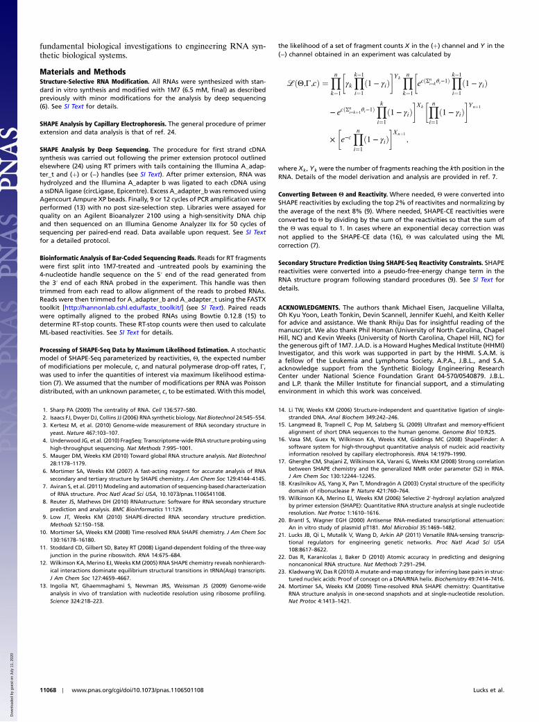
We have developed a high-throughput technique, SHAPE-Seq, that can simultaneously measure quantitative, single nucleo-tide-resolution secondary and tertiary structure information forhundreds of RNA molecules of arbitrary sequence. SHAPE-Seq combines selective 2′-hydroxyl acylation analyzed by primerextension (SHAPE) chemistry (6) with a multiplexed hierarchicalbar coding and deep sequencing strategy, enabling parallel invitro structure probing experiments in one test tube (Fig. 1).We have also developed a maximum-likelihood (ML) estimationstrategy for inferring nucleotide reactivities that rigorously incor-porates information from co-sequenced control experiments (7).Combined with bioinformatics software to process, bin, and mapraw sequence reads, this creates a fully automated data analysispipeline. Furthermore, the SHAPE reactivities that are the out-put of this pipeline are well established and can be immediatelyused in existing RNA folding algorithms to determine the struc-tures for each RNA molecule (8, 9).
In this work, we show that SHAPE-Seq is able to accuratelyinfer both secondary and tertiary structural information for themodel RNA fold of the Bacillus subtilis RNase P specificitydomain. Furthermore, we show that SHAPE-Seq can infer thisinformation from hundreds of bar-coded copies of the RNaseP RNA in a single sample. Finally we use this technique to simul-taneously infer local structural changes in RNase P due to singlepoint mutations and to determine the structures of two variants ofthe Staphylococcus aureus plasmid pT181 transcriptional attenua-tor, all within the same mixture.
ResultsThe SHAPE-Seq Pipeline. The goal of SHAPE-Seq is to accuratelyinfer nucleotide-resolution structural information through simul-taneous SHAPE probing of a mixture of RNA species (Fig. 1).To explicitly distinguish the species, each RNA in the experiment
Author contributions: J.B.L., S.A.M., C.T., S.L., S.A., G.P.S., L.P., J.A.D., and A.P.A. designedresearch; J.B.L., S.A.M., C.T., S.L., and S.A. performed research; J.B.L., S.A.M., C.T.,S.L., and S.A. contributed new reagents/analytic tools; J.B.L., S.A.M., C.T., S.L., S.A., andL.P. analyzed data; and J.B.L., S.A.M., C.T., S.L., S.A., G.P.S., L.P., J.A.D., and A.P.A. wrotethe paper.
The authors declare no conflict of interest.
Freely available online through the PNAS open access option.
See Commentary on page 10933.1To whom correspondence may be addressed at: School of Chemical and BiomolecularEngineering, Cornell University, 120 Olin Hall, Ithaca, NY 14853. E-mail: [email protected].
2To whom correspondence may be addressed. E-mail: [email protected] whom correspondence may be addressed at: E. O. Lawrence Berkeley NationalLaboratory, 1 Cyclotron Road, MS Stanley-922, Berkeley, CA 94720. E-mail: [email protected].
This article contains supporting information online at www.pnas.org/lookup/suppl/doi:10.1073/pnas.1106501108/-/DCSupplemental.
www.pnas.org/cgi/doi/10.1073/pnas.1106501108 PNAS ∣ July 5, 2011 ∣ vol. 108 ∣ no. 27 ∣ 11063–11068
STAT
ISTICS
BIOPH
YSICSAND
COMPU
TATIONALBIOLO
GY
SEECO
MMEN
TARY
Dow
nloa
ded
by g
uest
on
July
11,
202
0
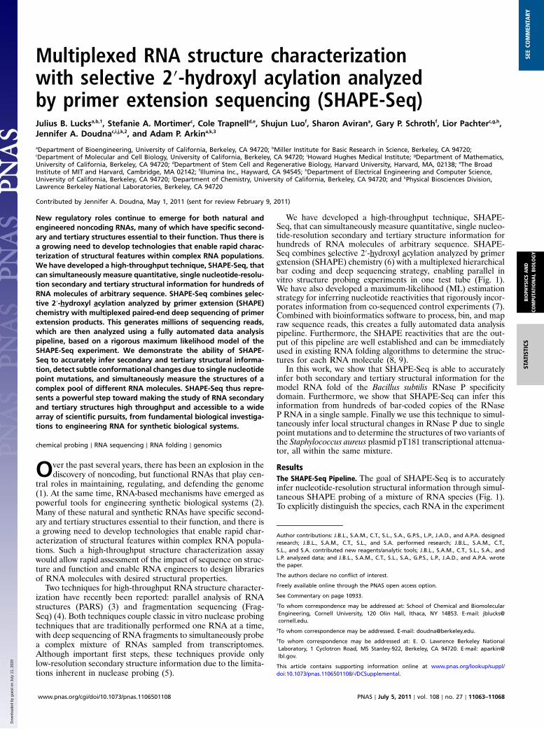
is bar-coded with a unique nucleotide sequence near the 3′ endof the RNA (Fig. S1). These RNAs are then mixed and foldedunder the desired in vitro conditions, which can include any ofthe wide array of buffers (10), ligands (11), temperatures (12),and other variables already established for conventional SHAPE.Once folded, the pool is split into two samples, one of which (þ)is treated with a SHAPE reagent [here 1M7 (6)], and the other(−) is treated with a control solvent. These pools then undergoconversion to cDNA through a reverse transcription (RT) processthat is blocked by 1M7 modification (6), generating bar-codeddistributions of different length cDNAs that represent locationsof 1M7 modification (þ), or processes such as transcriptase drop-off that cause bias in reverse transcription (−).
The (þ) and (−) pools are kept separate during the RTstep sothat they can be tagged with an additional bar code attached tothe 5′ tail of the RT primer, called a “handle” (Fig. 1A). The han-dles identify cDNA fragments as coming from the (þ) or (−)channels when the cDNA pools are simultaneously sequenced to-gether as a single mixture. Sequencing of the cDNA is performedusing paired-end Illumina sequencing (13). To add the requiredIllumina sequencing adapters to cDNA products, one of theadapters was included in the tail of the RT primers, and the otherwas added through a single-stranded DNA ligation step after theRNA was removed by NaOH hydrolysis (see Materials and Meth-ods). The single-stranded DNA ligation step is performed atelevated temperatures with a thermostable ligase, and with ablocking group on the 3′ end of the adapter to prevent adapterconcatemerization (14). After nine rounds of PCR amplification,the libraries are sequenced on an Illumina Genome Analyzer IIxplatform in paired-end mode. Only 50 nucleotides need to be se-quenced on each end because the two pieces of informationneeded—the SHAPE modification position and the RNA iden-tity (bar code)—are on opposite ends of the cDNA molecules.This obviated the need for a size-selection step, which has limitedthe structural information obtainable using other methods (4).
After sequencing, the reads are binned according to the handlesequence (Fig. 1B). The Illumina platform uses randomness inthe first four nucleotide incorporations to calibrate for spectraloverlap and cluster identification. Because the handles are thefirst nucleotides sequenced, we chose sets of handle sequences
to represent the (þ) and (−) reads, RRRY (R ¼ A;G; Y ¼ C;T)for (þ) and YYYR for (−). This guaranteed that at each positionof the handle, an equal mixture of A, T, C, and G is sequenced.Reads were first separated by handle, then bar code, and alignedto the appropriate RNA molecule sequence using the Bowtiealignment package (15), creating nucleotide-resolution countdistributions in the (þ) and (−) channels.
The digital nature of direct cDNA sequencing allows SHAPE-Seq data to be amenable to rigorous and fully automatedmathematical analysis. In conventional SHAPE experiments,fluorescently labeled cDNAs are typically quantified by capillaryelectrophoresis (SHAPE-CE), which requires a series of manualdata analysis steps associated with correcting channel mobilities,aligning, and integrating the analog electropherogram intensitiesinto (þ) and (−) distributions (16). The (þ) and (−) distributionsare subtracted to give the final output of the SHAPE experiment:a SHAPE “reactivity” for each nucleotide that represents the pro-pensity for 1M7 adduct formation at that position. Previous workcomparing SHAPE reactivities to NMR order parameters hasshown that reactivities correlate strongly with local spatial disor-der and are thus a measure of structural dynamics (17). In gen-eral, high reactivities are interpreted as nucleotides that are onaverage unstructured and low reactivities are interpreted as nu-cleotides that are constrained by canonical or noncanonical, sec-ondary or tertiary interactions. Before the subtraction of the twodistributions, two corrections are typically applied: The (þ) chan-nel intensities are adjusted by an exponential decay factor thatcorrects for fragment distribution decay resulting from the uni-directional RT process stopping at the first encountered adduct,and the (−) channel is scaled by a constant factor so that unreac-tive sites have a reactivity of zero when the two channels are sub-tracted. In addition to being manual, both of these steps requireexpert knowledge making it in general prohibitive to apply thestandard SHAPE data analysis pipeline to hundreds of raw(þ) and (−) distributions generated by SHAPE-Seq.
To overcome this barrier, we developed a rigorous, automatedmathematical framework that can be applied to find the optimalset of reactivities that are most consistent with the observed (þ)and (−) distributions [see Materials and Methods (7)]. The modeluses ML estimation to output a set of reactivities, Θ, and theestimated average number of modifications per cite, c.
A
N
OO O
NO2
1M7 =
Illumina Genome Analyzer Paired-end read
= adapter b
= adapter t
DMSOControl
(RNA–Seq)
inputRNAs
(+) handle primer (–) handle primer
RNA hydrolysisand
ssDNA ligation
ReverseTranscription5’-OH
5’-P 3’-C3
5’-OH
5’-P 3’-C3
B
Read Alignment and Separation by Bar codes
Handle Binning
CAAAGAAAAAAACACTGAGTTGTTTTTATAATCTTG
Cou
nts
(+) Handles
(-) Handles
CAAAGAAAAAAACACTGAGTTGTTTTTATAATCTTGRea
ctiv
ity
Automated Maximum Likelihood Estimation
For each RNA
(+) RRRY (-) YYYR
AU
A A G G A U UC C A A U G U AA U
G CA
U AC GG C
C AU AC GA UC GG U
C UC UC GU A
G AA A
C C
A UG CA U
A UA UA UA UA UC GA U
C UU G
G A
A
A
A
SHAPE-Seq Constrained Secondary Structure Prediction
AAACAGAT
AGAT
AAACCCCA
TTCG
CCCATTCG
Fig. 1. Overview of SHAPE-Seq. (A) Experimental pipeline. A DNA bar code is added to the 3′ end of template molecules, enabling SHAPE chemistry andsequencing library generation to be done on a mixture of bar-coded RNAs. (B) Bioinformatics and analysis pipeline. The automated pipeline separates reads byhandle pools and bar code and maps the reads onto RNA sequences. Raw read counts at each nucleotide position in the (þ) and (−) channel are fed into a MLestimation calculation to determine the reactivities at each nucleotide, Θ. Θ can be scaled and used in programs such as RNAStructure (9) to infer secondarystructure from SHAPE-Seq data.
11064 ∣ www.pnas.org/cgi/doi/10.1073/pnas.1106501108 Lucks et al.
Dow
nloa
ded
by g
uest
on
July
11,
202
0
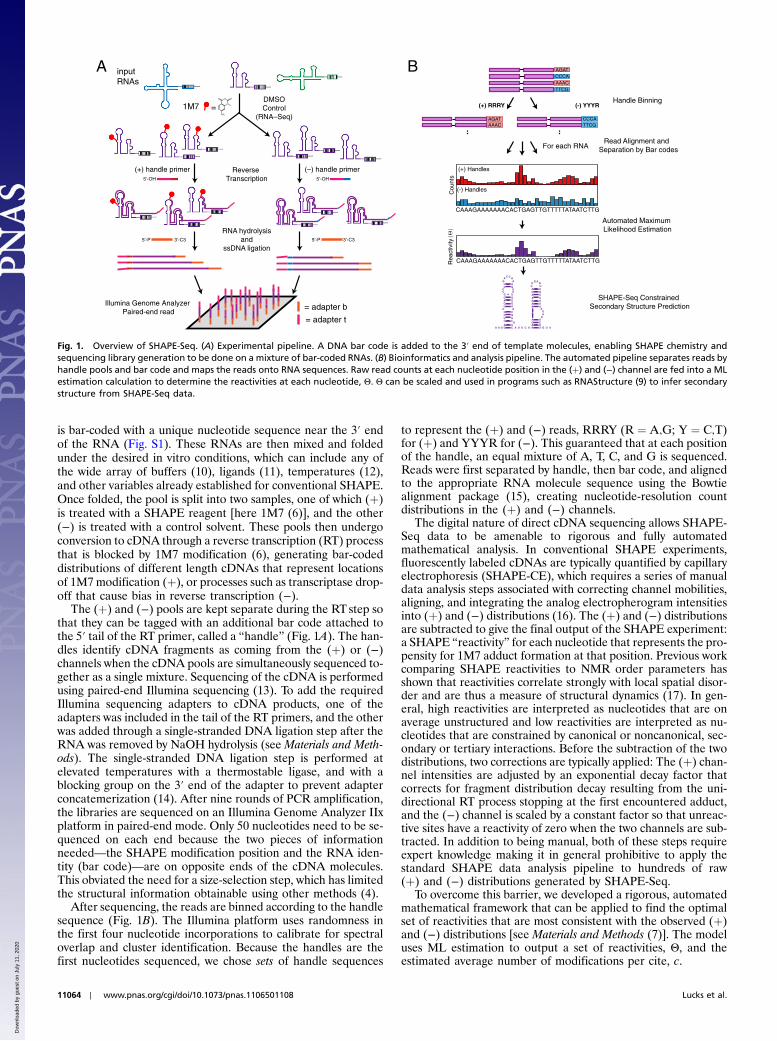
SHAPE-Seq Accurately Infers the Secondary and Tertiary Structure of aHighly Conserved Catalytic RNA. As an initial test of the SHAPE-Seq platform, we probed the specificity domain of the highly con-served catalytic RNA, RNase P, from B. subtilis, which has beenextensively characterized using conventional SHAPE with capil-lary electrophoresis (6). Furthermore, as determined by X-raycrystallography, the RNase P fold is highly structured, withwell-defined tertiary interactions (18), making it an ideal candi-date for an initial test of the method.
Fig. 2 shows an overlay of SHAPE-Seq reactivities on theknown structure of RNase P. Over 7.5 million sequencing readswere used to compute Θ values for each nucleotide of the mole-cule, which were then converted into conventional SHAPE reac-tivities (see Materials and Methods). Immediately apparent aretwo regions of highly reactive nucleotides in the loops of helicesP8 and P9 as expected because these loops are known to beunpaired. In contrast, the P8 and P9 stems show extremely lowreactivities due to extensive base pairing. Over 200,000 fragmentswere mapped in this region with near perfect cancellationbetween the (þ) and (−) channels, highlighting that even lowreactivities are derived from large amounts of information. Thisholds true across the entire RNase P as seen by comparing raw(þ) and (−) counts (Fig. S2).
A plot of SHAPE-Seq versus SHAPE-CE reactivities for everynucleotide of RNase P shows a high degree of correlationbetween the two (R ¼ 0.72) indicating that SHAPE-Seq accu-rately recapitulates SHAPE structure information (Fig. 2A,Inset). Pearson’s R correlation was determined both with andwithout inclusion of nucleotide G100 (asterisk in Fig, 2A, Inset).Nucleotide G100, although characterized as highly reactive inboth methods, showed a much higher reactivity in SHAPE-Seq.Because it is a highly flexible nucleotide within a highly flexibleloop, this does not change the consistency of these data with theknown RNase P structure. It was therefore considered as anoutlier based on structural interpretation. Two other nucleotides,U119 and U120, show similar behavior but to a lesser extent.These nucleotides are also within a highly flexible loop and arealso characterized as highly reactive according to SHAPE-CE.Although there are quantitative differences between SHAPE-Seq and SHAPE-CE at these positions, the overall structuralinterpretation is not affected by the method used.
It should be noted that several regions of the SHAPE-Seqreactivity spectrum, namely the nucleotides A130 and A194,and the P10.1 loop were not as reactive as observed by SHAPE-CE. Nucleotides A130 and A194 are single nucleotide bulges thatstack with other purines in the molecule (Fig. S3). This interac-tion is expected to cause a decrease in nucleotide flexibility andreactivity to 1M7. This is indeed what is observed in SHAPE-Seq(approximately 30% reactivity). This is nominally less than thereactivity obtained using SHAPE-CE and could be a result ofthe extra protocol steps required for SHAPE-Seq causing lesssensitivity to this type of structural effect. Importantly, however,because SHAPE-Seq displays reactivity in this region, it does notalter the interpretation of the RNase P structure. The P10.1 loopis a stable UUCG tetraloop, which contains a stabilizing GUwob-ble closing the loop and constraining these nucleotides comparedto other single-stranded regions in the RNA (Fig. S3). The lowerreactivity observed in SHAPE-Seq (approximately 15% reactive)compared to SHAPE-CE (approximately 20–70% reactive) doesnot alter the overall interpretation of the data. Despite beingunpaired on the secondary structure map, the P12 loop is knownto form a well-defined tertiary interaction with the P10.1 helixand has been previously observed to be unreactive in conven-tional SHAPE-CE experiments (6). This is indeed observed inthe SHAPE-Seq reactivity spectrum (Fig. 2A).
Fig. 2B shows an overlay of SHAPE-Seq data onto the knownthree-dimensional crystal structure of RNase P. The SHAPE-Seqreactivity data are remarkably consistent, with highly reactivenucleotides mapping onto positions of high flexibility, especiallyunpaired nucleotides that are not participating in tertiary con-tacts (Fig. S3). This demonstrates the power of SHAPE-Seq toinfer both secondary and tertiary structural information.
Bar Coding AllowsMultiplexed Structure Characterization.One of theadvantages of SHAPE-Seq is the ability to simultaneously deter-mine structural information from many RNAs at once throughdirect sequencing of 3′ RNA bar codes (Fig. 1). To test this,we added 256 different bar-coded versions of the WT RNaseP RNA into the same pool as the un-bar-coded WT RNase PRNA discussed above and carried out the SHAPE-Seq pipeline.The bar codes consisted of all four-nucleotide sequences andwere placed in the 3′ structure cassette commonly used in
C
JS D
iver
genc
e
Bar-Code Reads (x 105)
R
A
86
92
P7
P8
P9
J11/12
J12/11
P12
P10.1
5'
102
107
113
128
134
140
148
156
161
168
176
185
195
203210
216
220
225
237
1.0
0.3
0.1
3'
0.7
0
B
0 1 2 3
0.8
0.9
1.0
0.02
0.04
0.06R= 0.65 (0.72)
SHAPE–Seq
SH
AP
E–C
E
0 5 10
0
2
*
Fig. 2. SHAPE-Seq mapping of the RNase P specificity domain. (A) Overlay of SHAPE-Seq reactivities on an RNase P structure diagram (18). Θ were convertedinto reactivities by the 2%∕8% rule for purposes of visualization. (Inset) Correlation between SHAPE-Seq and SHAPE-CE reactivities. Solid line represents aslope of one. Nucleotide G100 (asterisk) was not included for the R value indicated in parenthesis. (B) Crystal structure of RNase P [from ref. 18)] with nucleo-tides color-coded by reactivity as in A. (C) The Jensen-Shannon divergence (Top) and correlation coefficient R (Bottom) calculated between each bar code andthe WT RNase P Θ, plotted versus the total number of reads mapped for each bar code. The average JS divergence and R are 0.02 and 0.99, respectively.
Lucks et al. PNAS ∣ July 5, 2011 ∣ vol. 108 ∣ no. 27 ∣ 11065
STAT
ISTICS
BIOPH
YSICSAND
COMPU
TATIONALBIOLO
GY
SEECO
MMEN
TARY
Dow
nloa
ded
by g
uest
on
July
11,
202
0
SHAPE experiments (Fig. S1). These were introduced with de-generate primers before in vitro transcription of the RNA pool(see Materials and Methods).
Over 8.6 million bar-coded reads were mapped. For each barcode, Θ and c were calculated automatically according to theSHAPE-Seq data analysis pipeline (Fig. 1). The total numberof sequenced fragments for each bar code was uneven (rangingfrom 10,488 to 308,978), most likely due to biases in randomprimer synthesis (Fig. S4). However, this gave us an opportunityto study how many fragments need to be mapped to accuratelyreconstruct SHAPE reactivity profiles. The distribution of cwas tightly peaked around a value of 0.76, which closely matchedthe WT RNase P SHAPE-Seq data where c ¼ 0.73 (Fig. S5).
The reactivities Θ from the 256 bar-coded molecules werecompared to the WT RNase P SHAPE-Seq reactivity profilein two ways. To directly compare SHAPE-Seq reactivity profiles,we computed the Jensen-Shannon (JS) divergence betweenΘbar code and ΘWT for each bar code (see SI Text). The JS diver-gence takes into account the reactivity information at every nu-cleotide and has a value in between 0 and 1. It is a symmetricmeasure of the similarity between two distributions with identicaldistributions having JS divergence ¼ 0. As shown in Fig. 2C, thehistogram of the JS divergences for each bar code is extremelytightly peaked with an average JS divergence of 0.02. As anothermeasure, we computed Pearson correlation coefficients, R,between the SHAPE-Seq Θ of bar-coded and WT RNase P.The correlation coefficient measures the degree to which thetwo datasets lie on a line and ranges from −1 for anticorrelateddata to 1 for perfectly correlated data. Fig. 2C shows a histogramof R for all bar codes, which again is tightly peaked with an aver-age of 0.99.
As shown in Fig. 2, both of these measures have a very weakdependence on the total number of fragments mapped for eachbar code. The GGGG bar code was the least represented with10,488 total hits spread over 204 nucleotides and had a SHAPE-Seq reactivity profile that was extremely similar to the non-bar-coded RNase P (R ¼ 0.99, JSD ¼ 0.04) (Fig. S6). By dividing bythe total fragment counts observed for each bar code, we estimatethat an upper bound of 0.1 pmol of RNA is needed to recover aSHAPE-Seq reactivity profile for RNase P. This is in contrast tocurrent SHAPE-CE protocols that require a minimum of 3 pmolof each RNA (19). In terms of the amount of RNA used in this
experiment, with 0.1 pmol of each RNA, it would be possible toinfer accurate SHAPE-Seq reactivities of over 800 bar-codedRNA species.
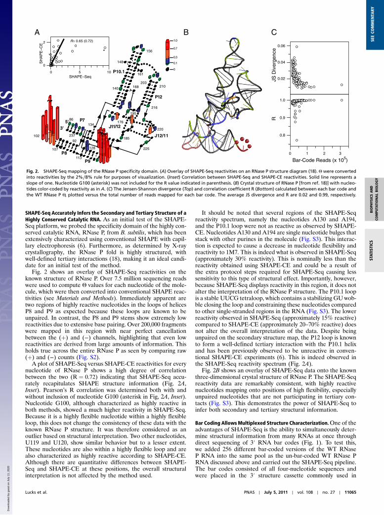
SHAPE-Seq Resolves Local Structural Changes due to Point Mutations.With bar coding, we have the ability to identify structural changesdue to single point mutations in any given RNAmolecule. As partof a seven-member SHAPE-Seq library, we generated five bar-coded variants of the native RNase P molecule from the previouslibrary. These included the WTand the following point mutants:ΔA130, A130U, A131, and A194U (Table S1). These specificRNase P mutations were chosen at bulged nucleotides in theRNA to provide local, subtle changes in secondary structure.The SHAPE-Seq pipeline was applied to this mixture of RNAsto determine the changes in reactivity at positions 130 and 194 inthe RNA due to the four point mutations (Fig. 3). Almost allother positions in the RNA remain unchanged compared tothe wild-type RNase P RNA, and all RNAs had a similar numberof mapped reads ranging from 1,441,075 to 1,224,576 (Fig. S7).
The reactivity at positions 130 and 194 show only subtlechanges for each of the four mutants as expected with only a cou-ple of exceptions (Fig. 3). The reactivity of the A130U mutantremains unchanged at both position 130 and 194, indicating thata U at position 130 maintains a similar structural role in the RNAand does not disrupt the native stacking interaction at that posi-tion. The ΔA130 mutant has a gap in reactivity at position 130 asexpected and shows little change in reactivity at position A194.That combined with fact that the RNAmaintains a similar overallreactivity pattern indicates that removal of the stacking interac-tion at position 130 does not greatly impact the structural integ-rity of the RNA. The A131 RNA, which contains two bulged Anucleotides at position 130, shows a large increase in reactivity atone of the A’s and a very similar reactivity to wild type at the otherA. This indicates that one of the A’s maintains a similar structuralrole to the wild-type A130 position by stacking with another pur-ine nucleotide, whereas the other bulged A is much more flexibleand is most likely not participating in any constraining interac-tions with neighboring nucleotides. Finally, the A194U mutantRNA shows a very similar reactivity at position 130 and a slightlylower reactivity at position 194. The cause of this decrease isunclear; however, it could be explained by enhanced stacking,or a G-U wobble interaction at this position (Fig. S3), causingthe nucleotide to be more constrained and therefore less reactive.
The overall reactivities of the WT RNase P and the four mu-tants obtained using the SHAPE-Seq pipeline are similar to thoseobtained using the SHAPE-CE method (Fig. 3). A more detailedinspection of reactivity profiles around the mutation sites(Fig. S7) show that the changes in reactivity follow the same gen-eral trend in both SHAPE-Seq and SHAPE-CE with two notableexceptions. First, as mentioned above, SHAPE-CE shows higherreactivities for mutations at the 130 and 194 sites. Second, there isa reversal in the reactivity trend for RNase PA194U at the muta-tion position between SHAPE-Seq and SHAPE-CE. Althoughthere is an inconsistency between the two techniques at this singleposition, the interpretation of the point mutations at every otherposition are the same by either method. However, SHAPE-Seqallows the experiment to be done in a single tube and with rig-orous, automated mathematical analysis.
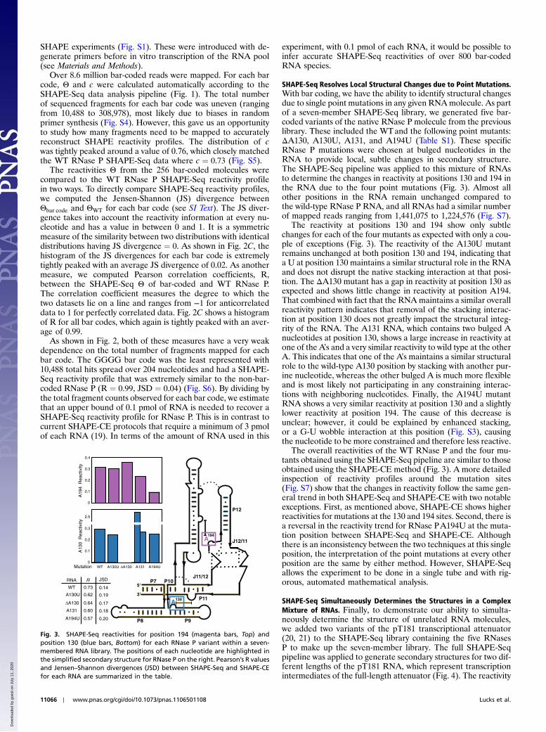
SHAPE-Seq Simultaneously Determines the Structures in a ComplexMixture of RNAs. Finally, to demonstrate our ability to simulta-neously determine the structure of unrelated RNA molecules,we added two variants of the pT181 transcriptional attenuator(20, 21) to the SHAPE-Seq library containing the five RNasesP to make up the seven-member library. The full SHAPE-Seqpipeline was applied to generate secondary structures for two dif-ferent lengths of the pT181 RNA, which represent transcriptionintermediates of the full-length attenuator (Fig. 4). The reactivity
A
J11/12
J12/11
A
P7 P10
P8 P9
P11
5'
3'
P12
WT A130U ∆A130 A131 A194U
A13
0 R
eact
ivity
Mutation
A19
4 R
eact
ivity
RNA R
WT
A130U
∆A130
A131
A194U
0.73
130
194
0
0.1
0.2
0.3
0.4
0
0.1
0.2
0.3
2.5
0.62
0.64
0.60
0.57
0.14
0.19
0.17
0.18
0.20
JSD
Fig. 3. SHAPE-Seq reactivities for position 194 (magenta bars, Top) andposition 130 (blue bars, Bottom) for each RNase P variant within a seven-membered RNA library. The positions of each nucleotide are highlighted inthe simplified secondary structure for RNase P on the right. Pearson’s R valuesand Jensen–Shannon divergences (JSD) between SHAPE-Seq and SHAPE-CEfor each RNA are summarized in the table.
11066 ∣ www.pnas.org/cgi/doi/10.1073/pnas.1106501108 Lucks et al.
Dow
nloa
ded
by g
uest
on
July
11,
202
0
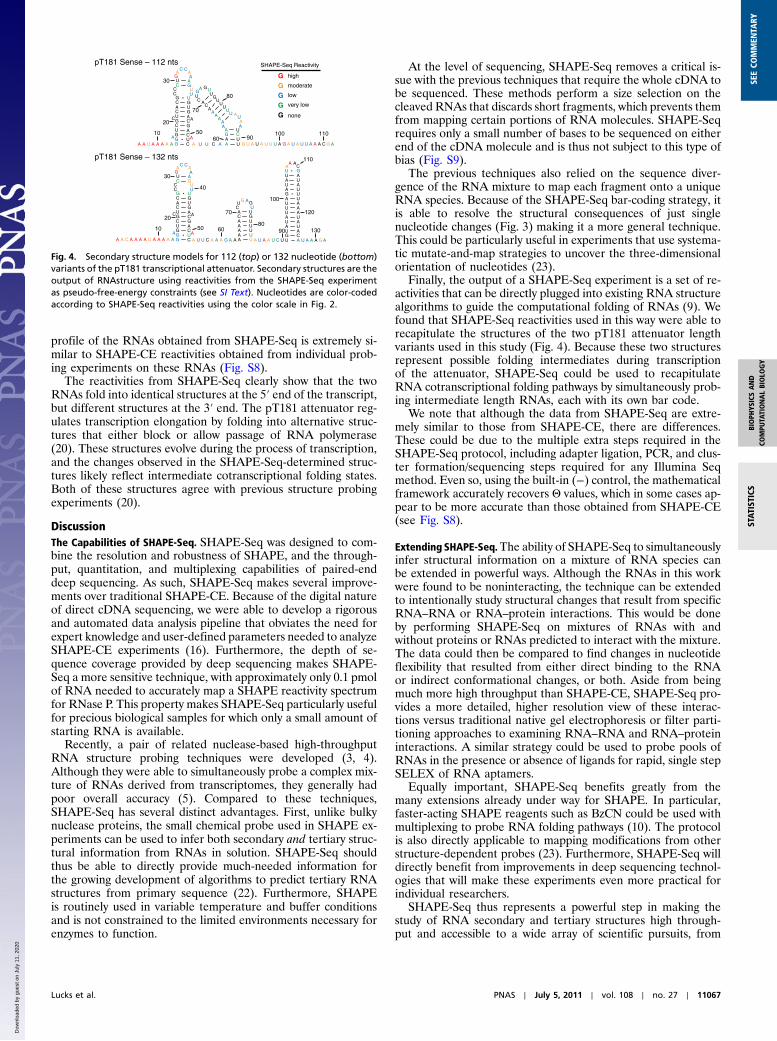
profile of the RNAs obtained from SHAPE-Seq is extremely si-milar to SHAPE-CE reactivities obtained from individual prob-ing experiments on these RNAs (Fig. S8).
The reactivities from SHAPE-Seq clearly show that the twoRNAs fold into identical structures at the 5′ end of the transcript,but different structures at the 3′ end. The pT181 attenuator reg-ulates transcription elongation by folding into alternative struc-tures that either block or allow passage of RNA polymerase(20). These structures evolve during the process of transcription,and the changes observed in the SHAPE-Seq-determined struc-tures likely reflect intermediate cotranscriptional folding states.Both of these structures agree with previous structure probingexperiments (20).
DiscussionThe Capabilities of SHAPE-Seq. SHAPE-Seq was designed to com-bine the resolution and robustness of SHAPE, and the through-put, quantitation, and multiplexing capabilities of paired-enddeep sequencing. As such, SHAPE-Seq makes several improve-ments over traditional SHAPE-CE. Because of the digital natureof direct cDNA sequencing, we were able to develop a rigorousand automated data analysis pipeline that obviates the need forexpert knowledge and user-defined parameters needed to analyzeSHAPE-CE experiments (16). Furthermore, the depth of se-quence coverage provided by deep sequencing makes SHAPE-Seq a more sensitive technique, with approximately only 0.1 pmolof RNA needed to accurately map a SHAPE reactivity spectrumfor RNase P. This property makes SHAPE-Seq particularly usefulfor precious biological samples for which only a small amount ofstarting RNA is available.
Recently, a pair of related nuclease-based high-throughputRNA structure probing techniques were developed (3, 4).Although they were able to simultaneously probe a complex mix-ture of RNAs derived from transcriptomes, they generally hadpoor overall accuracy (5). Compared to these techniques,SHAPE-Seq has several distinct advantages. First, unlike bulkynuclease proteins, the small chemical probe used in SHAPE ex-periments can be used to infer both secondary and tertiary struc-tural information from RNAs in solution. SHAPE-Seq shouldthus be able to directly provide much-needed information forthe growing development of algorithms to predict tertiary RNAstructures from primary sequence (22). Furthermore, SHAPEis routinely used in variable temperature and buffer conditionsand is not constrained to the limited environments necessary forenzymes to function.
At the level of sequencing, SHAPE-Seq removes a critical is-sue with the previous techniques that require the whole cDNA tobe sequenced. These methods perform a size selection on thecleaved RNAs that discards short fragments, which prevents themfrom mapping certain portions of RNA molecules. SHAPE-Seqrequires only a small number of bases to be sequenced on eitherend of the cDNA molecule and is thus not subject to this type ofbias (Fig. S9).
The previous techniques also relied on the sequence diver-gence of the RNA mixture to map each fragment onto a uniqueRNA species. Because of the SHAPE-Seq bar-coding strategy, itis able to resolve the structural consequences of just singlenucleotide changes (Fig. 3) making it a more general technique.This could be particularly useful in experiments that use systema-tic mutate-and-map strategies to uncover the three-dimensionalorientation of nucleotides (23).
Finally, the output of a SHAPE-Seq experiment is a set of re-activities that can be directly plugged into existing RNA structurealgorithms to guide the computational folding of RNAs (9). Wefound that SHAPE-Seq reactivities used in this way were able torecapitulate the structures of the two pT181 attenuator lengthvariants used in this study (Fig. 4). Because these two structuresrepresent possible folding intermediates during transcriptionof the attenuator, SHAPE-Seq could be used to recapitulateRNA cotranscriptional folding pathways by simultaneously prob-ing intermediate length RNAs, each with its own bar code.
We note that although the data from SHAPE-Seq are extre-mely similar to those from SHAPE-CE, there are differences.These could be due to the multiple extra steps required in theSHAPE-Seq protocol, including adapter ligation, PCR, and clus-ter formation/sequencing steps required for any Illumina Seqmethod. Even so, using the built-in (−) control, the mathematicalframework accurately recovers Θ values, which in some cases ap-pear to be more accurate than those obtained from SHAPE-CE(see Fig. S8).
Extending SHAPE-Seq.The ability of SHAPE-Seq to simultaneouslyinfer structural information on a mixture of RNA species canbe extended in powerful ways. Although the RNAs in this workwere found to be noninteracting, the technique can be extendedto intentionally study structural changes that result from specificRNA–RNA or RNA–protein interactions. This would be doneby performing SHAPE-Seq on mixtures of RNAs with andwithout proteins or RNAs predicted to interact with the mixture.The data could then be compared to find changes in nucleotideflexibility that resulted from either direct binding to the RNAor indirect conformational changes, or both. Aside from beingmuch more high throughput than SHAPE-CE, SHAPE-Seq pro-vides a more detailed, higher resolution view of these interac-tions versus traditional native gel electrophoresis or filter parti-tioning approaches to examining RNA–RNA and RNA–proteininteractions. A similar strategy could be used to probe pools ofRNAs in the presence or absence of ligands for rapid, single stepSELEX of RNA aptamers.
Equally important, SHAPE-Seq benefits greatly from themany extensions already under way for SHAPE. In particular,faster-acting SHAPE reagents such as BzCN could be used withmultiplexing to probe RNA folding pathways (10). The protocolis also directly applicable to mapping modifications from otherstructure-dependent probes (23). Furthermore, SHAPE-Seq willdirectly benefit from improvements in deep sequencing technol-ogies that will make these experiments even more practical forindividual researchers.
SHAPE-Seq thus represents a powerful step in making thestudy of RNA secondary and tertiary structures high through-put and accessible to a wide array of scientific pursuits, from
pT181 Sense – 132 nts
A A U A A A A A G CG UAG CU AC GG CCU AC GA UC GG U
CCC GU A
GA
C CAA
UU
A
AA U U C A A U
A UG CA U
AA
UA
UA
UA
UA
UC
GA
UC
UG
A GU
AUAA
G U A U A U U U A G A U A U U A A A C G A
pT181 Sense – 112 nts
A A C A A A A U A A A A A G CG UAG CU AC GG CCU AC GA UC GG U
CCC GU A
GA
C CAA
UU
A
A
A U U C A A A G A A A UA UA UA UA UC GA U
CU
G AGU
A U A A U C U U AG CU AA UU AA UU AU AU AA UG UA UU AA UU AU GA
A AC
U A A A G A
10
20
30
5060
70
80
90100 110
10
20
30
50
40
60
70
8090
100
110
120
130
GGGG
high
moderate
low
very low
G none
SHAPE-Seq Reactivity
Fig. 4. Secondary structure models for 112 (top) or 132 nucleotide (bottom)variants of the pT181 transcriptional attenuator. Secondary structures are theoutput of RNAstructure using reactivities from the SHAPE-Seq experimentas pseudo-free-energy constraints (see SI Text). Nucleotides are color-codedaccording to SHAPE-Seq reactivities using the color scale in Fig. 2.
Lucks et al. PNAS ∣ July 5, 2011 ∣ vol. 108 ∣ no. 27 ∣ 11067
STAT
ISTICS
BIOPH
YSICSAND
COMPU
TATIONALBIOLO
GY
SEECO
MMEN
TARY
Dow
nloa
ded
by g
uest
on
July
11,
202
0
fundamental biological investigations to engineering RNA syn-thetic biological systems.
Materials and MethodsStructure-Selective RNA Modification. All RNAs were synthesized with stan-dard in vitro synthesis and modified with 1M7 (6.5 mM, final) as describedpreviously with minor modifications for the analysis by deep sequencing(6). See SI Text for details.
SHAPE Analysis by Capillary Electrophoresis. The general procedure of primerextension and data analysis is that of ref. 24.
SHAPE Analysis by Deep Sequencing. The procedure for first strand cDNAsynthesis was carried out following the primer extension protocol outlinedelsewhere (24) using RT primers with tails containing the Illumina A_adap-ter_t and (þ) or (−) handles (see SI Text). After primer extension, RNA washydrolyzed and the Illumina A_adapter b was ligated to each cDNA usinga ssDNA ligase (circLigase, Epicentre). Excess A_adapter_b was removed usingAgencourt Ampure XP beads. Finally, 9 or 12 cycles of PCR amplification wereperformed (13) with no post size-selection step. Libraries were assayed forquality on an Agilent Bioanalyzer 2100 using a high-sensitivity DNA chipand then sequenced on an Illumina Genome Analyzer IIx for 50 cycles ofsequencing per paired-end read. Data available upon request. See SI Textfor a detailed protocol.
Bioinformatic Analysis of Bar-Coded Sequencing Reads. Reads for RT fragmentswere first split into 1M7-treated and -untreated pools by examining the4-nucleotide handle sequence on the 5′ end of the read generated fromthe 3′ end of each RNA probed in the experiment. This handle was thentrimmed from each read to allow alignment of the reads to probed RNAs.Reads were then trimmed for A_adapter_b and A_adapter_t using the FASTXtoolkit [http://hannonlab.cshl.edu/fastx_toolkit/] (see SI Text). Paired readswere optimally aligned to the probed RNAs using Bowtie 0.12.8 (15) todetermine RT-stop counts. These RT-stop counts were then used to calculateML-based reactivities. See SI Text for details.
Processing of SHAPE-Seq Data by Maximum Likelihood Estimation. A stochasticmodel of SHAPE-Seq parameterized by reactivities, Θ, the expected numberof modificiations per molecule, c, and natural polymerase drop-off rates, Γ,was used to infer the quantities of interest via maximum likelihood estima-tion (7). We assumed that the number of modifications per RNA was Poissondistributed, with an unknown parameter, c, to be estimated.With this model,
the likelihood of a set of fragment counts X in the (þ) channel and Y in the(−) channel obtained in an experiment was calculated by
LðΘ;Γ;cÞ ¼Ynk¼1
�γk
Yk−1i¼1
ð1 − γiÞ�Yk Yn
k¼1
�ecðΣ
nι¼kθι−1Þ
Yk−1i¼1
ð1 − γiÞ
− ecðΣnι¼kþ1
θι−1ÞYki¼1
ð1 − γiÞ�Xk�Yni¼1
ð1 − γiÞ�Ynþ1
×�e−c
Yni¼1
ð1 − γiÞ�Xnþ1
;
where Xk , Yk were the number of fragments reaching the kth position in theRNA. Details of the model derivation and analysis are provided in ref. 7.
Converting Between Θ and Reactivity. Where needed, Θ were converted intoSHAPE reactivities by excluding the top 2% of reactivites and normalizing bythe average of the next 8% (9). Where needed, SHAPE-CE reactivities wereconverted to Θ by dividing by the sum of the reactivities so that the sum ofthe Θ was equal to 1. In cases where an exponential decay correction wasnot applied to the SHAPE-CE data (16), Θ was calculated using the MLcorrection (7).
Secondary Structure Prediction Using SHAPE-Seq Reactivity Constraints. SHAPEreactivities were converted into a pseudo-free-energy change term in theRNA structure program following standard procedures (9). See SI Text fordetails.
ACKNOWLEDGMENTS. The authors thank Michael Eisen, Jacqueline Villalta,Oh Kyu Yoon, Leath Tonkin, Devin Scannell, Jennifer Kuehl, and Keith Kellerfor advice and assistance. We thank Rhiju Das for insightful reading of themanuscript. We also thank Phil Homan (University of North Carolina, ChapelHill, NC) and Kevin Weeks (University of North Carolina, Chapel Hill, NC) forthe generous gift of 1M7. J.A.D. is a Howard Hughes Medical Institute (HHMI)Investigator, and this work was supported in part by the HHMI. S.A.M. isa fellow of the Leukemia and Lymphoma Society. A.P.A., J.B.L., and S.A.acknowledge support from the Synthetic Biology Engineering ResearchCenter under National Science Foundation Grant 04-570/0540879. J.B.L.and L.P. thank the Miller Institute for financial support, and a stimulatingenvironment in which this work was conceived.
1. Sharp PA (2009) The centrality of RNA. Cell 136:577–580.2. Isaacs FJ, Dwyer DJ, Collins JJ (2006) RNA synthetic biology.Nat Biotechnol 24:545–554.3. Kertesz M, et al. (2010) Genome-wide measurement of RNA secondary structure in
yeast. Nature 467:103–107.4. Underwood JG, et al. (2010) FragSeq: Transcriptome-wide RNA structure probing using
high-throughput sequencing. Nat Methods 7:995–1001.5. Mauger DM, Weeks KM (2010) Toward global RNA structure analysis. Nat Biotechnol
28:1178–1179.6. Mortimer SA, Weeks KM (2007) A fast-acting reagent for accurate analysis of RNA
secondary and tertiary structure by SHAPE chemistry. J Am Chem Soc 129:4144–4145.7. Aviran S, et al. (2011) Modeling and automation of sequencing-based characterization
of RNA structure. Proc Natl Acad Sci USA, 10.1073/pnas.1106541108.8. Reuter JS, Mathews DH (2010) RNAstructure: Software for RNA secondary structure
prediction and analysis. BMC Bioinformatics 11:129.9. Low JT, Weeks KM (2010) SHAPE-directed RNA secondary structure prediction.
Methods 52:150–158.10. Mortimer SA, Weeks KM (2008) Time-resolved RNA SHAPE chemistry. J Am Chem Soc
130:16178–16180.11. Stoddard CD, Gilbert SD, Batey RT (2008) Ligand-dependent folding of the three-way
junction in the purine riboswitch. RNA 14:675–684.12. Wilkinson KA,Merino EJ, Weeks KM (2005) RNA SHAPE chemistry reveals nonhierarch-
ical interactions dominate equilibrium structural transitions in tRNA(Asp) transcripts.J Am Chem Soc 127:4659–4667.
13. Ingolia NT, Ghaemmaghami S, Newman JRS, Weissman JS (2009) Genome-wideanalysis in vivo of translation with nucleotide resolution using ribosome profiling.Science 324:218–223.
14. Li TW, Weeks KM (2006) Structure-independent and quantitative ligation of single-stranded DNA. Anal Biochem 349:242–246.
15. Langmead B, Trapnell C, Pop M, Salzberg SL (2009) Ultrafast and memory-efficientalignment of short DNA sequences to the human genome. Genome Biol 10:R25.
16. Vasa SM, Guex N, Wilkinson KA, Weeks KM, Giddings MC (2008) ShapeFinder: Asoftware system for high-throughput quantitative analysis of nucleic acid reactivityinformation resolved by capillary electrophoresis. RNA 14:1979–1990.
17. Gherghe CM, Shajani Z, Wilkinson KA, Varani G, Weeks KM (2008) Strong correlationbetween SHAPE chemistry and the generalized NMR order parameter (S2) in RNA.J Am Chem Soc 130:12244–12245.
18. Krasilnikov AS, Yang X, Pan T, Mondragón A (2003) Crystal structure of the specificitydomain of ribonuclease P. Nature 421:760–764.
19. Wilkinson KA, Merino EJ, Weeks KM (2006) Selective 2′-hydroxyl acylation analyzedby primer extension (SHAPE): Quantitative RNA structure analysis at single nucleotideresolution. Nat Protoc 1:1610–1616.
20. Brantl S, Wagner EGH (2000) Antisense RNA-mediated transcriptional attenuation:An in vitro study of plasmid pT181. Mol Microbiol 35:1469–1482.
21. Lucks JB, Qi L, Mutalik V, Wang D, Arkin AP (2011) Versatile RNA-sensing transcrip-tional regulators for engineering genetic networks. Proc Natl Acad Sci USA108:8617–8622.
22. Das R, Karanicolas J, Baker D (2010) Atomic accuracy in predicting and designingnoncanonical RNA structure. Nat Methods 7:291–294.
23. KladwangW, Das R (2010) Amutate-and-map strategy for inferring base pairs in struc-tured nucleic acids: Proof of concept on a DNA/RNA helix. Biochemistry 49:7414–7416.
24. Mortimer SA, Weeks KM (2009) Time-resolved RNA SHAPE chemistry: QuantitativeRNA structure analysis in one-second snapshots and at single-nucleotide resolution.Nat Protoc 4:1413–1421.
11068 ∣ www.pnas.org/cgi/doi/10.1073/pnas.1106501108 Lucks et al.
Dow
nloa
ded
by g
uest
on
July
11,
202
0