Highly Efficient FRET From Single Nitrogen-Vacancy Center
6
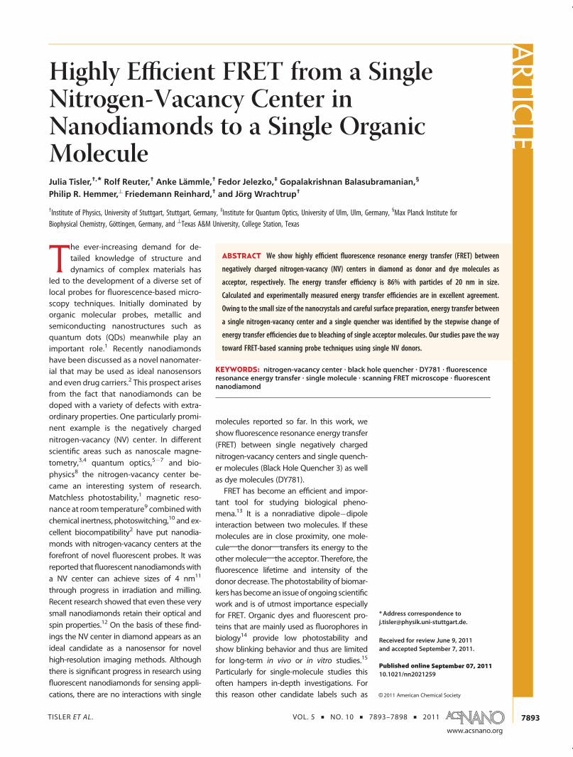
TISLER ET AL . VOL. 5 ’ NO. 10 ’ 7893–7898 ’ 2011 www.acsnano.org 7893 September 07, 2011 C 2011 American Chemical Society Highly Efficient FRET from a Single Nitrogen-Vacancy Center in Nanodiamonds to a Single Organic Molecule Julia Tisler, †, * Rolf Reuter, † Anke La ¨ mmle, † Fedor Jelezko, ‡ Gopalakrishnan Balasubramanian, § Philip R. Hemmer, ^ Friedemann Reinhard, † and Jo ¨ rg Wrachtrup † † Institute of Physics, University of Stuttgart, Stuttgart, Germany, ‡ Institute for Quantum Optics, University of Ulm, Ulm, Germany, § Max Planck Institute for Biophysical Chemistry, Göttingen, Germany, and ^ Texas A&M University, College Station, Texas T he ever-increasing demand for de- tailed knowledge of structure and dynamics of complex materials has led to the development of a diverse set of local probes for fluorescence-based micro- scopy techniques. Initially dominated by organic molecular probes, metallic and semiconducting nanostructures such as quantum dots (QDs) meanwhile play an important role. 1 Recently nanodiamonds have been discussed as a novel nanomater- ial that may be used as ideal nanosensors and even drug carriers. 2 This prospect arises from the fact that nanodiamonds can be doped with a variety of defects with extra- ordinary properties. One particularly promi- nent example is the negatively charged nitrogen-vacancy (NV) center. In different scientific areas such as nanoscale magne- tometry, 3,4 quantum optics, 57 and bio- physics 8 the nitrogen-vacancy center be- came an interesting system of research. Matchless photostability, 1 magnetic reso- nance at room temperature 9 combined with chemical inertness, photoswitching, 10 and ex- cellent biocompatibility 2 have put nanodia- monds with nitrogen-vacancy centers at the forefront of novel fluorescent probes. It was reported that fluorescent nanodiamonds with a NV center can achieve sizes of 4 nm 11 through progress in irradiation and milling. Recent research showed that even these very small nanodiamonds retain their optical and spin properties. 12 On the basis of these find- ings the NV center in diamond appears as an ideal candidate as a nanosensor for novel high-resolution imaging methods. Although there is significant progress in research using fluorescent nanodiamonds for sensing appli- cations, there are no interactions with single molecules reported so far. In this work, we show fluorescence resonance energy transfer (FRET) between single negatively charged nitrogen-vacancy centers and single quench- er molecules (Black Hole Quencher 3) as well as dye molecules (DY781). FRET has become an efficient and impor- tant tool for studying biological pheno- mena. 13 It is a nonradiative dipoledipole interaction between two molecules. If these molecules are in close proximity, one mole- cule;the donor;transfers its energy to the other molecule;the acceptor. Therefore, the fluorescence lifetime and intensity of the donor decrease. The photostability of biomar- kers has become an issue of ongoing scienti fic work and is of utmost importance especially for FRET. Organic dyes and fluorescent pro- teins that are mainly used as fluorophores in biology 14 provide low photostability and show blinking behavior and thus are limited for long-term in vivo or in vitro studies. 15 Particularly for single-molecule studies this often hampers in-depth investigations. For this reason other candidate labels such as * Address correspondence to [email protected]. Received for review June 9, 2011 and accepted September 7, 2011. Published online 10.1021/nn2021259 ABSTRACT We show highly efficient fluorescence resonance energy transfer (FRET) between negatively charged nitrogen-vacancy (NV) centers in diamond as donor and dye molecules as acceptor, respectively. The energy transfer efficiency is 86% with particles of 20 nm in size. Calculated and experimentally measured energy transfer efficiencies are in excellent agreement. Owing to the small size of the nanocrystals and careful surface preparation, energy transfer between a single nitrogen-vacancy center and a single quencher was identified by the stepwise change of energy transfer efficiencies due to bleaching of single acceptor molecules. Our studies pave the way toward FRET-based scanning probe techniques using single NV donors. KEYWORDS: nitrogen-vacancy center . black hole quencher . DY781 . fluorescence resonance energy transfer . single molecule . scanning FRET microscope . fluorescent nanodiamond ARTICLE
-
Upload
jan-havlik -
Category
Documents
-
view
56 -
download
3
Transcript of Highly Efficient FRET From Single Nitrogen-Vacancy Center
TISLER ET AL . VOL. 5 ’ NO. 10 ’ 7893–7898 ’ 2011
www.acsnano.org
7893
September 07, 2011
C 2011 American Chemical Society
Highly Efficient FRET from a SingleNitrogen-Vacancy Center inNanodiamonds to a Single OrganicMoleculeJulia Tisler,†,* Rolf Reuter,† Anke Lammle,† Fedor Jelezko,‡ Gopalakrishnan Balasubramanian,§
Philip R. Hemmer,^ Friedemann Reinhard,† and Jorg Wrachtrup†
†Institute of Physics, University of Stuttgart, Stuttgart, Germany, ‡Institute for Quantum Optics, University of Ulm, Ulm, Germany, §Max Planck Institute forBiophysical Chemistry, Göttingen, Germany, and ^Texas A&M University, College Station, Texas
The ever-increasing demand for de-tailed knowledge of structure anddynamics of complex materials has
led to the development of a diverse set oflocal probes for fluorescence-based micro-scopy techniques. Initially dominated byorganic molecular probes, metallic andsemiconducting nanostructures such asquantum dots (QDs) meanwhile play animportant role.1 Recently nanodiamondshave been discussed as a novel nanomater-ial that may be used as ideal nanosensorsand even drug carriers.2 This prospect arisesfrom the fact that nanodiamonds can bedoped with a variety of defects with extra-ordinary properties. One particularly promi-nent example is the negatively chargednitrogen-vacancy (NV) center. In differentscientific areas such as nanoscale magne-tometry,3,4 quantum optics,5�7 and bio-physics8 the nitrogen-vacancy center be-came an interesting system of research.Matchless photostability,1 magnetic reso-nance at room temperature9 combinedwithchemical inertness, photoswitching,10 and ex-cellent biocompatibility2 have put nanodia-monds with nitrogen-vacancy centers at theforefront of novel fluorescent probes. It wasreported thatfluorescent nanodiamondswitha NV center can achieve sizes of 4 nm11
through progress in irradiation and milling.Recent research showed that even these verysmall nanodiamonds retain their optical andspin properties.12 On the basis of these find-ings the NV center in diamond appears as anideal candidate as a nanosensor for novelhigh-resolution imaging methods. Althoughthere is significant progress in research usingfluorescent nanodiamonds for sensing appli-cations, there are no interactions with single
molecules reported so far. In this work, weshow fluorescence resonance energy transfer(FRET) between single negatively chargednitrogen-vacancy centers and single quench-er molecules (Black Hole Quencher 3) as wellas dye molecules (DY781).FRET has become an efficient and impor-
tant tool for studying biological pheno-mena.13 It is a nonradiative dipole�dipoleinteraction between two molecules. If thesemolecules are in close proximity, one mole-cule;the donor;transfers its energy to theother molecule;the acceptor. Therefore, thefluorescence lifetime and intensity of thedonor decrease. The photostability of biomar-kers hasbecomean issueof ongoing scientificwork and is of utmost importance especiallyfor FRET. Organic dyes and fluorescent pro-teins that are mainly used as fluorophores inbiology14 provide low photostability andshow blinking behavior and thus are limitedfor long-term in vivo or in vitro studies.15
Particularly for single-molecule studies thisoften hampers in-depth investigations. Forthis reason other candidate labels such as
* Address correspondence [email protected].
Received for review June 9, 2011and accepted September 7, 2011.
Published online10.1021/nn2021259
ABSTRACT We show highly efficient fluorescence resonance energy transfer (FRET) between
negatively charged nitrogen-vacancy (NV) centers in diamond as donor and dye molecules as
acceptor, respectively. The energy transfer efficiency is 86% with particles of 20 nm in size.
Calculated and experimentally measured energy transfer efficiencies are in excellent agreement.
Owing to the small size of the nanocrystals and careful surface preparation, energy transfer between
a single nitrogen-vacancy center and a single quencher was identified by the stepwise change of
energy transfer efficiencies due to bleaching of single acceptor molecules. Our studies pave the way
toward FRET-based scanning probe techniques using single NV donors.
KEYWORDS: nitrogen-vacancy center . black hole quencher . DY781 . fluorescenceresonance energy transfer . single molecule . scanning FRET microscope . fluorescentnanodiamond
ARTIC
LE

TISLER ET AL . VOL. 5 ’ NO. 10 ’ 7893–7898 ’ 2011
www.acsnano.org
7894
quantum dots meanwhile have their firm place inbiological research. QDs are brighter and more photo-stable than organic molecules or proteins16 and arealso used as donors in FRET experiments.17 Fluorescentnanodiamondswith particle sizes being on the order ofthe smallest energy transfer distances18 together withtheir inherent photostability potentially are ideal can-didates as FRET labels.Recently, FRET between several NVs and several
IRDye molecules with an energy transfer efficiency of7%19 has been reported. Here we describe energytransfer between single NV centers and single quench-er molecules with an energy transfer efficiency of 95%.
RESULTS AND DISCUSSION
For the present study two different samples oflabeled nanodiamonds are prepared: one sample withdye (DY781-NHS) covalently linked by amide bonds tothe surface of amino-silanized nanodiamonds andanother with Black Hole Quencher (BHQ3-NH2) stron-gly attached to the surface by electrostatic interactionwith the negatively charged nanodiamond surface atpH 6. In order to covalently bind the DY781 to thesurface of the nanodiamonds, an oxidizing acid treat-ment to remove sp2 carbon was followed by boranereduction to create hydroxyl groups and also to stabi-lize the NV� charge state.20�23 An aminosilanizationtreatment of the nanodiamond surfacewas carried out,and the FRET acceptor was bound using its N-hydro-xysuccinimide (NHS) ester derivative.24
The DY781 sample showed fast bleaching of the dyemolecules, and as a result of that, a different sampleusing BHQ as FRET acceptorwas prepared. The BHQwasattached to the naodiamond surface via adsorption, andthis resulted in a higher coverage rate than DY781.Zeta potential measurements show a value of �40
mV at pH 7, indicating a high surface coverage withnegative charges after the oxidizing treatment. As aresult, the positively charged BHQ adsorbs strongly byelectrostatic interaction. The treated nanodiamondswere then spin-coated on a glass slide.For the experiment we used a combination of an
atomic force microscope and a confocal microscopewith fluorescence lifetime imaging (FLIM) capability.This allows for simultaneous measurement of thetopography, confocal image, and lifetime image ofthe same area. We used a pulsed laser with a wave-length of λ = 532 nm for excitation and two filters fordetecting the fluorescence of the NV center and of thedye (FRET signal). The bandpass filter from 650 to750 nm separates NV0 from NV�. In addition, thespectrum of NV0 is shifted to the blue so that the FRETefficiency is very low due to the small overlap integralbetween NV0 and BHQ. The NV center acts as a donorfor both quencher and dye. To confirm single NVcenters, autocorrelation curves were recorded (data
not shown). Two scans (before and after bleaching ofthe acceptor molecules) were performed for everyinvestigated sample area, and fluorescence lifetimeand intensity data of the NV center were recordedeach time. After the first scan, the dyemolecules on thesurface of the nanodiamonds were bleached by illu-minating every particle with high laser power. Theenergy transfer efficiencywas calculated by comparinglifetime and intensity data of the NV center before andafter bleaching of the acceptor molecules.In a first step the adsorption of dyemolecules on the
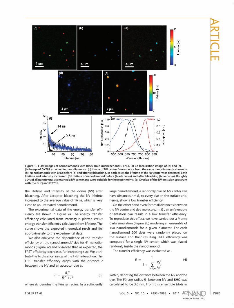
surface of nanodiamonds was confirmed with co-localization microscopy (Figure 1a). When comparingimages where dye fluorescence is measured (Figure 1b)with those where NV fluorescence was carried out(Figure 1c), the NV channel (red) and the dye channel(green) nicely co-localize, indicating attachment ofdyes on the surface (Figure 1a). It was found thatDY781 bleaches very fast (as compared to the BHQ3).Nevertheless, we were able to detect DY781 fluores-cence (Figure 1b) and hence a decrease in NV lifetimeas well. Also in this case, on bleaching of DY781, onlythe signal of the NV center remains with a longerfluorescence lifetime (Figure 1c). The observed energytransfer furthermore confirms attachment of the dyeon the nanodiamond surface.TheNV center emission ranges from600 to 750 nm25
and overlaps with the absorption maximum of DY781at 783 nm (see Figure 1g). Using eq 1 we calculated theFörster radius between the NV center and DY781 to be3.8 nm.
R0 ¼ 0:211(K2n�4QD J(λ))1=6 (1)
κ is the orientation factor, which is assumed to beκ2 = 2/3 for an average orientation factor. n is therefractive index (2.4 for diamond), andQD the quantumefficiency, with QD = 0.3. J(λ) is the spectral overlapbetween the emission spectra of the NV center and theabsorption spectra of the acceptor.Figure 1d and e show FLIM of a sample with nano-
diamonds coveredwith BHQbefore and after bleaching.The fluorescence of the NV center and intensity as wellas lifetime increased, after the quenchermolecules havebeen bleached, which clearly indicates FRET.To confirm this in a quantitative manner, we calcu-
lated the energy transfer efficiency using the recordedlifetime and intensity of the NV center before and afterbleaching of the quencher. For a FRET process theefficiency calculated from fluorescence intensity andlifetime are correlated. To verify this, we calculated theenergy transfer efficiency E:
E ¼ 1 � τDAτD
¼ 1 � IDAID
(2)
Here τda and Ida are the lifetime and intensity betweendonor and acceptor before bleaching, and τd and Id are
ARTIC
LE
TISLER ET AL . VOL. 5 ’ NO. 10 ’ 7893–7898 ’ 2011
www.acsnano.org
7895
the lifetime and intensity of the donor (NV) afterbleaching. After acceptor bleaching the NV lifetimeincreased to the average value of 16 ns, which is veryclose to an untreated nanodiamond.The experimental data of the energy transfer effi-
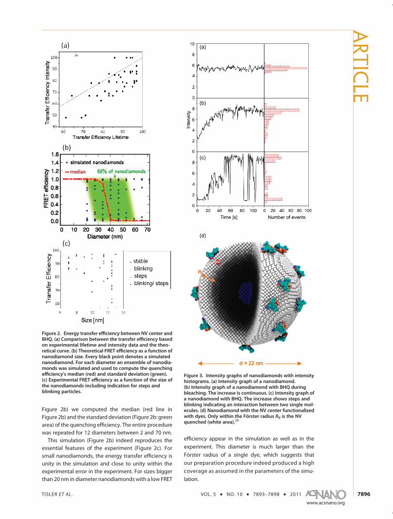
ciency are shown in Figure 2a. The energy transferefficiency calculated from intensity is plotted versus
energy transfer efficiency calculated from lifetime. Thecurve shows the expected theoretical result and fitsapproximately to the experimental data.We also analyzed the dependence of the transfer
efficiency on the nanodiamonds' size for 41 nanodia-monds (Figure 2c) and observed that, as expected, theFRET efficiency decreases for increasing size. We attri-bute this to the short range of the FRET interaction. TheFRET transfer efficiency drops with the distance r
between the NV and an acceptor dye as
E ¼ R06
R06 þ r6
(3)
where R0 denotes the Förster radius. In a sufficiently
large nanodiamond, a randomly placed NV center canhave distances r. R0 to every dye on the surface and,hence, show a low transfer efficiency.On the other hand even for small distances between
the NV center and dye molecule, r < R0, an unfavorableorientation can result in a low transfer efficiency.To reproduce this effect, we have carried out a MonteCarlo simulation (Figure 2b) modeling an ensemble of150 nanodiamonds for a given diameter. For eachnanodiamond 200 dyes were randomly placed onthe surface and their resulting FRET efficiency wascomputed for a single NV center, which was placedrandomly inside the nanodiamond.The transfer efficiency was evaluated as
E ¼ 1
1þ ∑d∈dyes
R06
rd6
(4)
with rd denoting the distance between the NV and thedye. The Förster radius R0 between NV and BHQ wascalculated to be 3.6 nm. From this ensemble (dots in
Figure 1. FLIM images of nanodiamonds with Black Hole Quencher and DY781. (a) Co-localization image of (b) and (c).(b) Image of DY781 attached to nanodiamonds. (c) Image of NV center fluorescence from the same nanodiamonds shown in(b). Nanodiamonds with BHQ before (d) and after (e) bleaching. In both cases the lifetime of the NV center was detected. Bothlifetime and intensity increased. (f) Lifetime of nanodiamond before (black curve) and after bleaching (blue curve). Roughly30%of all nanocrystals contained aNV center andwere suitable for the experiments. (g) Overlap of theNV emission spectrumwith the BHQ and DY781.
ARTIC
LE
TISLER ET AL . VOL. 5 ’ NO. 10 ’ 7893–7898 ’ 2011
www.acsnano.org
7896
Figure 2b) we computed the median (red line inFigure 2b) and the standard deviation (Figure 2b: greenarea) of the quenching efficiency. The entire procedurewas repeated for 12 diameters between 2 and 70 nm.This simulation (Figure 2b) indeed reproduces the
essential features of the experiment (Figure 2c). Forsmall nanodiamonds, the energy transfer efficiency isunity in the simulation and close to unity within theexperimental error in the experiment. For sizes biggerthan 20 nm in diameter nanodiamondswith a low FRET
efficiency appear in the simulation as well as in theexperiment. This diameter is much larger than theFörster radius of a single dye, which suggests thatour preparation procedure indeed produced a highcoverage as assumed in the parameters of the simu-lation.
Figure 3. Intensity graphs of nanodiamonds with intensityhistograms. (a) Intensity graph of a nanodiamond.(b) Intensity graph of a nanodiamond with BHQ duringbleaching. The increase is continuous. (c) Intensity graph ofa nanodiamond with BHQ. The increase shows steps andblinking indicating an interaction between two single mol-ecules. (d) Nanodiamond with the NV center functionalizedwith dyes. Only within the Förster radius R0 is the NVquenched (white area).26
Figure 2. Energy transfer efficiency between NV center andBHQ. (a) Comparison between the transfer efficiency basedon experimental lifetime and intensity data and the theo-retical curve. (b) Theoretical FRET efficiency as a function ofnanodiamond size. Every black point denotes a simulatednanodiamond. For each diameter an ensemble of nanodia-monds was simulated and used to compute the quenchingefficiency's median (red) and standard deviation (green).(c) Experimental FRET efficiency as a function of the size ofthe nanodiamonds including indication for steps andblinking particles.
ARTIC
LE

TISLER ET AL . VOL. 5 ’ NO. 10 ’ 7893–7898 ’ 2011
www.acsnano.org
7897
More insight into the local environment of a center canbe gained by recording time traces of the intensity andlifetime during the bleaching process that was measuredfor every NV center. These curves were acquired byilluminatingeveryparticlewithhigh laserpower. Thereforequencher molecules are progressively bleached, and thedetected NV intensity as well as its lifetime increasedproportionally to the remaining number of dyemoleculeson the surface. Figure 3 shows some examples of theintensity curves and the corresponding intensity histo-gram. The first example is a NV center without quencherand shows the well-known time trace without blinking orbleaching. In the following graphs there is a continuousincrease in intensity, and inpanel 3c the intensity increasesin steps. The percentage increase of both lifetime andintensity was the same for all curves and was verified by
I1
k2 þ k3
� �¼ ak1k2
1k2 þ k3
(5)
I is the intensity, a is a constant, k1 is the excitation rate, k2is the fluorescing rate, and k3 is the quenching rate. Thelifetime τ can be expressed as 1/(k2 þ k3). Equation 5gives a relation between the quenched intensity andlifetime and shows that the percentage increase of life-time and intensity was the same, indicating a real FRETsignal as opposed to blinking caused by photoionizationof the NV,18 which usually does not shorten the lifetime.Thirty-four percent of the particles showed steps
(Figure 3c), while the rest showed a continuous increasein which finally all dye molecules were fully bleached(Figure 3b). Steps indicate an interaction between asingleNVanda single quenchermolecule thatbleached,visible in a sudden increase in intensity and lifetime ofthe NV. Naively one might expect that all curves shouldshow stepwise increase in intensity at least at the end,when almost all molecules on the surface have beenbleached. In this case the NV center is within the Försterradius, R0. Figure 3d shows a 22 nm diamond functio-nalized with dyes where the energy transfer efficiency iscolor coded. If the NV is in the white shell, it is closeenough to the surface for FRET to anadsorbedmolecule;if it is in the core (the blue area) and too far away fromthe surface, no quenching should appear. The volumeofthat core divided by the whole volume of the nanodia-mond results in the probability of the quenched parti-cles η, where we expect to see steps:
η ¼ 1 � (r � R0)3
r3(6)
We estimate that 70% of the particles should showsteps for an average size of 20 nm. The observation that
this is not the case together with the observation thatevery particle showed quenching on the other hand isdue to the high coverage of quencher molecules onthe surface of the nanodiamonds. Many molecules arenot in the range of the Förster radius, but the NV isclose enough to the surface (black area) to be quenched by the high number of acceptors showing acontinuous increase. This, on the other hand, can beused to estimate the number of molecules on thesurface. The nanodiamond in Figure 3c shows twosteps; that is, two molecules are within the Försterradius. The particle has a radius of 7 nm. If one dividesthe surface of a nanodiamond with radius 7 nm by thearea that has two quencher molecules, about 30molecules on the surface can be estimated.For someNVs the curves showablinkingbehavior (see
Figure 3c). As shown before, the NV center in diamond isstable. Also the reference sample showed only 4% ofblinking NVs, compared to the sample with the treatednanodiamonds, where 26% showed such a behavior. Theblinking was visible in the intensity as well as the lifetimecurves. This suggests that the blinking is not caused bythe NV but by the acceptor molecules. The acceptormolecule switches on and off before it is completelybleached. In Figure 2c the energy transfer efficiency as afunction of the particle radius is shown with statistics ofthe nanodiamonds showing these effects. The figureshows that in some cases the steps are accompaniedby blinking. Note that especially for the smallest particlesof 7 nm in size this behavior is observed. Every particlemeasured with this radius showed steps and blinking.This is a further indication that blinking of NVs asobserved in our case is a signature for FRET.
CONCLUSION
In conclusion we showed that we were able tofunctionalize diamond nanocrystals for covalent at-tachment of acceptor molecules on their surface. Insome cases this causes strong interaction between asingle NV center and a single quencher molecule withnear unit FRET transfer efficiency between the NV andquenchermolecule. Because the NV center in diamondprovides several advantages such as photostability andbiocompatibility, it is an ideal candidate for FRETinvestigations. Besides it was shown that nanocrystalscan be produced down to sizes of 4 nm;27 thus thenitrogen-vacancy center is a biomarker that can beused for FRET studies in biological applications. Finallythe observed FRET efficiencies may foster develop-ments of FRETmicroscopes based on single NV centersas donors, which would be truly novel devices.28
METHODS
The used nanodiamonds were irradiated with electrons andannealed as described previously.12 The fluorescent defect
centers containing diamond nanocrystals were oxidized in air
at 480 �C for 12 h to selectively remove sp2-bonded carbon,
followed by cleaning in amixture of concentrated nitric, sulfuric,
ARTIC
LE

TISLER ET AL . VOL. 5 ’ NO. 10 ’ 7893–7898 ’ 2011
www.acsnano.org
7898
and perchloric acid in a volume ratio of 1:1:1. This cleaning stepwas done under reflux for 10 h with 30 min of sonification in anultrasonic bath after each 2 h of reflux. Extensive rinsing withMilli-Q water led to a stable, clear, and brownish solution.
Experimental Setup. The experiment was performed using ahome-built scanning confocal microscope combined with anAFM (MFP-3D Asylum Research). Nitrogen-vacancy defectswere excited with a frequency doubled CW Nd:YAG laser(Coherent Compass) and for lifetime measurements with aVanguard Quasi-CW DPSS laser (Newport Spectra-Physics) fo-cused onto the sample with a high-NA objective (OlympusPlanAPO, NA51.35). Lunimescence light was collected by thesame objective and filtered from the excitation light using adichroic beamsplitter (640 DCXR, Chroma). Photon counting ofthe filtered light was performed using two avalanche photo-diodes (SPQR-14, Perkin-Elmer). Fluorescence autocorrelationhistograms were recorded using a fast multichannel analyzer(Fastcomtec, P7889). Fluorescence lifetime and the autocorrela-tion histograms were recorded using TCSPC electronics(PicoHarp 300, PicoQuant GmbH) together with a two-channelrouter controlled by SymPhoTime V 4.0 software.
Acknowledgment. This work was financially supported bythe Volkswagenstiftung as well as the EU projects DINAMO aswell as SQUTEC and Baden-Württemberg Stiftung through theprojects Internationale Spitzenforschung II and Methoden fürdie Lebenswissenschaften. We thank Philip R. Hemmer forhelpful comments on the manuscript, Roman Kolesov forexperimental assistance, and Andrew Aird for help with thepreparation of the figures and the manuscript.
REFERENCES AND NOTES1. Michalet, X.; Pinaud, F. F.; Bentolila, L. A.; Tsay, J. M.; Doose,
S.; Li, J. J.; Sundaresan, G.; Wu, A. M.; Gambhir, S. S.; Weiss, S.Quantum Dots for Live Cells, in Vivo Imaging, and Diag-nostics. Science 2005, 307, 538–544.
2. Schrand, A. M.; Huang, H.; Carlson, C.; Schlager, J. J.; Osawa,E.; Hussain, S. M.; Dai, L. Are Diamond NanoparticlesCytotoxic? J Phys.Chem. B 2007, 111, 2–7.
3. Balasubramanian, G.; Chan, I. Y.; Kolesov, R.; Al-Hmoud, M.;Tisler, J.; Shin, C.; Kim, C.; Wojcik, A.; Hemmer, P. R.; Krueger,A.; et al. Nanoscale Imaging Magnetometry with DiamondSpins under Ambient Conditions. Nature 2008, 455,648–651.
4. Maze, J. R.; Stanwix, P. L.; Hodges, J. S.; Hong, S.; Taylor, J. M.;Cappellaro, P.; Jiang, L.; Dutt, M. V. G.; Togan, E.; Zibrov,A. S.; et al. Nanoscale Magnetic Sensing with an IndividualElectronic Spin in Diamond. Nature 2008, 455, 644–647.
5. Beveratos, A.; Brouri, R.; Gacoin, T.; Poizat, J.-P.; Grangier, P.Nonclassical Radiation from Diamond Nanocrystals. Phys.Rev. A 2001, 64, 061802.
6. Kurtsiefer, C.; Mayer, S.; Zarda, P.; Weinfurter, H. StableSolid-State Source of Single Photons. Phys. Rev. Lett. 2000,85, 290.
7. Wu, E.; Jacques, V.; Zeng, H.; Grangier, P.; Treussart, F.; Roch,J.-F. Narrow-Band Single-Photon Emission in the NearInfrared for Quantum Key Distribution. Opt. Express2006, 14, 1296–1303.
8. Chang, Y.-R.; Lee, H.-Y.; Chen, K.; Chang, C.-C.; Tsai, D.-S.; Fu,C.-C.; Lim, T.-S.; Tzeng, Y.-K.; Fang, C.-Y.; Han, C.-C.; et al.Mass Production and Dynamic Imaging of FluorescentNanodiamonds. Nat. Nanotechnol. 2008, 3, 284–288.
9. Gruber, A.; Dräbenstedt, A.; Tietz, C.; Fleury, L.; Wrachtrup,J.; Borczyskowski, C. von. Scanning Confocal Optical Mi-croscopy and Magnetic Resonance on Single Defect Cen-ters. Science 1997, 276, 2012–2014.
10. Han, K. Y.; Kim, S. K.; Eggeling, C.; Hell, S. W. MetastableDark States Enable Ground State Depletion Microscopy ofNitrogen Vacancy Centers in Diamond with Diffraction-Unlimited Resolution. Nano Lett. 2010, 10, 3199–3203.
11. Boudou, J.-P.; Curmi, P. A.; Jelezko, F.; Wrachtrup, J.; Aubert,P.; Sennour, M.; Balasubramanian, G.; Reuter, R.; Thorel, A.;Gaffet, E. High Yield Fabrication of Fluorescent Nanodia-monds. Nanotechnology 2009, 20, 235602.
12. Tisler, J.; Balasubramanian, G.; Naydenov, B.; Kolesov, R.;Grotz, B.; Reuter, R.; Boudou, J.-P.; Curmi, P. A.; Sennour, M.;Thorel, A.; et al. Fluorescence and Spin Properties ofDefects in Single Digit Nanodiamonds. ACS Nano 2009,3, 1959–1965.
13. Lakowicz, J. R. Principles of Fluorescence Spectroscopy;Springer: Berlin, 2006.
14. Giepmans, B. N. G.; Adams, S. R.; Ellisman, M. H.; Tsien, R. Y.The Fluorescent Toolbox for Assessing Protein Locationand Function. Science 2006, 312, 217–224.
15. Fu, C.-C.; Lee, H.-Y.; Chen, K.; Lim, T.-S.; Wu, H.-Y.; Lin, P.-K.;Wei, P.-K.; Tsao, P.-H.; Chang, H.-C.; Fann, W. Characteriza-tion and Application of Single Fluorescent Nanodiamondsas Cellular Biomarkers. Proc. Natl. Acad. Sci. U. S. A. 2007,104, 727–732.
16. Pohl, A.; Gubarevich, T.; Lapina, V.; Appelhans, D.; Rödel, G.;Pompe, W.; Schreiber, J.; Opitz, J.; Mkandawire, M. Selec-tive Targeting of Green Fluorescent Nanodiamond Con-jugates to Mitochondria in HeLa Cells. J. Biophotonics2009, 2, 596–606.
17. Rogach, A. L.; Klar, T. A.; Lupton, J. M.; Meijerink, A.;Feldmann, J. Energy Transfer with Semiconductor Nano-crystals. J. Mater. Chem. 2009, 19, 1208.
18. Bradac, C.; Gaebel, T.; Naidoo, N.; Sellars, M. J.; Twamley, J.;Brown, L. J.; Barnard, A. S.; Plakhotnik, T.; Zvyagin, A. V.;Rabeau, J. R. Observation and Control of Blinking Nitro-gen-Vacancy Centres in Discrete Nanodiamonds. Nat.Nanotechnol. 2010, 5, 345–349.
19. Mohan, N.; Tzeng, Y.; Yang, L.; Chen, Y.; Hui, Y. Y.; Fang, C.;Chang, H. Sub-20-nm Fluorescent Nanodiamonds asPhotostable Biolabels and Fluorescence Resonance En-ergy Transfer Donors. Adv. Mater. 2010, 22, 843–847.
20. Hauf, M. V.; Grotz, B.; Naydenov, B.; Dankerl, M.; Pezzagna,S.; Meijer, J.; Jelezko, F.; Wrachtrup, J.; Stutzmann, M.;Reinhard, F.; et al. Chemical Control of the Charge Stateof Nitrogen-Vacancy Centers in Diamond. Phys. Rev. B2011, 83, 081304.
21. Fu, K.-M. C.; Santori, C.; Barclay, P. E.; Beausoleil, R. G.Conversion of Neutral Nitrogen-Vacancy Centers to Nega-tively Charged Nitrogen-Vacancy Centers through Selec-tive Oxidation. Appl. Phys. Lett. 2010, 96, 121907.
22. Rondin, L.; Dantelle, G.; Slablab, A.; Grosshans, F.; Treussart,F.; Bergonzo, P.; Perruchas, S.; Gacoin, T.; Chaigneau, M.;Chang, H.-C.; et al. Surface-Induced Charge State Conver-sion of Nitrogen-Vacancy Defects in Nanodiamonds. Phys.Rev. B 2010, 82, 115449.
23. Petráková, V.; Nesládek, M.; Taylor, A.; Fendrych, F.; Cígler,P.; Ledvina, M.; Vacík, J.; �Stursa, J.; Ku�cka, J. LuminescenceProperties of Engineered Nitrogen Vacancy Centers in aClose Surface Proximity. Phys. Status Solidi A 2011, 1–6.
24. Krueger, A.; Stegk, J.; Liang, Y.; Lu, L.; Jarre, G. BiotinylatedNanodiamond: Simple and Efficient Functionalization ofDetonation Diamond. Langmuir 2008, 24, 4200–4204.
25. Gaebel, T.; Domhan, M.; Wittmann, C.; Popa, I.; Jelezko, F.;Rabeau, J.; Greentree, A.; Prawer, S.; Trajkov, E.; Hemmer,P. R.; Wrachtrup, J. Photochromism in Single Nitrogen-Vacancy Defect in Diamond. Appl. Phys. B: Laser Opt. 2005,82, 243–246.
26. Humphrey, W.; Dalke, A.; Schulten, K. VMD: Visual Molec-ular Dynamics. J. Mol. Graphics 1996, 14, 33–38.
27. Morita, Y.; Takimoto, T.; Yamanaka, H.; Kumekawa, K.;Morino, S.; Aonuma, S.; Kimura, T.; Komatsu, N. A Facileand Scalable Process for Size-Controllable Separation ofNanodiamond Particles as Small as 4 nm. Small 2008, 4,2154–2157.
28. Berman, G. P.; Chernobrod, B. M. Spin Microscope Basedon Optically Detected Magnetic Resonance. J. Appl. Phys.2005, 97, 014903.
ARTIC
LE