![Introduktion - Uppsala University · Huvudsats [Verb PRN] [Verb PRN] [PRN Verb] Bisats [Verb PRN] [PRN Verb] [PRN Verb] Engelska L2 Franska L2 n=22 n= 22 och en kontrollgrupp tyska](https://static.fdocuments.net/doc/165x107/60610a51f880885061013ebd/introduktion-uppsala-huvudsats-verb-prn-verb-prn-prn-verb-bisats-verb-prn.jpg)
Hierarchies in light sensing and dynamic interactions...
15
LIFE SCIENCES Copyright © 2017 The Authors, some rights reserved; exclusive licensee American Association for the Advancement of Science. No claim to original U.S. Government Works. Distributed under a Creative Commons Attribution NonCommercial License 4.0 (CC BY-NC). Hierarchies in light sensing and dynamic interactions between ocular and extraocular sensory networks in a flatworm Nishan Shettigar, 1,2 Asawari Joshi, 1 Rimple Dalmeida, 1,2 Rohini Gopalkrishna, 1 Anirudh Chakravarthy, 1 Siddharth Patnaik, 1 Manoj Mathew, 3 Dasaradhi Palakodeti, 1 * Akash Gulyani 1 * Light sensing has independently evolved multiple times under diverse selective pressures but has been exam- ined only in a handful among the millions of light-responsive organisms. Unsurprisingly, mechanistic insights into how differential light processing can cause distinct behavioral outputs are limited. We show how an orga- nism can achieve complex light processing with a simple “eye” while also having independent but mutually interacting light sensing networks. Although planarian flatworms lack wavelength-specific eye photoreceptors, a 25 nm change in light wavelength is sufficient to completely switch their phototactic behavior. Quantitative photoassays, eye-brain confocal imaging, and RNA interference/knockdown studies reveal that flatworms are able to compare small differences in the amounts of light absorbed at the eyes through a single eye opsin and convert them into binary behavioral outputs. Because planarians can fully regenerate, eye-brain injury-regeneration studies showed that this acute light intensity sensing and processing are layered on simple light detection. Unlike intact worms, partially regenerated animals with eyes can sense light but cannot sense finer gradients. Planarians also show a “reflex-like,” eye-independent (extraocular/whole-body) response to low ultraviolet A light, apart from the “processive” eye-brain–mediated (ocular) response. Competition experiments between ocular and extraocular sensory systems reveal dynamic interchanging hierarchies. In intact worms, cerebral ocular response can override the reflex-like extraocular response. However, injury-regeneration again offers a time window wherein both responses coexist, but the dominance of the ocular response is reversed. Overall, we demonstrate acute light intensity–based behavioral switching and two evolutionarily distinct but interacting light sensing networks in a regenerating organism. INTRODUCTION The ability to sense and respond to light has evolved more than once and significantly influenced fitness of life forms across nature. Al- though vision in image-forming vertebrate eyes has been a subject of longstanding fascination, light sensing can evolve in multiple ways. Of the millions of species that respond to light, quantitative studies on the functional and behavioral consequences of light sensing are limited to only a few organisms, especially among invertebrates. However, a better appreciation of the diversity in light-induced be- havior may be key to understanding eye evolution (1–3). It has been reasoned that evolutionary selection is thought to act not just on sensory structures but much more directly and robustly on light- mediated behavior (1). Any ability to turn information present in incident light into specific behavioral outputs can lead to a fitness advantage. Therefore, quantitative studies on how different orga- nisms process light and respond to specific light inputs are likely to be extremely valuable. Light sensing systems have been classified in different ways based on the types of photoreceptors, sensing structures, the neural net- works underlying sensory apparatus as well as innovations that en- hance specific behavioral functions (1–5). Simple cup-shaped eyes or eye pits having a pigment cell for directional screening and an array of photoreceptors represent an important eye class (1, 2, 4). Such eyes, at a minimum, sense the presence and direction of light and may be capable of more advanced functions, classified as low-resolution vision (1). This eye type is present in multiple species, spread across the animal kingdom, and likely constitutes an important evolutionary advance (1, 3). However, although some information exists on elements of “low-resolution vision”–like gross spatial mapping (2), these eyes are severely understudied. Little is known on how these simple eyes can convert diverse but defined light stimuli into clear be- havioral outputs. Many planarian flatworms have such simple, lensless, cup-shaped eyes broadly classified under low-resolution vision and offer consid- erable attraction for studying light sensing and behavior (1, 4, 6). Flatworms, such as Schmidtea mediterranea and Dugesia species, have prototypic rhabdomeric eyes, with pigment cells and bipolar photoreceptor neurons (PRNs) (fig. S1) (6–10) with a single opsin reported as the primary photosensor (11, 12). The planarian eye is also a true cerebral eye, with the two eyespots coupled to an early ex- ample of a bilobed, brain-like structure (13, 14). Planarians may also be valuable for the study of eye evolution and function because they are members of Lophotrochozoa (12, 15, 16), one of the most under- studied groups in animal phylogeny when compared to other major superphyletic assemblages of the Bilateria, namely, Deuterostomia and Ecdysozoa (15, 16). Apart from this, planarians have remarkable whole-body regeneration potential (17, 18), including the ability to regenerate their brain (dorsal ganglion) and eyes within days (19, 20). Although the process of regeneration has attracted attention (20, 21), much less is known in terms of how regeneration is linked to recovery of sensory function. Therefore, planarians offer unique 1 Institute for Stem Cell Biology and Regenerative Medicine (inStem), National Centre for Biological Sciences, GKVK Post, Bangalore 560065, India. 2 Shanmugha Arts, Science, Technology and Research Academy (SASTRA) University, Tirumalai- samudram, Thanjavur 613401, India. 3 National Centre for Biological Sciences, GKVK Post, Bangalore 560065, India. *Corresponding author. Email: [email protected] (A.G.); dasaradhip@instem. res.in (D.P.) SCIENCE ADVANCES | RESEARCH ARTICLE Shettigar et al., Sci. Adv. 2017; 3 : e1603025 28 July 2017 1 of 14 on September 1, 2018 http://advances.sciencemag.org/ Downloaded from
Transcript of Hierarchies in light sensing and dynamic interactions...

SC I ENCE ADVANCES | R E S EARCH ART I C L E
L I F E SC I ENCES
1Institute for Stem Cell Biology and Regenerative Medicine (inStem), NationalCentre for Biological Sciences, GKVK Post, Bangalore 560065, India. 2ShanmughaArts, Science, Technology and Research Academy (SASTRA) University, Tirumalai-samudram, Thanjavur 613401, India. 3National Centre for Biological Sciences,GKVK Post, Bangalore 560065, India.*Corresponding author. Email: [email protected] (A.G.); [email protected] (D.P.)
Shettigar et al., Sci. Adv. 2017;3 : e1603025 28 July 2017
Copyright © 2017
The Authors, some
rights reserved;
exclusive licensee
American Association
for the Advancement
of Science. No claim to
original U.S. Government
Works. Distributed
under a Creative
Commons Attribution
NonCommercial
License 4.0 (CC BY-NC).
Hierarchies in light sensing and dynamic interactionsbetween ocular and extraocular sensory networks ina flatworm
Nishan Shettigar,1,2 Asawari Joshi,1 Rimple Dalmeida,1,2 Rohini Gopalkrishna,1 Anirudh Chakravarthy,1Siddharth Patnaik,1 Manoj Mathew,3 Dasaradhi Palakodeti,1* Akash Gulyani1*
http://advances.sD
ownloaded from
Light sensing has independently evolved multiple times under diverse selective pressures but has been exam-ined only in a handful among the millions of light-responsive organisms. Unsurprisingly, mechanistic insightsinto how differential light processing can cause distinct behavioral outputs are limited. We show how an orga-nism can achieve complex light processing with a simple “eye” while also having independent but mutuallyinteracting light sensing networks. Although planarian flatworms lack wavelength-specific eye photoreceptors,a 25 nm change in light wavelength is sufficient to completely switch their phototactic behavior. Quantitativephotoassays, eye-brain confocal imaging, and RNA interference/knockdown studies reveal that flatworms areable to compare small differences in the amounts of light absorbed at the eyes through a single eye opsin andconvert them into binary behavioral outputs. Because planarians can fully regenerate, eye-brain injury-regenerationstudies showed that this acute light intensity sensing and processing are layered on simple light detection. Unlikeintact worms, partially regenerated animals with eyes can sense light but cannot sense finer gradients. Planariansalso show a “reflex-like,” eye-independent (extraocular/whole-body) response to low ultraviolet A light, apart fromthe “processive” eye-brain–mediated (ocular) response. Competition experiments between ocular and extraocularsensory systems reveal dynamic interchanging hierarchies. In intact worms, cerebral ocular response can overridethe reflex-like extraocular response. However, injury-regeneration again offers a time window wherein bothresponses coexist, but the dominance of the ocular response is reversed. Overall, we demonstrate acute lightintensity–based behavioral switching and two evolutionarily distinct but interacting light sensing networks in aregenerating organism.
cien
on September 1, 2018
cemag.org/
INTRODUCTIONThe ability to sense and respond to light has evolved more than onceand significantly influenced fitness of life forms across nature. Al-though vision in image-forming vertebrate eyes has been a subjectof longstanding fascination, light sensing can evolve inmultiple ways.Of themillions of species that respond to light, quantitative studies onthe functional and behavioral consequences of light sensing arelimited to only a few organisms, especially among invertebrates.However, a better appreciation of the diversity in light-induced be-havior may be key to understanding eye evolution (1–3). It has beenreasoned that evolutionary selection is thought to act not just onsensory structures but much more directly and robustly on light-mediated behavior (1). Any ability to turn information present inincident light into specific behavioral outputs can lead to a fitnessadvantage. Therefore, quantitative studies on how different orga-nisms process light and respond to specific light inputs are likelyto be extremely valuable.
Light sensing systems have been classified in different ways basedon the types of photoreceptors, sensing structures, the neural net-works underlying sensory apparatus as well as innovations that en-hance specific behavioral functions (1–5). Simple cup-shaped eyes oreye pits having a pigment cell for directional screening and an array
of photoreceptors represent an important eye class (1, 2, 4). Sucheyes, at a minimum, sense the presence and direction of light andmay be capable ofmore advanced functions, classified as low-resolutionvision (1). This eye type is present in multiple species, spread acrossthe animal kingdom, and likely constitutes an important evolutionaryadvance (1, 3). However, although some information exists onelements of “low-resolution vision”–like gross spatial mapping (2),these eyes are severely understudied. Little is known on how thesesimple eyes can convert diverse but defined light stimuli into clear be-havioral outputs.
Many planarian flatworms have such simple, lensless, cup-shapedeyes broadly classified under low-resolution vision and offer consid-erable attraction for studying light sensing and behavior (1, 4, 6).Flatworms, such as Schmidtea mediterranea and Dugesia species,have prototypic rhabdomeric eyes, with pigment cells and bipolarphotoreceptor neurons (PRNs) (fig. S1) (6–10) with a single opsinreported as the primary photosensor (11, 12). The planarian eye isalso a true cerebral eye, with the two eyespots coupled to an early ex-ample of a bilobed, brain-like structure (13, 14). Planarians may alsobe valuable for the study of eye evolution and function because theyare members of Lophotrochozoa (12, 15, 16), one of the most under-studied groups in animal phylogeny when compared to other majorsuperphyletic assemblages of the Bilateria, namely, Deuterostomiaand Ecdysozoa (15, 16). Apart from this, planarians have remarkablewhole-body regeneration potential (17, 18), including the abilityto regenerate their brain (dorsal ganglion) and eyes within days(19, 20). Although the process of regeneration has attracted attention(20, 21), much less is known in terms of how regeneration is linkedto recovery of sensory function. Therefore, planarians offer unique
1 of 14

SC I ENCE ADVANCES | R E S EARCH ART I C L E
http://advances.sciencemag.org
Dow
nloaded from
opportunities to study how simple eye and light-sensing structures,which are important in the study of eye evolution, can process dis-tinct light stimuli.
Although conceptual understanding of simple eyes is limited, evenmore elusive is the interplay between different kinds of light responses,having distinct evolutionary histories (22, 23). Apart from eye-basedsensing, metazoans can also sense light independent of specific eyestructures (22, 24, 25). Therefore, light responses have also beenclassified as ocular (eye-mediated) and extraocular (eye-independent)(25–27). Extraocular or eye-independent light sensing has been re-ported in many different organisms, including recent examples likeDrosophila larvae (27), Caenorhabditis elegans (28), and Platynereis(23). Extraocular photoreception provides a distinct framework inwhich light canmodulate behavior andmay also hold important clueson the overall evolution of light sensing in nature. Although eye-based(ocular) (29, 30) and extraocular (23, 27, 28) light sensing systems havebeen examined separately, almost nothing is known about relation-ships and interactions between ocular and extraocular systems inthe same organism.
Here, we show that both ocular and extraocular photoresponsesare prominent in planarians, with the distinct light responsesshowing striking hierarchies. We discovered that a simple cup-shaped eye and the associated network are able to resolve acute dif-ferences in light stimuli with striking precision and fidelity; evensmall changes in light input lead to complete switching of light-induced behavior. Further, injury-regeneration offers a way to tempo-rally uncouple gross sensing from the ability to resolve finer differences,offering unprecedented new insights into the buildup of comparativeneural processing in eye-brain regeneration. We further show howsimple injury-regeneration experiments also allow us to examine,probably for the first time, the dynamic interactions between thetwo independent (ocular and extraocular) light sensing networksin the same organism.
on Septem
ber 1, 2018/
RESULTSBehavioral photoswitching in wavelength choice assaysPlanarians are known to be light-aversive, with broad sensitivity to vis-ible light (6, 10, 31, 32). We confirmed that S. mediterranea are nega-tively phototactic (movement away from light)when illuminated by anysingle wavelength in the range of 365 to 625 nm (fig. S2, A and C). Thisdemonstrates that planarians are aversive to each of these light wave-lengths individually. We then examined planarian behavior in dual-wavelength/binary choice assays. This line of inquiry was significant be-cause planarians have only one known opsin and hence would not beexpected to easily discriminate light wavelengths in binary choice as-says. In this assay, planarians are simultaneously subjected to two lightinputs, of distinct wavelengths and carefully controlled intensities, andwormmovement is recorded (Fig. 1A). To our surprise, planarians ap-pear to fully discriminate between light inputs of distinct wavelengths inbinary choice assays (Fig. 1, A to C). For instance, if provided a choicebetween blue (450 nm) and green (545 nm) light of equal intensity, vir-tually all worms move away from the blue light toward green, with adiscrimination index (DI) approaching 1 (0.85 ± 0.022). Although pla-narians are aversive to each of these light wavelengths individually (fig.S2C), the animals are able to compare the simultaneously providedinputs and make a clear behavioral choice. Similarly, if provided achoice between red light (625 nm) and green light (545 nm), planar-ians consistently move away from green (Fig. 1B).
Shettigar et al., Sci. Adv. 2017;3 : e1603025 28 July 2017
We then examined the precision of this apparent discrimination.We found that planarians appear to efficiently differentiate light in-puts ~25 nm apart in wavelength (Fig. 1C). DI is consistently high(>0.9) whether the choice assay is performed between 500 and 590 nm,500 and 545 nm, or 500 and 525 nm, with the worms avoiding 500 nmin each of these cases (Fig. 1C and movies S1 and S2). Further, theseprecise (~25 nm) behavioral choices are seen across the visiblespectrum. Planarians consistently “discriminate” between 450 and425 nm, 525 and 545 nm, 545 and 590 nm, and 590 and 625 nm lightinputs (DI > 0.85; Fig. 1D). Comprehensive binary choice assaysshowed peak avoidance to be at 450 to 500 nm, with avoidance drop-ping off on either side of this peak (in choice assays: 425 nmpreferred to450 nm, 525 nm preferred to 500 nm, 545 nm preferred to 525 nm, andso on; Fig. 1D).
Acute light intensity sensing in planariansApparent wavelength discrimination by planarians in binary choiceassays was surprising. Previous works point to only one major opsinas the primary sensor in the eye PRNs (11, 12, 31) with no evidence ofwavelength-specific photoreceptors. We therefore asked whether thediscrimination observed is a consequence of true “wavelength detec-tion” or simply a comparative sensing of individual light wave-lengths, all detected by a broad-spectrum photosensor. For this, wefirst examined whether planarian “wavelength choice” could bereversed through light intensity compensation. This is indeed thecase. For instance, although planarians initially preferred the 525 nmover the 500 nm light, increasing the intensity (photon flux = photonfluence rate = number of photons per unit area per unit time) of the525 nm light input (500 nmwas kept constant) led to a neutralization(DI ~ 0) followed by the eventual overturning of choice (Fig. 2A).Significantly, the increase in photon fluence rate required for a rever-sal in choice scales with increasing difference in wavelength. Thephoton fluence rate required to reverse the 545/500 nm choice is~2× the photon fluence rate required to reverse the 525/500 nmchoice (Fig. 2A). Upon further examination, we found that it waspossible to systematically abrogate choice through light intensitycompensation across a wide wavelength range. Increasing the pho-ton fluence rate of the “less-aversive” wavelength in multiple binarychoice assays neutralized the clear choice that was observed whenequal photon fluence rate was used (Fig. 2A). The generation of aclear “isoluminance point” through light intensity compensationfor different wavelength pairs used in binary choice assays suggestedthat the observed planarian behavior is a form of acute intensitysensing. Phototactic choice is likely being achieved through compar-ative analysis of the amount of light absorbed at the eye, primarilythrough a broad-sensing opsin.
Conceptually, spectral differences can be converted into changesin “effective intensities” of light sensed at the eye depending on theabsorption spectrum of the photoreceptor. We therefore surmisedthat the spectral properties of this single opsin photoreceptor shouldbe encoded in the choice behavior. Specifically, the prediction wouldbe that the peak of the absorption spectra would be the most aversivestimuli in binary choice assays and that more light would be requiredto neutralize choice as the spectral distance from the peak increases.Therefore, we measured the minimum light (number of photons perunit area per unit time, specifically photon fluence rates Fl) requiredto neutralize choice for each light wavelength in binary choice assaysagainst a constant 500 nm light input (F500). According to the principleof classical “action” spectroscopy, at the point of choice neutralization,
2 of 14

SC I ENCE ADVANCES | R E S EARCH ART I C L E
on Septem
ber 1, 2018http://advances.sciencem
ag.org/D
ownloaded from
the photoresponse (proportional to the product of extinction co-efficient and number of photons incident) for the two inputs shouldbe identical (33) and can be used to build an action spectra. The datashowed that across the visible region, the amount of light required toscramble choice gradually increases away from 450 to 500 nm (peakaversion). Further, the wavelength plot of the photon fluence ratios(F500/Fl) at choice neutralization yielded a behavioral action profile(see Fig. 2B andMaterials andMethods) that resembled the absorptionspectra of a single photoreceptor. This action response curve could bematched for a preliminary comparison to the predicted absorptionspectra of the “alpha band” of a single hypothetical opsin (Fig. 2B),based on Govardovskii’s template (34).
It is remarkable that the measurement of light intensity modulationin wavelength choice assays could yield a response curve or behavioral“action spectrum.” In addition, the unimodal shape of this choice neu-tralization response profile strongly supports the model of planariansusing a single primary photoreceptor for eye-based sensing. To addressthis further, we then examined the spatial distribution of the primaryopsin in the planarian eye. Previous works show that light intensity–independent, true “color” vision requires multiple opsin pigments andmultiple opsins tend to show spatial segregation in PRNs in eye
Shettigar et al., Sci. Adv. 2017;3 : e1603025 28 July 2017
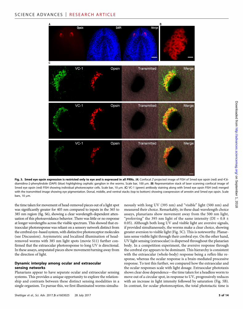
structures (35–38). Fluorescence in situ hybridization (FISH) imagingof the Smed eye opsin mRNA shows that the primary opsin likelyexpresses throughout the eye (Fig. 3, A and B). Costaining with labeledanti-arrestin (VC-1) antibody allowed us to mark every single PRN,and imaging with confocal microscopy revealed that every singlePRN does express the primary eye opsin (Fig. 3, B and C, and moviesS3 and S4). These data strongly suggest that all PRNs likely use thesame primary opsin photoreceptor for sensing. We then performedRNA interference (RNAi)–based knockdown of the primary eye opsingene (11, 12, 39). Knockdown of this single opsin mRNA attenuateslight sensing response across the visible spectrum in planarians (fig. S3),providing further evidence that acute discrimination shown by planar-ians in binary choice assays does not depend on wavelength-specificphotoreceptors.
Hierarchical light processing revealed through regenerationOur data so far suggest that the organisms are able to resolve smalldifferences in effective intensities of light sensed at the eye and convertthese differences into binary behavioral outputs. This behavioral fidel-ity points to highly efficient post-sensory comparative processingmediated through the visual neural network. To address this, we
Fig. 1. Planarians show the ability to resolve light inputs of distinct wavelengths. (A) Schematic of the binary phototactic choice assay performed. Two simulta-neous light inputs of specific wavelengths were provided, and the movement of planarians was measured to determine the choice made—either movement away froml2 (R1 localization), away from l1 (R3 localization), or no choice (R2 localization). (B) DI shown here for binary choice assays performed with either of the following lightpairs: blue and green (450 and 545 nm), green and red (545 and 625 nm), or blue and red (450 and 625 nm). Results of measurements on 10 worms, n = 6. Error barsindicate SEM. DI as indicated here and detailed in Materials and Methods. (C) Data from binary phototactic choice assay between a 500 nm light input and a secondwavelength (525/545/590 nm). DI = (NR3 − NR1)/total number of worms, where NR is the number of worms in a region. A DI value of 1 or −1 indicates a completeaversion or preference for the 500 nm light relative to the second wavelength. A value of 0 indicates no preference. All choice measurements were performed byproviding constant light intensity of 316 mW/cm2 (± 10) for both inputs. Results of measurements on 30 worms, n = 5. (D) Wavelength discrimination assay performedwith wavelengths from 425 to 625 nm with ~25 nm spectral separation. Results of measurements on 10 worms, n = 4. (B to D) Error bars indicate SEM.
3 of 14

SC I ENCE ADVANCES | R E S EARCH ART I C L E
on Septem
ber 1, 2018http://advances.sciencem
ag.org/D
ownloaded from
exploited the ability of planarians to regenerate their eye and the brainafter amputation. Specifically, we examined the return of phototacticability, including fine intensity discrimination (assayed throughwavelength choice) as a function of regeneration. After amputation,planarians show head regeneration approximately over a week (40).Eye spots (pigmentation) become visible after ~4 days (Fig. 4A). Ifwe examine the return of single-input light sensing and response ina light-dark assay, planarians show a robust negative phototactic re-sponse by ~5 days after amputation (fig. S4); this response is stable
Shettigar et al., Sci. Adv. 2017;3 : e1603025 28 July 2017
thereon. Remarkably, however, in binary choice assays at days 5 and6 after amputation, planarians do not show clear phototactic choicesseen with intact worms. Consistent choice behavior with multiple,closely spaced inputs is seen significantly later in regeneration (Fig. 4B).
Regeneration experiments show a hierarchy in light sensing andprocessing wherein the acute “effective intensity” discrimination islayered on a gross ability of the worms to detect light. In regenera-tion, by day 5 after amputation, a basic eye structure is established(20), and the visual network is able to signal to the locomotor sys-tem, leading to gross light-dark phototaxis (Fig. 4B). However, atthis stage, the worms do not show acute intensity discrimination.The return of this finer discrimination, over subsequent days, likelyrequires further rebuilding in the visual network such that compar-ative processing of two very similar inputs can be accomplished.Imaging of the planarian neural network shows that significantchanges occur in the regenerating planarian brain at specific timepoints that correlate with functional recovery of light sensing. Laserscanning confocal immunofluorescence microscopy with a synapticmarker reveals that by day 5 after amputation, a basic framework ofthe regenerated ventral nerve cord, cephalic ganglion (brain), andthe eye gets assembled, consistent with behavior. However, there isa substantial enrichment and enlargement of the planarian cephalicganglia between days 5 and 7 after amputation. This can be seenfrom representative imaging planes (Fig. 4D) and analysis of allz-stacks corresponding to the two cephalic lobes of the dorsal gan-glion (movies S5 to S9) during regeneration. This progressive en-richment of neural network was further tested through a carefulexamination of functional recovery. Figure 4C shows that althoughcoarse “discrimination” is restored to an extent by day 6 (90 nmdifferences in l; DI = 0.67 ± 0.014), the ability to do finer discrim-ination is significantly delayed. An ability to discriminate betweeninputs 25 nm apart is only seen 12 days after head removal (Fig. 4C).These data showing progressive enhancement of behavioral com-plexity with time provide a significant new insight that “functionallyrelevant regeneration” continues much after gross repair of organsystems has occurred.
Eye-independent, directional light avoidance in planariansPlanarians are highly light-aversive and show complex light discrimi-nation through their cerebral eyes.We then examined whether planar-ians have other modes of light sensing, including eye-independentmodes. For this, we observed the response of head-removed (ampu-tated) planarians to light. We found that S. mediterranea showed astriking extraocular, whole-body response to long-wavelength ultra-violet (UV) light (Fig. 5A and movie S10). Although there are earlyreports suggesting whole-body light sensing in planarians (10, 32),we quantitatively examined this sensing in the context of our newfindings. Movie S10 shows that although freshly decapitated (head-removed) planarians show no significant movement in “visible/white” light, illumination with long-wavelength UV light (360 to400 nm) leads to robust movement of the headless worm away fromlight. This movement of headless worms, in response to UV, is remi-niscent of motion exhibited by intact worms. To test the precisewavelength sensitivity of this extraocular photoavoidance, we examinedthe response of head-amputated planarians when stimulated with lightwavelengths ranging from 365 to 625 nm (fig. S5). Head-amputatedworms responded primarily to wavelength from ~365 to ~395 nm inphotoavoidance assays, with significantly reduced or no response at405 nm and beyond. Modified photoavoidance assays showed that
Fig. 2. Light intensity–based neutralization of wavelength choice yields an“action” spectrum that resembles an opsin absorption curve. (A) Overturningof choice through light intensity dosage. Binary choice experiments were initiallyperformed with equal amounts of photon flux (photon fluence rate − X = 1.59 ×10¹⁴/s per cm2). Photon flux of less-aversive input was then increased as indi-cated. Results of measurements on 10 worms, n = 4. (B) Putative action spectrumdetermined by measuring the amount of light required for choice neutralizationin binary choice assays with respect to 500 nm (near peak). Plotted here is a ratioof photon fluence rates (F500/Fl) versus wavelength (l) in nanometers at the pointof choice neutralization. The photon fluence rate at 500 nm was kept constant(1.59 × 10¹⁴/s per cm2), whereas the amount of light for other wavelengths (Fl)was modulated to determine minimum light required for choice neutralization inbinary assays. For all wavelengths other than 450 nm, an increase in photon flu-ence rate was required to neutralize choice with 500 nm. Results of measure-ments on 10 worms, n = 4. This obtained profile was matched with thepredicted spectrum of the alpha band of an opsin with a lmax of 475 nm, basedon Govardovskii’s template (34).
4 of 14
SC I ENCE ADVANCES | R E S EARCH ART I C L E
on Septem
ber 1, 2018http://advances.sciencem
ag.org/D
ownloaded from
the time taken formovement of head-removed pieces out of a light spotwas significantly greater for 405 nm compared to inputs in the 365 to385 nm region (fig. S6), showing a clear wavelength-dependent atten-uation of this photoavoidance behavior. There was little or no responseat longer wavelengths across the visible spectrum. This showed that ex-traocular photoresponse was reliant on a sensory network distinct fromthe cerebral eye–based system,with distinctive photoreceptormolecules(see Discussion). Asymmetric and localized illumination of head-removed worms with 385 nm light spots (movie S11) further con-firmed that the extraocular photoresponse to long UV is directional.In these assays, amputated pieces showmovement/turning away fromthe direction of light.
Dynamic interplay among ocular and extraocularsensing networksPlanarians appear to have separate ocular and extraocular sensingsystems. This provides a unique opportunity to explore the relation-ship and contrasts between these distinct sensing modalities in asingle organism. To pursue this, we first illuminated worms simulta-
Shettigar et al., Sci. Adv. 2017;3 : e1603025 28 July 2017
neously with long UV (395 nm) and “visible” light (500 nm) andmeasured their choice. Remarkably, in these dual-wavelength choiceassays, planarians show movement away from the 500 nm light,“preferring” the 395 nm light of the same intensity (DI = 0.8 ±0.05). Although both long UV and visible light are aversive signals,if provided simultaneously, the worms make a clear choice, showinggreater aversion to visible light (Fig. 5C). This is noteworthy. Planar-ians sense visible light through their cerebral eye. On the other hand,UV light sensing (extraocular) is dispersed throughout the planarianbody. In a competition experiment, the aversive response throughthe cerebral eye appears to be dominant. This hierarchy is consistentwith the extraocular (whole-body) response being a reflex-like re-sponse, whereas the ocular response is a brain-mediated processiveresponse. To test this further, we compared how the extraocular andthe ocular responses scale with light dosage. Extraocular phototaxisshows clear dose dependence—the time taken for a headless worm tomove out of a circular spot, in response to UV, progressively reduceswith an increase in light intensity followed by saturation (Fig. 5B).In contrast, for ocular photoreception, the total phototactic time is
Fig. 3. Smed eye opsin expression is restricted only to eye and is expressed in all PRNs. (A) Confocal Z-projected image of FISH of Smed eye opsin (red) and 4′,6-diamidino-2-phenylindole (DAPI) (blue) highlighting cephalic ganglion in the worms. Scale bar, 100 mm. (B) Representative stack of laser scanning confocal image ofSmed eye opsin (red) FISH showing individual photoreceptor cells. Scale bar, 10 mm. (C) VC-1 (green) antibody staining along with Smed eye opsin FISH (red) mergedwith the transmitted image showing eye pigmentation. Dorsal, middle, and ventral stacks (top to bottom) showing coexpression of arrestin and Smed eye opsin. Scalebars, 10 mm.
5 of 14

SC I ENCE ADVANCES | R E S EARCH ART I C L E
on Septem
ber 1, 2018http://advances.sciencem
ag.org/D
ownloaded from
Fig. 4. Mapping recovery of planarian phototactic abilities during regeneration. (A) Images showing head regeneration in S. mediterranea from days 1 to 8. (B) Wave-length discrimination assay performed on regenerating worms, with blue and green (450 and 545 nm), green and red (545 and 625 nm), and blue and red (450 and625 nm) light inputs. Measurements with 10 worms each, n = 6. Gray dashed line indicates the return of single-wavelength input (500 nm) negative phototacticability (see also fig. S4). (C) Comparative recovery of finer wavelength discrimination ability in regeneration. Binary choice assays with the light wavelength pairs500/590, 500/545, and 500/590 nm over regeneration. Measurements on 10 worms each, n = 4. (B and C) Error bars indicate SEM. ns, not significant. (D) Immu-nostaining of planarian cephalic ganglion using SYNORF1 antibody during the course of regeneration from days 3 to 7. Scale bar, 100 mm. *P < 0.05.
Shettigar et al., Sci. Adv. 2017;3 : e1603025 28 July 2017 6 of 14

SC I ENCE ADVANCES | R E S EARCH ART I C L E
on Septem
ber 1, 2018http://advances.sciencem
ag.org/D
ownloaded from
Fig. 5. Planarian extraocular photoresponse and hierarchical relationship with ocular light response. (A) Snapshots of amputated worms showing lightavoidance under UV light (350 to 400 nm) and no light avoidance under white light (W). (B) Dose dependence of extraocular photoresponse. Time taken for a headlesstail piece (24 hours after amputation) to move out of a circular spot of the 365 or 395 nm light (n = 30; see details in the Supplementary Materials). (C) Interplay betweenocular and extraocular light response in regeneration. DI for choice assays between 395 and 500 nm light in anterior and posterior regenerates during time course ofregeneration. DI for intact worms is also shown (right). Error bars indicate SEM. (D) Conceptual model indicates multiple facets and diversity in planarian light sensing.The ocular response mediated through the cerebral eye is a “processive” response, including the capacity for wavelength discrimination. This discrimination appearsdependent on comparative processing of input signals through the visual network. On the other hand, the extraocular response to UV light appears to be a morerudimentary response with the ventral nerve cord and peripheral network likely functioning as an integrator, collecting and mediating a whole-body response leadingto coordinated phototaxis. Both ocular and extraocular light sensing modalities engage the same motor machinery.
Shettigar et al., Sci. Adv. 2017;3 : e1603025 28 July 2017 7 of 14
SC I ENCE ADVANCES | R E S EARCH ART I C L E
on Sept
http://advances.sciencemag.org/
Dow
nloaded from
insensitive to dosage (fig. S7). Because extraocular response involveswhole-body sensing, the speed of response appears to depend on thetotal light sensed across the worm. However, in contrast, because theocular response appears to be subject to processing by the neuralnetwork, the total time required for movement is insensitive to lightdosage (Fig. 5D). In addition, head-removed worms fail to show anyspectral choice (fig. S8) unlike intact worms that show processing-based discrimination in binary choice assays.
We then examined this hierarchical relationship between thebrain-mediated ocular and the whole-body extraocular responsesduring eye-brain regeneration. Figure 5C shows aversive choice be-tween UV light (395 nm) and visible light (500 nm) plotted as afunction of time after head removal. As expected, freshly cut decapi-tated worms subjected to UV/visible choice show movement awayfrom UV. Head-removed, eyeless worms cannot sense visible light;hence, UV response dominates. Expectedly, this choice pattern ismaintained until 4 days after decapitation—there is no functionaleye or any response to visible light (fig. S4). However, by day 5 afterinjury, planarians regenerate a “functional” eye and a phototactic re-sponse to visible light (fig. S4). Therefore, by day 5, the eye-brain isfunctionally connected to the locomotor systems, and planarians re-spond to stimuli sensed at the eye. However, at this stage (day 5), theextraocular response retains its dominance over the nascent cerebral re-sponse in choice assays, wherein regenerating worms still show move-ment away fromUV (toward visible light) (Fig. 5C). This is opposite tothe behavior with intact worms. Remarkably, this result implies that re-generating worms that have sufficiently regenerated a functional eyeand brain capable of phototaxis behave similarly to headless, tail piecesin ocular-extraocular choice experiments. Subsequently, there is atransition about day 7 after decapitation, wherein the choice isreversed. By day 7 after injury, the regenerating planarians resembleintact worms, wherein the ocular, brain-mediated response is strongerand can override the extraocular response (Fig. 5C). These data re-veal the remarkable plasticity in the cross-talk between the two distinctresponses—ocular and extraocular—mediated by two independent butinteracting neural networks in the same organism.
ember 1, 2018
DISCUSSIONPlanarian flatworms are highly light-aversive organisms often foundin dark, aqueous environments shielded from direct light (41). Al-though reports of photoavoidance responses to visible and UV lightin planarians exist, a conceptual and mechanistic understanding oflight sensing in these organisms is limited. How do planarian flat-worms respond to light?What are the different ways in which planar-ians, with prototypic “simple” eyes, process information encoded inlight stimuli? Several of these questions have remained open. Our datashow that planarians have at least two independent light responses,ocular and extraocular, anchored in distinct sensory networks. Theocular response to visible light is mediated through the cerebral eye(fig. S1A), whereas the extraocular response is a whole-body response,with head-removed planarians able to respond to even small doses oflongUV light (Fig. 5B). Ourwork uncovers new light sensing and pro-cessing modalities while also showing how independent light sensingsystems can interact.
In examining the ocular (eye-based) sensing, we made new dis-coveries. In binary wavelength choice assays, planarians showconsistent phototactic choice with exceptional fidelity throughoutthe visible spectrum. Even a 25 nm change in wavelength inputs is
Shettigar et al., Sci. Adv. 2017;3 : e1603025 28 July 2017
sufficient to fully switch the phototactic behavior. Although differ-ential single-input photophobic responses have been reported (10),this is the first report on how planarians or similar organisms canacutely sense closely spaced spectral stimuli and convert them intothese distinct behavioral outputs. Conventionally, specific photo-receptors are required for true wavelength detection (35–37). However,planarians reportedly have only a single, broad-sensing opsin photo-receptor in the eye (11, 31). Our data show that this single opsin isexpressed throughout the eye, within every individual PRN (Fig. 3, Band C, and movies S3 and S4). In organisms with wavelength-specificopsins, these tend to be spatially confined to subsets of PRNs in the eye,allowing for true wavelength detection (38). Besides, our RNAi knock-down of the primary eye opsin leads to reduced photosensitivity acrossthe spectral range (fig. S3). These data strongly suggest a single opsinphotoreceptor in the eye.
So howdo the animals respond to small changes in lightwavelengthswithout specific photoreceptors? Broad-sensing photoreceptors shouldbe refractory to small spectral changes. Photoreceptors are extremelysensitive “photon counters,” but once an opsin is photoexcited by oneof two spectrally similar stimuli, the wavelength information is “lost”(35). The phototactic choice seen in our assays is not true “wavelengthdiscrimination” but amanifestation of an acute intensity sensing using asingle photosensor. We find that simple modulation of light intensitiesis sufficient to neutralize and even reverse wavelength choice (Fig. 2A).Further, measurements of intensities (photon fluence rate) requiredto neutralize wavelength choice (behavior) yield a putative actionspectrum, demonstrating the robustness of response (Fig. 2B). Al-though this unimodal action spectrum is consistent with a singleeye opsin, further microspectroscopic analysis would be requiredto precisely define the absorption profile of the eye.
We propose that worms sense small wavelength changes by com-paring the effective intensity of light inputs sensed at the eye. In thesimplest scenario, the signal sensed would scale with the actual ab-sorption of light by the eye. For any given wavelength, the light ab-sorbed would be a function of the intensity of incident light andextinction coefficient of the photoreceptors. Given two equal intensity(photon fluence rate) light inputs of different wavelengths (evenwith asingle opsin photoreceptor type), there will be differences in theamount of light sensed at the eye, reflecting the intrinsic absorptionspectrum of the opsin. Remarkably, when illuminated with closelyspaced spectral inputs, the animals are able to convert small differ-ences in effective intensities into virtually binary behavioral choices.Differences in effective intensities likely lead to changes in aggregatePRN response and signaling to a putative downstream processingcenter. The networks are able to parse these differences in effectiveintensities through comparative processing, leading to phototacticchoice. This choice can also be described as acute gradient sensing.Gradient sensing is seen in other organisms, such as Platynereis (42)and Drosophila larvae (43). Although planarians appear too acutelysensitive, this ability to sense light gradients likely represents awidespread evolutionary advance. Further work would be requiredusing RNAi and imaging methods to look for mechanistic and struc-tural underpinnings of acute gradient sensing.
In contrast to this “processive” cerebral eye light sensing, the high-sensitivity planarian whole-body extraocular photoresponse appears tobe a reflex-like response (Fig. 5, B andD, and fig. S6). Unlikewith ocularresponse, the extraocular response times scale strongly with light inten-sity; also, wavelength choice patterns, seen with eye-mediated sensing,are absent. The extraocular response network appears to integrate
8 of 14

SC I ENCE ADVANCES | R E S EARCH ART I C L E
on Septem
ber 1, 2018http://advances.sciencem
ag.org/D
ownloaded from
whole-body sensing events, finally leading to directional phototaxis(Fig. 5, A and D). Even the photoreceptor(s) mediating the extraocularresponse is distinct from the eye opsin, with different spectral ranges.High sensitivity to long UV (proxy for filtered sunlight) is consistentwith the environmental niche of many planarians. Being highly light-aversive, a whole-body response may be beneficial. Many planarianscan also propagate through fission (41, 44). A robust, phototactic ex-traocular response would allow newly fissioned, vulnerable tail piecesto avoid direct or bright light.
How is UV-A sensed and processed? Opsins are some of the mostwidely expressed light sensing proteins in the animal kingdom (26). Ex-traocular sensing using opsins has been seen in several organismsspread across different phyla (26). However, novel photoreceptors can-not be discounted. For instance, Drosophila larvae (27) and C. elegans(28) reportedly use unconventional light receptors resembling gusta-tory receptors for extraocular sensing (named Gr28b in Drosophilaand LITE-1 in C. elegans). Reports also suggest that transient receptorpotential ankyrin 1 (TRPA1) channels may also respond to UV light(27, 45). To list the possible extraocular photoreceptor molecules inSchmidtea, we conducted a preliminary investigation using bio-informatics. A pipeline designed to look for opsin-like proteins in theexisting planarian transcriptome yields six candidates (fig. S9), exclud-ing the previously identified eye opsin. We were unable to find anyhomologs for LITE-1 and Gr28b in our analysis, but found one TRPchannel candidate when comparing Schmidtea TRP channel homologswith UV-sensitive human and Drosophila TRPA1 channels (fig. S10).Our preliminary analysis, based onmolecules that have been implicatedin extraocular photoreception in other organisms (opsins, gustatoryreceptor–like, andTRP channels), yields some candidate photoreceptor(s).Further work would be required to identify the light receptor(s).
Planarian tails show coordinated movement away from UV, sug-gesting an independent neuronal signaling–dependent locomotor re-sponse. Where does the photosensing occur? In some organismsshowing extraocular sensing, photoreceptor molecules have been re-ported in neurons (23, 27, 28). Classical nervous system studies andimmunostaining have showed the existence of subepidermal nerveplexus in planarians (46–48). Therefore, it is conceivable that lightsensing molecules present in these neurons lead to extraocular re-sponse. There have been reports on ectopic eyes present in polychaetes(49); however, there is little or no information on these photoreceptivestructures in planarians. Investigating the structural and functionalunderpinnings of this high-sensitivity extraocular response wouldbe fascinating.
Because planarians show full eye-brain regeneration after injury,it is possible to address the nature of light sensing and processing inunprecedented ways. Examining functional recovery of light sensingrevealed a hierarchical, multilayered sensing paradigm. For eye-mediated light sensing, our data from wavelength choice experimentsperformed over eye-brain regeneration showed how gross sensingcould be temporally delineated from finer sensing (Fig. 4A). Thereis a period of time when regenerating worms (unlike intact worms)can sense light but cannot show clear behavioral choices in binarywavelength assays. Thus, the effective intensity–based acute gradientsensing is layered on a basic ability to detect light, with a progressivebuildup of sensing and processing capacity.We show that the ability todiscriminate closely spaced inputs (~25 nm changes) recovers signif-icantly later than crude discrimination abilities (Fig. 4C). This revealsthat functional recovery continues much after gross regeneration ofthe eye-brain structures is complete. Full functional recovery likely re-
Shettigar et al., Sci. Adv. 2017;3 : e1603025 28 July 2017
quires fine-tuning of sensing and processing capabilities, involvingpruning and patterning of the neural networks. Regeneration datasupport our model that acute sensing likely involves significant post-sensory comparative processing. Confocal imaging of planarian brainusing a synapticmarker over regeneration is consistentwith progressivebuildup of neural capacity (Fig. 4D). Although gross regeneration ofbasic brain structures appears to be complete by about day 5 after de-capitation, significant enrichment of synaptic density coincides with therecovery of acute sensing and processing abilities. Light sensing assaysestablished here may provide a new way to functionally “map” neuralregeneration (Fig. 6A).
Existence of two independent light sensing networks in planarians,alongwith eye-brain regeneration capacity, again allows unique lines ofinquiry. For intact worms, in competition experiments, the cerebral oc-ular response can override the reflex-like extraocular response, settingup abaselinehierarchy among the responses (Fig. 5C).Ocular-extraocularchoice experiments during regeneration reveal remarkable plasticity inthese hierarchies. During eye-brain regeneration, there is a period of~36 hours wherein both the extraocular and ocular responses “func-tionally” coexist, but the developing eye-brain axis is unable to overridethe reflex-like extraocular response (Figs. 5C and 6B). Thereafter,following a clear transition, the dominance of the ocular or brain-mediated response is reestablished (Figs. 5C and 6B). If the extraocularresponse is a more rudimentary, ancient response predating the eyeand a processive brain, then does regeneration coincidently mimic as-pects of evolutionary trajectories?
Ocular-extraocular choice experiments over regeneration also pro-vide new insights into how the eye-brain network interacts with thewhole-body light sensing network (Figs. 5C and 6A). A headless wormcan undergo locomotion in response to UV. By about day 5 in regen-eration (after head removal), a cerebral eye and a minimal brain areestablished, which link to the ventral nerve cord, whole-body nervoussystem, and the locomotionmachinery. This link of the visual networkto the locomotor network is sufficient to generate movement in re-sponse to light. However, it appears that the signaling flux from thecerebral eye and brain at this point is still insufficient to override theextraocular or reflex-type response anchored in the whole-body neu-ral network. After day 7, we propose that this signal from the eye-brainto the whole-body system strengthens, allowing overriding of the ex-traocular response (Fig. 6B). Appearance of a threshold whereinbrain-mediated response begins to dominate a whole-body responsehas implications for how the eye and the cephalic ganglion regenerateand how connectivity between two distinct neural networks, the ce-phalic ganglion and the ventral nerve cord in this case, is establishedand fine-tuned during regeneration. To our knowledge, this may bethe first report of its kind, describing such clear and dynamically inter-changing hierarchies between two distinct light sensory systems, allrevealed through relatively simple phototactic choice assays per-formed over regeneration.
In summary, this study significantly affects our fundamentalunderstanding of light sensing responses. Our work highlights the re-markable diversity and plasticity in light sensing and processing thatremain unexplored in nature. Because planarians have eye structuresand neural networks that appear simple yet similar to those observedin other animals, these acute and distinct light sensing and processingabilities may be more widespread in nature. This work also sets thestage for a comprehensive examination of light-induced behavior inflatwormswherein newquestions based on comparative visual process-ing and regeneration of neural networks can now be addressed. Our
9 of 14

SC I ENCE ADVANCES | R E S EARCH ART I C L E
on Septem
ber 1, 2018http://advances.sciencem
ag.org/D
ownloaded from
Fig. 6. Hierarchical light sensing and processing in planarians revealed through recovery of function with regeneration. (A) Schematic showing a timeline ofreturn of different phototactic abilities during head regeneration in S. mediterranea. By day 5 after amputation, worms sense light but have no ability to finer intensitydiscrimination (assayed through wavelength choice), which is acquired gradually significantly later. See (B) for ocular control. (B) Model for hierarchical relationshipbetween networks regulating ocular and extraocular sensing, including switching during regeneration. During days 1 to 4 after amputation, worms show only extra-ocular response anchored in ventral nerve cord (VNC) and peripheral neurons. By about day 5, ocular response recovers in worms, but signaling flux from cerebral visualnetworks is unable to override signals resulting from extraocular photosensing. Ocular and extraocular photosensing coexist, but extraocular sensing is dominant incompetition assays. Day 7 onward, flux from visual network strengthens, allowing the ocular response to be dominant, as in intact worms. Black arrow indicates visiblelight input, whereas violet arrow indicates long UV light input.
Shettigar et al., Sci. Adv. 2017;3 : e1603025 28 July 2017 10 of 14

SC I ENCE ADVANCES | R E S EARCH ART I C L E
results demonstrating the interplay between ocular and extraocularlight sensing networks are likely without precedent and reveal the richand multilayered light responses in planarians that can now beexamined further.
on Septem
ber 1, 2018http://advances.sciencem
ag.org/D
ownloaded from
MATERIALS AND METHODSLight source and measurementsWavelength-specific light-emitting diode (LED) light sources wereprocured from Roithner Lasertechnik. The specific LEDs used were365 nm (XSL-365-5E), 395 nm (RLS-UV395), 425 nm (LED425-6-30), 450 nm (LED450-01), 500 nm (LED500-10-30), 525 nm(LED525-01), 545 nm (LED545-04), 590 nm (LED590-03), and 625 nm(B5B-435-TL). Each LED is referred by its peak wavelength in thestudy. A suitable LED circuit along with a regulatable power sourceto drive the LEDs was assembled. This allowed precise control ofLED light intensities. Custom-designedOptoLED illumination system(Cairn Research) (fig. S11) was also used for extraocular-mediated be-havior experiments. Light power measurements were made usingNewport power meter 1918-R and 918D detector. Light measure-ments for all initial binary choice experimentsweremade atmicrowattper square centimeter (mW/cm2; intensity). All behavior experimentswere performedwith approximatemeasurement error of ±10 mW. Forthe experiments involving neutralization (and overturning) ofwavelength choice, the unit of light measurement and intensity wasphoton flux or photon fluence rate (number of photons per unit timeper unit area). This was done to ensure consistency with the theory ofaction spectroscopy and to build a wavelength-dependent responsefunction using light intensity required for neutralization of choice be-havior. The stability of the signal was checked for the duration of theexperiment in all cases.
Planarian maintenanceS. mediterranea was maintained at standard laboratory conditions(50). Worms were fed beef liver extract once in 3 days. Wormswere starved for at least 2 days before the start of any experiment.In addition, behavior experiments were performed in a dark roommaintained at 20°C.
Planarian negative phototaxis (single-input sensing)Earlier work shows that planarians respond broadly to visible light, withsubtle differences in response to colors (10). Using single-wavelengthphototaxis assays, we confirmed that planarians (S. mediterranea) arenegatively phototactic to visible light (fig. S2C). Sensitivity attenuates atwavelengths ≥625 nm (fig. S2C). The setup for the single-input, light-dark phototaxis has been illustrated in figs. S2A and S11. Planarianswere starved for at least 2 days for all phototaxis experimentation onintact or regenerating worms. Light-dark phototaxis assays with planar-ians were performed on a glass slide (75 mm × 25 mm × 1.25 mm).About 5 ml of medium was added to the slide, and a single planarianwas placed in the center region (R2, 10 mm wide; fig. S2A) using aPasteur pipette. Following this, LED light was turned on (all intensitieswere set to ~316 mW/cm2) such that illumination was spatially polar-ized creating a lit (R1) and a relatively dark zone (R3). After 2min, theregion (fig. S2A) in which the planarian is present is determined. Thisis repeated for several worms, and a DI is calculated. DI was calculatedby using the formula: DI = (NR3−NR1)/totalN (whereNR is the num-ber of worms in a region, andN is the number of worms). ADI value of1 or −1 indicates a complete aversion or preference for the provided
Shettigar et al., Sci. Adv. 2017;3 : e1603025 28 July 2017
wavelength of light, respectively. A value of 0 indicates no preference.Phototaxis during regeneration was also performed as described above,except that the position of the worm was determined after 3 min.
Wavelength discrimination choice assaysAll planarian choice assays were performed according to the sche-matic shown in Fig. 1A. LED lights were used as above. For all binarywavelength choice assays with equal intensities, two equal-intensitylight inputs at a constant light intensity of 316 (±10) mW/cm2 wereused. Similar to the single-input phototactic assay described above,each worm was placed in the center region and allowed to make achoice with a cutoff time of 2 min. Outcomes can be movement toregion 1, region 2, or no choice. Similarly, binary choice experimentswere performed during regeneration, and the position of the worm(for calculation of DI) was determined after 3 min.
For experiments testing overturning of wavelength choicethrough light intensity dosage (Fig. 2A), the initial effective photonfluence rate (number of photons per unit time per unit area) was setat X (1.59 × 10¹⁴/s per cm2) (for both wavelengths). Increasing dos-age of the less-aversive input was provided as 2X, 4X, or 8X as indi-cated in the figures. For building a behavioral response profile toaddress intensity-based wavelength choice neutralization (Fig. 2B),we measured the increase in photons (photon fluence rate) requiredto neutralize (randomize) choice relative to the 500 nm light at Xphoton fluence rate (1.59 × 10¹⁴/s per cm2). Choice neutralizationexperiments were performed with 425, 450, 525, and 545 nm as sec-ond wavelengths. According to the principle of a classical actionspectroscopy, at the point of choice neutralization, the photo-response (R; proportional to the product of extinction coefficientand number of photons incident) for the two inputs should be iden-tical (33) and can be used to build an action spectrum
R500 ¼ Rl ¼> e500 : el ¼ 1=F500 : 1=Fl
(33) where R is the photoresponse, e is the extinction coefficient, andF is the photon fluence rate (number of photons per unit time perunit area). Therefore, the plot of ratio of photon fluence rates(F500/Fl) versus wavelength (l) in nanometers at the point of choiceneutralization should resemble the absorption spectra of the photo-receptor (33). As described above, the photon fluence rate at 500 nmwas kept constant (1.59 × 10¹⁴/s per cm2), whereas the amount oflight for other wavelengths (Fl) was modulated to determineminimum light required for choice neutralization in binary assays. Thisobtained profile was matched with the predicted spectrum of the alphaband of an opsin with a lmax of 475 nm, based on Govardovskii’stemplate (34).
Extraocular photoreception and phototaxisFor determining wavelengths that affect extraocular phototaxis, pla-narian tail pieces (24 hours after decapitation; fig. S2B) were subjectedto phototaxis assays similar to the single-input phototaxis assays. Wemeasured the extraocular response using LEDs that showed their peakresponse at the following wavelengths from 365 to 625 nm. The inten-sity of all the light sources was kept constant at 126 (±10) mW/cm2. Toexamine the dosage (light intensity) dependence of extraocular pho-toreception (Fig. 5B), we performed a modified phototaxis assay.Worms were placed in a petri dish 24 hours after decapitation withplanarian medium and allowed to relax for ~30min without any lightor mechanical disturbance. A 1-cm light circle was made to fall on the
11 of 14

SC I ENCE ADVANCES | R E S EARCH ART I C L E
on Septem
ber 1, 2018http://advances.sciencem
ag.org/D
ownloaded from
worm, and the time it took the worm to escape from the circle of lightwas calculated and plotted against the intensity of light used. For lightdosage experiments with extraocular response, the light wavelengthsused were 365 and 395 nm, and the light intensity was varied from ~31to ~506 mW/cm2. To understand the attenuation of extraocular responseon the visible side of the spectrum, we performed photoavoidanceexperiments with 365, 385, and 405 nmwavelengths at a photon fluxof 2.33 × 10¹⁴/s per cm2 and recorded the response time.
Ocular versus extraocular binary choice assaysBinary choice assays were performed using 395 and 500 nm light in-puts using intact worms by maintaining constant light intensities of126 (±10) mW/cm2. Similarly, to study the ocular-extraocular choiceover regeneration, we subjected anterior regenerates (tail-forminghead) and posterior regenerates (head-forming tail) to similar binaryphototactic choice assays over a period of 8 days after head amputation.
Fluorescence in situ hybridizationThe heads of asexual planarians 5 to 10 mm in length were cut andprocessed for FISH as per the protocol described by Pearson et al.(51) with significant modifications. The reduction step after fixationwas omitted, and the worms were stored in methanol after dehydra-tion. Bleaching was performed for 2 hours using a formamide-basedsolution (1.2% H2O2, 5% formamide, and 0.5× SSC) as described byKing and Newmark (52) after rehydration. The post-hybridizationwashes, antibody blocking, incubation of peroxidase-conjugated anti-digoxigenin antibody, and post–antibody incubation washes wereperformed as per the protocol by King and Newmark (52). The de-velopment of the fluorescent signal was performed using a tyramidesignal amplification (TSA) reaction as per the protocol by King andNewmark (52). The tyramide conjugates were synthesized according tothe protocol described byHopman et al. (53) fromN-hydroxysuccinimideesters of Alexa Fluor 647 (A2006, Thermo Fisher Scientific). The TSAreaction was followed by at least five washes of 20 min each withPBSTx (phosphate-buffered saline with 0.3% Triton X-100) before fur-ther processing or mounting with a Mowiol-based mounting mediumprepared. For experiments involving immunostaining followed byFISH, the protocol mentioned below was followed.
Whole-body immunofluorescence and imagingConfocal immunofluorescence microscopy was performed to visu-alize changes in the planarian nervous system over regeneration.Immunostaining of the planarian nervous system was carried outas described by Sánchez Alvarado and Newmark (39) with modifi-cations using SYNORF1 (3C11, Developmental Studies HybridomaBank) or VC-1 [anti-arrestin (a gift from K. Agata)] as the primaryantibody. Horse serum (10%) was used as the blocking solution. Adonkey anti-mouse Alexa Fluor 488/555 (A-21206/A31570, LifeTechnologies) secondary antibody was used at dilution 1:500, withan incubation time of 90 min at room temperature. Imaging wasperformed on an LSM 780 Zeiss confocal microscope and analyzedusing ImageJ V. 1.48 (National Institutes of Health, Bethesda) (54).All stereomicroscope images were taken using Olympus SZX16.
RNAi-mediated knockdownPlanarian eye opsin (Smed eye opsin) sequence was obtained from thepublished planarian eye transcriptome (12). A region of the transcriptwas cloned using the following primers: Smed eye_opsin, AACACTC-GATGGGCTTGGG(forward) andCCCTGTGAGAGCCATAAGGG
Shettigar et al., Sci. Adv. 2017;3 : e1603025 28 July 2017
(reverse). From the cloned region, double-stranded RNA (dsRNA)wassynthesized as described by Rouhana et al. (55) with modifications.Planarians were amputated as shown in fig. S2B. Three 69-nl injec-tions with either opsin or control (green fluorescent protein) dsRNA(2 mg/ml) were made on days 1 to 3 of anterior-regenerating worms.Subsequently, these regenerates were examined for light response ondays 5 to 7 after amputation. Specifically, the regeneratingwormsweresubjected to single-input, light-dark choice using the light inputs (425,500, and 625 nmat 63 mW/cm2) as described earlier. DI was calculatedfor opsin knockdown and control worms and then plotted.
Movie recordingAll behavioral movies were recorded with Point Grey camera (GS3-U3-41C6NIR-C or BFLY-PGE-20E4M-CS). Fujinon 3.8- to 13-mmVarifocal C-mount lens were attached to the camera along with aninfrared long-pass filter if required. All of these were mounted onan adjustable mounting pole as shown in fig. S11. Optic fiber usedfor probing in movie S11 was procured from Thorlabs as a part ofcustomized OptoLED light system. Movie S10 was recorded on anOlympus MVX10 microscope using DP71 camera.
Bioinformatic analysisTo identify opsin homologs in planarians, we used 821 opsin se-quences from Porter et al. (56) as a reference data set. Tomake a non-redundant planarian transcriptome data set, we used CD-HIT (57) tocluster multiple transcriptomes (58–61). Reverse blast was performedwith (evalue = 1 × 10⁻¹⁰; coverage, 35%) 821 opsin sequences to iden-tify homologous sequences in S. mediterranea. We identified 172 pu-tative opsin-like candidates. A total of 143 of 172 transcripts that hadPFAM domain PF00001.16 (opsin G protein–coupled receptor classi-fication) were taken for further analysis. We built a hidden Markovmodel (HMM) for 821 opsin sequences and compared it with the143 transcript profiles. Of 143 transcripts, 130 matched the HMMprofile with evalue <1 × 10⁻¹⁰. We used TmHMM (www.cbs.dtu.dk/services/TMHMM-2.0/) to predict transmembrane (TM) helices for130 transcripts.We only considered transcripts with seven TMheliceswith optimum loop length for further analysis. Further, 66 transcriptswith seven TM helices were selected. From previous reports on opsinsequences from other organisms, we know that Lys (K) residue in theseventh (last) TMhelix is the amino acid residue towhich retinal bindsvia a Schiff base linkage (62). This TM helix has an NPxxY(x)6Fsequence motif (62). We took the seventh (last) TM helix for 66 S.mediterranea transcripts along with 821 opsin sequence and per-formed multiple sequence alignment using MacVector. To predictthe sequence motif, we used MEME (http://meme-suite.org/). Six of66 sequences had Lys (K) residue and complete/partial (one aminoacid missing/substitution) NPxxY(x)6F motif conservation. We pre-dicted tertiary structures of the six opsin sequences using homologymodeling (https://swissmodel.expasy.org/). Squid opsin (2z73) wasobtained as a closest hit to ourmodeled three-dimensional (3D) struc-ture of six opsins. Energy-minimized 3D structure was aligned usingPyMOL (https://www.pymol.org/). In addition, we obtained a rootmean square deviation of 0.167 Å, suggesting high structural simi-larity. We did phylogenetic analysis of the identified six putativeopsins from S. mediterranea using MEGA7 (maximum likelihood,50 bootstraps). The obtained phylogenetic tree was visualized usingTreeDyn (www.phylogeny.fr/one_task.cgi?task_type=treedyn). Seven TRPchannels were identified by Inoue et al. (63) in Dugesia japonica. Weidentified the homologs of these sequences in S. mediterranea using
12 of 14

SC I ENCE ADVANCES | R E S EARCH ART I C L E
PlanMine database (http://planmine.mpi-cbg.de) (64). Phylogeneticanalysis of seven Smed TRP channels with human and DrosophilaTRPA1 channels was then performed using TreeDyn.
Statistical analysisStatistical analyses were performed using a nonparametric Wilcoxonsigned-rank test.
http://advances.sciencemD
ownloaded from
SUPPLEMENTARY MATERIALSSupplementary material for this article is available at http://advances.sciencemag.org/cgi/content/full/3/7/e1603025/DC1fig. S1. Planarian light sensing apparatus.fig. S2. Planarians are aversive to light across a broad wavelength range.fig. S3. Planarian eye opsin knockdown attenuates light sensing across the color spectrum.fig. S4. Recovery of single-input light sensing and response during head regeneration.fig. S5. Wavelength dependence of extraocular photoreception.fig. S6. Extraocular response attenuates in visible light (~405 nm and longer).fig. S7. Aggregate time taken for eye-mediated phototaxis is insensitive to light intensity.fig. S8. Lack of wavelength discrimination in extraocular photoresponse.fig. S9. Phylogenetic tree (maximum likelihood) of planarian opsin sequence.fig. S10. Phylogenetic analysis of Smed TRP channels along with known UV-sensitive TRPA1channels (human and Drosophila).fig. S11. Light setup for phototaxis experiments and recording.movie S1. Planarian worm shows wavelength discrimination.movie S2. Planarians show robust wavelength discrimination (group response).movie S3. Imaging Smed eye opsin expression.movie S4. Smed eye opsin is expressed in all photoreceptor cells.movie S5. Regenerating cephalic ganglion (day 3).movie S6. Regenerating cephalic ganglion (day 4).movie S7. Regenerating cephalic ganglion (day 5).movie S8. Regenerating cephalic ganglion (day 6).movie S9. Regenerating cephalic ganglion (day 7).movie S10. Extraocular photoreception and phototaxis in planarians.movie S11. Light avoidance (extraocular) response to long UV is directional.
on Septem
ber 1, 2018ag.org/
REFERENCES AND NOTES1. D.-E. Nilsson, Eye evolution and its functional basis. Vis. Neurosci. 30, 5–20 (2013).2. D.-E. Nilsson, The evolution of eyes and visually guided behaviour. Philos. Trans. R. Soc.
Lond. B Biol. Sci. 364, 2833–2847 (2009).3. N. Randel, G. Jékely, Phototaxis and the origin of visual eyes. Philos. Trans. R. Soc. Lond. B
Biol. Sci. 371, 20150042 (2016).4. D. Arendt, J. Wittbrodt, Reconstructing the eyes of Urbilateria. Philos. Trans. R. Soc. Lond. B
Biol. Sci. 356, 1545–1563 (2001).5. D. Arendt, H. Hausen, G. Purschke, The ‘division of labour’ model of eye evolution. Philos.
Trans. R. Soc. Lond. B Biol. Sci. 364, 2809–2817 (2009).6. E. Saló, R. Batistoni, chap. 2, The Planarian Eye: A Simple and Plastic System with Great
Regenerative Capacity, in Animal Models in Eye Research, P. A. Tsonis, Ed. (Elsevier, 2008),pp. 15–26.
7. H. M. Brown, T. E. Ogden, The electrical response of the planarian ocellus. J. Gen. Physiol.51, 237–253 (1968).
8. K. S. Carpenter, M. Morita, J. B. Best, Ultrastructure of the photoreceptor of the planarianDugesia dorotocephala. Cell Tissue Res. 148, 143–158 (1974).
9. Y. Kishida, Electron microscopic studies on the planarian eye. I. Fine structures of thenormal eye. Sci. Rep. Kanazawa Univ. 12, 75–110 (1967).
10. T. R. Paskin, J. Jellies, J. Bacher, W. S. Beane, Planarian phototactic assay revealsdifferential behavioral responses based on wavelength. PLOS ONE 9, e114708 (2014).
11. D. Pineda, J. Gonzalez, M. Marsal, E. Salo, Evolutionary conservation of the initial eyegenetic pathway in planarians. Belg. J. Zool. 131, 77–82 (2001).
12. S. W. Lapan, P. W. Reddien, Transcriptome analysis of the planarian eye identifies ovo as aspecific regulator of eye regeneration. Cell Rep. 2, 294–307 (2012).
13. H. B. Sarnat, M. G. Netsky, The brain of the planarian as the ancestor of the human brain.Can. J. Neurol. Sci. 12, 296–302 (1985).
14. K. Agata, Y. Soejima, K. Kato, C. Kobayashi, Y. Umesono, K. Watanabe, Structure of theplanarian central nervous system (CNS) revealed by neuronal cell markers. Zoolog. Sci. 15,433–440 (1998).
15. K. Tessmar-Raible, D. Arendt, Emerging systems: Between vertebrates and arthropods,the Lophotrochozoa. Curr. Opin. Genet. Dev. 13, 331–340 (2003).
Shettigar et al., Sci. Adv. 2017;3 : e1603025 28 July 2017
16. O. Simakov, T. A. Larsson, D. Arendt, Linking micro- and macro-evolution at the cell typelevel: A view from the lophotrochozoan Platynereis dumerilii. Brief. Funct. Genomics 12,430–439 (2013).
17. P. W. Reddien, A. Sánchez Alvarado, Fundamentals of planarian regeneration. Annu. Rev.Cell Dev. Biol. 20, 725–757 (2004).
18. E. Saló, The power of regeneration and the stem-cell kingdom: Freshwater planarians(Platyhelminthes). Bioessays 28, 546–559 (2006).
19. F. Cebrià, M. Nakazawa, K. Mineta, K. Ikeo, T. Gojobori, K. Agata, Dissecting planariancentral nervous system regeneration by the expression of neural-specific genes.Dev. Growth Differ. 44, 135–146 (2002).
20. T. Inoue, H. Kumamoto, K. Okamoto, Y. Umesono, M. Sakai, A. Sánchez Alvarado, K. Agata,Morphological and functional recovery of the planarian photosensing system duringhead regeneration. Zoolog. Sci. 21, 275–283 (2004).
21. T. Takano, J. N. Pulvers, T. Inoue, H. Tarui, H. Sakamoto, K. Agata, Y. Umesono,Regeneration-dependent conditional gene knockdown (Readyknock) in planarian:Demonstration of requirement for Djsnap-25 expression in the brain for negativephototactic behavior. Dev. Growth Differ. 49, 383–394 (2007).
22. T. W. Cronin, S. Johnsen, Extraocular, non-visual, and simple photoreceptors: Anintroduction to the symposium. Integr. Comp. Biol. 56, 758–763 (2016).
23. B. Backfisch, V. B. Veedin Rajan, R. M. Fischer, C. Lohs, E. Arboleda, K. Tessmar-Raible,F. Raible, Stable transgenesis in the marine annelid Platynereis dumerilii sheds new lighton photoreceptor evolution. Proc. Natl. Acad. Sci. U.S.A. 110, 193–198 (2013).
24. M. L. Porter, Beyond the eye: Molecular evolution of extraocular photoreception. Integr.Comp. Biol. 56, 842–852 (2016).
25. D. M. Steven, The dermal light sense. Biol. Rev. Camb. Philos. Soc. 38, 204–240 (1963).26. K.-W. Yau, R. C. Hardie, Phototransduction motifs and variations. Cell 139, 246–264 (2009).27. Y. Xiang, Q. Yuan, N. Vogt, L. L. Looger, L. Y. Jan, Y. N. Jan, Light-avoidance-mediating
photoreceptors tile the Drosophila larval body wall. Nature 468, 921–926 (2010).28. A. Ward, J. Liu, Z. Feng, X. Z. S. Xu, Light-sensitive neurons and channels mediate
phototaxis in C. elegans. Nat. Neurosci. 11, 916–922 (2008).29. E. Pennisi, Evolution of developmental diversity. Evo-devo devotees eye ocular origins
and more. Science 296, 1010–1011 (2002).30. S. Yamaguchi, C. Desplan, M. Heisenberg, Contribution of photoreceptor subtypes to
spectral wavelength preference in Drosophila. Proc. Natl. Acad. Sci. U.S.A. 107, 5634–5639(2010).
31. H. M. Brown, H. Ito, T. E. Ogden, Spectral sensitivity of the planarian ocellus. J. Gen.Physiol. 51, 255–260 (1968).
32. G. H. Parker, The Reactions of Planarians: With and Without Eyes, to Light, vol. 115 ofContributions from the Zoölogical Laboratory of the Museum of Comparative Zoölogy atHarvard College (Harvard University, 1900).
33. E. Schäfer, L. Fukshansky, W. Shropshire Jr., in Photomorphogenesis (Springer, 1983).34. V. I. Govardovskii, N. Fyhrquist, T. Reuter, D. G. Kuzmin, K. Donner, In search of the visual
pigment template. Vis. Neurosci. 17, 509–528 (2000).35. T. H. Goldsmith, Optimization, constraint, and history in the evolution of eyes. Q. Rev. Biol.
65, 281–322 (1990).36. A. Kelber, L. S. V. Roth, Nocturnal colour vision—Not as rare as we might think. J. Exp. Biol.
209, 781–788 (2006).37. F. Pichaud, A. Briscoe, C. Desplan, Evolution of color vision. Curr. Opin. Neurobiol. 9,
622–627 (1999).38. J. Rister, C. Desplan, The retinal mosaics of opsin expression in invertebrates and
vertebrates. Dev. Neurobiol. 71, 1212–1226 (2011).39. A. Sánchez Alvarado, P. A. Newmark, Double-stranded RNA specifically disrupts gene
expression during planarian regeneration. Proc. Natl. Acad. Sci. U.S.A. 96, 5049–5054(1999).
40. T. Sandmann, M. C. Vogg, S. Owlarn, M. Boutros, K. Bartscherer, The head-regenerationtranscriptome of the planarian Schmidtea mediterranea. Genome Biol. 12, R76 (2011).
41. R. A.Medved, E. F. Legner, Feeding and reproduction of the planarian,Dugesia dorotocephala(Woodworth), in the presence of Culex peus Speiser. Environ. Entomol. 3, 637–641 (1974).
42. N. Randel, A. Asadulina, L. A. Bezares-Calderón, C. Verasztó, E. A. Williams, M. Conzelmann,R. Shahidi, G. Jékely, Neuronal connectome of a sensory-motor circuit for visualnavigation. eLife 3, e02730 (2014).
43. E. A. Kane, M. Gershow, B. Afonso, I. Larderet, M. Klein, A. R. Carter, B. L. de Bivort,S. G. Sprecher, A. D. T. Samuel, Sensorimotor structure of Drosophila larva phototaxis.Proc. Natl. Acad. Sci. U.S.A. 110, E3868–E3877 (2013).
44. C. M. Child, Physiological isolation of parts and fission in Planaria. Dev. Genes Evol. 30,159–205 (1910).
45. N. W. Bellono, L. G. Kammel, A. L. Zimmerman, E. Oancea, UV light phototransductionactivates transient receptor potential A1 ion channels in human melanocytes. Proc. Natl.Acad. Sci. U.S.A. 110, 2383–2388 (2013).
46. C. González-Estévez, D. A. Felix, M. D. Smith, J. Paps, S. J. Morley, V. James, T. V. Sharp,A. A. Aboobaker, SMG-1 and mTORC1 act antagonistically to regulate response to injuryand growth in planarians. PLOS Genet. 8, e1002619 (2012).
13 of 14

SC I ENCE ADVANCES | R E S EARCH ART I C L E
on http://advances.sciencem
ag.org/D
ownloaded from
47. K. G. Ross, K. C. Omuro, M. R. Taylor, R. K. Munday, A. Hubert, R. S. King, R. M. Zayas, Novelmonoclonal antibodies to study tissue regeneration in planarians. BMC Dev. Biol. 15, 2(2015).
48. J. Baguñà, R. Ballester, The nervous system in planarians: Peripheral and gastrodermalplexuses, pharynx innervation, and the relationship between central nervous systemstructure and the acoelomate organization. J. Morphol. 155, 237–252 (1978).
49. G. Purschke, D. Arendt, H. Hausen, M. C. M. Müller, Photoreceptor cells and eyes inAnnelida. Arthropod Struct. Dev. 35, 211–230 (2006).
50. N. J. Oviedo, C. L. Nicolas, D. S. Adams, M. Levin, Establishing and maintaining a colony ofplanarians. CSH Protoc. 2008, pdb.prot5053 (2008).
51. B. J. Pearson, G. T. Eisenhoffer, K. A. Gurley, J. C. Rink, D. E. Miller, A. Sánchez Alvarado,Formaldehyde-based whole-mount in situ hybridization method for planarians. Dev. Dyn.238, 443–450 (2009).
52. R. S. King, P. A. Newmark, In situ hybridization protocol for enhanced detection of geneexpression in the planarian Schmidtea mediterranea. BMC Dev. Biol. 13, 8 (2013).
53. A. H. N. Hopman, F. C. S. Ramaekers, E. J. M. Speel, Rapid synthesis of biotin-,digoxigenin-, trinitrophenyl-, and fluorochrome-labeled tyramides and their applicationfor in situ hybridization using CARD amplification. J. Histochem. Cytochem. 46, 771–777(1998).
54. J. Schindelin, I. Arganda-Carreras, E. Frise, V. Kaynig, M. Longair, T. Pietzsch, S. Preibisch,C. Rueden, S. Saalfeld, B. Schmid, J.-Y. Tinevez, D. J. White, V. Hartenstein, K. Eliceiri,P. Tomancak, A. Cardona, Fiji: An open-source platform for biological-image analysis.Nat. Methods 9, 676–682 (2012).
55. L. Rouhana, J. A. Weiss, D. J. Forsthoefel, H. Lee, R. S. King, T. Inoue, N. Shibata, K. Agata,P. A. Newmark, RNA interference by feeding in vitro-synthesized double-stranded RNA toplanarians: Methodology and dynamics. Dev. Dyn. 242, 718–730 (2013).
56. M. L. Porter, J. R. Blasic, M. J. Bok, E. G. Cameron, T. Pringle, T. W. Cronin, P. R. Robinson,Shedding new light on opsin evolution. Proc. Biol. Sci. 279, 3–14 (2012).
57. W. Li, A. Godzik, Cd-hit: A fast program for clustering and comparing large sets of proteinor nucleotide sequences. Bioinformatics 22, 1658–1659 (2006).
58. B. L. Cantarel, I. Korf, S. M. C. Robb, G. Parra, E. Ross, B. Moore, C. Holt, A. Sánchez Alvarado,M. Yandell, MAKER: An easy-to-use annotation pipeline designed for emerging modelorganism genomes. Genome Res. 18, 188–196 (2008).
59. M. J. Blythe, D. Kao, S. Malla, J. Rowsell, R. Wilson, D. Evans, J. Jowett, A. Hall, V. Lemay,S. Lam, A. A. Aboobaker, A dual platform approach to transcript discovery for theplanarian Schmidtea mediterranea to establish RNAseq for stem cell and regenerationbiology. PLOS ONE 5, e15617 (2010).
60. S.-Y. Liu, C. Selck, B. Friedrich, R. Lutz, M. Vila-Farré, A. Dahl, H. Brandl, N. Lakshmanaperumal,I. Henry, J. C. Rink, Reactivating head regrowth in a regeneration-deficient planarianspecies. Nature 500, 81–84 (2013).
61. S. M. C. Robb, K. Gotting, E. Ross, A. Sánchez Alvarado, SmedGD 2.0: The Schmidteamediterranea genome database. Genesis 53, 535–546 (2015).
Shettigar et al., Sci. Adv. 2017;3 : e1603025 28 July 2017
62. S. D’Aniello, J. Delroisse, A. Valero-Gracia, E. K. Lowe, M. Byrne, J. T. Cannon, K. M. Halanych,M. R. Elphick, J. Mallefet, S. Kaul-Strehlow, C. J. Lowe, P. Flammang, E. Ullrich-Lüter,A. Wanninger, M. I. Arnone, Opsin evolution in the Ambulacraria. Mar. Genomics 24 (Pt. 2),177–183 (2015).
63. T. Inoue, T. Yamashita, K. Agata, Thermosensory signaling by TRPM is processed by brainserotonergic neurons to produce planarian thermotaxis. J. Neurosci. 34, 15701–15714 (2014).
64. H. Brandl, H. Moon, M. Vila-Farré, S.-Y. Liu, I. Henry, J. C. Rink, PlanMine—A mineableresource of planarian biology and biodiversity. Nucleic Acids Res. 44, D764–D773 (2016).
Acknowledgments: We thank V. Lakshmanan for his help in bioinformatics analysis. We aregrateful to M. K. Mathew, M. Panicker, A. Ramesh, S. Sane, T. Mukherjee, R. Muddashetty,R. Sambasivan, S. Mayor, and U. S. Bhalla for their discussions and advice. We thankS. Ramaswamy and the Technologies for the Advancement of Science. We are also grateful tothe Central Imaging and Flow Cytometry Facility, Sequencing facility, the mechanical andelectronic workshops, and other facilities at the National Centre for Biological Sciences(NCBS)–Institute for Stem Cell Biology and Regenerative Medicine (inStem) campus. Funding:The support from inStem core funds and Department of Biotechnology (DBT), India, isgratefully acknowledged. Work in A.G. laboratory is also supported by the following grants:102/IFD/SAN/999/2014-2015 (DBT), BT/IN/Germany–BMBF/03/AG/2015-16 (DBT), and INT/SWISS/SNSF/P-50/2015 (Department of Science and Technology). D.P. is supported byWelcome/DBT India Alliance (GrantID: 500160/Z/09/Z). N.S. is grateful to the University GrantsCommission for the fellowship support. Author contributions: N.S. designed and performedthe experiments, analyzed the data, and assisted in manuscript writing. A.J. and S.P.assisted N.S. in performing behavioral experiments and experimental setups. R.D. and A.C.performed immunostaining and assisted in imaging. R.G. helped perform choice overturningand dose dependence experiments. M.M. assisted with light setups and the experimentaldesign. D.P. provided expertise in planarian biology, experimental design, and bioinformaticanalysis. A.G. conceived the project, designed the experiments, directed the study, analyzedthe data, and wrote the manuscript with contributions from the other authors. Competinginterests: The authors declare that they have no competing interests. Data and materialsavailability: All data needed to evaluate the conclusions in the paper are present in the paperand/or the Supplementary Materials. Additional data related to this paper may be requestedfrom the authors.
Submitted 1 December 2016Accepted 27 June 2017Published 28 July 201710.1126/sciadv.1603025
Citation: N. Shettigar, A. Joshi, R. Dalmeida, R. Gopalkrishna, A. Chakravarthy, S. Patnaik,M. Mathew, D. Palakodeti, A. Gulyani, Hierarchies in light sensing and dynamic interactionsbetween ocular and extraocular sensory networks in a flatworm. Sci. Adv. 3, e1603025 (2017).
Se
14 of 14
ptember 1, 2018

networks in a flatwormHierarchies in light sensing and dynamic interactions between ocular and extraocular sensory
Mathew, Dasaradhi Palakodeti and Akash GulyaniNishan Shettigar, Asawari Joshi, Rimple Dalmeida, Rohini Gopalkrishna, Anirudh Chakravarthy, Siddharth Patnaik, Manoj
DOI: 10.1126/sciadv.1603025 (7), e1603025.3Sci Adv
ARTICLE TOOLS http://advances.sciencemag.org/content/3/7/e1603025
MATERIALSSUPPLEMENTARY http://advances.sciencemag.org/content/suppl/2017/07/24/3.7.e1603025.DC1
PERMISSIONS http://www.sciencemag.org/help/reprints-and-permissions
Terms of ServiceUse of this article is subject to the
registered trademark of AAAS.is aScience Advances Association for the Advancement of Science. No claim to original U.S. Government Works. The title
York Avenue NW, Washington, DC 20005. 2017 © The Authors, some rights reserved; exclusive licensee American (ISSN 2375-2548) is published by the American Association for the Advancement of Science, 1200 NewScience Advances
on Septem
ber 1, 2018http://advances.sciencem
ag.org/D
ownloaded from