Hemostasis 2 - Fiziologiefiziologie.ro/en/2015-2016/lectures/Blood Physiology 5_2016.pdf ·...
110
HEMOSTASIS 2 Dr. Suzana Voiculescu, Assist. Prof., Discipline of Physiology and Fundamental Neuroscience
Transcript of Hemostasis 2 - Fiziologiefiziologie.ro/en/2015-2016/lectures/Blood Physiology 5_2016.pdf ·...

HEMOSTASIS 2
Dr. Suzana Voiculescu,
Assist. Prof., Discipline of Physiology and Fundamental Neuroscience

REVIEW- CLASSIC VS MODERN

CELL BASED MODEL OF HEMOSTASIS
Specific cellular receptors for coagulation
proteins promote hemostasis which occurs
in three overlapping phases
Initiation – involves tissue-factor bearing cell
and production of small amounts of thrombin
Amplification – involves platelet activation and
sets the stage for large scale thrombin
production
Propagation – the activated platelet surface
protects factor XI and results in an explosive
burst of thrombin formation

CELL BASED MODEL OF HEMOSTASIS

NATURAL ANTICOAGULANT SYSTEM


PROTEIN C
62-kD vitamin K-dependent glycoprotein
Synthesized in the liver as a single-chain
zymogen
Clipped into a serine-protease-like enzyme on
phospholipid cell surfaces by thrombin

PROTEIN C
Activated protein C (APC) binds protein S for
further activity
Protein C also has pro-fibrinolytic, anti-
inflammatory and anti-ischemic properties
PROTEIN S
Require negatively charged phospholipids and
Ca2+ for normal anti-coagulant activity
Complexes with protein C and acts by
proteolyzing Factor VIII and Factor V, which in
turn prevents activation of factor X and
prothrombin

PROTEIN S AND C DEFIECENCY
Genetic- homo/heterozygous
Acquired
Liver disease
DIC
Vitamin K deficiency
Septic shock

CLINICAL
Deep vein thrombosis is the most common
Pulmonary embolism
Fetal loss

THROMBOMODULIN
specific endothelial cell receptor that forms a
1:1 stoichiometric complex with thrombin
Cofactor in the thrombin-induced activation of
protein C
responsible for the conversion of protein C to
the activated protein C .

ANTICOAGULANT THERAPY
Injectable- heparin and heparin derivatives
Unfractioned heparin
Low molecular weight heparins
Oral- antivitamin K inhibitors
Coumadins- most used= WARFARIN

HEPARIN AND HEPARAN SULFATE
Heparan sulfate- is a GAG
Similar structure to heparin
Endogenous heparin- secretory granules of mast cells (probably being negatively charged- retains histamine inside)
Similar synthetic pathways
Function of heparan sulfate in coagulation- anticoagulant- receptor for ATIII activation thrombin inhibition

HEPARAN SULPHATE
vast structural diversity
bind and interact with a wide variety of
proteins, such as growth factors, chemokines,
morphogens, extracellular matrix components,
and enzymes
modulate different biological processes


TYPES OF HEPARIN
Unfractioned- high molecular weight; injectable
iv, hospitalised patient usually, APTT monitoring
needed (administred using a heparin pump)
Low molecular weight heparin- subcutaneously,
home therapy, no APTT monitoring, less
bleeding as side effect

UNFRACTIONED HEPARIN AS A DRUG
Needs ATIII as a cofactor
Inhibits Xa and thrombin
Clinical uses
Venous thrombosis disorders
Pulmonary embolism
Prophylaxis and treatment- thrombosis in major surgeries
Extracorporeal circulation
Blood samples drowned for lab purpuses- AC
Blood transfusions- in vitro AC
LOW MOLECULAR WEIGHT HEPARIN USE
No APTT monitoring needed
Lower molecular weight
May be administrated subcutaneously- may be
used for unhospitalized patients (once/twice a
day)
Less bleeding
Tends to replace heparin in venous thrombosis,
pulm embolism, acute coronary syndomes

ADVANTAGES OF LMWH OVER UH
Less bleeding (less inhibition on platelet
function)
Longer plasma half life (4-6 over 1 h)
No monitoring for LMWH
Less incidence of thrombocytopenia

HEPARIN TREATMENT COMPLICATIONS
Haemorrhage
Thrombocytopenia (most common drug-
induced thrombocytopenia)
Type I- nonimmune (2 days after treat)
Type II- immune (4-10 days after- HIT)
Osteoporosis (when long- term admin)

ORAL ANTICOAGULANT THERAPY
Vitamin K competitors- coumadin (Warfarin)
Warfarin inhibits the vitamin K-dependent
synthesis of biologically active forms of the
calcium-dependent clotting factors II, VII, IX and
X, as well as the regulatory factors protein C,
protein S.

VITAMIN K DEPENDENT FACTORS
VITAMIN K
Lipidic vitamin- diet (green veg and oils); colic
bacteria synthesis
Haemostasis
Bone matrix proteins, osteocalcin, undergo
gamma carboxylation with calcium much the
way coagulation factors do; needs VK

VITAMIN K DEF
according to the age
most often in infancy (lack of VK reaching the fetus across the placenta, the low level of VK in breast milk, and low colonic bacterial synthesis)
large amount of VK given to a pregnant patient can lead to jaundice in a newborn
Adults- 1 week reserve, abs in terminal ileum in lymph (bile salts, microvilli)

VITAMIN K (VK)
Cofactor
needed for the conversion of 10-12 glutamic
acid residue on the NH2 -terminal of precursor
coagulation proteins into the action form of
gamma-carboxyglutamic acid
VK-dependent gamma-glutamyl carboxylase
allows the VK-dependent proteins to bind to
surface phospholipids

In the liver- vitamin K helps the carboxylation of glutamic acid residues on immature coag factors; in this process it gets oxidized
Antivitamin K drugs inhibit reductase by inhibition, vitamin K stays in an inactive form

ANTI VITAMIN K
Anticoagulant effect starts in 24-36 h from
initiation – stops in 36-72 h after it is stopped
Oral treatment
At first- injectable treatment overlaps the oral
one (at least 4 days)

ANTIVITAMIN K CLINICAL USES
Venous thrombosis
Stroke
Thrombembolism
Cardiac valve replacement
Post myocardial infarction

MONITORING HEMOSTASIS
Primary
Bleeding time
Rumpel Leede
PLT count
Secondary
Clotting time
Howell Gram time
APTT
PT (Quick time)

HOWELL GRAM INTRINSIC AND COMMON
test explores the intrinsic and common pathways; for this we add CaCl2 to citrate plasma and start monitoring the time needed to clot.
Normal values= 60 -120s
High:
TR disfunction / thrombocytopenia
intrinsic pathway factor deficit (XII,XI,IX,VIII- hemophilia)
common pathway deficit- X, V, II, I (hypofibrinogenemia/ afibrinogenemia)
-anti- clotting therapy- heparin

APTT ACTIVATED PARTIAL THROMBOPLASTIN
TIME INTRINSIC AND COMMON PATHWAYS
To citrate plasma (with low amount of platelets- centrifuge for 15 min- 5000 rpm) we add Ca Cl2, kaolin, cephalin and start monitoring the time needed to clot.
Cephalin is a partial tromboplastin (only phospholipid)
Kaolin or silica is a negatively charged molecule activates factor XII
Normal values = 30- 40 s
Causes of abnormal high values- the same as Howell- except for the platelet- derived causes
It is affected by unfractioned heparin- used to monitor therapy

APTT

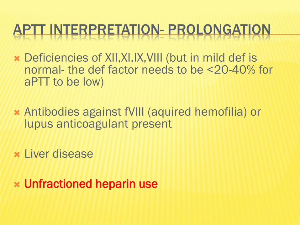
APTT INTERPRETATION- PROLONGATION
Deficiencies of XII,XI,IX,VIII (but in mild def is normal- the def factor needs to be <20-40% for aPTT to be low)
Antibodies against fVIII (aquired hemofilia) or lupus anticoagulant present
Liver disease
Unfractioned heparin use

APTT
May also be affected by:
Coumarin high dosage therapy
Lupus anticoagulant presence- antibody with in
vivo prothrmobotic activity, but in vitro
antithrombotic activity (interferes with the PL
used in anticoagulat tests procedure)

PROTHROMBIN TIME (QUICK METHOD-PT/QT)
EXTRINSIC AND COMMON
To citrate plasma (with high amount of
platelets) we add Ca Cl2, thromboplastin and
start monitoring the time needed to clot.
Elevated in
Deficit of I, II, V, VII, X factors
Liver failure
K vitamin deficiency/ anti vit K anticoag therapy
Normal values = 12-15 s


PROTHROMBIN TIME
Used to monitor therapy with oral anticoagulant drugs- vitamin K blocking agents- coumarins (warfarin)!!!
By INR fraction which should be measured constantly in these patients
The therapy is used in patients with high thrombotic risk

INR
International normalized ratio
ISI- depends on the tissue factor, it is established
by the manufacturer (usually between 1- 2)
Normal range for a healthy person= 0.9- 1.3
Warfarin therapy monitoring- depends on
pathology


THROMBIN TEST
Explores the last coagulation step, except for factor XIII
Thrombin is added to the plasma directly time needed for clot to form
Normal values- 15-18 s
High in:
Heparin therapy
Abnormal fibrinogen (qualitative and quantitative)
Lupus anticoagulant present

HEMOPHILIA
group of hereditary genetic disorders that impair the body's ability to control blood clotting or coagulation
Excessive bleeding, minor injury
Sex- linked
Type A- VIII factor deficit
Type B- IX factor deficit
Not sex- linked
Type C- XI factor deficit
Severity varies due to level of active clotting factor (<1 %- severe)

FIBRINOLYSIS
As the clots forms, it incorporates plasmin molecules.
Plasmin is an enzyme formed from plasminogen by tissue plasminogen activator (t-PA) and urokinase type plasminogen activator (u-PA)
Plasmin breaks down fibrin polymers into fibrin fragments = fibrinolysis
Fibrinolysis helps removing the clot as the repair processes occur.

FIBRINOLYSIS
Damage to the tissues releases TPA, which together with activated components from the coagulation pathways and protein C, activates plasminogen to plasmin.
Plasmin acts on the insoluble fibrin to form a series of soluble products: FDPs.
Fibrin that has been stabilized (crosslinked) by factor XIII gives rise to crosslinked FDPs (XDPs), as well as X Y D and E fragments.
The XDPs (D-dimer, D-dimer-E fragments), and oligomers of fragments X and Y, can be detected using antibody coated latex beads.


PLASMINOGEN
Zymogen plasmin
GP
Syntesized in liver
Lyses fibrin into fibrin degradation products (FDP)

PHYSIOLOGICAL ACTIVATORS OF PLASMIN
tPA- released from endoth cells responsable
for intravascular fibrinolysis- mainly activates
plasminogen bound to fibrin
uPA- high conc in urine responsible for
extravascular fibrinolysis
PHYSIOLOGICAL INHIBITORS OF PLASMIN
PAI-1 is the physiological plasminogen
activatior inhibitor 1- (tPA)- from platelets
Alpha 2 antitripsin- inhibits plasmin directly


FIBRINOLYTIC THERAPY= THROMBOLYTIC
Used to dissolve blood clots- act by
plasminogen activation
3 major classes:
tPA used in miocardial infarction therapy
uPA
sPA (streptokinase activator)

HEMOSTATIC BALLANCE
The regulation of hemostatic and fibrinolytic processes is dynamic Balance between
Pro- and anti-hemostatic mediators
Pro- and anti-fibrinolytic mediators
Balance can be upset if any components are Inadequate
Excessive
Development of thrombi Excessive local or systemic activation of coagulation
Sustained bleeding Excessive local or systemic fibrinolytic activity

When hemostasis is delayed Either platelet disorder or a coagulation defect
Bleeding episode may be prolonged
Imbalance created between An abnormally slow hemostatic rate
A normal rate of fibrinolysis
An inadequate fibrinolytic response May retard lysis of a thrombus and even contribute
to its extension


CLINICAL CASE 1
23 year old male.
Over the past week noted increasing fatigue,
sore throat, earaches, headaches, and episodic
fever and chills. Unable to run his customary 25
miles per week.
Erythematous throat and tonsils.
Swollen cervical lymph nodes.


DIAGNOSIS INFECTIOUS MONONUCLEOSIS
“kissing disease”
Viral- Ebstein Barr virus
90% asymptomatic
Pharyngitis, fatigue, malaise, fever

CASE STUDY 2
70 year old female.
Symptoms of dyspnea on exertion, easy
fatigability for past 2 to 3 months.
Physical exam- palor


BLOOD SMEAR
Morphologic Alterations Results of the blood smear exam were:
RBC morphology: 2+ hypochromasia 3+ microcytosis 2+ anisocytosis 2+ elliptocytes and target cells occ teardrops and fragments
WBC morphology: Within normal limits
(one lymphocyte shown here)
PLT morphology: Within normal limits

Iron studies were performed, and results were:
serum ferritin <10 ng/mL (RI 12-86)
serum iron 24 µg/dL (RI 65-175)
TIBC 729 µg/dL (RI 250-410) saturation 3 % (RI 20-55)

DIAGNOSIS- IRON DEFICIENCY ANEMIA
Clinical Course
Diagnostic procedures included upper GI endoscopy, colonoscopy,
and small bowel biopsy. All were negative.
The patient received packed RBC transfusions and was started on iron therapy.
The etiology of her iron deficiency anemia could not be determined,
but it was most likely nutritional.

CASE STUDY 3
25 year old male.
Recurrent upper respiratory infections with
fever, nausea, and submandibular swelling for
several months prior to admission.
Noted that cuts on his hands did not heal well.
Physical Exam
Submandibular adenopathy.
No other organomegaly.


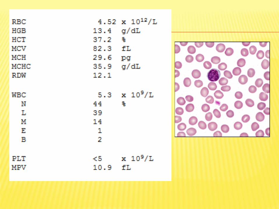
RBC morphology: Normochromic 1+ polychromasia 1+ macrocytosis
WBC morphology: The abnormal cells are medium-sized blasts. The nuclei are often irregular in shape, and some have invaginations or deep clefts.
Most have a fine chromatin pattern and one or more prominent nucleoli.
The cytoplasm is basophilic, and thin Auer rods are seen.
PLT morphology: Within normal limits

FURTHER LABORATORY STUDIES BONE
MARROW BIOPSY:
Aspirate:
The differential showed 93.6% blasts similar to
those in the blood.
DIAGNOSIS ACUTE MYELOBLASTIC LEUKEMIA
Chemotherapy was started, and remission was induced within one month.
The post-induction marrow showed normal regeneration, with 2.5% blasts.
The patient was placed on consolidation chemotherapy, and followed in Oncology Clinic.
He was still in remission 4 years after diagnosis.
Note: The patient's MCV returned to the normal
range shortly after induction was started, and the cause of his mild macrocytosis was not investigated.

CASE STUDY 4
40 year old female.
Brought to Emergency Room with symptoms of severe frontal headache and associated confusion. Noted to have decreased energy level and a 15 pound weight loss over the previous three months.
Physical Exam
Pale appearing, but otherwise within normal limits. No organomegaly.


SMEAR
RBC morphology: Normochromic 2+ anisocytosis 2+ oval macrocytes 1+ teardrop cells
WBC morphology: Dysgranulopoietic changes, including hyposegmentation, pseudo-Pelger-Huët nuclei, and hypogranularity are seen in some neutrophils (one shown here).
PLT morphology: Within normal limits

CASE STUDY 5
34 year old female. Two day history of
ecchymoses, petechiae, and hematuria. She
had noted headaches, nausea, and increasing
dysphoria over the past week.
Physical Exam: Mild scleral icterus. Scattered
ecchymoses and petechiae. Appeared anxious
and agitated.

SMEAR
RBC morphology: normochromic 2+ polychromasia 3+ anisocytosis 3+ fragments 2+ spherocytes
WBC morphology: Within normal limits (one lymphocyte shown here) PLT morphology: Within normal limits

HEMOSTASIS:
Bleeding time= 10 min
INR 0.91 (RI 0.85-1.15)
Howel Gram 2 min
PTT 24.8 sec (RI 23-34)
TT 15.8 sec (RI 13-18)

Chemistry:
BUN 41 mg/dL (RI 9-23)
Creatinine 0.8 mg/dL (RI 0.3-1.0)
Bilirubin Conj. 0.5 mg/dL (RI 0.0-0.3)
Total 2.8 mg/dL (RI 0.0-1.3)
Haptoglobin <5 mg/dL (RI 50-150)
Urinalysis: Large amount of blood present
Protein positive (100 mg/dL)

IMMUNE THROMBOCYTOPENIC PURPURA
Plasma exchange was commenced promptly after admission.
Initially, she became more acutely ill, and developed neurologic symptoms (combative and irritable, with fluctuating levels of consciousness).
She was continued on plasma exchange and given other appropriate therapy.
Over the next several days, her physical and mental status improved,
signs of hemolysis diminished, and her PLT count gradually increased.
She was discharged to be followed in Hematology Clinic.

CASE STUDY 6
History
54 year old female.
One year history of fatigue, weight loss, and
increasingly severe back pain.
Physical Exam
She appeared pale, but otherwise her physical
exam was within normal limits.


PROTEIN ELECTROPHORESIS
Total protein 11.0 g/dL (RI 5.2-8.3)
Serum protein electrophoresis:
Albumin 3.2 g/dL (RI 3.0-5.0)
Globulins:
Alpha1 0.4 (RI 0.1-0.5)
Alpha2 1.0 (RI 0.5-1.2)
Beta 0.8 (RI 0.5-1.1)
Gamma 5.6 (RI 0.6-1.7)

Immunoglobulins, quantitative:
IgA 9 mg/dL (RI 85-450)
IgG 5800 mg/dL (RI 800-1700)
IgM 25 mg/dL (RI 60-370)
Fibrinogen 650 mg/dl
ESR 70 mm/h

MULTIPLE MYELOMA

CASE STUDY 7
History
75 year old male.
Symptoms of severe headache and generalized pruritis.
Physical Exam
Spleen palpable 10 cm. below left costal margin. Liver palpable 3 cm. below right costal margin. The rest of the exam was within normal limits.

PULMONARY FUNCTION:
Oxygen saturation: 97% (RI 94-100)

POLYCYTHEMIA VERA

90
Case study 8
34 year old male. Seen for treatment of superficial skin wounds resulting from a
shotgun accident while grouse hunting. Family physician noted slight pallor, jaundice,
and scleral icterus. History of cholecystectomy five years prior to admission. At that time
the patient was told he had Gilbert's syndrome. He stated he had always had "low
blood," and that his father and paternal grandfather both had "liver ailments."
Physical Exam: Somewhat pale yellowish skin with scattered small surface wounds-
mostly over the face, scalp and upper extremities. Moderate scleral icterus. Spleen
palpable 3 cm below the left costal margin.
CBC (with microscopic differential)
RBC 3.93 x 10[12]/L HGB 11.3 g/dL HCT
33.1 % MCV 84.1 fL
MCH 28.8 pg MCHC 34.4 g/dL
RDW 18.7
WBC 5.0 x 10[9]/L
N 53 % , L 31%, M 8%, E 6%, B 2%
PLT 362 x 10[9]/L

91
Morphologic Alterations
Results of the blood smear exam were:
RBC morphology:
normochromic
2+ polychromasia
2+ anisocytosis
2+ spherocytes
1+ echinocytes
WBC morphology:Within normal limits (one lymphocyte shown
here)
PLT morphology: Within normal limit
Question What further laboratory studies, if any, are indicated?

92
Further Laboratory Studies
Hematology:
Reticulocytes 14.3 % Absolute 562 x 10[6]/L Osmotic fragility (unincubated)
Initial hemolysis 0.65% NaCL
Complete hemolysis 0.40% NaCL Control: Initial 0.50%;
Complete 0.20% Osmotic fragility (incubated) Initial hemolysis 0.85% NaCL
Complete hemolysis 0.60% NaCL Control: Initial 0.60%; Complete 0.20%
Chemistry:
Bilirubin Conj. 0.5 mg/dL (RI 0.0-0.3) Total 5.8 mg/dL (RI 0.0-1.3)
Question What is the most likely diagnosis?
93
Clinical Course
The patient was referred to a hematologist to evaluate the
advisibility of a splenectomy
Answer 3
Diagnosis Hereditary spherocytosis

PRACTICE QUESTIONS

95
Case study 9
37 year old male. Lifelong history of a seizure disorder, treated since age two. At a
routine check with his neurologist, he complained of fatigue, exertional dyspnea, and
lightheadedness over the past 2-3 months. He appeared pale, but otherwise his
physical exam was within normal limits. He was found to have a decreased
hemoglobin, and was referred to Hematology Clinic.
CBC
RBC 1.26 x 10[12]/L
HGB 5.7 g/dL HCT 16.3 %
MCV 130 fL MCH 45.2 pg
MCHC 34.9 g/dL RDW 18.1
WBC 6.2 x 10[9]/L
N 73 % L 21 M 1 E 4 B 1
PLT 219 x 10[9]/L
Question
What morphologic alterations are seen in this blood smear field?

96
Answer 1
RBC morphology:
Normochromic
3+ macrocytosis
3+ anisocytosis
Numerous oval macrocytes
Occ teardrop cells and fragments
WBC morphology: Many neutrophils show nuclear hypersegmentation
PLT morphology: Within normal limits
Question 2
What further laboratory studies, if any, are indicated?

97
Answer 2 Further Laboratory Studies
Bone marrow biopsy Aspirate : Erythroid hyperplasia with megaloblastic maturation.
Large polychromatic and orthochromatic megaloblasts show nuclear karyorrhexis
and other dyserythropoietic changes. Multiple Howell Jolly bodies are seen in both
megaloblasts and oval macrocytes. eutrophils show premature nuclear
segmentation, with giant metamyelocytes and band forms.
Sections:Appear hypercellular
Chemistry:
Serum folate <1.0 µg/L (RI 3.5-15)
RBC folate 131 µg/L (RI 160-600)
Serum B12 136 ng/L (RI 250-900)
Question 3
What is the most likely diagnosis?

98
Clinical Course
The patient was given large doses of folic acid, and within 6 days his reticulocyte
count was 15.2%. One month later, his hemoglobin was 12.7 g/dL, MCV was 92 fL,
and his blood smear morphology was normal. The anticonvulsant drug he had been
taking is known to interfere with folate metabolism. In addition, the patient had
been trying to lose weight, and over the past few months his diet had consisted
mainly of TV dinners, with little or no fresh vegetables or fruits. A nutritional consult
was arranged, and he was instructed to add folic acid to his daily medications.
Note: Patients with folic acid deficiency occasionally show decreased levels of
vitamin B12. Because of the patient's history and lack of typical neurologic
symptoms, concurrent pernicious anemia was considered very unlikely.
Answer 3
Diagnosis Megaloblastic anemia due to folate deficiency

BLOOD
1. is a type of connective tissue
2. is involved in the process of homeothermy
3. bicarbonate buffer system is the most
important extracellular system
4. ADH helps at the reabsorption of Na+ in the
distal nephron

HEMATOCRIT
1. somatic hematocrit is higher than the venous
one
2. represents the % of formed elements in the
blood
3. blood viscosity is inversely proportional to the
hematocrit value
4. splenic venous blood has the highest value

PLASMA PROTEINS
1. albumin is the main contributor to oncotic
pressure
2. the normal concentration of albumin is 7 g/dl
of blood
3. ceruloplasmin is a copper carring protein
4. albumin is a positive acute phase protein
IMMUNOGLOBULINS
1. IgM is released during primary humoral
response
2. IgG may cross the placenta
3. IgA is important in local mucosal immunity
4. IgE trigger alergic reactions

ESR
1. 1. is directly proportional with blood viscosity
2. 2. low ESR can be encountered in anemia
3. 3. it’s measured after centrifuging the blood at
12000 RPM
4. 4. detects non-specific inflammation

RED BLOOD CELLS
1. 1 g of Hb can transport 1.34 mL of oxygen
2. MCV =120fL showes that there are
macrocytes present in the blood
3. 70% of the iron in the organism is found in
hemoglobin
4. when pCO2 is low, red blood cells release O2
more easely to the tissues
WHICH OF THE FOLLOWING ARE PART OF THE
SPECIFIC IMMUNE ANSWERS
1. first line of defense mechanisms
2. second line of defense
3. all three lines of defense
4. third line of defense

CHOOSE THE RIGHT MACROPHAGES FUNCTIONS
1. antigen presenting cells
2. initiation of humoral immune answer
3. initiation of celular immune answer
4. they are the first cells to respond to tissue
infection

ANTIBODIES WORK AS
1. opsonins
2. antitoxins
3. agglutinate bacteria
4. stimulate perforin- pores formation in
antigenic cell membrane

FORMS OF CO2 TRANSPORT IN THE BLOOD ARE
carbaminoHB
bound to albumin
bicarbonates
carboxyHB

CHOOSE THE RIGHT CONDITIONS WHICH SHIFT
THE OXYHB DISSOCIATION CURVE TO THE RIGHT
Hb has low affinity for oxigen
high 2,3 DPG
low pH
low temperature

WHAT WOULD BE THE ERYTHROCYTE INDICES
PROFILE FOR A PERSON WITH NORMOCHROMIC
MACROCYTIC ANEMIA
1. MCHC= 28 g/100 ml
2. MCHC= 35 g/100 ml
3. MCV= 80 fL
4. MCV= 120 fL