HEMOLYSIS & JAUNDICE: An Overview and Jaundice PPP 18.pdf · •Acute Hepatocellular damage...
29
HEMOLYSIS & JAUNDICE: An Overview University of Papua New Guinea School of Medicine and Health Sciences Division of Basic Medical Sciences Discipline of Biochemistry and Molecular Biology PBL MBBS SEMINAR VJ Temple 1
Transcript of HEMOLYSIS & JAUNDICE: An Overview and Jaundice PPP 18.pdf · •Acute Hepatocellular damage...
HEMOLYSIS & JAUNDICE: An Overview University of Papua New Guinea
School of Medicine and Health SciencesDivision of Basic Medical Sciences
Discipline of Biochemistry and Molecular BiologyPBL MBBS SEMINAR
VJ Temple
1
What do you understand by Intravascular Hemolysis?
• Destruction of RBC (Hemolysis) normally occurs in Reticuloendothelial system:
• Extra-vascular compartment: Extravascular Hemolysis
• In some diseases, Hemolysis of RBC occurs within the Vascular System:
• Intravascular compartment: Intravascular Hemolysis
• During Intravascular Hemolysis Free Hemoglobin and Heme are released in Plasma;
2

• Free Hb and Heme in plasma can result in their excretion via the Kidneys with substantial loss of Iron
• Loss of Iron is prevented by Specific Plasma Proteins:
• Transferrin, and
• Haptoglobins (Hp)
• Both are involved in scavenging mechanisms
• Transferrin: binds and transports Iron in plasma and thus permits Reutilization of Iron;
• Haptoglobins (Hp): 2-Globulins produced in the Liver;
3

What happens to Free Hb during Intravascular Hemolysis?
• Sequence of events that occurs when Free Hb appears in plasma (Fig. 1):
• Hb is Oxygenated in Pulmonary Capillaries,
• OxyHb dissociates into -OxyHb Dimers,
• -OxyHb Dimers are bound by circulating plasma Haptoglobins,
• Haptoglobins have High Affinity for -OxyHb Dimers;
• One molecule of Haptoglobin binds Two -OxyHb Dimers,
4
• DeoxyHb does not dissociate into Dimers under normal physiological settings, thus it is not bound by Haptoglobins;
• Complex formed when Haptoglobin interacts with -OxyHb Dimers is too large to be filtered via the Glomerular Membrane;
• During Intravascular Hemolysis:
• Free Hb, appears in Renal Tubules and in Urine, causing Black-Water Fever,
• This occurs when binding capacity of circulating Haptoglobin molecules have been exceeded;
5

Figs. 1 & 2
6
What are the functions of Haptoglobin?
• Haptoglobins (Hp):
• Prevent loss of Free Hb via the Kidneys,
• Binds and transports -OxyHb Dimers to liver and Lymphoreticular system for catabolism,
• Heme in Free Hb is relatively resistant to the action of Heme Oxygenase
• Heme Oxygenase easily catalyzes breakdown of Heme in the Haptoglobin--OxyHb Complex,
7
How significant is plasma Haptoglobin as a diagnostic tool?
• Measurement of Plasma Haptoglobin level is used clinically to indicate severity of Intravascular Hemolysis;
• Patients with significant Intravascular Hemolysis have low levels of Haptoglobin because of removal of Haptoglobin--OxyHb complexes by Reticuloendothelial system;
• Plasma Haptoglobin level falls rapidly during severe Intravascular Hemolysis (Hemolytic Anemia)
• Level of Haptoglobin in Plasma will be very low;
8

• Haptoglobin levels in plasma can also be low during Severe Extra-vascular Hemolysis, when large amount of Hb in Reticuloendothelial System leads to release of Free Hemoglobin in plasma,
• Decreased Plasma Haptoglobin level may occur in Liver disease,
• Plasma Haptoglobin level increases in:
• Acute Infections,
• Trauma,
• Nephrotic syndrome (Why?)
• Because Haptoglobin is one of the Acute-Phase Reactants,
9

HEMOLYSIS AND G-6-P D DEFICIENCY:What reaction does Glucose-6-Phosphate Dehydrogenase catalyzes?
• Glucose-6-Phosphate Dehydrogenase (G-6-PD) catalyzes the first reaction in HMP-shunt
• NADPH is produced in reaction catalyzed by G-6-PD,
• HMP shunt in RBC is important for maintaining the Integrity of RBC membrane (Why?)
• Because the NADPH produced is used to protect the integrity of RBC membrane by maintaining normal cellular levels of Reduced Glutathione (GSH);
• GSH: Reduced Glutathione
• GSSG: Oxidized Glutathione 10

How do GSH and G-6-PD interact to protect RBC membrane from damage by Oxidants?
• Oxidants can damage RBC membrane causing Hemolysis,
• GSH is a reducing agent that removes Oxidants in RBC,
• Process is as follows:
• GSH interacts with Oxidants to form GSSG,
• Reaction is catalyzed by Glutathione Peroxidase(Selenium is required),
• GSSG then reacts with NADPH + H+ to form GSH,
• Reaction is catalyzed by Glutathione Reductasethat utilizes NADPH
11
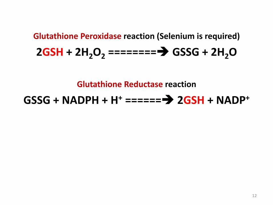
Glutathione Peroxidase reaction (Selenium is required)
2GSH + 2H2O2 ======== GSSG + 2H2O
Glutathione Reductase reaction
GSSG + NADPH + H+ ====== 2GSH + NADP+
12

• Major source of NADPH is the G-6-PD reaction in HMP shunt,
• HMP shunt is the major pathway for producing NADPH in mature RBC,
• Decreased in the level of GSH in RBC results in accumulation of Oxidants, causing impairment of essential metabolic processes and Hemolysis;
13

What are some consequences of G-6-PD deficiency?
• Mature RBC is sensitive to Oxidative damage if function of HMP shunt is Impaired (e.g. G-6-PD deficiency);
• Oxidants (e.g. Primaquine and other Anti-malarial drugs) can interact with GSH to produce high amount of GSSG, which must be converted to GSH using NADPH from HMP shunt;
• Mature RBC of individuals deficiency in G-6-PD cannotgenerate sufficient NADPH to convert GSSG to GSH;
• Resulting in accumulation of GSSG in the RBG;
• This impairs the ability of RBC to dispose of Oxidants and Free Radicals (Reactive Oxygen Species);
14
• Accumulation of Oxidants and Free Radicals causes Oxidation of critical –SH groups in proteins and Peroxidation of Lipids in RBC membrane, causing Hemolysis; (Figs. 2 & 3)
• Drugs or Chemicals capable of generating Oxidants should not be given to individuals with G-6-PD deficient; (WHY?)
• Because they can cause rapid fall in GSH level in mature RBC, leading to Intravascular Hemolysis;
15

• Effect of G-6-PD deficiency is greatest in Older RBC, because of their inability to synthesize Proteins and produce more G-6-PD,
• Mature RBC cannot synthesize proteins because they do not contain Nucleus;
• Hemolysis is higher in Older RBC,
• This explains the high percentage of circulating Young RBC in blood of patients with Intravascular Hemolysis
• Hemolysis may be accompanied by unconjugated hyperbilirubinemia leading to jaundice;
16

Fig. 2: Glucose-6-Phosphate Dehydrogenase (G-6-PD) reaction
17
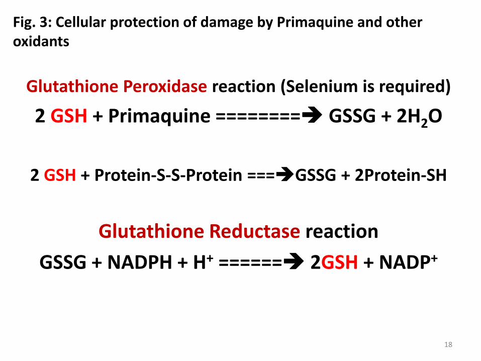
Fig. 3: Cellular protection of damage by Primaquine and other oxidants
Glutathione Peroxidase reaction (Selenium is required)
2 GSH + Primaquine ======== GSSG + 2H2O
2 GSH + Protein-S-S-Protein ===GSSG + 2Protein-SH
Glutathione Reductase reaction
GSSG + NADPH + H+ ====== 2GSH + NADP+
18

HYPERBILIRUBINEMIA AND JAUNDICE
What is Hyperbilirubinemia?
• Hyperbilirubinemia:
• Accumulation of Bilirubin in blood, when level of Bilirubin exceeds 1.0 mg/dL (17.1 mol/L),
19

What are the different types of Hyperbilirubinemia?
• Pre-hepatic Hyperbilirubinemia:
• Due to over-production of bilirubin causing increased level of unconjugated bilirubin in plasma:
• May occur in:
• Hemolytic anemia,
• Hemolytic disease of the new-born, due to Rhesus Incompatibility,
• Ineffective Erythropoiesis (e.g., Pernicious Anemia),
• Bleeding into tissues (Trauma),
• Rhabdomyolysis;
20
• Hepatocellular Hyperbilirubinemia:
• May be due to:
• Hepatocellular damage caused by:
• Infective agents,
• Drugs or Toxins,
• Cirrhosis is usually a late complication
• Low activity /Failure of Conjugating mechanism
• UDP-Glucuronyl-Transferase within Hepatocytes,
21

• Cholestatic Hyperbilirubinemia:
• may be Intra-hepatic or Extra-hepatic
• Causes Conjugated Hyperbilirubinemia and Bilirubinuria
• Intra-hepatic Cholestasis commonly due to:
• Acute Hepatocellular damage (Infectious Hepatitis)
• Primary Biliary Cirrhosis, Drugs
• Extra-hepatic Cholestasis is most often due to:
• Gallstones,
• Carcinoma of Head of Pancreas,
• Carcinoma of Biliary Tree,
• Bile duct compression from other courses;22

How is Hyperbilirubinemia related to Jaundice?
• Jaundice: due to Hyperbilirubinemia,
• It occurs when plasma Bilirubin exceeds 2.5mg/dL,
• Bilirubin diffuses into skin and Sclera, which then become yellow (Jaundice or Icterus),
• Yellow discoloration of eyes in Jaundice patients is due to affinity of protein Elastin (in Sclera) for Bilirubin,
• Hypercarotenemia (high beta-carotene level in blood) does not cause yellow discoloration of the eyes because Elastin does not bind beta-carotene,
23

What are the two types of Hyperbilirubinemia?
• Hyperbilirubinemia: separated based on type of Bilirubin (Conjugated Bilirubin or Unconjugated Bilirubin) present in Plasma:
• Retention Hyperbilirubinemia: overproduction of bilirubin, leading to accumulation in blood;
• Regurgitation Hyperbilirubinemia: hepatic reflux of bilirubin into blood caused by biliary obstruction,
• Unconjugated bilirubin is Hydrophobic, thus it can cross Blood-Brain Barrier and enter Central Nervous System,
24

• Encephalopathy due to Hyperbilirubinemia (Kernicterus) can occur only in Unconjugated Hyperbilirubinemia – as in Retention Hyperbilirubinemia;
• Conjugated Bilirubin is Hydrophilic, thus it can appear in Urine;
• Choluric Jaundice (Choluria: biliary pigments in urine):
• Occurs in Regurgitation Hyperbilirubinemia, WHY??
• Because of high Conjugated Bilirubin in blood;
• Acholuric Jaundice (Acholuria: no biliary pigments in urine)
• Occurs in Retention Hyperbilirubinemia, WHY??
• Because of high Unconjugated bilirubin in blood, 25

How are the causes of Jaundice classified?
• Causes of Jaundice are simply classified as:
• Pre-hepatic Jaundice (e.g., Hemolytic anemia),
• Hepatic Jaundice (e.g., Hepatitis),
• Post-hepatic Jaundice (e.g., Obstruction of the common bile duct);
26

What laboratory tests are used for diagnosis of different classes of Jaundice?
• Several lab tests including the following:
• Liver Function Tests (LFT),
• Plasma Total Bilirubin and Conjugated Bilirubin,
• Urinary Urobilinogen,
• Urinary Bilirubin,
• Inspection of Stool Samples
• Examples: Table 1
27

Table 1: Laboratory results for Healthy patient and patients with different causes of Jaundice
Patients Serum Bilirubin
(mg/dl)
Urine
Bilirubin
Urine
Urobilinogen
(mg/24h)
Fecal
Urobilinogen
(mg/24h)
Normal Direct: 0.1–0.4
Indirect: 0.2–0.7
Absent 0 – 4 40 – 280
Hemolytic
Anemia
Elevation of
Indirect
Absent Increased Increased
Hepatitis Elevations of
Direct &
Indirect
Present Decreased Decreased
Obstructive
Jaundice
Elevation of
direct
Present Absent Trace to
absent28
REFERENCES
• Textbook of Biochemistry, with clinical correlations, Ed. By T. M. Devlin, 4th Ed.
• Harper’s Illustrated Biochemistry 26th Edition; 2003; Ed. By R. K. Murray et. al.
• Biochemistry, By V. L. Davidson & D. B. Sittman. 3rd Edition.
• Hames BD, Hooper NM, JD Houghton; Instant Notes in Biochemistry, Bios Scientific Pub, Springer; UK.
• VJ Temple Biochemistry 1001: Review and Viva Voce Questions and Answers Approach; Sterling Publishers Private Limited, 2012, New Delhi-110 – 020.
• G Beckett, S Walker, P Rae & P Ashby. Lecture Notes: Clinical Biochemistry 7th Ed. Blackwell Publishing, Australia 2008.
29