Hemodynamic Monitoring Emmy Pranggono, MD, FINASIM, PhD ...
38
Hemodynamic Monitoring Emmy Pranggono Internal Medicine Department, Pulmonary and Critical Care Division Universitas Padjajaran
Transcript of Hemodynamic Monitoring Emmy Pranggono, MD, FINASIM, PhD ...

Hemodynamic Monitoring
Emmy Pranggono
Internal Medicine Department, Pulmonary and Critical Care Division
Universitas Padjajaran

INTRODUCTION

Hemodynamic, make it easy
Hemodynamic management is
a very important
a corner stone of activity in high/care intensive care or emergency room, although it can be happened in the ward
Misleading of the treatment
increasing the morbidity and mortality
Understanding is mandatory.
One type of shock is circulatory shock which is
a complex clinical syndrome
if not corrected will be fatal

Hemodynamic, make it easy
Monitoring hemodynamic in shock as a guide and dictation of accurate clinical assessments
The issues raised in the new statements or recommendations for monitoring hemodynamic shock
individualization of blood pressure targets
prediction of fluid responsiveness
the use of echocardiography which is not invasive as the first means during the initial evaluation of circulatory shock. Bedside assessment of intravascular volume status in critically ill
to prevent the risk of organ failure and mortality.

Hemodynamic, make it easy
Clinicians often use invasive hemodynamic monitoring like Central venous pressure (CVP) is a hemodynamic parameter that is extensively used
CVP is a good indicator of right ventricular preloadbut the complication associated with CVP insertion includes
failure to place the catheter, arterial puncture, catheter malposition, pneumothorax, subcutaneous hematoma, hemothorax, asystolic cardiac arrest and catheter-related infection

Hemodynamic, make it save
There is a needs for a non-invasive and economical technique like ultrasound in the critically ill patient
helps to approach diagnosis and treatment of the critically ill patients.
to estimate the intravascular status by measuring IVC diameter uses the size
collapsibility of the inferior vena cava (IVC).
Fluid administration and pharmacological therapy with vasopressor or inotropes are used to maintain CO and improve organ perfusion while treating the underlying disease

Shock states
Initiated by inadequate tissue perfusion Impaired the oxygen supply to the mitochondria Resulting severe dysfunction of vital organs (cellular
dysoxia) Associated with elevated blood lactate levels.
Hypotension is no longer mandatory in defining a shock state (ESICMc-2014)
Shock can occur following either hypoperfusion or inadequate perfusion of end organs leading imbalance oxygen delivery (DO2) and oxygen
consumption (VO2) Elevated blood lactate
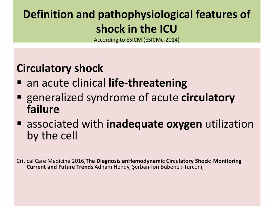
Definition and pathophysiological features of shock in the ICU
According to ESICM (ESICMc-2014)
Circulatory shock an acute clinical life-threatening generalized syndrome of acute circulatory
failure associated with inadequate oxygen utilization
by the cell
Critical Care Medicine 2016,The Diagnosis anHemodynamic Circulatory Shock: Monitoring Current and Future Trends Adham Hendy, Şerban-Ion Bubenek-Turconi,

The hemodynamic monitoringtechniques in shock
There are three main reasons for hemodynamic
monitoring in shock patients:
1. To identify the type of shock
2. To select the most appropriate therapy
3. To evaluate the response to that therapy

What is blood pressure?
As the heart contracts, blood is forced out of the left ventricle and into the aorta and distributing arteries
The force created by the heart's contraction generates pressure as blood pushes against the walls of the vessels
This is known as blood pressure and consists of two pressures readings: The maximum pressure (systolic) occurs as the heart contracts The minimum pressure (diastolic) occurs as the heart fills back
up with more blood
The normal blood pressure for an adult is 120/80 mmHg.

Blood Pressure

What is Mean Arterial Pressure (MAP)?
The average blood pressure during a single cardiac cycle
Necessary for adequate perfusion of the organs
A better indication of perfusion than systolic blood pressure
At least 60 mmHg to provide enough blood to
the coronary arteries, kidneys, and brain
The normal range is between 70 and 100 mmHg

MAP is directly affected by factors:
Amount of blood pumped out of the heart per minute (cardiac output)
Heart rate (beats per minute)
Blood pressure
Resistance to blood flow in the vessels

At normal resting heart rates, MAP can be approximated by the following equation:
MAP = (CO × SVR) + CVP
Because CVP is usually at or near 0 mmHg, this relationship is often simplified to:
MAP ≈ CO × SVR which is
CO depend on Heart Rate (HR) and Stroke Volume (SV)
CO = Cardiac Output
SVR = Systemic Vascular Resistance
CVP = Central Venous Pressure

Approach management of hypotension

How to Calculate MAP
The most accurate MAP can only be determined through an invasive central line
However, it can be measured by another equation using the systolic blood pressure (SBP) and diastolic blood pressure (DBP)

Four types of hemodynamic shock
Hypovolemic shock:
A direct loss of effective circulating blood volume (internal and/or external) which primary leads to decreased cardiac preload, stroke volume and consequently impaired end-organ perfusion.
Cardiogenic shock
A decreased systemic circulatory blood flow due to an intrinsic defect in cardiac function either the heart muscle and/or valvular dysfunction.

Four types of hemodynamic shock
Obstructive shock
Intra-cardiac or extra-cardiac mechanical obstruction to cardiac filling that decreases the cardiac output and consequently decreases end-organ perfusion.
Distributive shock:
A peripheral vascular dilatation causing a decreased systemic vascular resistance (SVR) associated with increased cardiac output and compromised perfusion of vital organs.
However many patients with circulatory failure have a combination of more than one form of shock as is seen in cases of septic shock.

Sepsis previously was defined as
a systemic inflammatory response syndrome (SIRS) in response to an infectious process
Septic shock
severe sepsis with persistent signs of end organ damage, hypotension (systolic blood pressure <90mmHg) and elevated serum lactate (>4mmol/l).
Considering that there was a need to re-examine theclassical definition of sepsis to distinguish sepsis from uncomplicated infection
ESICM and the Society of Critical Care Medicine (USA) published in February 2016
“The Third International Consensus Definitions for Sepsis and Septic Shock (Sepsis-3)

“The Third International Consensus Definitions for Sepsis and Septic Shock (Sepsis-3)
They redefined sepsis as “a life-threatening organ dysfunction caused by a dysregulated host response to infection”.
Hence, they advocated a change in the way sepsisand septic shock is approached,
away from a focus on the reaction to a systemic inflammation and towards a consideration of organ dysfunction
The latter can be identified in ICU patients as an
acute change in the Sequential [Sepsis-related] Organ Failure Assessment (SOFA) score>2 points, subsequent to the infection

Mortality septic shock vs sepsis alone
>40 % versus >10 %
The Sepsis-3 definition for the septic shock in adult patients is based on cumulative criteria
sepsis plus hypotension requiring the use of vasopressors to maintain MAP ≥ 65mm Hg
elevated blood lactate levels > 2 mmol/L
persisting after adequate fluid resuscitation

Diagnosis of shock
Patients should be assessed for the etiology of shock by an initial rapid clinical
evaluation based on a history taking
physical examination
appropriate initial laboratory tests.

Diagnosis of shock
Clinical signs and bedside observations
maintained arterial blood pressure
is still possible to find markers of inadequate tissueperfusion such as
increased blood lactate levels
low mixed venous oxygen saturation (SvO2)
low centralvenous oxygen saturation (ScvO2) values

Diagnosis of shock
For this reason, hypotension was excluded from themandatory definition of shock states, with the notable exception of septic shock
On the other hand, some early clinical findings such as
skin colour and skin temperature disturbances, heart rate, rhythm, electrocardiogram (ECG), capillary re-fill test, urineoutput, mental status, the effect of body position on the blood pressure, remain valuable signs of hemodynamicshock and pre-shock phases.
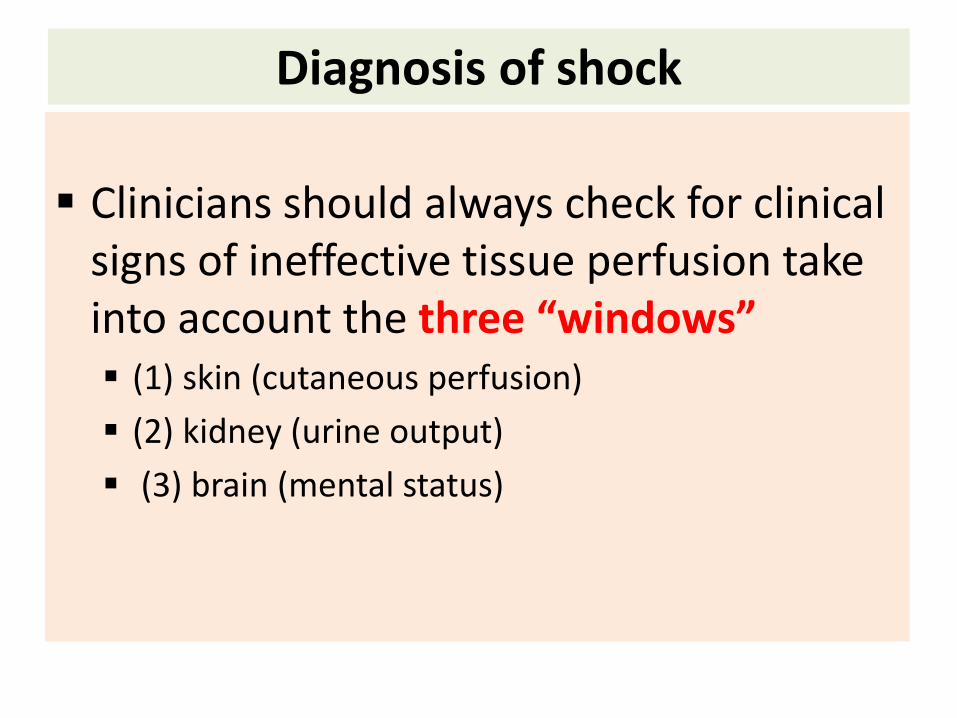
Diagnosis of shock
Clinicians should always check for clinical signs of ineffective tissue perfusion take into account the three “windows” (1) skin (cutaneous perfusion)
(2) kidney (urine output)
(3) brain (mental status)

2. Metabolic markers of regional and microcirculation
and the assessment of cellular function in shock
Arterial or venous blood lactate levels
The higher blood lactate level the higher mortality and morbidity Hyperlactatemia is a marker of
Imbalance between the oxygen consumption (VO2)and oxygen delivery (DO2) in any kind of shock
It is a much more precise indicator of ineffective perfusion than a base deficit or metabolic acidosis
the transition from aerobic to anaerobic metabolism

The new ESICMc-2014
Blood lactate levels > 2mEq/L should be consideredthe hallmark of an existing shock syndrome
Presence of hemodynamic shock plus hyperlactatemia > 4.0 mmol/L is associated with a mortality of 30% to 45%
Blood lactate levelel > 6 mmol/l predicts a mortality range between 80% and 90%

Obviously, the other circumstances associated withhigh lactate levels, are driven by ineffective localperfusion such as
local ineffective perfusion, drug effects, liver failure, malignancy, thiamine deficiency, seizures, or patients in bed struggling against restraints or shivering, have to be exclude.
Apart from blood lactate, SvO2 or ScvO2 values canoffer valid information about the balance between DO2 and VO2.
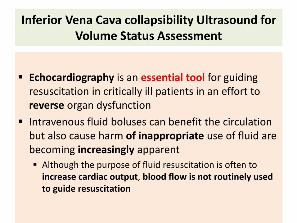
Inferior Vena Cava collapsibility Ultrasound for Volume Status Assessment
Echocardiography is an essential tool for guiding resuscitation in critically ill patients in an effort to reverse organ dysfunction
Intravenous fluid boluses can benefit the circulation but also cause harm of inappropriate use of fluid are becoming increasingly apparent
Although the purpose of fluid resuscitation is often to increase cardiac output, blood flow is not routinely used to guide resuscitation

Diameter Vena Cava Inferior
Vena cava inferior Atrium
kanan
Pressure of IntrathoracalIntraperikardial Intraabdominal,
Hipertensi Pulmonal
collapsibility
Ǿ 1,2 - 1,7 cm
1-8 mmHg
RAP 10-15 mmHg
collaps < 50%
RAP >15 mmg
No collaps
RAP 6-10 mmHg
collaps ≥ 50%
Ǿ VCI <1,2 cm
spontancolaps
Subcostal
view
M
mode

Inferior Vena Cava collapsibility Ultrasound for Volume Status Assessment
Although the purpose of fluid resuscitation is often to increase cardiac output, blood flow is not routinely used to guide resuscitation
Sufficient mean arterial pressure (MAP) is rightly targeted but is proportional to flow only for a given systemic vascular resistance (SVR)
Measurement of flow requires more equipment, time and expertise than standard parameters such as blood pressure and for an individual patient is not predictable
Mixed venous oxygen saturations and lactate are useful but only reveal a large mismatch between supply and demand, missing subtler shock states.

Inferior Vena Cava (IVC) collapsibility Ultrasound for Volume Status Assessment
The presence of signs that fluid delivery improves cardiac output does not mean that a greater cardiac output is necessary.
??? the patient improves with
fluid
additional vasopressors or inotropes
Echocardiography is an evidence-based approach and ideally suited to address this problem.
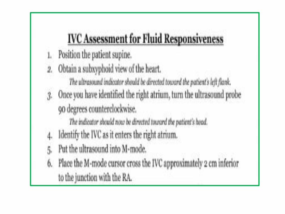


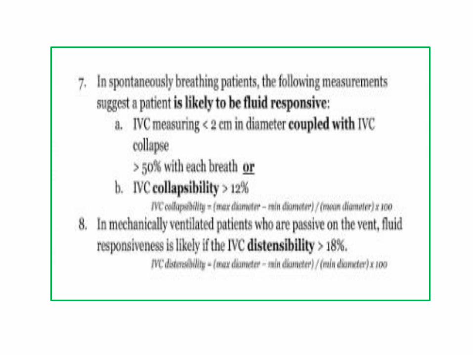
IVC and collapsibility

Conclusions
Hemodynamic monitoring is mandatory in critically ill patient
The superiority of the dynamic over the static parameter to predict fluid responsiveness
Echocardiography is an uninvasive methode and preferred technical method for the initial diagnosis and follow-up of patient in hemodynamic shock
Blood lactate is an important clinical bed-side biomarker during the follow-up of a patient in circulatory shock.

TERIMA KASIH