Hematologic Changes in Dengue Hemorrhagic Fever Hematologic changes [special... · Introduction:...
10
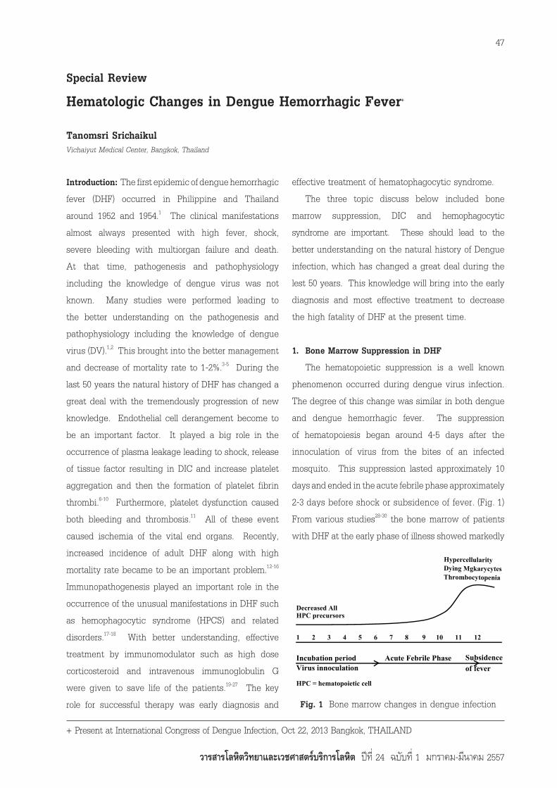
วารสารโลหิตวิทยาและเวชศาสตรบริการโลหิต ปที่ 24 ฉบับที่ 1 มกราคม-มีนาคม 2557 47 Introduction: The first epidemic of dengue hemorrhagic fever (DHF) occurred in Philippine and Thailand around 1952 and 1954. 1 The clinical manifestations almost always presented with high fever, shock, severe bleeding with multiorgan failure and death. At that time, pathogenesis and pathophysiology including the knowledge of dengue virus was not known. Many studies were performed leading to the better understanding on the pathogenesis and pathophysiology including the knowledge of dengue virus (DV). 1,2 This brought into the better management and decrease of mortality rate to 1-2%. 3-5 During the last 50 years the natural history of DHF has changed a great deal with the tremendously progression of new knowledge. Endothelial cell derangement become to be an important factor. It played a big role in the occurrence of plasma leakage leading to shock, release of tissue factor resulting in DIC and increase platelet aggregation and then the formation of platelet fibrin thrombi. 6-10 Furthermore, platelet dysfunction caused both bleeding and thrombosis. 11 All of these event caused ischemia of the vital end organs. Recently, increased incidence of adult DHF along with high mortality rate became to be an important problem. 12-16 Immunopathogenesis played an important role in the occurrence of the unusual manifestations in DHF such as hemophagocytic syndrome (HPCS) and related disorders. 17-18 With better understanding, effective treatment by immunomodulator such as high dose corticosteroid and intravenous immunoglobulin G were given to save life of the patients. 19-27 The key role for successful therapy was early diagnosis and effective treatment of hematophagocytic syndrome. The three topic discuss below included bone marrow suppression, DIC and hemophagocytic syndrome are important. These should lead to the better understanding on the natural history of Dengue infection, which has changed a great deal during the lest 50 years. This knowledge will bring into the early diagnosis and most effective treatment to decrease the high fatality of DHF at the present time. 1. Bone Marrow Suppression in DHF The hematopoietic suppression is a well known phenomenon occurred during dengue virus infection. The degree of this change was similar in both dengue and dengue hemorrhagic fever. The suppression of hematopoiesis began around 4-5 days after the innoculation of virus from the bites of an infected mosquito. This suppression lasted approximately 10 days and ended in the acute febrile phase approximately 2-3 days before shock or subsidence of fever. (Fig. 1) From various studies 28-30 the bone marrow of patients with DHF at the early phase of illness showed markedly + Present at International Congress of Dengue Infection, Oct 22, 2013 Bangkok, THAILAND Special Review Hematologic Changes in Dengue Hemorrhagic Fever + Tanomsri Srichaikul Vichaiyut Medical Center, Bangkok, Thailand Incubation period Acute Febrile Phase Subsidence of fever 1 2 3 4 5 6 7 8 9 10 11 12 HPC = hematopoietic cell Decreased All HPC precursors Hypercellularity Dying Mgkarycytes Thrombocytopenia Virus innoculation Fig. 1 Bone marrow changes in dengue infection
Transcript of Hematologic Changes in Dengue Hemorrhagic Fever Hematologic changes [special... · Introduction:...
วารสารโลหิตวิทยาและเวชศาสตรบริการโลหิต ป ที่ 24 ฉบับ ที่ 1 มกราคม-มีนาคม 2557
47
Introduction: The first epidemic of dengue hemorrhagic
fever (DHF) occurred in Philippine and Thailand
around 1952 and 1954.1 The clinical manifestations
almost always presented with high fever, shock,
severe bleeding with multiorgan failure and death.
At that time, pathogenesis and pathophysiology
including the knowledge of dengue virus was not
known. Many studies were performed leading to
the better understanding on the pathogenesis and
pathophysiology including the knowledge of dengue
virus (DV).1,2 This brought into the better management
and decrease of mortality rate to 1-2%.3-5 During the
last 50 years the natural history of DHF has changed a
great deal with the tremendously progression of new
knowledge. Endothelial cell derangement become to
be an important factor. It played a big role in the
occurrence of plasma leakage leading to shock, release
of tissue factor resulting in DIC and increase platelet
aggregation and then the formation of platelet fibrin
thrombi.6-10 Furthermore, platelet dysfunction caused
both bleeding and thrombosis.11 All of these event
caused ischemia of the vital end organs. Recently,
increased incidence of adult DHF along with high
mortality rate became to be an important problem.12-16
Immunopathogenesis played an important role in the
occurrence of the unusual manifestations in DHF such
as hemophagocytic syndrome (HPCS) and related
disorders.17-18 With better understanding, effective
treatment by immunomodulator such as high dose
corticosteroid and intravenous immunoglobulin G
were given to save life of the patients.19-27 The key
role for successful therapy was early diagnosis and
effective treatment of hematophagocytic syndrome.
The three topic discuss below included bone
marrow suppression, DIC and hemophagocytic
syndrome are important. These should lead to the
better understanding on the natural history of Dengue
infection, which has changed a great deal during the
lest 50 years. This knowledge will bring into the early
diagnosis and most effective treatment to decrease
the high fatality of DHF at the present time.
1. BoneMarrowSuppressioninDHF
The hematopoietic suppression is a well known
phenomenon occurred during dengue virus infection.
The degree of this change was similar in both dengue
and dengue hemorrhagic fever. The suppression
of hematopoiesis began around 4-5 days after the
innoculation of virus from the bites of an infected
mosquito. This suppression lasted approximately 10
days and ended in the acute febrile phase approximately
2-3 days before shock or subsidence of fever. (Fig. 1)
From various studies28-30 the bone marrow of patients
with DHF at the early phase of illness showed markedly
+Present at International Congress of Dengue Infection, Oct 22, 2013 Bangkok, THAILAND
SpecialReview
HematologicChangesinDengueHemorrhagicFever+
TanomsriSrichaikulVichaiyut Medical Center, Bangkok, Thailand
Incubation period Acute Febrile Phase Subsidenceof fever
1 2 3 4 5 6 7 8 9 10 11 12
HPC = hematopoietic cell
Decreased All HPC precursors
HypercellularityDying MgkarycytesThrombocytopenia
Virus innoculation
Fig 1 Bone Marrow Changes in Dengue Infection.
Fig.1 Bone marrow changes in dengue infection
Tanomsri Srichaikul
JHematolTransfusMedVol. 24 No. 1 January-March 2014
48
hypocellularity accompanying with decreased all the
hematopoietic cell precursors namely megakaryocytes,
erythroid and myeloid precursors. In vitro culture, the
colony forming units granulocyte and macrophage
(CFU-GM) were markedly decreased or almost absent.
The colony size were also smaller or appeared as
a cluster of cells. Following the hematopoietic
suppression, the recovery of hematopoiesis occurred
few days before shock or subsidence of fever. The
bone marrow appeared hypercellular, accompanied by
increase in number of megakaryocytes, erythroid and
myeloid precursors. Despite the increase in normal
number of megakaryocytes, these cells showed sign of
degeneration as manifested by nuclear fragmentation
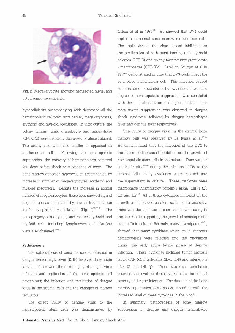
and/or cytoplasmic vacuolization. (Fig. 2)29-30,32 The
hemophagocytosis of young and mature erythroid and
myeloid cells including lymphocytes and platelets
were also observed.31-35
Pathogenesis
The pathogenesis of bone marrow suppression in
dengue hemorrhagic fever (DHF) involved three main
factors. These were the direct injury of dengue virus
infection and replication of the hematopoietic cell
progenitors; the infection and replication of dengue
virus in the stromal cells and the changes of marrow
regulators.
The direct injury of dengue virus to the
hematopoietic stem cells was demonstrated by
Nakoa et al in 1989.36 He showed that DV4 could
replicate in normal bone marrow mononuclear cells.
The replication of the virus caused inhibition on
the proliferation of both burst forming unit erythroid
colonies (BFU-E) and colony forming unit granulocyte
- macrophages (CFU-GM). Later on, Murgur et al in
199737 demonstrated in vitro that DV3 could infect the
cord blood mononuclear cell. This infection caused
suppression of progenitor cell growth in cultures. The
degree of hematopoietic suppression was correlated
with the clinical spectrum of dengue infection. The
most severe suppression was observed in dengue
shock syndrome, followed by dengue hemorrhagic
fever and dengue fever respectively.
The injury of dengue virus on the stromal bone
marrow cells was observed by La Russa et al.38-39
He demonstrated that the infection of the DV2 to
the stromal cells caused inhibition on the growth of
hematopoietic stem cells in the culture. From various
studies in vitro40-44 during the infection of DV to the
stromal cells, many cytokines were released into
the supernatant in culture. These cytokines were
macrophage inflammatory protein-1 alpha (MIP-1 a),
IL6 and IL8.40 All of these cytokines inhibited on the
growth of hematopoietic stem cells. Simultaneously,
there was the decrease in stem cell factor leading to
the decrease in supporting the growth of hematopoietic
stem cells in culture. Recently, many investigators45-51,
showed that many cytokines which could suppress
hematopoiesis were released into the circulation
during the early acute febrile phase of dengue
infection. These cytokines included tumor necrosis
factor (INF a), interleukins (IL-6, IL-8) and interferons
(INF a and INF g). There was close correlation
between the levels of these cytokines to the clinical
severity of dengue infection. The duration of the bone
marrow suppression was also corresponding with the
increased level of these cytokines in the blood.
In summary, pathogenesis of bone marrow
suppression in dengue and dengue hemorrhagic
Fig.2 Megakaryocyte showing negleected nuclei and
cytoplasmic vacuolization
Hematologic Changes in Dengue Hemorrhagic Fever
วารสารโลหิตวิทยาและเวชศาสตรบริการโลหิต ป ที่ 24 ฉบับ ที่ 1 มกราคม-มีนาคม 2557
49
fever involved three major mechanisms. These
were the direct infection of dengue virus in to the
hematopoietic stem cells and stromal cells of the
bone marrow, accompanied by the release of various
hematodepressive cytokines during the dengue virus
infection. All of these factors caused hematopoietic
suppression, resulting in granulocytopenia and
thrombocytopenia. The hematopoietic suppression
occurred transiently and recovered rapidly at the
late acute febrile period. Following this recovery the
number of neutrophils and platelets were furtherly
decreased until the day of shock or subcidence of
fever and then gradually returned to normal within 2
days at the convalescent period of the disease.52
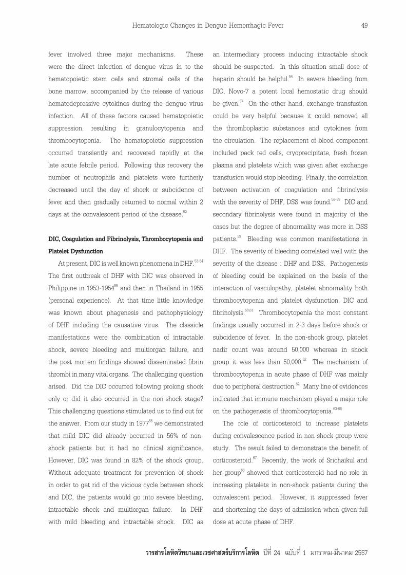
DIC,CoagulationandFibrinolysis,Thrombocytopeniaand
PlateletDysfunction
At present, DIC is well known phenomena in DHF.53-54
The first outbreak of DHF with DIC was observed in
Philippine in 1953-195455 and then in Thailand in 1955
(personal experience). At that time little knowledge
was known about phagenesis and pathophysiology
of DHF including the causative virus. The classicle
manifestations were the combination of intractable
shock, severe bleeding and multiorgan failure, and
the post mortem findings showed disseminated fibrin
thrombi in many vital organs. The challenging question
arised. Did the DIC occurred following prolong shock
only or did it also occurred in the non-shock stage?
This challenging questions stimulated us to find out for
the answer. From our study in 197756 we demonstrated
that mild DIC did already occurred in 56% of non-
shock patients but it had no clinical significance.
However, DIC was found in 82% of the shock group.
Without adequate treatment for prevention of shock
in order to get rid of the vicious cycle between shock
and DIC, the patients would go into severe bleeding,
intractable shock and multiorgan failure. In DHF
with mild bleeding and intractable shock. DIC as
an intermediary process inducing intractable shock
should be suspected. In this situation small dose of
heparin should be helpful.54 In severe bleeding from
DIC, Novo-7 a potent local hemostatic drug should
be given.57 On the other hand, exchange transfusion
could be very helpful because it could removed all
the thromboplastic substances and cytokines from
the circulation. The replacement of blood component
included pack red cells, cryoprecipitate, fresh frozen
plasma and platelets which was given after exchange
transfusion would stop bleeding. Finally, the correlation
between activation of coagulation and fibrinolysis
with the severity of DHF, DSS was found.58-59 DIC and
secondary fibrinolysis were found in majority of the
cases but the degree of abnormality was more in DSS
patients.59 Bleeding was common manifestations in
DHF. The severity of bleeding correlated well with the
severity of the disease : DHF and DSS. Pathogenesis
of bleeding could be explained on the basis of the
interaction of vasculopathy, platelet abnormality both
thrombocytopenia and platelet dysfunction, DIC and
fibrinolysis.60,61 Thrombocytopenia the most constant
findings usually occurred in 2-3 days before shock or
subcidence of fever. In the non-shock group, platelet
nadir count was around 50,000 whereas in shock
group it was less than 50,000.52 The mechanism of
thrombocytopenia in acute phase of DHF was mainly
due to peripheral destruction.62 Many line of evidences
indicated that immune mechanism played a major role
on the pathogenesis of thrombocytopenia.63-66
The role of corticosteroid to increase platelets
during convalescence period in non-shock group were
study. The result failed to demonstrate the benefit of
corticosteroid.67 Recently, the work of Srichaikul and
her group68 showed that corticosteroid had no role in
increasing platelets in non-shock patients during the
convalescent period. However, it suppressed fever
and shortening the days of admission when given full
dose at acute phase of DHF.
Tanomsri Srichaikul
JHematolTransfusMedVol. 24 No. 1 January-March 2014
50
HemophagocyticSyndromeandRelatedDisorders
The first report of hemophagocytic syndrome
in DHF was in 2007.19 The patient was 46 year old
female, who presented with multiorgan failure at the
recovery phase. She survived by receiving high dose
corticosteroid and IVIg therapy.
Hemophagocytic Syndrome (HPCS) is characterized
by four major manifestations. These were high fever,
progressive cytopenia, multiorgan failure with initially
present as hepatic failure, and finally the demonstration
of hemophagocytosis in the bone marrow and other
reticuloendothelial organs. This syndrome occurred
mainly in immunocompromised host, in both children
and adult but more in the adult. Two major disorders
could cause HPCS. These were lymphoid malignancy
and others and non-malignant disorders mostly from
infections such as Ebstein Bar virus (EBV), DHF and
others, bacteria protozoal and fungus, drugs, toxin and
immune diseases such as SLE and rheumatoid arthritis.
The prognosis of HPCS in virus infection was poor and
needed early treatment by high dose corticosteroid
and high dose intravenous immunoglobulin G to save
life of the patients.19-26
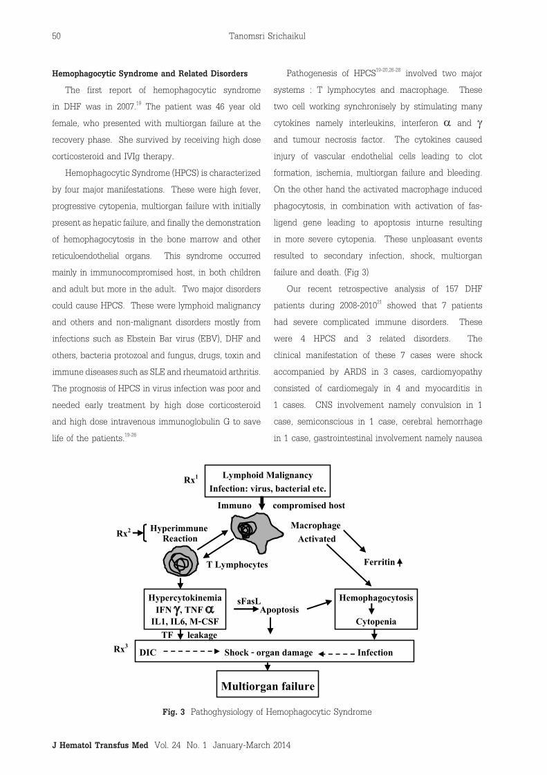
Pathogenesis of HPCS19-20,26-28 involved two major
systems : T lymphocytes and macrophage. These
two cell working synchronisely by stimulating many
cytokines namely interleukins, interferon a and g and tumour necrosis factor. The cytokines caused
injury of vascular endothelial cells leading to clot
formation, ischemia, multiorgan failure and bleeding.
On the other hand the activated macrophage induced
phagocytosis, in combination with activation of fas-
ligend gene leading to apoptosis inturne resulting
in more severe cytopenia. These unpleasant events
resulted to secondary infection, shock, multiorgan
failure and death. (Fig 3)
Our recent retrospective analysis of 157 DHF
patients during 2008-201021 showed that 7 patients
had severe complicated immune disorders. These
were 4 HPCS and 3 related disorders. The
clinical manifestation of these 7 cases were shock
accompanied by ARDS in 3 cases, cardiomyopathy
consisted of cardiomegaly in 4 and myocarditis in
1 cases. CNS involvement namely convulsion in 1
case, semiconscious in 1 case, cerebral hemorrhage
in 1 case, gastrointestinal involvement namely nausea
Lymphoid MalignancyInfection: virus, bacterial etc.
Immuno compromised host
Hyperimmune Reaction
Macrophage Activated
Ferritin
HypercytokinemiaIFN g, TNF a
IL1, IL6, M-CSF
Hemophagocytosis
Cytopenia
DIC Shock - organ damage Infection
Multiorgan failure
TF leakageRx3
Rx2
Rx1
ApoptosissFasL
T Lymphocytes
Fig 3 Pathophysiology of Hemophagocytic SyndromeFig.3 Pathoghysiology of Hemophagocytic Syndrome
Hematologic Changes in Dengue Hemorrhagic Fever
วารสารโลหิตวิทยาและเวชศาสตรบริการโลหิต ป ที่ 24 ฉบับ ที่ 1 มกราคม-มีนาคม 2557
51
vomiting lose of appetite in 1 case, severe abdominal
pain in 2 cases, severe gastrointestinal bleeding in
3 cases, severe hepatitis as manifested by jaundice,
rising of liver enzyme >500 unit in 3 cases and acute
renal failure : azothemia in 1 case, require hemodialysis
in 1 case and required blood transfusion in 3 cases.
Hematologic abnormalities consisted of acute anemia
in 4 cases, initial leukocytosis in 2 cases, severe
thrombocytopenia in 3 cases and DIC in 3 cases. The
illustration of case 1 (HPCS) and case 5 (prolonged
bradycardia) were shown.
In conclusion, from the retrospective analysis
of 157 adult DHF patients we demonstrated 7
cases presented with unusual severe complications
namely 4 HPCS and 3 related disorders. Immune
pathogenesis plays a big role in the development of
these complications. The treatment in these cases
consisted of high dose corticosteroid and high dose
intravenous immunoglobulin G. Five cases survived
with complete recovery whereas the other two cases
died. The first case died from delayed treatment with
co-infection. The second case died from intracranial
bleeding.
The detail history of each case were presented in
Table I, II and the illustration of case 1 was presented
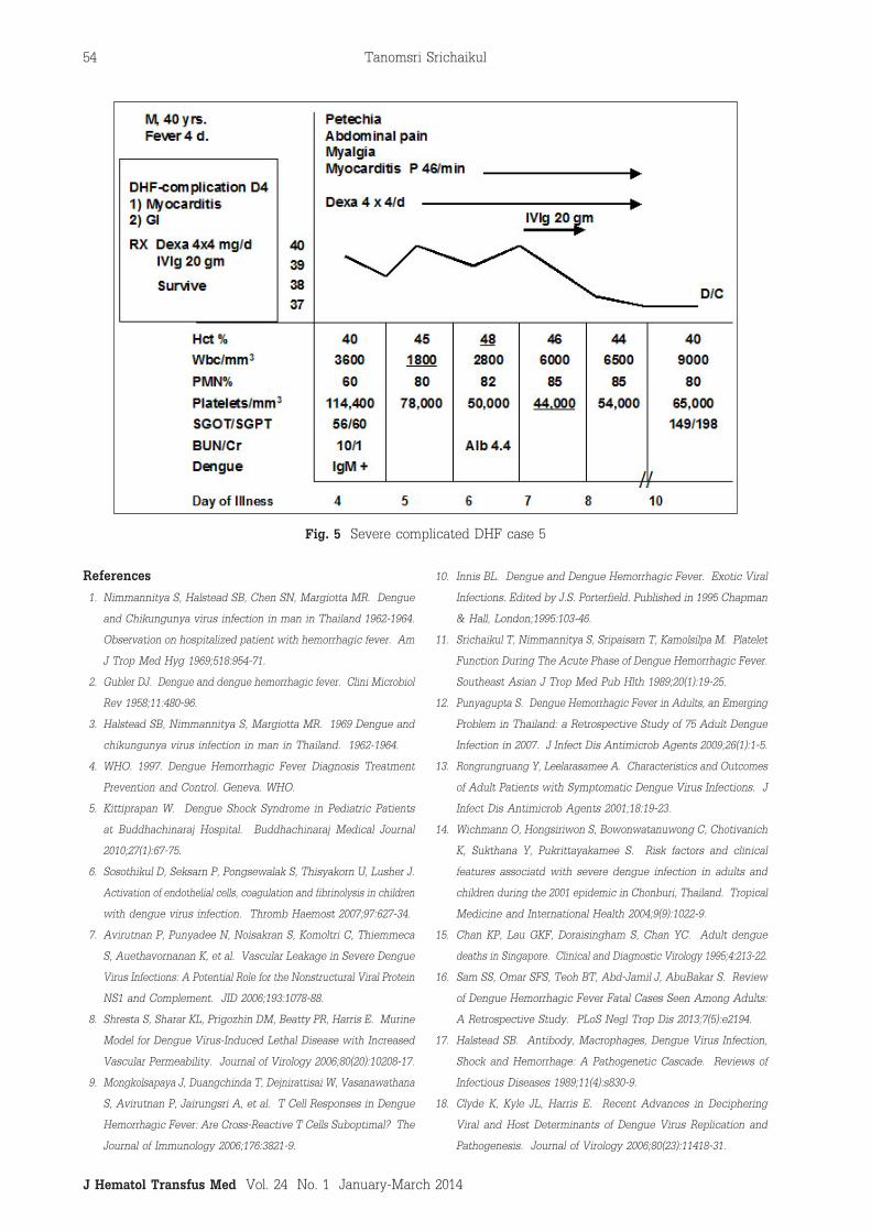
in Fig 4-1, 4-2, 4-3 and case 5 in Fig 5
The finding of myocarditis (prolonged bradycardia)
in case 5 was quite interesting. The patient reported
from Hong Kong in 2010, died from shock and
tachycardia. Resuscitation by fluid therapy was given
without corticosteroid or IVIg therapy.69 The literature
reviewed from 6154 DHF cases in Thailand found
that only 2 cases had myocarditis as manifested by
bradycardia and hypotension. Immune pathogenesis
could play an important role for this complication.
However, corticosteroid therapy and the outcome of
the patients were not mentioned.
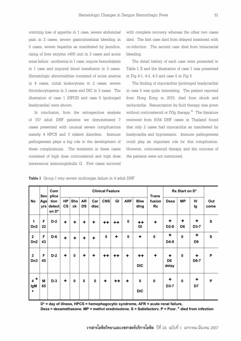
Table 1 Group I Very Severe Multiorgan Failure in 4 Adult DHF
No
1Dn2
2Dn2
3Dn3
4IgM +
SexAgeyrs
F22
F43
F45
M65
Complication
detecton D*
D-2
D-6
D-2
D-3
HPCS
Shock
0
0
ARDS
0
Cardiac
0
CNS
0
GI ARF
0
0
Bleeding
GI
DIC
0
DIC
Trans fusion Rx
0
0
Dexa
D2-8
D4-9
D6delay
D3-7
MP
D6
0
0
0
IV Ig
D3-7
D9
D6-7
D7
Out come
S
S
P
P
Clinical Feature Rx Start on D*
D* = day of illness, HPCS = hemophagocytic syndrome, AFR = acute renal failure,Dexa = dexamethasone, MP = methyl prednisolone, S = Satisfactory, P = Poor, * died from infection
+
+
+
+
+
+ +
+
+ +
+
+ ++
++
+
+
++
++
+
++
+
+
+
+
+
+ +
+
+
+
++
+
++
++
*
TableI Group I very severe multiorgan failure in 4 adult DHF
Tanomsri Srichaikul
JHematolTransfusMedVol. 24 No. 1 January-March 2014
52
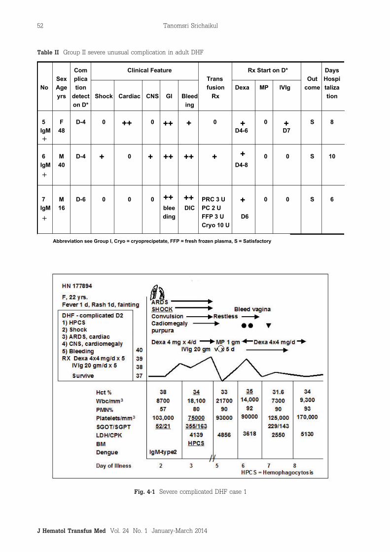
Table 2 Group II Severe Unusual Complication in 3 Adult DHF
No
5IgM
6IgM
7IgM
SexAgeyrs
F48
M40
M16
Complication
detecton D*
D-4
D-4
D-6
Shock
0
0
Cardiac
0
0
CNS
0
0
GI
bleeding
Bleeding
DIC
Trans fusion Rx
0
PRC 3 UPC 2 UFFP 3 UCryo 10 U
Dexa
D4-6
D4-8
D6
MP
0
0
0
IVIg
D7
0
0
Out come
S
S
S
DaysHospitalization
8
10
6
Clinical Feature Rx Start on D*
Abbreviation see Group I, Cryo = cryoprecipetate, FFP = fresh frozen plasma, S = Satisfactory
++
++
++
++
++
+
+
+
+
++
+
+
+
+
+
+ +
TableII Group II severe unusual complication in adult DHF
Table 2 Group II Severe Unusual Complication in 3 Adult DHF
No
5IgM
6IgM
7IgM
SexAgeyrs
F48
M40
M16
Complication
detecton D*
D-4
D-4
D-6
Shock
0
0
Cardiac
0
0
CNS
0
0
GI
bleeding
Bleeding
DIC
Trans fusion Rx
0
PRC 3 UPC 2 UFFP 3 UCryo 10 U
Dexa
D4-6
D4-8
D6
MP
0
0
0
IVIg
D7
0
0
Out come
S
S
S
DaysHospitalization
8
10
6
Clinical Feature Rx Start on D*
Abbreviation see Group I, Cryo = cryoprecipetate, FFP = fresh frozen plasma, S = Satisfactory
++
++
++
++
++
+
+
+
+
++
+
+
+
+
+
+ +
Fig.4-1 Severe complicated DHF case 1
Hematologic Changes in Dengue Hemorrhagic Fever
วารสารโลหิตวิทยาและเวชศาสตรบริการโลหิต ป ที่ 24 ฉบับ ที่ 1 มกราคม-มีนาคม 2557
53
A B
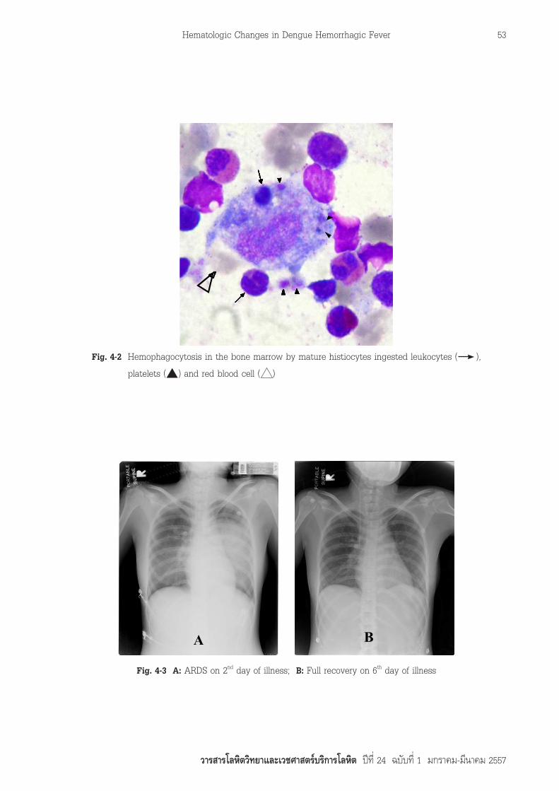
Fig 4-3 A ARDS on 2nd day of
Illness
B Full Recovery on 6th day
of Illness
Fig 4-2 Hemophagocytosis in the bone marrow by mature histiocytes
ingested leukocytes ( ), platelets ( ) and red blood cell ( )
Fig.4-2 Hemophagocytosis in the bone marrow by mature histiocytes ingested leukocytes ( ),
platelets ( ) and red blood cell ( )
Fig.4-3 A: ARDS on 2nd day of illness; B: Full recovery on 6th day of illness
Tanomsri Srichaikul
JHematolTransfusMedVol. 24 No. 1 January-March 2014
54
References1. Nimmannitya S, Halstead SB, Chen SN, Margiotta MR. Dengue
and Chikungunya virus infection in man in Thailand 1962-1964.
Observation on hospitalized patient with hemorrhagic fever. Am
J Trop Med Hyg 1969;518:954-71.
2. Gubler DJ. Dengue and dengue hemorrhagic fever. Clini Microbiol
Rev 1958;11:480-96.
3. Halstead SB, Nimmannitya S, Margiotta MR. 1969 Dengue and
chikungunya virus infection in man in Thailand. 1962-1964.
4. WHO. 1997. Dengue Hemorrhagic Fever Diagnosis Treatment
Prevention and Control. Geneva. WHO.
5. Kittiprapan W. Dengue Shock Syndrome in Pediatric Patients
at Buddhachinaraj Hospital. Buddhachinaraj Medical Journal
2010;27(1):67-75.
6. Sosothikul D, Seksarn P, Pongsewalak S, Thisyakorn U, Lusher J.
Activation of endothelial cells, coagulation and fibrinolysis in children
with dengue virus infection. Thromb Haemost 2007;97:627-34.
7. Avirutnan P, Punyadee N, Noisakran S, Komoltri C, Thiemmeca
S, Auethavornanan K, et al. Vascular Leakage in Severe Dengue
Virus Infections: A Potential Role for the Nonstructural Viral Protein
NS1 and Complement. JID 2006;193:1078-88.
8. Shresta S, Sharar KL, Prigozhin DM, Beatty PR, Harris E. Murine
Model for Dengue Virus-Induced Lethal Disease with Increased
Vascular Permeability. Journal of Virology 2006;80(20):10208-17.
9. Mongkolsapaya J, Duangchinda T, Dejnirattisai W, Vasanawathana
S, Avirutnan P, Jairungsri A, et al. T Cell Responses in Dengue
Hemorrhagic Fever: Are Cross-Reactive T Cells Suboptimal? The
Journal of Immunology 2006;176:3821-9.
10. Innis BL. Dengue and Dengue Hemorrhagic Fever. Exotic Viral
Infections. Edited by J.S. Porterfield. Published in 1995 Chapman
& Hall, London;1995:103-46.
11. Srichaikul T, Nimmannitya S, Sripaisarn T, Kamolsilpa M. Platelet
Function During The Acute Phase of Dengue Hemorrhagic Fever.
Southeast Asian J Trop Med Pub Hlth 1989;20(1):19-25.
12. Punyagupta S. Dengue Hemorrhagic Fever in Adults, an Emerging
Problem in Thailand: a Retrospective Study of 75 Adult Dengue
Infection in 2007. J Infect Dis Antimicrob Agents 2009;26(1):1-5.
13. Rongrungruang Y, Leelarasamee A. Characteristics and Outcomes
of Adult Patients with Symptomatic Dengue Virus Infections. J
Infect Dis Antimicrob Agents 2001;18:19-23.
14. Wichmann O, Hongsiriwon S, Bowonwatanuwong C, Chotivanich
K, Sukthana Y, Pukrittayakamee S. Risk factors and clinical
features associatd with severe dengue infection in adults and
children during the 2001 epidemic in Chonburi, Thailand. Tropical
Medicine and International Health 2004;9(9):1022-9.
15. Chan KP, Lau GKF, Doraisingham S, Chan YC. Adult dengue
deaths in Singapore. Clinical and Diagnostic Virology 1995;4:213-22.
16. Sam SS, Omar SFS, Teoh BT, Abd-Jamil J, AbuBakar S. Review
of Dengue Hemorrhagic Fever Fatal Cases Seen Among Adults:
A Retrospective Study. PLoS Negl Trop Dis 2013;7(5):e2194.
17. Halstead SB. Antibody, Macrophages, Dengue Virus Infection,
Shock and Hemorrhage: A Pathogenetic Cascade. Reviews of
Infectious Diseases 1989;11(4):s830-9.
18. Clyde K, Kyle JL, Harris E. Recent Advances in Deciphering
Viral and Host Determinants of Dengue Virus Replication and
Pathogenesis. Journal of Virology 2006;80(23):11418-31.
Fig.5 Severe complicated DHF case 5
Hematologic Changes in Dengue Hemorrhagic Fever
วารสารโลหิตวิทยาและเวชศาสตรบริการโลหิต ป ที่ 24 ฉบับ ที่ 1 มกราคม-มีนาคม 2557
55
19. Srichaikul T, Punyagupta S, Kanchanapoom T, Chanokovat C,
Likittanasombat K, Leelasiri A. Hemophagocytic Syndrome in
Dengue Hemorrhagic Fever with Severe Multiorgan Complications.
J Med Assoc Thai 2008;91(1):104-9.
20. ถนอมศรี ศรีชัยกุล, สมพนธ บุณยคุปต, วิเชียร มงคลศรีตระกูล, สุวรรณี
จิตตภักดีบดินทร. Hemophagocytic Syndrome: An Analysis of 7
Cases and the Literatures Review. J Hematol Transfus Med
2004;14(4):263-80.
21. Sorakhunpipitkul L, Punyagupta S, Srichaikul T, Tribuddharat S. Thai
Adult Dengue Hemorrhagic Fever During 2008-2010: Seven Cases
Presented with Severe Multiorgan Failure and Successfully Treated
with High Dose of Corticosteroids and Intravenous Immunoglobulin
G. J Infect Dis Antimicrob Agents 2011;28(2):99-103.
22. ลัดดา สรคุณพิพิธกุล, ถนอมศรี ศรีชัยกุล, สิทธิเทพ ธนกิจจารุ, กิตติศักดิ์
เกงสกุล, ไพบูลย ปุญญฤทธิ์. Hemophagocytic Syndrome จาก Epstein
Barr Virus ทำาใหเกิดระบบทางเดินหายใจลมเหลวและตอมน้ำาเหลืองโตมาก:
รายงานผูปวย 1 รายที่รอดชีวิตจากการรักษาดวย Acyclovir และ Pulse
Methylprednisolone ทางหลอดเลือดดำารวมกับ Intravenous Immunoglobulin
G และทบทวนวรสาร. J Hematol Transfus Med 2011;21(2):95-101.
23. ถนอมศรี ศรีชัยกุล. Multiorgan Failure in Dengue Hemorrhagic
Fever Related to Hemophagocytic Syndrome. J Hematol Transfus
Med 2007;17(4):371-6.
24. Pongtanakul B, Narkbunnam N, Veerakul G, Sanpakit K, Viprakasit
V,Tanphaichitr VT, et al. Dengue Hemorrhagic Fever in Patients
with Thalassemia. J Med Assoc Thai 2005;88(8):s80-5.
25. Veerakul G, Sanpakit K, Tanphaichitr VS, Mahasandana C,
Jirarattanasopa N. Secondary hemophagocytic lymphohistiocytosis
in children: an analysis of etiology and outcome. J Med Assoc
Thai;2002:85(2):s530-41.
26. Ray S, Kundu S, Saha M Chakrabarti P. Hemophagocytic Syndrome
in Classic Dengue Fever. J Glob Infect Dis 2011;3(4):399-401.
27. Wong KF, Chan JKC, Chan JCW, Lim WWL, Wong WK. Letter to
the Editor: Dengue Virus Infection-Associated Hemophagocytic
Syndrome. American J Hematol 1991;38:339-40.
28. Bierman HR and Nelson ER. Hematodepressive virus disease of
Thailand. Annals of Internal Medicine 1965;62:867-84.
29. Na-Nakorn S, Suingdumrong A, Pootrakul S, and Bhamarapravati N.
Bone marrow studies in Thai hemorrhagic fever. The American
Journal of the Medical Sciences 1966;35:54-5.
30. Nelson ER, Bierman HR and Chulajata R. Hematologic findings in
the 1960 hemorrhagic fever epidemic (dengue in Thailand). The
American Journal of Tropical Medicine and Hygiene 1964;13:642-9.
31. Nelson ER, Bierman HR and Chulajata R. Hematologic phagocytosis
in postmortem bone marrow of dengue hemorrhagic fever. The
American Journal of the Medical Sciences. 1966;252:68-74.
32. Bhamarapravati N, Tuchinda P. Boonyapakavik V. Pathology of
Thailand hemorrhagic fever: a study of 100 autopsy cases. Annals
of Tropical Medicine and Parasitology 1967; 61:500-10.
33. Kho LK, Wulur H and Himawan T. Blood and bone marrow in
dengue hemorrhagic fever. Paediatrica Indonesiana 1972;12:31-9.
34. Putintseva E, Vega G and Fernandez L. Alteration in thrombopoiesis
in patients with thrombocytopenia produced by dengue hemorrhagic
fever. Nouvelle Revue Francaise Hematologic 1986;28:269-73.
35. Lin SF, Liu HW, Chang CS, Yen JH, Chen TP. Hematological
aspects of dengue fever. Kaohsiung Journal of Medical Sciences
1989;5:12-6.
36. Nakao S, Lai CJ and Young NS. Dengue virus, a flavi virus,
propagates in human bone marrow progenitors and hematopoietic
cell lines. Blood. 1989;74;1235-40.
37. Murgue B, Cassar O, Guigon M, Chungue E. Dengue virus
inhibit human hematopoietic progenitor growth in vitro. Journal
of Infectious Disease 1997;175:1497-501.
38. La Russa VF, Putnak JR and Knight RD. Dengue virus infection
of stromal cells in Dexter cultures of human marrow (abstract)
Experimental Hematology 1991;19:479.
39. La Russa VF, Cutting MA, Kanshal S et al. Generation of stromal
cell colonies from CD34+ cells (abstract). Blood 1993;82:98a.
40. Maze R, Sherry B, Kwon Bs, Cerami A and Brox Meyer HE.
Myelosuppressive effect in vivo of purified recombinant murine
macrophage inflammatory protein-1 alpha. The Journal of
Immunology 1992;149:1004-9.
41. Oppenheim J. Zacharie C, Mukuaida N and Matsuskinna K.
Properties of the novel proinflammatory supergene ‘intercrine’
cytokine family. Annual Review of Immunology 1991;9:617-48.
42. Dunlop D, Wright E, Lorimore S. et al. Demonstration of stem
cell inhibition and myeloprotective effects of SCI/rh MIP 1a in
vivo. Blood 1992;79:2221-5.
43. Williams DE, Devries P, Namen AE, et al. The steel factor.
Developmental Biology 1992;151:368-76.
44. Williams DE, and Lyman SD. Characterization of the gene product
of the steel locus. Progress in Growth Factor Research 1991;3:235-42.
45. Laur F, Murgue B, Deparis X, Roche C, Cassar O, chungue E.
Plasma levels of tumour necrosis factor alpha and transforming
growth factor beta-1 in children with dengue – 2 virus infection in
French Polynesia. Transection Royal society of Tropical Medicine
and Hygiene. 1998;92:654-6.
46. Avirutnan P, Malasit P, seliger B, Bhakdi S, Husmann M. Dengue
virus infection of human endothelial cells leads to chemokine
production, complement activation and apoptosis. Journal of
Immunology 1998;161:6338-46.
47. Raghupathy R, Chaturvedi UC, AI-Sayer H et al. Elevated
levels of IL-8 in Dengue Hemorrhagic fever. Journal of Medical
Virology 1998;56:280-5.
48. Kurane I, Innis BL, Nimmannitya S and et al. Activation of T
Iymphocytes in dengue virus infections. High levels of soluble
interleukin 2 receptor, soluble CD8, interleukin 2, and interferon
- g in sera of children with dengue. The Journal of Clinical
Investigation. 1991;88:1473-80.
49. Kurane I, Innis BL, Nimmannitys S, Nisalak A, Meager A and
Ennis FA. High levels of interferon alpha in the sera of children
Tanomsri Srichaikul
JHematolTransfusMedVol. 24 No. 1 January-March 2014
56
with dengue virus infection. The American Journal of Tropical
Medicine and Hygiene 1993;48:222-9.
50. Hober D, Poli L, Roblin B et al. Serum levels of tumour necrosis
factor - a (TNF-2), interleukin 6 (IL-6) and interleukin – 1b (IL-1b)
in dengue infected patients. The American Journal of Tropical
Medicine and Hygiene 1993;48:324-31.
51. Yadar M, Kamath KR, Iyngkaran N and Sinniah M. Dengue
Hemorrhagic fever and dengue shock syndrome : are they tumour
necrosis factor – mediated disorders? Federation of European
Medical Sciences of Microbiology and Immunology (Netherland)
1991;89:45-50.
52. Srichaikul T. Haematology in dengue and dengue haemorrhagic
fever. Bailliere’s Clinical Haematology 2000;13(2):1-16.
53. Srichaikul T, Punyagupta S, Nitiyanant P, Alkarawong K. Disseminated
intravascular coagulation in adult dengue haemorrhagic fever:
Report of three cases. Southeast Asian J Trop Med Pub Hlth
1975;6(1):106-14.
54. Srichaikul T. Disseminated intravascular coagulation in dengue
haemorrhagic fever. Southeast Asian J Trop Med Pub Hlth
1987;18(3):303-11.
55. Tavodova M. Dengue Fever. South Sudan Medical Journal
2012;5(1):13-6.
56. Srichaikul T, Nimmanitaya S, Artchararit N, Siriasawakul T,
Sungpeuk P. Fibrinogen metabolism and disseminated intravascular
coagulation in dengue hemorrhagic fever. American J Trop Med
& Hygiene 1977;26(3):525-32.
57. Chuansumrit A, Teeraratkul S, Wanichkul S, Treepongkaruna S,
Sirachainan N, Pakakasama S, et al. Recombinant-activated factor VII
for control and prevention of hemorrhage in nonhemophilic pediatric
patients. Blood Coagulation and Fibrinolysis 2010;21(4):354-62.
58. Mairuhu ATA, Gillavry MRM, Setiati TE, Soemantri A, Cate HT,
Brandjes DPM, Gorp ECMV. Is clinical outcome of dengue-virus
infections influenced by coagulation and fibrinolysis? A critical
review of the evidence. The Lancet Infectious Diseases 2003;3:33-41.
59. Kalayanarooj S, Uthaisang E, Nimmannitya S. A study on coagulation
and fibrinolytic system changes in dengue hemorrhagic fever
patients. Studies/Collaborative Studies on Dengue infections/
Dengue Hemorrhagic Fever:312-4.
60. Srichaikul T. Pathogenesis of Bleeding in DHF: Role of Platelet and
Coagulation Abnormalities. J Med Assoc Thai 1989;72(4):239-42.
61. Krishnamurti C, Kalayanarooj S, Cutting MA, Peat RA, Rothwell
SW, Reid TJ, et al. Mechanisms of Hemorrhage in dengue without
circulatory collapse. Am J Trop Med Hyg 2001;65(6):840-7.
62. Mitrakul C, Poshyachinda M, Futrakul P, Sangkawibhan, Ahandrik
S. Hemostatic and platelet kinetic studies in dengue hemorrhagic
fever. Am J Trop Med & Hygiene 1977;26(5):975-84.
63. Phanichyakarn P, Israngkura P, Krisarin C, Pongpanich B,
Dhanamitta S, Valyasevi A. Studies on dengue haemorrhagic
fever IV. Fluorescent staining of the immune complexes on
platelets. Journal Medical Association of Thailand 1977;60:307-11.
64. Boonpucknavig S, Vuttiviroj O, Bunnag C, Bhamarapravati N
Nimmannitya S. Demonstration of dengue antibody complexes on
the surface of platelets from patients with dengue hemorrhagic fever.
American Journal of Tropical Medicine and Hygiene 1979;28:881-4.
65. Wang S, HeR, Patarapotikul J, Innis BL, Anderson R. Antibody
enhanced binding of dengue – 2 virus to human platelets. Virology
1995;213:254-7.
66. Bokisch VA, Top FH, Russell PK, Dixon F, Muller-Eberhard
J. The potential pathogeneic role of complement in dengue
hemorrhagic shock syndrome. The New England Journal of
Medicine 1973;289:996-1000.
67. Prasad KPR, Alahakoon DGS, Gunawardane JN. Effect of prednisolone
during defervescence in dengue haemorrhagic fever: an open
label controlled study. Galle Medical Journal 2008;13(1):19-21.
68. Srichaikul T, Punyagupta S, Sorakhunpipitkul L, Udomsubpayakul
U. Adjunctive Corticosteroid Therapy in 149 Grade II (Non-Shock)
Adult DHF Patients: An Analysis during January 2008-February
2010. J Med Assoc Thai 2011;94(12):1419-23.
69. Goh PL. Dengue perimyocarditis: a case report. Hong Kong J
of Emerg Med 2010;17(1):58-60.