
Gut Microbial Dysbiosis is Correlated with Stroke Severity ...
26
Gut Microbial Dysbiosis is Correlated with Stroke Severity Markers in Aged Rats Following Stroke Tyler C. Hammond University of Kentucky College of Medicine Sarah Messmer University of Kentucky College of Medicine Jaqueline A. Frank University of Kentucky College of Medicine Douglas Lukins University of Kentucky College of Medicine Rita Colwell CosmosID, INC Ai-Ling Lin University of Missouri-St Louis Keith Pennypacker ( [email protected] ) University of Kentucky https://orcid.org/0000-0002-4618-0903 Research Keywords: stroke, microbiome, inァammation, imaging Posted Date: November 8th, 2021 DOI: https://doi.org/10.21203/rs.3.rs-1044260/v1 License: This work is licensed under a Creative Commons Attribution 4.0 International License. Read Full License
Transcript of Gut Microbial Dysbiosis is Correlated with Stroke Severity ...

Gut Microbial Dysbiosis is Correlated with StrokeSeverity Markers in Aged Rats Following StrokeTyler C. Hammond
University of Kentucky College of MedicineSarah Messmer
University of Kentucky College of MedicineJaqueline A. Frank
University of Kentucky College of MedicineDouglas Lukins
University of Kentucky College of MedicineRita Colwell
CosmosID, INCAi-Ling Lin
University of Missouri-St LouisKeith Pennypacker ( [email protected] )
University of Kentucky https://orcid.org/0000-0002-4618-0903
Research
Keywords: stroke, microbiome, in�ammation, imaging
Posted Date: November 8th, 2021
DOI: https://doi.org/10.21203/rs.3.rs-1044260/v1
License: This work is licensed under a Creative Commons Attribution 4.0 International License. Read Full License
1
Gut microbial dysbiosis is correlated with stroke severity 1
markers in aged rats following stroke 2
Tyler C. Hammond,1,2 Sarah Messmer,3,4 Jacque A. Frank,3,4 Doug Lukins,5 Rita Colwell,6 3
Ai-Ling Lin,1,7* and Keith R. Pennypacker3,4* 4
5
Abstract 6
Background: An imbalanced gut microbial community, or dysbiosis, has been shown to occur 7
following stroke. It is possible that this dysbiosis negatively impacts stroke recovery and 8
rehabilitation. Species level resolution measurements of the gut microbiome following stroke 9
are needed to develop and test precision interventions such as probiotic or fecal microbiota 10
transplant therapies that target the gut microbiome following stroke. Previous studies have used 11
16S rRNA amplicon sequencing in young male mice to obtain broad profiling of the gut 12
microbiome at the genus level following stroke, but further investigations will be needed with 13
whole genome shotgun sequencing in aged rats of both sexes to obtain species level resolution 14
in a model which will better translate to the demographics of human stroke patients. 15
Results: 39 aged male and female rats underwent middle cerebral artery occlusion. Fecal 16
samples were collected before stroke and three days post stroke to measure gut microbiome. 17
Machine learning was used to identify the top ranked bacteria which were changed following 18
stroke. MRI imaging was used to obtain infarct and edema size and cerebral blood flow (CBF). 19
ELISA was used to obtain inflammatory markers. 20
Dysbiosis was demonstrated by an increase in pathogenic bacteria such as Butyricimonas 21
virosa (15.52 fold change, p<0.0001), Bacteroides vulgatus (7.36 fold change, p<0.0001), and 22
Escherichia coli (47.67 fold change, p<0.0001). These bacteria were positively associated with 23
infarct and edema size and with the inflammatory markers Ccl19, Ccl24, IL17a, IL3, and 24
complement C5; they were negatively correlated with CBF. Conversely, beneficial bacteria 25
such as Ruminococcus flavefaciens (0.14 fold change, p<0.0001), Akkermansia muciniphila 26
(0.78 fold change, p<0.0001), and Lactobacillus murinus (0.40 fold change, p<0.0001) were 27
decreased following stroke and associated with all the previous parameters in the opposite 28

2
direction of the pathogenic species. There were not significant microbiome differences 29
between the sexes. 30
Conclusion: The species level resolution measurements found here can be used as a foundation 31
to develop and test precision interventions targeting the gut microbiome following stroke. 32
Probiotics that include Ruminococcus flavefaciens, Akkermansia muciniphila, and 33
Lactobacillus murinus should be developed to target the deficit following stroke to measure 34
the impact on stroke severity. 35
36
Author affiliations: 37
1 Sanders-Brown Center on Aging, University of Kentucky, Lexington, KY 40536 USA 38
2 Department of Neuroscience, University of Kentucky College of Medicine, Lexington, KY 39
40536 USA 40
3 The Center for Advanced Translational Stroke Science, University of Kentucky, Lexington, 41
KY 40536 USA 42
4 Department of Neurology, University of Kentucky College of Medicine, Lexington, KY 43
40536 USA 44
5 Department of Radiology, University of Kentucky College of Medicine, Lexington, KY 45
40536 USA 46
6 CosmosID, Inc., Rockville, MD 20850 USA 47
7 Department of Radiology, Division of Biological Sciences and Institute for Data Science & 48
Informatics, University of Missouri, Columbia, MO 65211 49
*The authors have equal contribution to the study. 50
51
Correspondence to: 52
Keith R. Pennypacker, PhD 53
BBSRB, Office B457, University of Kentucky, Lexington, KY 40536 54
56
Ai-Ling Lin, PhD 57
1101 Hospital Drive, University of Missouri, Columbia, MO 65211 58
60
Running title: Gut bacteria tied with stroke severity 61
Keywords: stroke, microbiome, inflammation, imaging 62

3
Background 63
Over 795,000 people suffer a stroke every year in the United States alone1. Recent advances in 64
acute stroke therapies have lowered stroke mortality, but survivors are often left severely 65
impaired2. Rehabilitation therapies are beneficial at inducing neuroplasticity to overcome these 66
impairments, but over 40% of stroke survivors are left with moderate to severe disabilities that 67
markedly reduce quality of life3. Novel multimodal approaches are needed to promote plasticity 68
and sensorimotor function through a combination of current rehabilitation therapies with other 69
treatments designed to foster neuroplasticity. 70
Accumulating evidence suggests that gut microbes modulate brain plasticity via the 71
bidirectional gut-brain axis and may play a role in stroke rehabilitation4. A severely imbalanced 72
microbial community, or dysbiosis, has been shown to occur following stroke, causing a 73
systemic flood of neuro- and immunomodulatory substances due to increased gut permeability 74
and decreased gut motility5. These substances can impact neuroinflammation as commensal 75
bacteria invade the bloodstream and as intestinal lymphocytes migrate from gut-associated 76
lymphoid tissue to the brain6. Fecal microbiota transplant has been shown to normalize brain 77
lesion-induced dysbiosis and to improve stroke outcome in mice6. The microbiome is 78
modifiable as it is influenced by environmental factors such as diet and exercise and could 79
potentially be a therapeutic target in stroke rehabilitation through nutritional and 80
pharmacological interventions and physical therapy7,8. To our knowledge, no studies have 81
measured the species level resolution necessary to develop precision interventions such as 82
probiotics or fecal microbiota transplants that target the gut microbiota following stroke. 83
Furthermore, no microbiome studies have been performed on aged rats of both sexes, which 84
are better matched to the demographics of human stroke patient than the young male mice used 85
in most studies. The microbiome changes found in this study need to be examined and 86
correlated with clinical imaging markers of stroke and inflammatory markers to understand 87
better whether the microbiome could be a therapeutic target in stroke rehabilitation. 88
Here we identify the gut-brain axis changes that occur following stroke in aged rats using high 89
resolution whole genome shotgun sequencing and correlate them with clinical imaging markers 90
of stroke including MRI-based infarct size, edema size, and cerebral blood flow (CBF) as well 91
as inflammatory markers. We found that microbial communities are disrupted in an aged rat 92
population following stroke, showing significantly different beta diversity, increased alpha 93
diversity, and changes in the relative abundance of 5 of the 6 major phyla found in the gut. 94

4
Changes in thirteen bacterial species as detected by machine learning were highly associated 95
with stroke and changes in these species were also associated with increased infarct and edema 96
size and decreased CBF. Changes in the microbiome due to stroke were also associated with 97
increases in 49 inflammatory markers. 98
Materials and methods 99
Ethics approval and animals 100
Aged male and female rats (18‐month‐old Sprague‐Dawley rats (ENVIGO, Indianapolis, IN) 101
were used for all procedures. The aged female rats on average weighed between 245g and 425g, 102
and aged male rats approximately weighed between 505g and 705g. The study was conducted 103
in accordance with the National Institutes of Health Guide for the Care and Use of Laboratory 104
Animals and study protocols were approved by University of Kentucky’s (UK) Institutional 105
Animal Care and Use Committee. Animals were housed in a climate-controlled room on a 12‐106
hr light and dark cycle (0700–1,900) with access to food and water. Per Division of Laboratory 107
Animal Resources (DLAR) cage requirements at UK’s vivarium facility, the animals can be 108
paired in one cage if the animal weight is under 650 grams. We typically house two animals 109
(males or females) per cage upon arrival to DLAR. Once the rats are over 650 grams, they are 110
then split into a separate cage by themselves. Fecal samples were collected for all animals at 111
24 hours before surgery and 72 hours post-surgery and for 4 animals at 30 days post-surgery. 112
The rats underwent MRI at 72 hours to measure infarct and edema volumes and CBF then 113
euthanized. 114
Middle cerebral artery occlusion 115
22 of the rats received a permanent Middle Cerebral Artery Occlusion (p-MCAO) and 17 of 116
the rats received a 5-hour transient Middle Cerebral Artery Occlusion (5t-MCAO). All animals 117
were induced with oxygen containing 5% isoflurane, then shaved, prepped with Hibiclens 118

5
(chlorohexidine scrub) prior to 70% EtOH and then a betadine solution. Maintenance isoflurane 119
was maintained at 2.5% in O2 was delivered via a nosecone placed in line with the binner 120
tubeQ (gas delivery tube) of the anesthesia circuit. Under bnear sterileQ conditions and with 121
the use of a Zeiss operating microscope (Carl Zeiss AG, Gottingen, Germany) at 4 to 25 122
magnification, the procedure was performed. First, the skin was opened with a midline vertical 123
incision, and the underlying submandibular gland bluntly dissected in the midline to produce 124
left and right lobes, which were retracted laterally. Division of the omohyoid muscle, then 125
dissection medial to the right sternocleidomastoid (SCM) muscle was used to expose the 126
common carotid artery (CCA), which was separated from the vagus nerve. Elastic hooks (Lone 127
Star Medical Products, Houston, TX, USA) tethered to metal stays on the customized surgery 128
table were used to retract the skin and the SCM muscle. In the p-MCAO, a hand-held 129
electrocautery (Aaron Medical, St. Petersburg, FL, USA) is used to cauterize the superior 130
thyroid artery (STA), a collateral off the ECA, and the occipital artery (OA), a collateral off 131
the ICA. Two 5-0 silk sutures (Surgical Specialties, Reading, PA, USA) were used to ligate the 132
external carotid artery (ECA) as distal as possible to the ECA/ICA bifurcation, and a second 133
tie that was applied just proximal to the first, leaving enough space in between the two ties to 134
cut the artery with micro scissors. At this point, blunt dissection was used to isolate the internal 135
carotid artery (ICA) and its collateral, the pterygopalatine artery. Next, microvascular 136
aneurysm clips (Mizuho, Beverly, MA, USA) were applied to the CCA and the ICA. A 5-0 137
PDS II monofilament embolus (Ethicon, Cornelia, GA, USA), was introduced into an 138
arteriotomy hole–produced with a 26-gauge hypodermic needle–in the reflected ECA stump 139
and fed distally into the ICA. At this time, a collar suture at the base of the ECA stump was 140
tightened around the embolus, and the ICA clamp was removed. The embolus was advanced 141
20 mm from the carotid bifurcation, with care taken to avoid entrance into the pterygopalatine 142
artery. 143

6
For the transient occlusion, the same steps were done as stated with the pMCAO, with the 144
exception that Doccol Corporation silicone rubber-coated monofilaments were used for the 145
occlusion of the middle cerebral artery (MCA). Multiple sized Doccol monofilaments are used 146
in the MCAO surgery depending on the sex and weight of the rat. Two 18-inch length of 5-0 147
silk suture were used for the ligation of the external carotid artery (ECA) to secure the ECA 148
stump, and the entry point of the monofilament into the ECA/ICA bifurcation. The third 5-0 149
silk suture was used to secure the monofilament within the ECA. A micro-serrefines arterial 150
clamp (FST, Fine Science Tools, #18055-01) was used to occlude the internal carotid artery 151
(ICA) and common carotid artery (CCA) prior to advancement of the monofilament into the 152
MCA. After 5 hours, the embolus was gently removed and the collar suture at the base of the 153
ECA stump tightened. The skin was closed with 3-0 nylon suture (Ethicon, Cornelia, GA, 154
USA), anesthesia discontinued, and the animal allowed to recover. Animals used for control 155
underwent a neck dissection and coagulation of the external carotid artery, but no manipulation 156
or occlusion of the common or internal carotid arteries. 157
Post‐surgical fluid management and pain control 158
Immediately post‐operatively the animals received 2 ml of sterile saline (0.9%) subcutaneous. 159
An additional 1 ml of saline was given if extra blood loss occurred during surgery. The animals 160
were injected with sterile filtered PBS pH 7.4 at 6 (for the p-MCAO), 24, 48, and 72 hours 161
post‐MCAO. The animals were weighed every morning post‐MCAO to determine dehydration. 162
Hydration status was checked by pinching up or “tenting” the skin over the nape of the neck. 163
The skin should immediately relax into its normal position. If the skin remains tented longer 164
than normal, the rat was deemed dehydrated, and saline was given. Per DLAR guidelines, rats 165
can receive up to 10 ml at a time and no more than 2 ml at any one location per 6 hr. If 166
warranted, additional saline (1–2 ml) will be given in addition to 6, 24, 48, and 72 hr. Also, we 167

7
added an additional water bottle in each cage to allow more avail‐ ability to free water for the 168
rats to consume and moistened food was provided on the bottom of the cage to encourage 169
feeding and additional water intake. Post‐surgical pain control was managed with carprofen, 170
which is based on weight of the animal. Animal weights are taken prior to surgery (pMCAO) 171
and daily until animals are euthanized at 72 hr. (post MRI). The animals received a dosage of 172
carprofen 5mg/kg prior to surgery and every 24 hr. for three days post‐pMCAO until 72 hr. 173
when they were euthanized (post MRI). Termination of survival criteria include that all animals 174
were weighed and monitored, especially for dehydration and pain, each morning post surgery. 175
This includes specific attention to the animal as a whole, as well as incision sights. If symptoms 176
such as pain, fatigue, loss of energy, excess energy, ruffled hair coat, reluctance to move, failure 177
to groom or feed, hypoactivity, hyperactivity, restlessness, self‐trauma, aggressiveness, ataxia, 178
pale mucous membranes, cyanosis, rapid, shallow and/or labored breathing, cachexia, 179
porphyria, soiled anogenital area, inactivity, failure to respond to stimuli, lack of 180
inquisitiveness, vocalization, and/or hunched posture were observed, the research team 181
obtained advice from the vivarium veterinary staff on how best to intervene to alleviate 182
discomfort; if that was not possible the animal was euthanatized. Additional checks were made 183
in the afternoon if there was any rat of concern. The animals were removed from the study if 184
adverse signs persisted despite carprofen and treatment past 24 hr. If the signs fail to resolve, 185
the vivarium veterinarian was consulted and decided the time course when such animals were 186
euthanized. Additionally, weight loss greater than 20% (emaciated appearance, rapid weight 187
loss over two days) was considered an endpoint. Rapid weight loss was considered greater than 188
10% a day for two days. 189
Microbiome Sequencing 190
8
Fecal samples were collected for all animals at 24 hours before surgery and 72 hours post-191
surgery and for 4 animals at 30 days post-surgery. Genomic DNA were extracted from 0.25 192
grams of stool using ZymoBIOMICS™ DNA Mini Kit and shipped to CosmosID for DNA 193
quantification using fluorometer Qubit 3.0. Libraries were constructed and the PCR products 194
were purified using 1.0X speed beads and eluted in 15 µL of nuclease-free water and quantified 195
by PicoGreen fluorometric assay (100X final dilution). The libraries were pooled and loaded 196
onto a high sensitivity chip run on the Caliper LabChipGX (Perkin Elmer, Waltham, MA) for 197
size estimation and sequenced using Illumina NextSeq/HiSeq platform. Unassembled 198
sequencing reads were analyzed by CosmosID bioinformatics platform (CosmosID Inc., 199
Rockville, MD) 9-12 for microbiome analysis. Heatmaps, stacked bar graphs, and Principal 200
Component Analysis (PCA) plots were generated to visualize the diversity and abundance of 201
each microbial taxa. Alpha- and beta-diversity were calculated to determine the number of 202
species present in a cohort and diversity similarities between groups. 203
Magnetic resonance imaging 204
MRI images were acquired on a 7T Bruker Clinscan horizontal bore system (7.0T, 30 cm, 300 205
Hz) equipped with a triple‐axis gradient system (630 mT/m and 6,300 T m‐1 s ‐1) with a closed 206
cycle. PCASL (pseudo conintous arterial spin labelling) images were acquired coronally to 207
determine CBF with a fat saturated, double refocused echo planar sequence: TR 4000 ms, TE 208
26 ms, Matrix 74 x 56, FOV 26 mm x 19.7 mm, Slice 1.2 mm, Slices 6, 120 Tagged-Untagged 209
Pairs, 10 M0 Images, Tagging Plane Offset 12mm, Bolus duration 1.86sec, Post Labeling Delay 210
0sec, and Acquisition Time of 10 min. T2 weighted images were acquired coronally with a 211
RARE sequence: TR 6000 ms, TE 29 ms, Turbo Factor 5, Matrix 190 x 190, FOV 240 mm x 212
240 mm, Slice 0.4 mm, Slices 44, and Acquisition Time of 9 min. Male rats were anesthetized 213
with an average of 2.25% isoflurane in oxygen, while female rats were anesthetized with an 214

9
average of 1.75% isoflurane in oxygen using an MRI compatible CWE Inc. equipment 215
(Ardmore, PA). They were held in place on a Bruker scanning bed with a tooth bar, ear bars, 216
and tape. Body temperature, heart rate, and respiratory rate were continuously monitored 217
throughout the MRI scans (SA Instruments, Inc., Stony Brook, NY). The animal's body 218
temperatures were maintained at 37°C with a water heating system built into the scanning bed. 219
The scanning procedure took approximately 40-60 mins. per animal. 220
The MR images were analyzed by a blinded neuroradiologist who visually identified infarct 221
volume and edema volume. These volumes were counted, and this number was normalized to 222
the number of images counted to provide a per section count. The volume of brain parenchyma 223
demonstrating infarct volume visibly affected was calculated by manual segmentation using 224
ITK‐SNAP software (www.itksnap.org, version 3.6)13. The volume of brain parenchyma 225
visibly affected by T2 hyperintensity (edema volume) was calculated in a similar fashion. The 226
data are given as absolute volume in cubic millimeters. The calculation was based on all slices 227
from each MR sequence. Cerebral perfusion values of the area of lesion within the ipsilateral 228
hemisphere, and the equivalent region within the contralateral hemisphere were generated 229
using the quantification as previously described.14,15 230
231
Biochemical analysis 232
In following STAIR guidelines, clinically relevant biomarkers were determined in our aged 233
male and female rats16. Blood was taken from the jugular vein at three different time points: 234
immediately prior to MCAO surgery and 5 mins after reperfusion of the MCA in the pMCAO, 235
and 5 hours post MCAO procedure in the 5t-MCAO. Blood was immediately placed on ice and 236
centrifuged at 2000 g for 15 minutes. Plasma was extracted and stored separately, both pellet 237
and plasma were frozen at -80˚C for later analysis. RNA extraction and Amplification followed 238

10
the methods of Martha et.al 202017. Briefly, total RNA was extracted from the pellet portion 239
via a Nucleospin Blood Kit (Macherey-Nagel, Düren, Germany), RNA quantity was estimated 240
using a Qubit 4 Fluorometer (Thermo-Fisher; Waltham, MA), cDNA was synthesized using a 241
RT² PreAMP cDNA synthesis Kit from Qiagen and expression of 84 genes were measured 242
using an ABI StepOne Plus (Germantown, MD) and a RT² Profiler Rat Chemokine and 243
Receptor Array from Qiagen. Delta Delta CT was calculated using the fold change of the gene 244
expression measurement from pre to 3-day. 245
Statistical analysis 246
Descriptive microbiome analyses were performed with CosmosID bioinformatics software to 247
generate alpha diversity, beta diversity, and relative abundance data. Alpha diversities amongst 248
groups were compared using Wilcoxon Rank Sum test. Beta diversities amongst groups were 249
compared using PermANOVA. Relative abundance data was compared to measures of stroke 250
severity as determined by imaging (infarct size, edema size, CBF) using general linear models 251
within the MaAsLin 2 R package18. Random forest was used to determine top bacterial species 252
that were changed following stroke using the randomForest R package19. All imaging variables 253
in the study were transformed to meet assumptions of normality. The transformation 254
procedures began with Shapiro‐Wilks and for measures with p < 0.05, the variables were square 255
root transformed. A p‐value of 0.05 was set a priori to determine statistical significance. 256
Results 257
We analyzed all rats before and after middle cerebral artery occlusion and considered sex, 258
surgery type, and treatment with LIF or PBS in the analysis. We administered a leukemia 259
inhibitory factor (LIF) treatment on half of the rats based on previous work suggesting that LIF 260
is an anti-inflammatory that regulates the immune/inflammatory response to stroke20. The rats 261
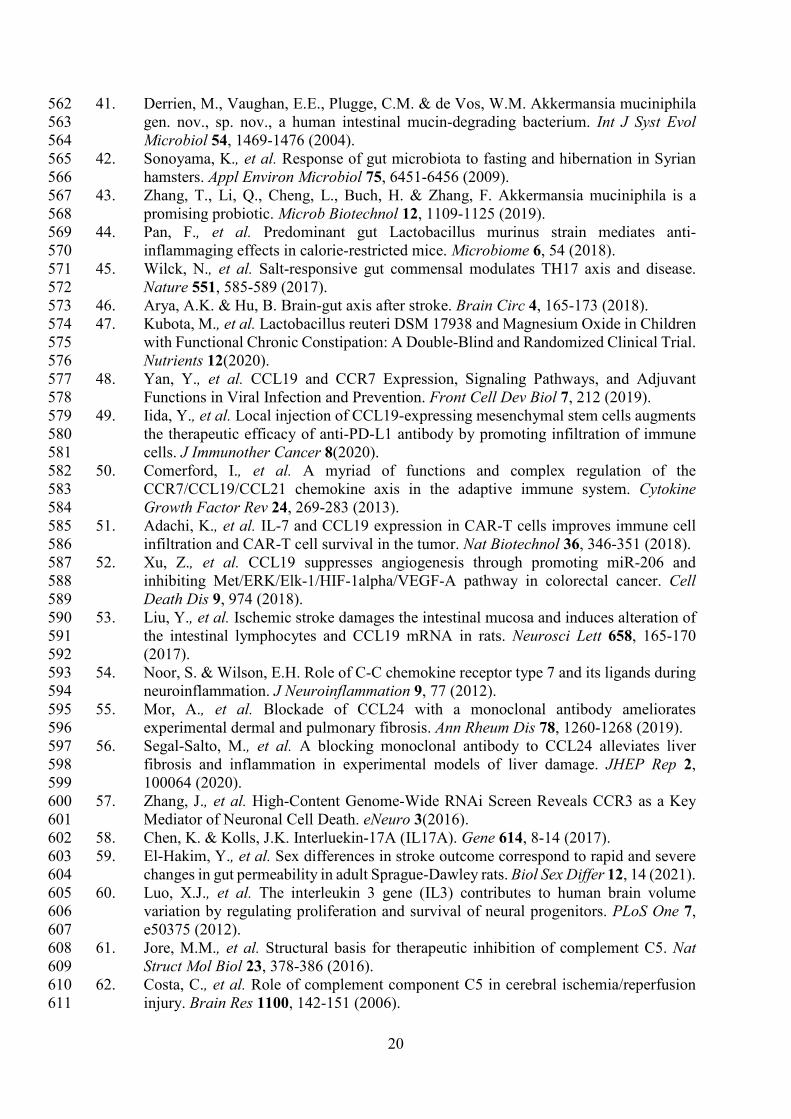
had an average of 96.50 mm3 infarct size, 131.0 mm3 edema size, and 1.31 ml/g/min CBF from 262
11
a permanent occlusion and 31.46 mm3 infarct size, 102.1 mm3 edema size, and 2.16 ml/g/min 263
CBF from a transient occlusion. Infarct and edema volumes were not significantly different 264
between sex, treatment group, or occlusion type. No significant difference in CBF was detected 265
between sex or treatment, but, as expected, a significant difference occurred between 266
permanent and transient occlusion in CBF (Fig. 1). 267
The aged rat gut microbiome is disrupted following stroke 268
We performed an analysis on the gut microbial communities of the aged rats before and after 269
stroke. Comparing the alpha diversity before and after stroke, we found that richness and 270
evenness increased from 3.818 on the Shannon diversity index21 to 4.178 (Fig. 2A). There were 271
no differences in the change of alpha diversity between sex, treatment, or occlusion type. 272
Comparing the beta diversity before and after stroke, we found that the microbial communities 273
were significantly different between baseline and stroke (p=0.0001), but no significant 274
microbial community differences were detected based on sex, treatment, or occlusion type. 275
(Fig. 2B and Supplementary Table 1). 276
We investigated specific differences in the relative abundance of the major bacterial phyla in 277
the gut (Fig. 3). We found increases in proteobacteria and Bacteroidetes and decreases in 278
firmicutes, verrucomicrobia, and actinobacteria following stroke (Supplementary Table 2A). 279
This translates to a sharp decrease in the firmicutes to bacteroidetes ratio. Using linear 280
regression, the major bacterial phyla predict infarct size with an R2=0.3866 and edema size 281
with an R2=0.6022 (Supplementary Table 2B). 282
The top 13 disrupted bacterial species following stroke 283
We investigated specific differences in the relative abundance of the major bacterial species in 284
the gut. There was a total of 29 species increased and 23 species decreased following stroke 285
(Table 1). Supplementary Table 3 gives a detailed description of all the taxa that were 286
increased (red) or decreased (green) following stroke. Using random forest machine learning 287
classification, we found the most important bacterial species that predict stroke verse baseline 288
with an 85.14% accuracy. They include an increase in Butyricimonas virosa, Bacteroides 289
vulgatus, Escherichia coli, Bacteroides uniformis, Bacteroides dorei, Parabacteroides 290
distasonis, and Alistipes indistinctus and a decrease in Ruminococcus flavefaciens, 291
Akkermansia muciniphila, Ruminococcus_u_s, [Clostridium] clostridioforme, Lactobacillus 292
murinus, and Lachnospiraceae bacterium 3-1. Using linear regression with backwards 293

12
elimination (Table 2), we found that increases in Ruminococcus_u_s and Alistipes indistinctus 294
and decreases in Lachnospiraceae bacterium 3-1 predict infarct volume with an R2=0.4433. 295
Increases in Butyricinomas virosa, Bacteroides uniformis, and Ruminococcus_u_s and 296
decreases in Ruminococcus flavefaciens predict edema with an R2=0.6230. Finally, decreases 297
in Alistipes indistinctus predict CBF with an R2=0.1825. 298
We investigated potential interactions between bacterial species in predicting infarct size, 299
edema size, and CBF (Supplementary Table 4). Using a feasible solution algorithm (FSA) 300
for finding interactions, we found that decreases in Lachnospiraceae bacterium A2 and 301
Lactobacillus murinus predict infarct size, but a combination of the two predicts a dramatic 302
increase in the prediction value with an R2=0.6206. Decreases in Lachnospiraceae bacterium 303
A4 and Lactobacillus murinus predict edema size, but a combination of the two have stronger 304
predictive ability with an R2=0.6454. Decreases in Adlercreutzia equolifaciens and 305
Desulfovibrio desulfuricans predict CBF, but again, a combination of the two has a stronger 306
prediction with an R2=0.8093. 307
Bacterial community disruptions following stroke are correlated 308
with stroke severity markers 309
We investigated the correlation of all the bacterial species with infarct size and edema size 310
(Table 3). Using the MaAsLin 2 R package18, which automatically normalizes and transforms 311
all variables in preparation for linear regression, we correlated metagenomic sequencing with 312
imaging variables of stroke severity. Twenty-seven bacterial species were positively correlated 313
and 19 negatively correlated with infarct volume. Thirty species were positively correlated, 314
and 31 species were negatively correlated with edema volume. No species were correlated with 315
CBF. 316
Bacterial community disruptions following stroke are correlated 317
with rises in inflammatory markers 318
We investigated the association of inflammatory markers with gut microbiome changes (Table 319
4). Using an Rt2 PCR array22 to test the difference between inflammatory genes expressed 320
before and after stroke in a subsample of the rats, we found all the markers that were associated 321
with the changes in gut microbiome. There were 22 bacterial species changed with stroke that 322

13
were also correlated with changes in inflammatory markers. There were 49 total inflammatory 323
markers that were increased in association with bacterial changes (Supplementary Table 5). 324
Discussion 325
To our knowledge, we are the first to report on the gut microbial changes with species level 326
resolution in aged male and female rats and to correlate these changes with clinical MRI 327
imaging markers of stroke and inflammatory markers. Following stroke, we found that alpha 328
diversity significantly increased, beta diversity significantly changed, and 5 of the 6 major 329
bacterial phyla were altered. Using machine learning, the top 13 bacterial species that predict 330
whether a sample came from the baseline or post-stroke time point. These bacterial species had 331
independent significant correlations with infarct size, edema size, and CBF. We also identified 332
several species whose interactions with one another were significant in correlating with stroke 333
imaging outcomes. Finally, we found 49 inflammatory markers that correlated with the changes 334
in microbiome from stroke. These changes are representative of a shift from beneficial to 335
pathogenic bacterial species following stroke which results in an increased inflammatory 336
response. 337
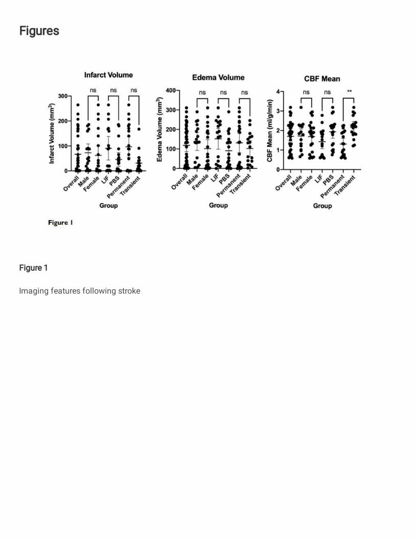
Figure 4 summarizes the changes in gut microbial communities in response to stroke. 338
Following stroke there is a significant shift in the gut microbiome, with alterations to 52 major 339
bacterial species. These bacterial fluctuations shift the environment to a more inflammatory 340
state that adversely affect injury. The microbial community dysbiosis is likely due to the 341
increased gut permeability and decreased gut motility in addition to the immunodepression 342
caused by the amplified stress response (increased sympathetic nervous system response and 343
hypothalamic-pituitary-adrenal (HPA) axis response) following stroke23. Previous groups have 344
reported a decrease in alpha diversity following stroke in a mouse model6 and an increase in a 345
human model24. Our findings are consistent with others who have seen that microbial 346
communities differ before and after stroke based on measures of beta diversity25. We did not 347
find any significant differences in the microbiome between males and females. Some groups 348
have found sex differences in the microbiome that are largely attributed to hormone 349
differences26. It is possible that we did not see these differences because the female rats we 350
used are aged and reproductively senescent. 351
We saw increases in proteobacteria following stroke. In previous studies, proteobacteria have 352
been associated with increased cognitive impairment following stroke27. Dysbiosis related to 353
14
metabolic disorders, inflammation, and cancer is often related to an increase in 354
proteobacteria28,29. This is possibly due to increased oxygen content in the gut following 355
increases in inflammation, providing an optimal environment for these facultative anaerobes30. 356
We also saw decreases in firmicutes and increases in bacteroidetes species. Decreased 357
firmicutes have also been associated with Alzheimer’s disease31. Obesity is often characterized 358
by a significantly increased firmicutes to bacteroidetes (F/B) ratio32; interestingly, our study 359
found that stroke has the opposite effect on F/B ratio. Actinobacteria was significantly 360
decreased following stroke. Actinobacteria downregulates inflammation by production of IL-361
4 and IL-1333 and is known to have anti-biofilm properties against pathogenic bacteria34. It is 362
possible that a decrease in actinobacteria allows other pathogenic bacteria to flourish. 363
Of the bacteria we found that are increased following stroke, many were of the bacteroides 364
species. Bacteroides species have the ability to reduce oxygen levels and breakdown food 365
products to liberate fucose and sialic acid residues from glycoproteins that can be consumed 366
by other microorganisms, including pathogens. Higher bacteroides species are associated with 367
type I diabetes35. Bacteroides vulgatus and Bacteroides dorei reduce gut microbial 368
lipopolysaccharide production and inhibit atherosclerosis36, but they are also associated with 369
insulin resistance, altered bile acid metabolism, and reduced interleukin-22 secretion37. 370
Butyricimonas virosa, Escherichia coli, and Parabacteroides distasonis were also elevated 371
following stroke. An increase of Butyricimonas virosa has also been seen in divers with high 372
occupational exposure to a hyperoxic environment38, which is very different from the hypoxic 373
environment of stroke. Escherichia coli is a very common commensal bacteria that has the 374
potential to cause extraintestinal infections based on its genome content and phenotypic traits39 375
and is famous for causing post-stroke infections, especially pneumonia. Parabacteroides 376
distasonis has been shown to alleviate obesity and metabolic dysfunctions via production of 377
succinate and secondary bile acids40, which is interesting since stroke is often associated with 378
obesity and metabolic dysfunctions. 379
380
Many bacteria which are generally considered beneficial were decreased following stroke 381
including akkermansia, lactobacillus, and ruminococcus species. Akkermansia muciniphila is 382
a mucin-degrading bacterium41 that can be increased with fasting42 that is known to improve 383
host metabolic functions and immune responses43. Lactobacillus murinus can combat 384
inflammaging44, and a reduction of L. murinus due to high salt consumption has been 385

15
associated with an increase in proinflammatory TH17 cells45, which have been correlated with 386
post stroke dysbiosis and secondary injury46. Lactobacillus reuteri was also significantly 387
reduced following stroke. A randomized control trial in children showed administration of L. 388
reuteri as a probiotic to be useful in treating constipation in children47. Constipation is a 389
common morbidity in stroke, and administration of this species could help to alleviate 390
symptoms. Ruminococcus flavefaciens has also been shown to decrease the therapeutic effects 391
of antidepressants, having implications for the treatment of post-stroke depression. 392
Many of the bacterial changes were associated with increases in inflammatory markers. The 393
major markers that were increased were CCL19, CCL24, IL-17A, IL-3, and complement factor 394
C5. CCL19 is a chemokine that is commonly upregulated as a result of viral infections48, and 395
attracts dendritic cells and T lymphocytes49; it promotes thymocyte development, secondary 396
lymphoid organogenesis, high affinity antibody responses, regulatory and memory T cell 397
function, and lymphocyte egress from tissues organs50,51. CCL19 suppresses angiogenesis and 398
can inhibit proliferation, migration, and sprouting responses of tumors52. CCL19 has previously 399
been found to be upregulated following stroke after damage to the intestinal epithelium53 and 400
has been shown to facilitate T-cell migration to the insult site and microglial activation 401
following stroke54. CCL24 plays an important role in pathological processes of skin and lung 402
inflammation and fibrosis55 and regulates inflammatory and fibrotic activities through its 403
receptor, CCR356. CCR3 is a mediator of neural cell death57. In host defense, IL-17A has been 404
shown to be mostly beneficial against infection caused by extracellular bacteria and fungi58 and 405
IL-17A has been shown to be increased following stroke, especially in males59. IL3 is strongly 406
associated with brain volume variation and plays pivotal roles in the expansion and 407
maintenance of the neural progenitor pool and the number of surviving neurons60; our work 408
has previously identified IL3 increased in the spleen with our aged rat model of stroke20. 409
Activation of complement C5 generates the potent anaphylatoxin C5a and leads to pathogen 410
lysis, inflammation, and cell damage61. Activated C5 complement components are a part of the 411
cerebral tissue inflammation following ischemia62. 412
413
This study lays an important foundation upon which precision interventions can be developed 414
to target the gut microbiome in stroke rehabilitation. Future studies should attempt to 415
manipulate the microbiome to change stroke outcomes. This could be achieved through diet 416
interventions, antibiotic therapy, probiotics, or fecal microbiota transplant. For example, a 417

16
future probiotics study should include the use of Ruminococcus flavefaciens, Akkermansia 418
muciniphila, and Lactobacillus murinus as these were deficient in our population. Stroke 419
severity measures from imaging and inflammatory markers could be used as outcomes to 420
compare to the current study. While the present study identified associations of various 421
inflammatory markers with changes in gut microbial composition, it would also be useful to 422
perform mechanistic studies to determine how the microbiota change the expression of these 423
markers and what their downstream effects are. Finally, human studies will be needed to 424
determine whether the microbial changes seen in animals following stroke are similar to the 425
changes seen in animals. Such results can then be used to alter the gut microbiome to favor 426
positive clinical outcomes after stroke. 427
428
Conclusion 429
We found that alpha diversity significantly increased following stroke irrespective of sex, 430
treatment, or occlusion type. Beta diversity was also significantly different, with increases in 431
proteobacteria and decreases in the firmicutes to bacteroidetes ratio. Random forest analysis 432
revealed the top 13 species changes as a result of stroke including increases in Butyricimonas 433
virosa and Escherichia coli and decreases in Akkermansia muciniphila and Bacteroides dorei. 434
Correlation analysis revealed that these species changes were associated with increased infarct 435
and edema sizes following stroke. Furthermore, the bacterial changes were associated with 436
increases in inflammatory markers, notably Ccl19, Ccl24, IL17a, IL3, and complement C5. 437
Declarations 438
Ethics approval 439
The study was conducted in accordance with the National Institutes of Health Guide for the 440
Care and Use of Laboratory Animals and study protocols were approved by University of 441
Kentucky’s (UK) Institutional Animal Care and Use Committee. 442
Consent for publication 443

17
Not applicable 444
Availability of data and materials 445
All data generated or analyzed during this study are included in this published article 446
Competing interests 447
The authors declare that they have no competing interests 448
Funding 449
This research was supported by NIH/NIA grants RF1AG062480-01S1 to ALL and TRIAD 450
grant training T32AG057461 to TCH. 7T ClinScan small animal MRI scanner of UK was 451
funded by the S10 NIH Shared Instrumentation Program Grant (1S10RR029541-01). 452
Authors’ contributions 453
TCH processed the fecal samples, analyzed the data, and prepared the manuscript. SM 454
performed the stroke surgeries. JAF collected the fecal pellets, performed the imaging, and ran 455
the inflammatory analysis. DL interpreted the imaging findings. RC processed the microbiome 456
samples. A-LL and KRP oversaw the design and analysis of all experiments. All authors read 457
and approved the final manuscript. 458
Acknowledgements 459
Not applicable 460
461
Supplementary material 462
Supplementary material is available at Brain online 463
464

18
Reference List 465
466
1. Benjamin, E.J., et al. Heart Disease and Stroke Statistics-2017 Update: A Report From 467
the American Heart Association. Circulation 135, e146-e603 (2017). 468
2. Moy, E., et al. Leading Causes of Death in Nonmetropolitan and Metropolitan Areas- 469
United States, 1999-2014. Morbidity and mortality weekly report. Surveillance 470
summaries (Washington, D.C. : 2002) 66, 1-8 (2017). 471
3. Carandang, R., et al. Trends in incidence, lifetime risk, severity, and 30-day mortality 472
of stroke over the past 50 years. Jama 296, 2939-2946 (2006). 473
4. Leung, K. & Thuret, S. Gut Microbiota: A Modulator of Brain Plasticity and Cognitive 474
Function in Ageing. Healthcare (Basel, Switzerland) 3, 898-916 (2015). 475
5. Stanley, D., Moore, R.J. & Wong, C.H.Y. An insight into intestinal mucosal microbiota 476
disruption after stroke. Scientific reports 8, 568 (2018). 477
6. Singh, V., et al. Microbiota Dysbiosis Controls the Neuroinflammatory Response after 478
Stroke. The Journal of neuroscience : the official journal of the Society for 479
Neuroscience 36, 7428-7440 (2016). 480
7. Winek, K., Meisel, A. & Dirnagl, U. Gut microbiota impact on stroke outcome: Fad or 481
fact? Journal of cerebral blood flow and metabolism : official journal of the 482
International Society of Cerebral Blood Flow and Metabolism 36, 891-898 (2016). 483
8. Mailing, L.J., Allen, J.M., Buford, T.W., Fields, C.J. & Woods, J.A. Exercise and the 484
Gut Microbiome: A Review of the Evidence, Potential Mechanisms, and Implications 485
for Human Health. Exerc Sport Sci Rev 47, 75-85 (2019). 486
9. Ottesen, A., et al. Enrichment dynamics of Listeria monocytogenes and the associated 487
microbiome from naturally contaminated ice cream linked to a listeriosis outbreak. 488
BMC Microbiol 16, 275 (2016). 489
10. Ponnusamy, D., et al. Cross-talk among flesh-eating Aeromonas hydrophila strains in 490
mixed infection leading to necrotizing fasciitis. Proc Natl Acad Sci U S A 113, 722-727 491
(2016). 492
11. Hasan, N.A., et al. Microbial community profiling of human saliva using shotgun 493
metagenomic sequencing. PLoS One 9, e97699 (2014). 494
12. Lax, S., et al. Longitudinal analysis of microbial interaction between humans and the 495
indoor environment. Science 345, 1048-1052 (2014). 496
13. Yushkevich, P.A., et al. User-guided 3D active contour segmentation of anatomical 497
structures: significantly improved efficiency and reliability. Neuroimage 31, 1116-1128 498
(2006). 499
14. Lin, A.L., et al. APOE genotype-dependent pharmacogenetic responses to rapamycin 500
for preventing Alzheimer's disease. Neurobiol Dis 139, 104834 (2020). 501
15. Lin, A.L., Zhang, W., Gao, X. & Watts, L. Caloric restriction increases ketone bodies 502
metabolism and preserves blood flow in aging brain. Neurobiol Aging 36, 2296-2303 503
(2015). 504
16. Mehra, M., et al. Preclinical acute ischemic stroke modeling. J Neurointerv Surg 4, 505
307-313 (2012). 506
17. Martha, S.R., et al. Expression of Cytokines and Chemokines as Predictors of Stroke 507
Outcomes in Acute Ischemic Stroke. Front Neurol 10, 1391 (2019). 508
18. Mallick, H., et al. Multivariable Association Discovery in Population-scale Meta-omics 509
Studies. bioRxiv, 2021.2001.2020.427420 (2021). 510
19. Liaw, A. & Wiener, M. Classification and Regression by RandomForest. Forest 511
23(2001). 512

19
20. Davis, S.M., Collier, L.A., Messmer, S.J. & Pennypacker, K.R. The Poststroke 513
Peripheral Immune Response Is Differentially Regulated by Leukemia Inhibitory 514
Factor in Aged Male and Female Rodents. Oxid Med Cell Longev 2020, 8880244 515
(2020). 516
21. Longuet-Higgins, M.S. On the Shannon-Weaver index of diversity, in relation to the 517
distribution of species in bird censuses. Theor Popul Biol 2, 271-289 (1971). 518
22. Attal, J., Puissant, C. & Houdebine, L.M. An improvement of a rapid method using 519
Qiagen columns to purify plasmids. Biotechniques 8, 269-271 (1990). 520
23. Benakis, C., et al. The microbiome-gut-brain axis in acute and chronic brain diseases. 521
Curr Opin Neurobiol 61, 1-9 (2020). 522
24. Yin, J., et al. Dysbiosis of Gut Microbiota With Reduced Trimethylamine-N-Oxide 523
Level in Patients With Large-Artery Atherosclerotic Stroke or Transient Ischemic 524
Attack. J Am Heart Assoc 4(2015). 525
25. Park, M.J., et al. Reproductive Senescence and Ischemic Stroke Remodel the Gut 526
Microbiome and Modulate the Effects of Estrogen Treatment in Female Rats. Transl 527
Stroke Res 11, 812-830 (2020). 528
26. Ahmed, S. & Spence, J.D. Sex differences in the intestinal microbiome: interactions 529
with risk factors for atherosclerosis and cardiovascular disease. Biol Sex Differ 12, 35 530
(2021). 531
27. Ling, Y., et al. Gut Microbiome Signatures Are Biomarkers for Cognitive Impairment 532
in Patients With Ischemic Stroke. Front Aging Neurosci 12, 511562 (2020). 533
28. Shin, N.R., Whon, T.W. & Bae, J.W. Proteobacteria: microbial signature of dysbiosis 534
in gut microbiota. Trends Biotechnol 33, 496-503 (2015). 535
29. Rizzatti, G., Lopetuso, L.R., Gibiino, G., Binda, C. & Gasbarrini, A. Proteobacteria: A 536
Common Factor in Human Diseases. Biomed Res Int 2017, 9351507 (2017). 537
30. Rivera-Chavez, F., Lopez, C.A. & Baumler, A.J. Oxygen as a driver of gut dysbiosis. 538
Free Radic Biol Med 105, 93-101 (2017). 539
31. Vogt, N.M., et al. Gut microbiome alterations in Alzheimer's disease. Scientific reports 540
7, 13537 (2017). 541
32. Magne, F., et al. The Firmicutes/Bacteroidetes Ratio: A Relevant Marker of Gut 542
Dysbiosis in Obese Patients? Nutrients 12(2020). 543
33. Binda, C., et al. Actinobacteria: A relevant minority for the maintenance of gut 544
homeostasis. Dig Liver Dis 50, 421-428 (2018). 545
34. Azman, A.S., Mawang, C.I., Khairat, J.E. & AbuBakar, S. Actinobacteria-a promising 546
natural source of anti-biofilm agents. Int Microbiol 22, 403-409 (2019). 547
35. Wexler, A.G. & Goodman, A.L. An insider's perspective: Bacteroides as a window into 548
the microbiome. Nat Microbiol 2, 17026 (2017). 549
36. Yoshida, N., et al. Bacteroides vulgatus and Bacteroides dorei Reduce Gut Microbial 550
Lipopolysaccharide Production and Inhibit Atherosclerosis. Circulation 138, 2486-551
2498 (2018). 552
37. Qi, X., et al. Gut microbiota-bile acid-interleukin-22 axis orchestrates polycystic ovary 553
syndrome. Nat Med 25, 1225-1233 (2019). 554
38. Yuan, Y., et al. Changes in the gut microbiota during and after commercial helium-555
oxygen saturation diving in China. Occup Environ Med 76, 801-807 (2019). 556
39. Leimbach, A., Hacker, J. & Dobrindt, U. E. coli as an all-rounder: the thin line between 557
commensalism and pathogenicity. Curr Top Microbiol Immunol 358, 3-32 (2013). 558
40. Wang, K., et al. Parabacteroides distasonis Alleviates Obesity and Metabolic 559
Dysfunctions via Production of Succinate and Secondary Bile Acids. Cell Rep 26, 222-560
235 e225 (2019). 561
20
41. Derrien, M., Vaughan, E.E., Plugge, C.M. & de Vos, W.M. Akkermansia muciniphila 562
gen. nov., sp. nov., a human intestinal mucin-degrading bacterium. Int J Syst Evol 563
Microbiol 54, 1469-1476 (2004). 564
42. Sonoyama, K., et al. Response of gut microbiota to fasting and hibernation in Syrian 565
hamsters. Appl Environ Microbiol 75, 6451-6456 (2009). 566
43. Zhang, T., Li, Q., Cheng, L., Buch, H. & Zhang, F. Akkermansia muciniphila is a 567
promising probiotic. Microb Biotechnol 12, 1109-1125 (2019). 568
44. Pan, F., et al. Predominant gut Lactobacillus murinus strain mediates anti-569
inflammaging effects in calorie-restricted mice. Microbiome 6, 54 (2018). 570
45. Wilck, N., et al. Salt-responsive gut commensal modulates TH17 axis and disease. 571
Nature 551, 585-589 (2017). 572
46. Arya, A.K. & Hu, B. Brain-gut axis after stroke. Brain Circ 4, 165-173 (2018). 573
47. Kubota, M., et al. Lactobacillus reuteri DSM 17938 and Magnesium Oxide in Children 574
with Functional Chronic Constipation: A Double-Blind and Randomized Clinical Trial. 575
Nutrients 12(2020). 576
48. Yan, Y., et al. CCL19 and CCR7 Expression, Signaling Pathways, and Adjuvant 577
Functions in Viral Infection and Prevention. Front Cell Dev Biol 7, 212 (2019). 578
49. Iida, Y., et al. Local injection of CCL19-expressing mesenchymal stem cells augments 579
the therapeutic efficacy of anti-PD-L1 antibody by promoting infiltration of immune 580
cells. J Immunother Cancer 8(2020). 581
50. Comerford, I., et al. A myriad of functions and complex regulation of the 582
CCR7/CCL19/CCL21 chemokine axis in the adaptive immune system. Cytokine 583
Growth Factor Rev 24, 269-283 (2013). 584
51. Adachi, K., et al. IL-7 and CCL19 expression in CAR-T cells improves immune cell 585
infiltration and CAR-T cell survival in the tumor. Nat Biotechnol 36, 346-351 (2018). 586
52. Xu, Z., et al. CCL19 suppresses angiogenesis through promoting miR-206 and 587
inhibiting Met/ERK/Elk-1/HIF-1alpha/VEGF-A pathway in colorectal cancer. Cell 588
Death Dis 9, 974 (2018). 589
53. Liu, Y., et al. Ischemic stroke damages the intestinal mucosa and induces alteration of 590
the intestinal lymphocytes and CCL19 mRNA in rats. Neurosci Lett 658, 165-170 591
(2017). 592
54. Noor, S. & Wilson, E.H. Role of C-C chemokine receptor type 7 and its ligands during 593
neuroinflammation. J Neuroinflammation 9, 77 (2012). 594
55. Mor, A., et al. Blockade of CCL24 with a monoclonal antibody ameliorates 595
experimental dermal and pulmonary fibrosis. Ann Rheum Dis 78, 1260-1268 (2019). 596
56. Segal-Salto, M., et al. A blocking monoclonal antibody to CCL24 alleviates liver 597
fibrosis and inflammation in experimental models of liver damage. JHEP Rep 2, 598
100064 (2020). 599
57. Zhang, J., et al. High-Content Genome-Wide RNAi Screen Reveals CCR3 as a Key 600
Mediator of Neuronal Cell Death. eNeuro 3(2016). 601
58. Chen, K. & Kolls, J.K. Interluekin-17A (IL17A). Gene 614, 8-14 (2017). 602
59. El-Hakim, Y., et al. Sex differences in stroke outcome correspond to rapid and severe 603
changes in gut permeability in adult Sprague-Dawley rats. Biol Sex Differ 12, 14 (2021). 604
60. Luo, X.J., et al. The interleukin 3 gene (IL3) contributes to human brain volume 605
variation by regulating proliferation and survival of neural progenitors. PLoS One 7, 606
e50375 (2012). 607
61. Jore, M.M., et al. Structural basis for therapeutic inhibition of complement C5. Nat 608
Struct Mol Biol 23, 378-386 (2016). 609
62. Costa, C., et al. Role of complement component C5 in cerebral ischemia/reperfusion 610
injury. Brain Res 1100, 142-151 (2006). 611

21
612
Figure legends 613
Figure 1: Imaging features following stroke 614
Figure 2: Diversity changes following stroke. A) Alpha diversity as measured by the 615
Shannon diversity index detecting species richness and evenness is increased following stroke. 616
There is no difference in change across sex, treatment, or stroke type. B) Beta Diversity as 617
measured by Bray-Curtis method comparing how different samples are 618
Figure 3: Phyla changes as a result of stroke. Relative Abundance shows phyla composition 619
before and after stroke. 620
Figure 4. Summary Figure depicting changes in gut microbial communities in response 621
to stroke. 622
623
624
Figures
Figure 1
Imaging features following stroke

Figure 2
Diversity changes following stroke. A) Alpha diversity as measured by the Shannon diversity indexdetecting species richness and evenness is increased following stroke. There is no difference in changeacross sex, treatment, or stroke type. B) Beta Diversity as measured by Bray-Curtis method comparinghow different samples are

Figure 3
Phyla changes as a result of stroke. Relative Abundance shows phyla composition before and afterstroke.
Figure 4
Summary Figure depicting changes in gut microbial communities in response to stroke.
Supplementary Files
This is a list of supplementary �les associated with this preprint. Click to download.
RatSupplementalTablesandFigures.pdf


![Obesity-Linked Gut Microbiome Dysbiosis Associated …downloads.hindawi.com/journals/jdr/2018/3462092.pdf · microbiome dysbiosis causes obesity [2]; however, mecha-nism(s) on how](https://static.fdocuments.net/doc/165x107/5b973b0a09d3f206218c4c9c/obesity-linked-gut-microbiome-dysbiosis-associated-microbiome-dysbiosis-causes.jpg)


