Guest Editor: N. Moldovan Future dentistry: cell therapy ... · PDF fileFuture dentistry: cell...
12
Future dentistry: cell therapy meets tooth and periodontal repair and regeneration Javier Catón a, b , Nagihan Bostanci c , Eumorphia Remboutsika d , Cosimo De Bari e , Thimios A. Mitsiadis c, * a Clinical and Diagnostic Sciences, Dental Institute, King’s College London, London, UK b Human Anatomy & Embryology Department 1, Medical Faculty, University Complutense of Madrid, Madrid, Spain c Institute of Oral Biology, ZZMK, Faculty of Medicine, University of Zurich, Zurich, Switzerland d Stem Cell Biology Laboratory, Institute of Molecular Biology and Genetics ‘Alexander Fleming’, Vari, Greece e Division of Applied Medicine, School of Medicine & Dentistry, Institute of Medical Sciences, University of Aberdeen, Aberdeen, UK Received: July 16, 2010; Accepted: December 11, 2010 Abstract Cell-based tissue repair of the tooth and – tooth-supporting – periodontal ligament (PDL) is a new attractive approach that complements traditional restorative or surgical techniques for replacement of injured or pathologically damaged tissues. In such therapeutic approaches, stem cells and/or progenitor cells are manipulated in vitro and administered to patients as living and dynamic biological agents. In this review, we discuss the clonogenic potential of human dental and periodontal tissues such as the dental pulp and the PDL and their potential for tooth and periodontal repair and/or regeneration. We propose novel therapeutic approaches using stem cells or progenitor cells, which are targeted to regenerate the lost dental or periodontal tissue. Keywords: tooth • odontoblast • periodontal ligament • stem cells • dental pulp • implants J. Cell. Mol. Med. Vol 15, No 5, 2011 pp. 1054-1065 © 2011 The Authors Journal of Cellular and Molecular Medicine © 2011 Foundation for Cellular and Molecular Medicine/Blackwell Publishing Ltd doi: 10.1111/j.1582-4934.2010.01251.x Guest Editor: N. Moldovan Introduction Our understanding of tooth development and biology of tooth diseases has tremendously advanced in the past two decades. Tooth pathology occurs mainly as a result of periodontal disease or carious lesions. Recent insights on the reparative capability of periodontium and dental pulp in conjunction with progress in stem cell biology, molecular biology and material science will enable us to develop novel therapies using engineered biological compounds and cell based therapeutics. In order to achieve a dental engineering therapy it is necessary to address all the cells and tissues involved in the formation, maintenance and repair of the tooth. Resembling other epithelial appendages, the tooth develops from a series of epithelial/mesenchymal interactions. Once formed the mesenchymal components of the tooth (dentine and periodontal ligament [PDL]) persist and are capable of only limited repair in response to injury. Here we will explore how tissue engineering can take advantage of these repair processes enhancing them and achieve artificial but biological tooth repair. The need for tooth tissue engineering One of the unwanted effects of increase longevity in the population is the increase of dentition decay. By the end of last century nearly 25% of the US population aged 65 to 75 years old had lost their *Correspondence to: Thimios MITSIADIS, University of Zurich, Faculty of Medicine, Institute of Oral Biology, Plattenstrasse 11, 8032 Zurich, Switzerland. Tel.: 41 44 634 33 90 Fax: 41 44 634 43 10 E-mail: [email protected] Regenerative Medicine Review Series • Introduction - The need for tooth tissue engineering - Cellular components and development of tooth - Bases for tooth tissue engineering - The patient as a recipient and as the source of cells - Dental stem cell based tissue engineering - Dental pulp stem cells (DPSCs) - The clinical application of DPSCs in regeneration of the pulp/dentin complex - Periodontal tissues as a source and niche for stem cells - Past, current and future approaches in periodontal regeneration - Molecular mechanisms and factors regulating regeneration of periodontal tissues - Scaffolding and material science • Conclusions
Transcript of Guest Editor: N. Moldovan Future dentistry: cell therapy ... · PDF fileFuture dentistry: cell...

Future dentistry: cell therapy meets tooth and
periodontal repair and regeneration
Javier Catón a, b, Nagihan Bostanci c, Eumorphia Remboutsika d, Cosimo De Bari e, Thimios A. Mitsiadis c, *
a Clinical and Diagnostic Sciences, Dental Institute, King’s College London, London, UKb Human Anatomy & Embryology Department 1, Medical Faculty, University Complutense of Madrid, Madrid, Spain
c Institute of Oral Biology, ZZMK, Faculty of Medicine, University of Zurich, Zurich, Switzerlandd Stem Cell Biology Laboratory, Institute of Molecular Biology and Genetics ‘Alexander Fleming’, Vari, Greece
e Division of Applied Medicine, School of Medicine & Dentistry, Institute of Medical Sciences, University of Aberdeen, Aberdeen, UK
Received: July 16, 2010; Accepted: December 11, 2010
Abstract
Cell-based tissue repair of the tooth and – tooth-supporting – periodontal ligament (PDL) is a new attractive approach that complementstraditional restorative or surgical techniques for replacement of injured or pathologically damaged tissues. In such therapeuticapproaches, stem cells and/or progenitor cells are manipulated in vitro and administered to patients as living and dynamic biologicalagents. In this review, we discuss the clonogenic potential of human dental and periodontal tissues such as the dental pulp and the PDLand their potential for tooth and periodontal repair and/or regeneration. We propose novel therapeutic approaches using stem cells orprogenitor cells, which are targeted to regenerate the lost dental or periodontal tissue.
Keywords: tooth • odontoblast • periodontal ligament • stem cells • dental pulp • implants
J. Cell. Mol. Med. Vol 15, No 5, 2011 pp. 1054-1065
© 2011 The AuthorsJournal of Cellular and Molecular Medicine © 2011 Foundation for Cellular and Molecular Medicine/Blackwell Publishing Ltd
doi:10.1111/j.1582-4934.2010.01251.x
Guest Editor: N. Moldovan
Introduction
Our understanding of tooth development and biology of tooth diseases has tremendously advanced in the past two decades.Tooth pathology occurs mainly as a result of periodontal diseaseor carious lesions. Recent insights on the reparative capability ofperiodontium and dental pulp in conjunction with progress instem cell biology, molecular biology and material science willenable us to develop novel therapies using engineered biologicalcompounds and cell based therapeutics. In order to achieve a dental engineering therapy it is necessary to address all the cellsand tissues involved in the formation, maintenance and repair ofthe tooth. Resembling other epithelial appendages, the toothdevelops from a series of epithelial/mesenchymal interactions.
Once formed the mesenchymal components of the tooth (dentineand periodontal ligament [PDL]) persist and are capable of onlylimited repair in response to injury. Here we will explore how tissue engineering can take advantage of these repair processesenhancing them and achieve artificial but biological tooth repair.
The need for tooth tissue engineering
One of the unwanted effects of increase longevity in the populationis the increase of dentition decay. By the end of last century nearly25% of the US population aged 65 to 75 years old had lost their
*Correspondence to: Thimios MITSIADIS, University of Zurich, Faculty of Medicine, Institute of Oral Biology,Plattenstrasse 11, 8032 Zurich, Switzerland.
Tel.: �41 44 634 33 90Fax: �41 44 634 43 10E-mail: [email protected]
Regenerative Medicine Review Series
• Introduction- The need for tooth tissue engineering- Cellular components and development of tooth- Bases for tooth tissue engineering- The patient as a recipient and as the source
of cells- Dental stem cell based tissue engineering- Dental pulp stem cells (DPSCs)
- The clinical application of DPSCs in regeneration of the pulp/dentin complex
- Periodontal tissues as a source and niche for stem cells- Past, current and future approaches in periodontal regeneration- Molecular mechanisms and factors regulating regeneration
of periodontal tissues- Scaffolding and material science
• Conclusions

J. Cell. Mol. Med. Vol 15, No 5, 2011
1055© 2011 The AuthorsJournal of Cellular and Molecular Medicine © 2011 Foundation for Cellular and Molecular Medicine/Blackwell Publishing Ltd
natural teeth. This problem has a substantial effect in the popula-tion’s quality of life [1]. Preventive dental care reduces the setbackof dental decay, but the problem persists. The only solution totooth perish has been restorative prosthetics in the form ofimplants. Implant technology is indeed ancient, as the earliestknown dental prosthetics dates back to 2500 BC in Egypt and thefirst known tooth replacement was documented from the Mayanculture in AD 600 [2].
Despite the long history of dental implants there are still limitations in functionality and longevity of the implants mainlydue to alveolar bone loss. The tooth organ interacts actively withthe alveolar bone through the PDL. Implants lack the plasticity andthe biological interactions with the bone that the natural tooth has.PDL biology and bioengineering have advanced tremendouslyaddressing the problems with periodontal diseases and haveattempted to alleviate the tooth implants side effects [3].
The hard tissues of the tooth’s crown provide a barrier againstbacteria. When a traumatic injury or a carious lesion breaks downthis barrier, repair takes place to prevent invasion of the pulpchamber by bacteria. The capacity for pulp cells to resist andrepair injuries is fundamental for the maintenance of toothintegrity and homeostasis. In the adult pulp, cell division and thesecretory activity of odontoblasts are limited [4], but theseprocesses may be re-activated after injury. In the case of severetooth lesions the spontaneous regenerative power of the peri-odontium or the dental pulp is often insufficient resulting in toothlost. In these cases tissue engineering and regenerative medicinecould find indication.
Cellular components and development of tooth
The usage of stem cells systems as a tool for tissue engineeringhas great potential. Stem cell research has resulted in many clini-cal applications. Examples of cell based therapies include repair ofskin [5], bone [6, 7], articular cartilage [8], cardiac tissues [9, 10]and neuronal tissue in Parkinson’s disease [11, 12]. Through aseries of epithelial–mesenchymal interactions, teeth share similarpatterns of gene expression and morphological events with theearly stages of other epithelial appendages like lung, hair andbreast. The epithelial–mesenchymal interactions in tooth are regulated by bone morphogenetic protein (BMP)-2, BMP-4 andMidkine [13–16], whereas fibroblast growth factors (FGFs) areinvolved in cell proliferation and regulation of specific target genes[17–19]. During tooth initiation and morphogenesis Wnt3, Wnt7b,Wnt10a and Wnt10b in conjunction with sonic hedgehog (SHH)regulate cell proliferation, migration and differentiation [20, 21].From all these molecules, BMP4 and FGF8 constitute essentialearly oral epithelial signals that have a crucial role in activatingspecific homeobox genes in the underlying mesenchyme. It hasbeen proposed that these two molecules could control toothpatterning in rodents: BMP4 directs the shape of incisors andFGF8 the shape of molars [22]. The mesenchyme of the develop-ing incisors expresses a specific complement of genes (Msx1,Msx2 ) regulated by the influence of BMP4 from the epithelium. In
the molars the mesenchyme posses a different complement ofgenes (Dlx1, Dlx2, Barx1) regulated by FGF8 also from the overly-ing epithelium. The specific complement of these transcriptionfactors dictates the development of the tooth germs towards anincisorform or molariform shape [23]. Based on the restrictedexpression domains of signalling molecules and homeobox genesin the cranial neural crest cell-derived mesenchyme of the maxillaand mandible, a ‘co-operative genetic interaction’ model has beenproposed [22]. The presence of all these transcription factorsappears to be required for a transcriptional program responsiblefor the characteristic growth and morphology of teeth [23]. Themolecular mechanisms governing these events have been exten-sively reviewed in detail [24–28]. These molecular mechanismscan be used as bases to establish possible mechanisms neededfor tooth regeneration [29–31]. In the final stages of tooth devel-opment, enamel and dentine form as the outcome of the interac-tions of the mentioned molecules resulting in the differentiation of the oral ectoderm and cranial ectomesenchyme, respectively.These interactions progressively lead to transformation of thetooth germs into complex mineralized structures. Mesenchymalcells form the dental follicle and dental pulp, and the oral ectodermform the inner dental epithelium. In terms of mineralized tissue,pulp mesenchymal cells differentiate into odontoblasts and innerdental epithelium into ameloblasts. Odontoblasts are the cellsresponsible for the formation of mineralized dentine, whilstameloblasts are responsible for the formation of enamel. Once themineralization of the crown is completed the tooth starts to eruptin the oral cavity, while the root continues to develop. Hertwing’sepithelial root sheath, a derivative from the outer dental epitheliumand the inner dental epithelium will spear head the growth of theroot [32–34]. Root development will be accomplished togetherwith the organization of innervation, vascularization and anchoringto the surrounding alveolar bone. This anchoring process will beaccomplished mainly by the relationship of three main tissues inthe periodontium: cementum, alveolar bone and PDL (Fig. 1). PDLcontains a great variety of cells and extracellular matrix. The cellular components include osteoblasts, fibroblasts cemento-blasts, osteoclasts, cementoclasts, epithelial rests of Malassez andendothelial cells as well as several connective tissue cells [35].
Bases for tooth tissue engineering
In the natural course of the life of the tooth crown, the mineralizedtissues may be damaged, thus jeopardizing the integrity of thetooth. In the case of dentine this damage can be repaired naturally.Dentine repair can be achieved from surviving odontoblasts (reac-tionary dentinogenesis) or, in the case of severe damage cells ofthe dental pulp having stem cells properties are re-activated toform new odontoblasts that will replace the apoptotic odonto-blasts and repair the injury. Signalling molecules that areexpressed by pulp cells (e.g. BMPs, transforming growth factor[TGF]-�, insulin-like growth factor [IGF]-1) play an important rolein the pulp healing during dentine repair. Several studies haveshown the importance of these molecules in dentine repair
1056 © 2011 The AuthorsJournal of Cellular and Molecular Medicine © 2011 Foundation for Cellular and Molecular Medicine/Blackwell Publishing Ltd
[36–39]. The damage caused to the enamel cannot be repairednaturally because the cells responsible for its formation disappearafter the enamel is fully formed. Enamel is the hardest mineralizedtissue of the body and acts as a biological barrier that protects thepulp-dentine complex. Enamel interacts with dentine through thedentine/enamel junction. The replacement of enamel with bioma-terials, ceramics and precious metals has been well documentedand is of common practice in clinic. PDL repair and regenerationhas been extensively studied. The complexity of PDL cellular com-ponents, extracellular matrix and tissues interactions requires atight control in the cellular deposition. Cell-occlusive barriers thatrestrict the repopulation of epithelial and connective tissue infavour of PDL cells and cementoblasts have been used since themid-1970s [40]. These barriers range from cellulose in earlier pro-cedures to a more convenient usage of synthetic absorbable mate-rial. These materials have been used in conjunction with biologicalfactors like BMP2 to enhance the regeneration of bone [41].
The patient as a recipient and as the source of cells
The outcome of all tissue engineering approaches using autolo-gous cell preparations is influenced by the patient selectionbecause the patient is at the same time the source of the cells andthe recipient of his/her own treatment.
The identification of a proper indication and the selection ofpatients are crucial for the evaluation of the efficacy of a treatment.Patient-related factors, such as age, body weight, general healthstatus, size and site of the lesion may influence the outcome ofstem cell-based treatments. Tooth damage is the result of differ-ent mechanisms of injury combined with the incapacity of intrin-sic dental tissue repair. Because the reasons for this can be differ-ent in individual patients, it is conceivable that there is no idealapproach to dental tissue repair. However, as our understanding ofdental damage and repair mechanisms advance, we might be ableto adapt appropriate and more personalized treatment strategies.
In autologous cell-based approaches the patient is also thesource of the therapeutic preparation. Consequently, patient-relatedfactors may influence the quality and properties of the therapeuticpreparation. Factors include the age of the patient and the healthyor pathological condition of the dental pulp and PDL at the momentof operation. The influence of these factors on the efficacy of cellpreparations for cell-based dental treatments has not been inves-tigated exhaustively [42]. Although the entire procedure of stem cellisolation, expansion and preparation is perfectly standardized, cellpreparations from every individual patient have to be consideredas single/special batches and be quality controlled accordingly.The amount and complexity of quality controls make these proce-dures expensive, thereby limiting their routine applicability.Nonetheless, autologous cell therapies offer the advantages of min-imal risk of disease transmission and of immunological rejection.
Dental stem cell based tissue engineering
The cells represent the active component of cell-based therapies.Hence, besides the patient-related factors discussed above, anycell product will be affected also by the preparation technology.With regard to dental cells, various cell types are needed to forma tooth. In order to tissue engineer a tooth, which is a complexorgan, these cells need to come together in a spatially and tempo-rally controlled manner. Because the mature tooth cannot repair or make de novo enamel we will focus on the bioengineering ofdentine from pulp and the PDL complex including cementum andalveolar bone. Dental stem cells, originally derived from theectomesenchyme, are considered a new source of human adultstem cells for regenerative medicine. These can be obtained fromeither shed primary teeth or extracted permanent teeth (Fig. 2).These stem cells can be used to perform autologous cell replace-ment. The source of the cells is of most importance and the pos-sibility of harvesting the needed cells from the patient makes thisprocess very attractive. A key question that needs to be addressed
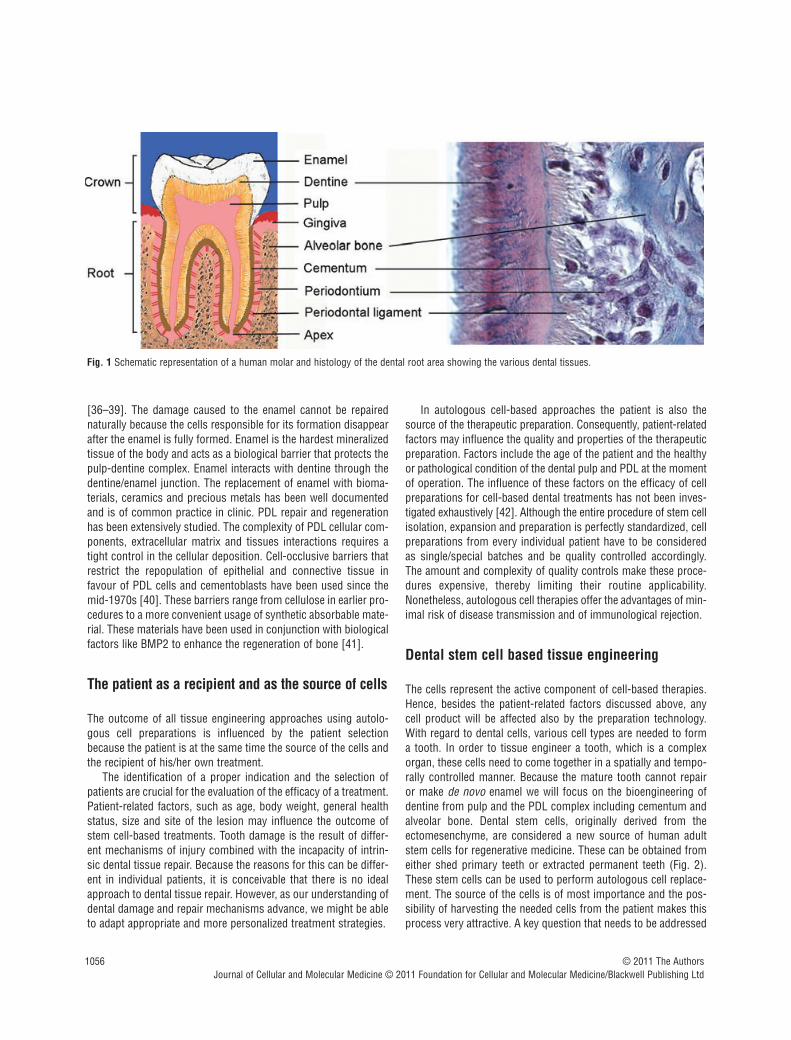
Fig. 1 Schematic representation of a human molar and histology of the dental root area showing the various dental tissues.

J. Cell. Mol. Med. Vol 15, No 5, 2011
1057© 2011 The AuthorsJournal of Cellular and Molecular Medicine © 2011 Foundation for Cellular and Molecular Medicine/Blackwell Publishing Ltd
is if these dental cells are indeed stem cells. Different studies haveprovided evidence that dental pulp and PDL cells have mesenchymalstem cell features, based on their ability to differentiate into carti-lage, bone, fat, muscle, muscle and neural tissue [43]. Apart fromthe dental pulp and PDL, mesenchymal stem cells have a diversedistribution in vivo as they can be derived from most, if not all,connective tissues including bone marrow, adipose, periosteum,synovial membrane, skeletal muscle, dermis, pericytes, blood, trabecular bone, human umbilical cord and lung [44].
Dental pulp stem cells (DPSCs)
The pulp has been long recognized as an organ with good repara-tive and regenerative capacity. Cells present in the dental pulp arecapable of terminally differentiate into odontoblast-like cells toform reparative dentine. Gene therapy approaches have beentested and demonstrated the higher odontogenic differentiationability of pulp cells transfected with growth/differentiation factor11 [45]. The use of the synthetic glucocorticoid dexamethasoneand growth factors like BMP2 to induce differentiation of pulp cellsinto odontoblast-like cells has been also examined [46, 47]. Adultdental pulp and the pulp of exfoliated deciduous human teeth havealso been identified as a potential stem cell source. DPSCs exhibita multipotent character and the potential to differentiate into chon-drocytes, adipocytes [48], osteoblasts/osteocytes [49, 50],myocytes [49], neuronal cells [51] and cardiomyocytes [52].DPSCs were firstly isolated from the human pulp tissue approxi-mately 10 years ago [53]. DPSCs were isolated from human adultthird molars with enzyme treatment of pulp tissues [53, 54]. Pulptissue from exfoliated deciduous human teeth was also used as asource of DPSCs [55]. These studies demonstrated that the den-tal pulp contains self-renewing, highly proliferative multipotentstem cells. It has been suggested that these cells reside withinperivascular niches [56, 57]. DPSCs are able to form a vascular-ized pulp-like tissue in vivo, which is surrounded by a layer ofodontoblast-like cells [53]. A special feature of DPSCs is theirpotential for odontoblastic differentiation, as characterized by
polarized cell bodies and accumulation of mineralized nodules [58,59]. Further studies showed that DPSCs do express several stemcell markers including c-kit, CD34, STRO-1 and CD146 [60, 61].However, the detection of other specific stem cell markersexpressed by dental pulp cells is under investigation. DPSCs havethe potential to form bone-like tissues when transplanted intoimmunocompromised animals [62]. Several DPSCs populationscan differentiate in vitro into adipocytes, neuronal-like cells andcardiomyocytes as defined by cell morphology and the expressionof respective gene markers [57, 63–65]. DPSCs can also undergoosteogenic, chondrogenic and myogenic differentiation in vitro[50, 66–68]. Recently, dental stem cells were shown to be repro-grammed into induced pluripotent stem cells (iPS) with a higherrate compared to other cell types of human origin tried so far [69].Furthermore, in vivo experimental evidence in animals suggeststhat DPSCs could provide a novel alternative cell population forrepair and/or regeneration of the heart [52], bone [62, 70], mus-cles [71], brain [51] and tooth [72–74]. Of note, last year the firstclinical application for alveolar bone reconstruction using DPSCswas successfully carried out in a patient [75].
The clinical application of DPSCs in regenerationof the pulp/dentin complex
Dental caries is a very common oral disease. It is essentially aninfection of the mineral tissues of the tooth, which eventuallyreaches the dental pulp of the tooth causing inflammation andpotentially tooth loss. The dental pulp has important functions toprovide nutrients, oxygen and nerve supply to the tooth. In addi-tion, a crucial property of the dental pulp is to foster cells of theimmune system that would tackle the infection, and to producereactionary or reparative dentin formation in response to externalstimuli, including bacterial invasion [76–78]. In clinical terms,regeneration of pulp is not yet a routine treatment modality inendodontics. Instead, entire pulp amputation is the choice of treat-ment, which is followed by the mechanical and chemical disinfec-tion pulp cavity and its filling with an artificial material. Despite
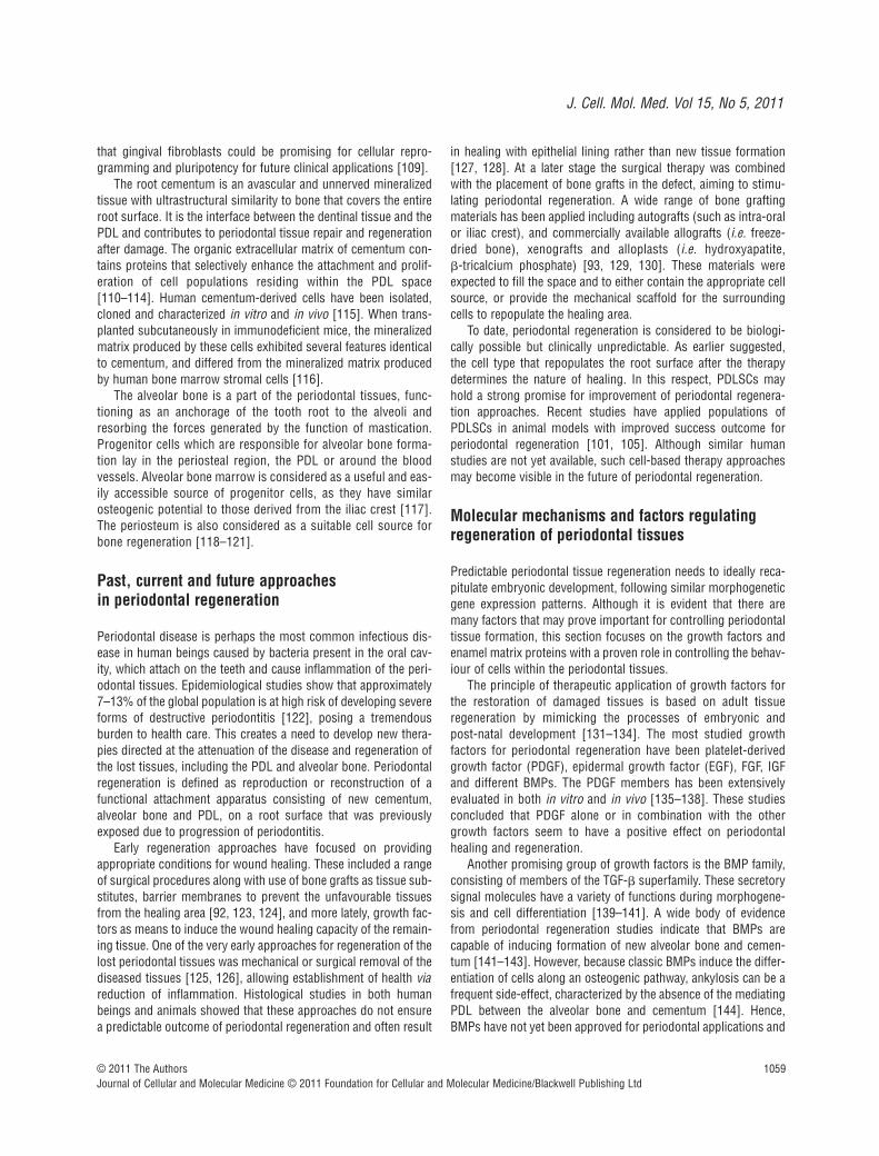
Fig. 2 The various steps leading to the formation of single cell colonies derived from human dental pulp.

1058 © 2011 The AuthorsJournal of Cellular and Molecular Medicine © 2011 Foundation for Cellular and Molecular Medicine/Blackwell Publishing Ltd
that the tooth is saved in its functional position, it is not vital anyfurther and could not fulfil to the full extent its role. The regenera-tion of the dentine-pulp interface would be the ideal therapy, aim-ing to re-build a dentinal bridge at the affected site, sealing off theinfection and other external stimuli. At present this is not yetachievable. There have been approaches to use growth factorswith the appropriate delivery systems, or other regenerative mate-rial, as agents for immediate capping of the exposed pulp [79–83].However, such situations require that the pulp has been exposedto a minimal extent, and inflammation is limited. This is hardly thecase in chronic pulpitis that is the most frequent development ofdeep carious lesions.
If regeneration of the dentine-pulp complex can be achieved,this would require the re-vascularization and re-innervation of thepulp and, importantly, the deposition of new dentine by odonto-blasts. The use of DPSCs holds a strong promise in this respect,as they can be isolated from the adult dental pulp tissue [53].When in contact with dentin in vitro, DPSCs can acquire odonto-blast-like cell morphology with a polarized cell body and a cellprocess extending into the existing dentinal tubules [54]. Thesecan be differentiated into odontoblasts that will form dentine [58,59, 84], endothelial cells that would support the re-vascularization[67, 85] and neurons which could support the re-innervation ofthe regenerated pulp tissue [63]. Transplantation of DPSCs inmice can regenerate pulp-dentine tissue complexes, with dentinaltubule-like structures [53, 86, 87]. A recent in vivo beagle dogstudy also demonstrated that a combination of calcium hydroxya-patite and DPSCs was able to regenerate dentin [88]. Althoughthese biological principles prove that pulp regeneration can beachieved via the DPSCs, further studies are required to investigatethe potential clinical application in human beings.
Periodontal tissues as a source and niche for stem cells
When considering dental stem cells and their niches, one has alsoto include the tissues surrounding the teeth. These are termed‘periodontal tissues’ or periodontium, and consist of the gingiva,the cementum of the tooth root, the surrounding alveolar bone, aswell as the interconnecting PDL.
Although the PDL is a physically small tissue, it is uniqueamong the various ligament and tendon systems of the body, inthe sense that it is the only soft tissue to span between two distinct hard tissues, namely the cementum of the root and thealveolar bone [89]. This tissue acts as a suspension for the tooth,adapting to the mechanical loads of the oral cavity. Collectively theperiodontal tissues are specifically tailored to support the tooth inits functional position. The pathological damage of the periodontaltissues due to a very common inflammatory condition is termedperiodontitis. This disease may eventually lead to exfoliation of theadult tooth, due to lack of supporting tissue. One of the majorchallenges in dentistry has been the regeneration of the disease-affected periodontal tissues, in a manner that recapitulates embry-onic tooth development.
Early observations indicated that the periodontal tissues have aregenerative capacity and that multipotent progenitor cells mayexist within the periodontium [40]. Extensive studies in experi-mental animal models have depleted the various periodontal tis-sues and investigated the regenerative capacities of the remainingtissues each time [90, 91]. It was concluded that the PDL tissuehas the regenerative capacity and can form not only the intercon-necting collagen tissue, but also alveolar bone and cementum[92–94]. From these early observations, it was concluded that thePDL tissue contains progenitor cells for fibroblasts, osteoblastsand cementoblasts [95]. Several studies demonstrated that PDLfrom mouse molars contains a slowly dividing population of pro-genitor cells that is located in perivascular sites [96, 97], as wellas in the periosteal and endosteal spaces of the alveolar bone [98].It was further demonstrated that PDL contains renewable and differentiated populations of cells [99, 100]. Similarly to the othertissues, PDL cells form a heterogeneous cell population com-posed predominantly of fibroblasts, but also of stem cells thatexhibit various developmental potentials. Recent advancement oftechnologies identifying and characterizing adult stem cells hasled to the first substantial evidence that a putative stem-cell pop-ulation exists within PDL. PDL stem cells (PDLSCs) were positivefor the stem cell markers STRO-1 and CD146 and were able to dif-ferentiate into cementoblasts, adipocytes and osteoblasts [101].After culture expansion, these human cells were transplanted intoimmunocompromised mice and were shown to contribute to peri-odontal tissue repair forming a PDL/cementum-like structure.Nevertheless, whether this tissue can function to the full extent asa normal PDL tissue is still under examination. Another study hasshown that cryopreserved PDL can retain stem cells-like charac-teristics, suggesting that long term storage is possible [102]. In vitro studies have also found that PDLSCs were capable of differentiating into mesodermal (i.e. adipocytes, osteoblasts,chondrocytes), ectodermal (i.e. neurons), and endodermal (i.e.hepatocytes) lineages [103]. In addition, rat PDLSCs have thecapacity to differentiate into vascular cells forming blood vessel-like structures [104]. These findings suggest that the PDL consti-tutes an important stem cell source not only for the regenerationof periodontal tissues [105], but also for the regeneration of othertissues and organs. Despite the strong promise that PDL holds forregenerative clinical dentistry, there is a however need for furthercharacterization of the PDLSCs.
Gingiva is the oral mucosal tissue that surrounds the teeth andcovers the alveolar bone. It functions as a protective mechanicalbarrier for the tooth supporting tissues and as a biological barrierconferring distinct immunity to oral infection. It is composed of anepithelial layer with an underlying vasculated connective tissue. Asthe epithelial layer has the capacity for continuous renewal, it isanticipated that this tissue could be a source of stem cells. Cellswith mesenchymal stem cell properties have recently been iso-lated from gingival tissues and characterized [106–108]. Thesecells have unique immunomodulatory functions, clonogenicity, aswell as self-renewal and multi-potent differentiation capacities.Very recently gingival fibroblasts have been evaluated as source ofiPS cells. These cells gave rise to high-quality iPS cells suggested
J. Cell. Mol. Med. Vol 15, No 5, 2011
1059© 2011 The AuthorsJournal of Cellular and Molecular Medicine © 2011 Foundation for Cellular and Molecular Medicine/Blackwell Publishing Ltd
that gingival fibroblasts could be promising for cellular repro-gramming and pluripotency for future clinical applications [109].
The root cementum is an avascular and unnerved mineralizedtissue with ultrastructural similarity to bone that covers the entireroot surface. It is the interface between the dentinal tissue and thePDL and contributes to periodontal tissue repair and regenerationafter damage. The organic extracellular matrix of cementum con-tains proteins that selectively enhance the attachment and prolif-eration of cell populations residing within the PDL space[110–114]. Human cementum-derived cells have been isolated,cloned and characterized in vitro and in vivo [115]. When trans-planted subcutaneously in immunodeficient mice, the mineralizedmatrix produced by these cells exhibited several features identicalto cementum, and differed from the mineralized matrix producedby human bone marrow stromal cells [116].
The alveolar bone is a part of the periodontal tissues, func-tioning as an anchorage of the tooth root to the alveoli andresorbing the forces generated by the function of mastication.Progenitor cells which are responsible for alveolar bone forma-tion lay in the periosteal region, the PDL or around the bloodvessels. Alveolar bone marrow is considered as a useful and eas-ily accessible source of progenitor cells, as they have similarosteogenic potential to those derived from the iliac crest [117].The periosteum is also considered as a suitable cell source forbone regeneration [118–121].
Past, current and future approaches in periodontal regeneration
Periodontal disease is perhaps the most common infectious dis-ease in human beings caused by bacteria present in the oral cav-ity, which attach on the teeth and cause inflammation of the peri-odontal tissues. Epidemiological studies show that approximately7–13% of the global population is at high risk of developing severeforms of destructive periodontitis [122], posing a tremendousburden to health care. This creates a need to develop new thera-pies directed at the attenuation of the disease and regeneration ofthe lost tissues, including the PDL and alveolar bone. Periodontalregeneration is defined as reproduction or reconstruction of afunctional attachment apparatus consisting of new cementum,alveolar bone and PDL, on a root surface that was previouslyexposed due to progression of periodontitis.
Early regeneration approaches have focused on providingappropriate conditions for wound healing. These included a rangeof surgical procedures along with use of bone grafts as tissue sub-stitutes, barrier membranes to prevent the unfavourable tissuesfrom the healing area [92, 123, 124], and more lately, growth fac-tors as means to induce the wound healing capacity of the remain-ing tissue. One of the very early approaches for regeneration of thelost periodontal tissues was mechanical or surgical removal of thediseased tissues [125, 126], allowing establishment of health viareduction of inflammation. Histological studies in both humanbeings and animals showed that these approaches do not ensurea predictable outcome of periodontal regeneration and often result
in healing with epithelial lining rather than new tissue formation[127, 128]. At a later stage the surgical therapy was combinedwith the placement of bone grafts in the defect, aiming to stimu-lating periodontal regeneration. A wide range of bone graftingmaterials has been applied including autografts (such as intra-oralor iliac crest), and commercially available allografts (i.e. freeze-dried bone), xenografts and alloplasts (i.e. hydroxyapatite, �-tricalcium phosphate) [93, 129, 130]. These materials wereexpected to fill the space and to either contain the appropriate cellsource, or provide the mechanical scaffold for the surroundingcells to repopulate the healing area.
To date, periodontal regeneration is considered to be biologi-cally possible but clinically unpredictable. As earlier suggested,the cell type that repopulates the root surface after the therapydetermines the nature of healing. In this respect, PDLSCs mayhold a strong promise for improvement of periodontal regenera-tion approaches. Recent studies have applied populations ofPDLSCs in animal models with improved success outcome forperiodontal regeneration [101, 105]. Although similar humanstudies are not yet available, such cell-based therapy approachesmay become visible in the future of periodontal regeneration.
Molecular mechanisms and factors regulatingregeneration of periodontal tissues
Predictable periodontal tissue regeneration needs to ideally reca-pitulate embryonic development, following similar morphogeneticgene expression patterns. Although it is evident that there aremany factors that may prove important for controlling periodontaltissue formation, this section focuses on the growth factors andenamel matrix proteins with a proven role in controlling the behav-iour of cells within the periodontal tissues.
The principle of therapeutic application of growth factors forthe restoration of damaged tissues is based on adult tissueregeneration by mimicking the processes of embryonic andpost-natal development [131–134]. The most studied growthfactors for periodontal regeneration have been platelet-derivedgrowth factor (PDGF), epidermal growth factor (EGF), FGF, IGFand different BMPs. The PDGF members has been extensivelyevaluated in both in vitro and in vivo [135–138]. These studiesconcluded that PDGF alone or in combination with the othergrowth factors seem to have a positive effect on periodontalhealing and regeneration.
Another promising group of growth factors is the BMP family,consisting of members of the TGF-� superfamily. These secretorysignal molecules have a variety of functions during morphogene-sis and cell differentiation [139–141]. A wide body of evidencefrom periodontal regeneration studies indicate that BMPs arecapable of inducing formation of new alveolar bone and cemen-tum [141–143]. However, because classic BMPs induce the differ-entiation of cells along an osteogenic pathway, ankylosis can be afrequent side-effect, characterized by the absence of the mediatingPDL between the alveolar bone and cementum [144]. Hence,BMPs have not yet been approved for periodontal applications and
1060 © 2011 The AuthorsJournal of Cellular and Molecular Medicine © 2011 Foundation for Cellular and Molecular Medicine/Blackwell Publishing Ltd
further experimentation is needed for improving the capacity forperiodontal regeneration.
Amelogenins are proteins secreted by ameloblasts and areconsidered to play a major role in regulating formation of enamel[145]. The rationale for the potential use of enamel proteins inperiodontal tissue regeneration therapies is justified by their pres-ence in initial cementum formation, during normal development ofthe tooth attachment apparatus [146, 147]. The commerciallyavailable enamel matrix protein product namely, Emdogain(Straumann AG, Basel, Switzerland), has been in the market forover a decade now [148, 149]. This is a purified acid extract ofproteins containing predominantly amelogenin from pig enamelmatrix. A large number of studies have investigated the mecha-nisms of action and clinical efficiency of Emdogain [131, 146,150–153]. Still, despite the encouraging clinical outcome, themechanism of action of Emdogain is not clear. It has been sug-gested that its capacity to initiate the processes of periodontalregeneration relies on the recruitment of cementoblasts onto theroot surface, hence stimulating the formation of root cementum.Several in vitro studies suggest that Emdogain could act as a sig-nalling molecule for epithelial–mesenchymal interactions, whichcan regulate the activity of follicle cells, PDL cells, odontoblasts,gingival fibroblasts and cementoblasts [142, 154–159]. Alongwith clinical and experimental animal confirming the clinical efficiency of Emdogain in periodontal regeneration procedures[158, 160, 161], recent systematic reviews are also in support ofthese findings [162–164].
Taken together, these molecular factors are likely to con-tribute to the regeneration of the periodontal tissues by targetingperiodontal progenitors that reside within putative stem cellniches of the periodontium. The potential effects include prolif-eration and differentiation along pathways parallel to tooth rootdevelopment. Nevertheless, this should not be over-interpreted.One should consider the multiplicity of the cells and complexityof tissues within the periodontal environment, their responsive-ness of cells to the type or dose of the growth factors used, aswell as the previously diseased tissue substrate due to inflam-mation. Therefore, as much as an identical recapitulation of
developmental events is desirable, it may be very difficult toachieve due to these reasons.
Further studies will be still needed to identify right cell pop-ulations (progenitor or stem cells), signalling molecules thatcontrol cell behaviour and scaffold material to act as carriers forthe cells and stimulating molecules (i.e. bioreactors). Oncethese parameters are more clearly defined and the respectivewound healing events are clarified, the clinical efficiency of suit-able treatment modalities would need to be further assessed.Moreover, as the oral microenvironment is not aseptic, any periodontal regenerative activity involving differentiation ofpotent stem cells is likely face bacterial challenge. Therefore, itis suggested that any regenerative approach involving stemcells in the oral cavity would need to take this aspect under consideration, in order to achieve predictable and optimal therapeutic outcome.
Scaffolding and material science
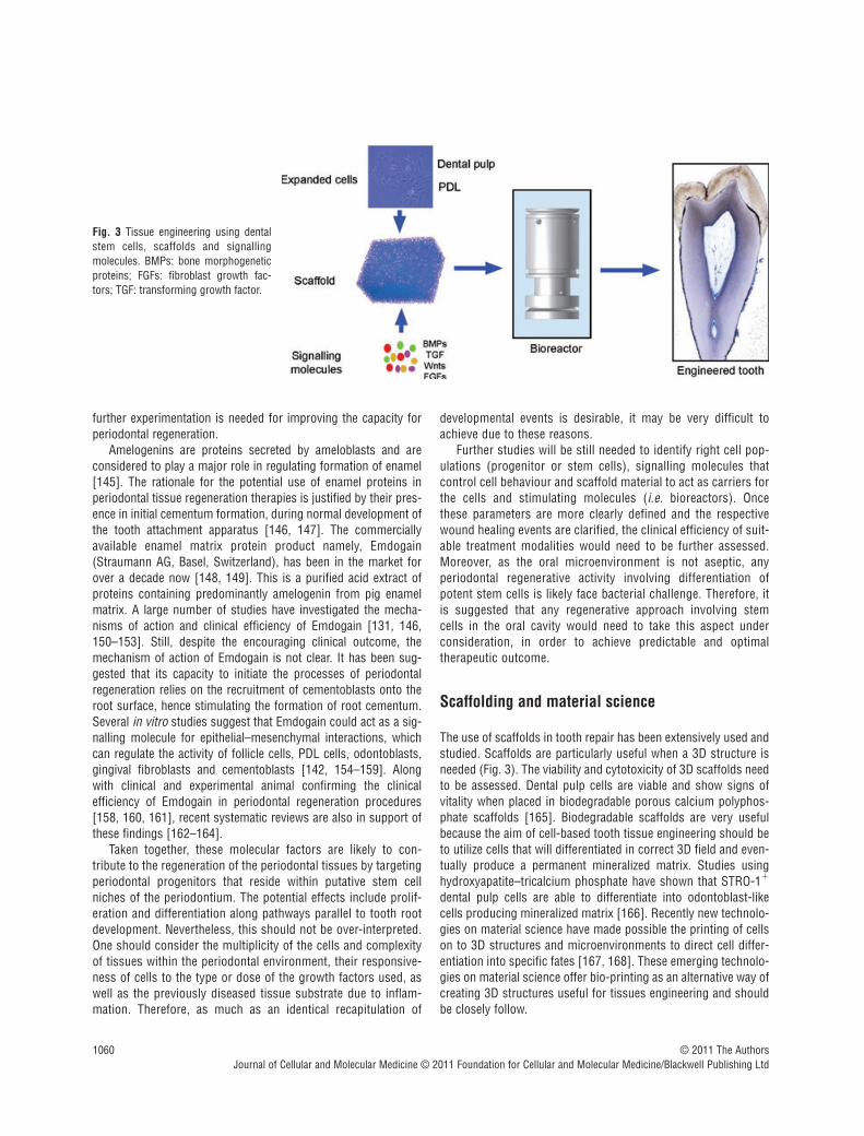
The use of scaffolds in tooth repair has been extensively used andstudied. Scaffolds are particularly useful when a 3D structure isneeded (Fig. 3). The viability and cytotoxicity of 3D scaffolds needto be assessed. Dental pulp cells are viable and show signs ofvitality when placed in biodegradable porous calcium polyphos-phate scaffolds [165]. Biodegradable scaffolds are very usefulbecause the aim of cell-based tooth tissue engineering should beto utilize cells that will differentiated in correct 3D field and even-tually produce a permanent mineralized matrix. Studies usinghydroxyapatite–tricalcium phosphate have shown that STRO-1�
dental pulp cells are able to differentiate into odontoblast-likecells producing mineralized matrix [166]. Recently new technolo-gies on material science have made possible the printing of cellson to 3D structures and microenvironments to direct cell differ-entiation into specific fates [167, 168]. These emerging technolo-gies on material science offer bio-printing as an alternative way ofcreating 3D structures useful for tissues engineering and shouldbe closely follow.
Fig. 3 Tissue engineering using dentalstem cells, scaffolds and signalling molecules. BMPs: bone morphogeneticproteins; FGFs: fibroblast growth fac-tors; TGF: transforming growth factor.
J. Cell. Mol. Med. Vol 15, No 5, 2011
1061© 2011 The AuthorsJournal of Cellular and Molecular Medicine © 2011 Foundation for Cellular and Molecular Medicine/Blackwell Publishing Ltd
Conclusions
Although the prospective of tooth tissue engineering is veryattractive, we are far from performing routine clinical procedures.Despite the large amount of interest in this field, no clinical trialshave been performed for dentine repair and very limited clinicalapplications are available in periodontal disease treatment. Cellbased bioengineering and material sciences have to define condi-tions for manufacturing consistent and reproducible products,which are quality controlled for safety and efficacy. In addition, celltherapies are in their infancy and many issues need to be takinginto account. The use of culture expanded cell populations needsto take into account the possibility of genetic and epigenetic insta-bility, which could possibly result in malignant transformation.The paracrine effects, interactions with the host and immuneresponse following cell transplantation also need to be taking intoconsideration. For example, the transplantation of bone marrowstem cells to cure diabetes mellitus in an animal model resulted inthe reduction of hyperglycaemia by the regeneration of the recipi-ent’s own pancreatic cells. This regeneration was initiated in
response to the transplanted cells [169]. In the case of tooth engi-neering, the possibility of autologous cell replacement and theusage of cells naturally occurring in the site of injury may mini-mize the risk of side effects. In addition, a better understanding ofthe biology of tooth repair opens the exciting prospect to developcell free approaches. The utilization of bioactive factors and bio-materials will support and enhance the intrinsic mechanisms oftooth repair.
Acknowledgements
N.B. was supported by a grant from the University of Zurich (Switzerland).C.D.B. is a Fellow of the Medical Research Council, UK.
Conflict of interest
The authors confirm that there are no conflicts of interest.
References
1. Murray PE, Garcia-Godoy F. The outlookfor implants and endodontics: a review ofthe tissue engineering strategies to createreplacement teeth for patients. Dent ClinNorth Am. 2006; 50: 299–315, x.
2. Ring ME. A thousand years of dentalimplants: a definitive history–part 2.Compend Contin Educ Dent. 1995; 16:1132, 4, 6 passim.
3. Taba M, Jin Q, Sugai JV, et al. Currentconcepts in periodontal bioengineering.Orthod Craniofac Res. 2005; 8: 292–302.
4. Mitsiadis TA, De Bari C, About I. Apoptosisin developmental and repair-related humantooth remodeling: a view from the inside.Exp Cell Res. 2008; 314: 869–77.
5. Naughton GK. From lab bench to market:critical issues in tissue engineering. AnnNY Acad Sci. 2002; 961: 372–85.
6. Quarto R, Mastrogiacomo M, CanceddaR, et al. Repair of large bone defects withthe use of autologous bone marrow stro-mal cells. N Engl J Med. 2001; 344: 385–6.
7. Vacanti CA, Bonassar LJ, Vacanti MP, et al. Replacement of an avulsed phalanxwith tissue-engineered bone. N Engl JMed. 2001; 344: 1511–4.
8. Brittberg M, Lindahl A, Nilsson A, et al.Treatment of deep cartilage defects in theknee with autologous chondrocyte trans-plantation. N Engl J Med. 1994; 331:889–95.
9. Stamm C, Westphal B, Kleine HD, et al.Autologous bone-marrow stem-cell trans-plantation for myocardial regeneration.Lancet. 2003; 361: 45–6.
10. Wollert KC, Meyer GP, Lotz J, et al.Intracoronary autologous bone-marrowcell transfer after myocardial infarction: theBOOST randomised controlled clinicaltrial. Lancet. 2004; 364: 141–8.
11. Bjorklund A. Cell therapy for Parkinson’sdisease: problems and prospects. NovartisFound Symp. 2005; 265: 174–86.
12. Lindvall O, Kokaia Z, Martinez-SerranoA. Stem cell therapy for human neurode-generative disorders-how to make it work.Nat Med. 2004; 10: S42–50.
13. Bluteau G, Luder HU, De Bari C, et al.Stem cells for tooth engineering. Eur CellMater. 2008; 16: 1–9.
14. Kratochwil K, Dull M, Farinas I, et al.Lef1 expression is activated by BMP-4 andregulates inductive tissue interactions intooth and hair development. Genes Dev.1996; 10: 1382–94.
15. Nadiri A, Kuchler-Bopp S, Haikel Y, et al.Immunolocalization of BMP-2/-4, FGF-4,and WNT10b in the developing mouse firstlower molar. J Histochem Cytochem.2004; 52: 103–12.
16. Vainio S, Karavanova I, Jowett A, et al. Identification of BMP-4 as a signalmediating secondary induction between
epithelial and mesenchymal tissues duringearly tooth development. Cell. 1993; 75:45–58.
17. Bei M, Maas R. FGFs and BMP4 induceboth Msx1-independent and Msx1-dependent signaling pathways in earlytooth development. Development. 1998;125: 4325–33.
18. Kettunen P, Laurikkala J, Itaranta P, et al.Associations of FGF-3 and FGF-10 withsignaling networks regulating tooth mor-phogenesis. Dev Dyn. 2000; 219: 322–32.
19. Kettunen P, Thesleff I. Expression andfunction of FGFs-4, -8, and -9 suggestfunctional redundancy and repetitive useas epithelial signals during tooth morpho-genesis. Dev Dyn. 1998; 211: 256–68.
20. Dassule HR, McMahon AP. Analysis ofepithelial-mesenchymal interactions in theinitial morphogenesis of the mammaliantooth. Dev Biol. 1998; 202: 215–27.
21. Khan M, Seppala M, Zoupa M, et al.Hedgehog pathway gene expression dur-ing early development of the molar toothroot in the mouse. Gene Expr Patterns.2007; 7: 239–43.
22. Mitsiadis TA, Smith MM. How do genesmake teeth to order through development?J Exp Zool B Mol Dev Evol. 2006; 306:177–82.
23. Cobourne MT, Mitsiadis T. Neural crestcells and patterning of the mammalian

1062 © 2011 The AuthorsJournal of Cellular and Molecular Medicine © 2011 Foundation for Cellular and Molecular Medicine/Blackwell Publishing Ltd
dentition. J Exp Zool B Mol Dev Evol. 2006;306: 251–60.
24. Caton J, Tucker AS. Current knowledge oftooth development: patterning and miner-alization of the murine dentition. J Anat.2009; 214: 502–15.
25. Thesleff I. Epithelial-mesenchymal sig-nalling regulating tooth morphogenesis. JCell Sci. 2003; 116: 1647–8.
26. Thesleff I. The genetic basis of toothdevelopment and dental defects. Am JMed Genet A. 2006; 140: 2530–5.
27. Thesleff I, Sharpe P. Signalling networksregulating dental development. Mech Dev.1997; 67: 111–23.
28. Tucker A, Sharpe P. The cutting-edge ofmammalian development; how the embryomakes teeth. Nat Rev Genet. 2004; 5:499–508.
29. Mitsiadis TA, Graf D. Cell fate determina-tion during tooth development and regen-eration. Birth Defects Res C EmbryoToday. 2009; 87: 199–211.
30. Thesleff I, Jarvinen E, Suomalainen M.Affecting tooth morphology and renewalby fine-tuning the signals mediating celland tissue interactions. Novartis FoundSymp. 2007; 284: 142–53; discussion53–63.
31. Thesleff I, Wang XP, Suomalainen M.Regulation of epithelial stem cells in toothregeneration. C R Biol. 2007; 330: 561–4.
32. Diekwisch TG. The developmental biologyof cementum. Int J Dev Biol. 2001; 45:695–706.
33. Sonoyama W, Seo BM, Yamaza T, et al.Human Hertwig’s epithelial root sheathcells play crucial roles in cementum for-mation. J Dent Res. 2007; 86: 594–9.
34. Yokohama-Tamaki T, Ohshima H,Fujiwara N, et al. Cessation of Fgf10 signaling, resulting in a defective dentalepithelial stem cell compartment, leads tothe transition from crown to root forma-tion. Development. 2006; 133: 1359–66.
35. Mariotti A. The extracellular matrix of theperiodontium: dynamic and interactive tis-sues. Periodontol 2000. 1993; 3: 39–63.
36. Mitsiadis TA, Rahiotis C. Parallelsbetween tooth development and repair:conserved molecular mechanisms follow-ing carious and dental injury. J Dent Res.2004; 83: 896–902.
37. Smith AJ, Lesot H. Induction and regula-tion of crown dentinogenesis: embryonicevents as a template for dental tissuerepair? Crit Rev Oral Biol Med. 2001; 12:425–37.
38. Smith AJ, Matthews JB, Hall RC.Transforming growth factor-beta1 (TGF-
beta1) in dentine matrix. Ligand activationand receptor expression. Eur J Oral Sci.1998; 106: 179–84.
39. Tziafas D, Smith AJ, Lesot H. Designingnew treatment strategies in vital pulp ther-apy. J Dent. 2000; 28: 77–92.
40. Melcher AH. On the repair potential ofperiodontal tissues. J Periodont. 1976; 47:256–60.
41. Lee YM, Nam SH, Seol YJ, et al.Enhanced bone augmentation by con-trolled release of recombinant human bonemorphogenetic protein-2 from bioab-sorbable membranes. J Periodont. 2003;74: 865–72.
42. De Bari C, Pitzalis C, Dell’Accio F.Reparative medicine: from tissue engineer-ing to joint surface regeneration. RegenMed. 2006; 1: 59–69.
43. Karaoz E, Dogan BN, Aksoy A, et al.Isolation and in vitro characterisation ofdental pulp stem cells from natal teeth.Histochem Cell Biol. 2010; 133: 95–112.
44. Rosenbaum AJ, Grande DA, Dines JS.The use of mesenchymal stem cells in tis-sue engineering: a global assessment.Organogenesis. 2008; 4: 23–7.
45. Nakashima M, Iohara K, Ishikawa M, et al. Stimulation of reparative dentin formation by ex vivo gene therapy usingdental pulp stem cells electrotransfectedwith growth/differentiation factor 11(Gdf11). Human Gene Ther. 2004; 15:1045–53.
46. Alliot-Licht B, Bluteau G, Magne D, et al.Dexamethasone stimulates differentiationof odontoblast-like cells in human dentalpulp cultures. Cell Tissue Res. 2005; 321:391–400.
47. Iohara K, Nakashima M, Ito M, et al.Dentin regeneration by dental pulp stemcell therapy with recombinant human bonemorphogenetic protein 2. J Dent Res.2004; 83: 590–5.
48. Waddington RJ, Youde SJ, Lee CP, et al.Isolation of distinct progenitor stem cellpopulations from dental pulp. CellsTissues Organs. 2009; 189: 268–74.
49. de Mendonca Costa A, Bueno DF, MartinsMT, et al. Reconstruction of large cranialdefects in nonimmunosuppressed experi-mental design with human dental pulpstem cells. J Craniofac Surg. 2008; 19:204–10.
50. Carinci F, Papaccio G, Laino G, et al.Comparison between genetic portraits ofosteoblasts derived from primary culturesand osteoblasts obtained from human pul-par stem cells. J Craniofac Surg. 2008; 19:616–25; discussion 26–7.
51. Nosrat IV, Widenfalk J, Olson L, et al.Dental pulp cells produce neurotrophicfactors, interact with trigeminal neurons invitro, and rescue motoneurons after spinalcord injury. Dev Biol. 2001; 238: 120–32.
52. Gandia C, Arminan A, Garcia-VerdugoJM, et al. Human dental pulp stem cellsimprove left ventricular function, induceangiogenesis, and reduce infarct size inrats with acute myocardial infarction.Stem Cells. 2008; 26: 638–45.
53. Gronthos S, Mankani M, Brahim J, et al.Postnatal human dental pulp stem cells(DPSCs) in vitro and in vivo. Proc NatlAcad Sci USA. 2000; 97: 13625–30.
54. Huang GT, Shagramanova K, Chan SW.Formation of odontoblast-like cells fromcultured human dental pulp cells on dentinin vitro. J Endod. 2006; 32: 1066–73.
55. Miura M, Gronthos S, Zhao M, et al.SHED: stem cells from human exfoliateddeciduous teeth. Proc Natl Acad Sci USA.2003; 100: 5807–12.
56. Shi S, Gronthos S. Perivascular niche ofpostnatal mesenchymal stem cells inhuman bone marrow and dental pulp. JBone Miner Res. 2003; 18: 696–704.
57. Gronthos S, Brahim J, Li W, et al. Stemcell properties of human dental pulp stemcells. J Dent Res. 2002; 81: 531–5.
58. About I, Bottero MJ, de Denato P, et al.Human dentin production in vitro. Exp CellRes. 2000; 258: 33–41.
59. Couble ML, Farges JC, Bleicher F, et al.Odontoblast differentiation of human den-tal pulp cells in explant cultures. CalcifTissue Int. 2000; 66: 129–38.
60. Laino G, Graziano A, d’Aquino R, et al.An approachable human adult stem cellsource for hard-tissue engineering. J CellPhysiol. 2006; 206: 693–701.
61. Menicanin D, Bartold PM, Zannettino AC,et al. Identification of a common geneexpression signature associated with imma-ture clonal mesenchymal cell populationsderived from bone marrow and dental tis-sues. Stem Cells Dev. 2010; 19: 1501–10.
62. Graziano A, d’Aquino R, Laino G, et al.Human CD34� stem cells produce bonenodules in vivo. Cell Prolif. 2008; 41: 1–11.
63. Arthur A, Rychkov G, Shi S, et al. Adulthuman dental pulp stem cells differentiatetoward functionally active neurons underappropriate environmental cues. StemCells. 2008; 26: 1787–95.
64. Arminan A, Gandia C, Bartual M, et al.Cardiac differentiation is driven by NKX2.5and GATA4 nuclear translocation in tissue-specific mesenchymal stem cells. StemCells Dev. 2009; 18: 907–18.

J. Cell. Mol. Med. Vol 15, No 5, 2011
1063© 2011 The AuthorsJournal of Cellular and Molecular Medicine © 2011 Foundation for Cellular and Molecular Medicine/Blackwell Publishing Ltd
65. Balic A, Aguila HL, Caimano MJ, et al.Characterization of stem and progenitorcells in dental pulps of the erupted andunerupted murine molars. Bone. 2010; 46:1639–51.
66. Laino G, d’Aquino R, Graziano A, et al. Anew population of human adult dental pulpstem cells: a useful source of living autol-ogous fibrous bone tissue (LAB). J BoneMiner Res. 2005; 20: 1394–402.
67. d’Aquino R, Graziano A, Sampaolesi M,et al. Human postnatal dental pulp cellsco-differentiate into osteoblasts andendotheliocytes: a pivotal synergy leadingto adult bone tissue formation. Cell DeathDiffer. 2007; 14: 1162–71.
68. Zhang W, Walboomers XF, Shi S, et al.Multilineage differentiation potential ofstem cells derived from human dental pulpafter cryopreservation. Tissue Eng. 2006;12: 2813–23.
69. Yan X, Qin H, Qu C, et al. iPS cells reprogrammed from mesenchymal-likestem/progenitor cells of dental tissueorigin. Stem Cells Dev. 2009; 19: 469–80.
70. Graziano A, d’Aquino R, Laino G, et al.Dental pulp stem cells: a promising tool forbone regeneration. Stem Cell Rev. 2008; 4:21–6.
71. Kerkis I, Ambrosio CE, Kerkis A, et al.Early transplantation of human immaturedental pulp stem cells from baby teeth togolden retriever muscular dystrophy(GRMD) dogs: local or systemic? J TranslMed. 2008; 6: 35.
72. Onyekwelu O, Seppala M, Zoupa M, et al. Tooth development: 2. Regeneratingteeth in the laboratory. Dent Update. 2007;34: 20–2, 5–6, 9.
73. Cordeiro MM, Dong Z, Kaneko T, et al.Dental pulp tissue engineering with stemcells from exfoliated deciduous teeth. J Endod. 2008; 34: 962–9.
74. Nedel F, Andre Dde A, de Oliveira IO, et al.Stem cells: therapeutic potential in dentistry.J Contemp Dent Pract. 2009; 10: 90–6.
75. d’Aquino R, De Rosa A, Lanza V, et al.Human mandible bone defect repair by thegrafting of dental pulp stem/progenitorcells and collagen sponge biocomplexes.Eur Cell Mater. 2009; 18: 75–83.
76. Bergenholtz G. Evidence for bacterial cau-sation of adverse pulpal responses inresin-based dental restorations. Crit RevOral Biol Med. 2000; 11: 467–80.
77. Bjorndal L. Presence or absence of tertiarydentinogenesis in relation to caries pro-gression. Adv Dent Res. 2001; 15: 80–3.
78. Durand SH, Flacher V, Romeas A, et al.Lipoteichoic acid increases TLR and func-
tional chemokine expression while reduc-ing dentin formation in in vitro differenti-ated human odontoblasts. J Immunol.2006; 176: 2880–7.
79. Tziafas D, Belibasakis G, Veis A, et al.Dentin regeneration in vital pulp therapy:design principles. Adv Dent Res. 2001; 15:96–100.
80. Tziafas D, Pantelidou O, Alvanou A, et al.The dentinogenic effect of mineral trioxideaggregate (MTA) in short-term cappingexperiments. Int Endod J. 2002; 35:245–54.
81. Rutherford RB. BMP-7 gene transfer toinflamed ferret dental pulps. Eur J Oral Sci.2001; 109: 422–4.
82. Sloan AJ, Rutherford RB, Smith AJ.Stimulation of the rat dentine-pulp com-plex by bone morphogenetic protein-7 in vitro. Arch Oral Biol. 2000; 45: 173–7.
83. Goldberg M, Six N, Decup F, et al.Bioactive molecules and the future of pulptherapy. Am J Dent. 2003; 16: 66–76.
84. Yang X, van der Kraan PM, van denDolder J, et al. STRO-1 selected rat den-tal pulp stem cells transfected with aden-oviral-mediated human bone morpho-genetic protein 2 gene show enhancedodontogenic differentiation. Tissue Eng.2007; 13: 2803–12.
85. Nakashima M, Iohara K, Sugiyama M.Human dental pulp stem cells with highlyangiogenic and neurogenic potential forpossible use in pulp regeneration.Cytokine Growth Factor Rev. 2009; 20:435–40.
86. Batouli S, Miura M, Brahim J, et al.Comparison of stem-cell-mediated osteo-genesis and dentinogenesis. J Dent Res.2003; 82: 976–81.
87. Iohara K, Zheng L, Ito M, et al.Regeneration of dental pulp after pulpo-tomy by transplantation of CD31(-)/CD146(-) side population cells from acanine tooth. Regen Med. 2009; 4: 377–85.
88. Ji YM, Jeon SH, Park JY, et al. Dentalstem cell therapy with calcium hydroxidein dental pulp capping. Tissue Eng Part A.2010; 16: 1823–33.
89. McCulloch CA, Lekic P, McKee MD. Roleof physical forces in regulating the formand function of the periodontal ligament.Periodontol 2000. 2000; 24: 56–72.
90. Karring T, Nyman S, Lindhe J. Healingfollowing implantation of periodontitisaffected roots into bone tissue. J ClinPeriodontol. 1980; 7: 96–105.
91. Nyman S, Karring T, Lindhe J, et al.Healing following implantation of peri-odontitis-affected roots into gingival
connective tissue. J Clin Periodontol.1980; 7: 394–401.
92. Nyman S, Gottlow J, Karring T, et al. Theregenerative potential of the periodontalligament. An experimental study in themonkey. J Clin Periodontol. 1982; 9:257–65.
93. Nielsen IM, Ellegaard B, Karring T.Kielbone in healing interradicular lesions inmonkeys. J Periodontal Res. 1980; 15:328–37.
94. Parlar A, Bosshardt DD, Unsal B, et al.New formation of periodontal tissuesaround titanium implants in a novel dentinchamber model. Clin Oral Implants Res.2005; 16: 259–67.
95. Gottlow J, Nyman S, Karring T, et al.New attachment formation as the result ofcontrolled tissue regeneration. J ClinPeriodontol. 1984; 11: 494–503.
96. Gould TR, Melcher AH, Brunette DM.Migration and division of progenitor cellpopulations in periodontal ligament afterwounding. J Periodontal Res. 1980; 15:20–42.
97. McCulloch CA. Progenitor cell populationsin the periodontal ligament of mice. AnatRec. 1985; 211: 258–62.
98. Aukhil I, Nishimura K, Fernyhough W.Experimental regeneration of the periodon-tium. Crit Rev Oral Biol Med. 1990; 1:101–15.
99. Davidson D, McCulloch CA. Proliferativebehavior of periodontal ligament cell pop-ulations. J Periodontal Res. 1986; 21:414–28.
100. Nemeth E, Kulkarni GW, McCulloch CA.Disturbances of gingival fibroblast popula-tion homeostasis due to experimentallyinduced inflammation in the cynomolgusmonkey (Macaca fascicularis): potentialmechanism of disease progression. JPeriodontal Res. 1993; 28: 180–90.
101. Seo BM, Miura M, Gronthos S, et al.Investigation of multipotent postnatal stemcells from human periodontal ligament.Lancet. 2004; 364: 149–55.
102. Seo BM, Miura M, Sonoyama W, et al.Recovery of stem cells from cryopre-served periodontal ligament. J Dent Res.2005; 84: 907–12.
103. Kawanabe N, Murata S, Murakami K, et al. Isolation of multipotent stem cells in human periodontal ligament usingstage-specific embryonic antigen-4.Differentiation. 2010; 79: 74–83.
104. Okubo N, Ishisaki A, Iizuka T, et al.Vascular cell-like potential of undifferenti-ated ligament fibroblasts to construct vas-cular cell-specific marker-positive blood

1064 © 2011 The AuthorsJournal of Cellular and Molecular Medicine © 2011 Foundation for Cellular and Molecular Medicine/Blackwell Publishing Ltd
vessel structures in a PI3K activation-dependent manner. J Vasc Res. 2010; 47:369–83.
105. Kim SH, Kim KH, Seo BM, et al. Alveolarbone regeneration by transplantation ofperiodontal ligament stem cells and bonemarrow stem cells in a canine peri-implantdefect model: a pilot study. J Periodont.2009; 80: 1815–23.
106. Zhang Q, Shi S, Liu Y, et al. Mesenchymalstem cells derived from human gingiva arecapable of immunomodulatory functionsand ameliorate inflammation-related tissuedestruction in experimental colitis. JImmunol. 2009; 183: 7787–98.
107. Zhang QZ, Su WR, Shi SH, et al. Humangingiva-derived mesenchymal stem cellselicit polarization of M2 macrophages andenhance cutaneous wound healing. StemCells. 2010; 28: 1856–68.
108. Tomar GB, Srivastava RK, Gupta N, et al.Human gingiva-derived mesenchymal stemcells are superior to bone marrow-derivedmesenchymal stem cells for cell therapy inregenerative medicine. Biochem BiophysRes Commun. 2010; 393: 377–83.
109. Egusa H, Okita K, Kayashima H, et al.Gingival fibroblasts as a promising sourceof induced pluripotent stem cells. PLoSONE. 5: e12743.
110. Wu D, Ikezawa K, Parker T, et al.Characterization of a collagenous cemen-tum-derived attachment protein. J BoneMiner Res. 1996; 11: 686–92.
111. Pitaru S, Savion N, Hekmati H, et al.Molecular and cellular interactions of acementum attachment protein with peri-odontal cells and cementum matrix compo-nents. J Periodontal Res. 1993; 28: 560–2.
112. Liu HW, Yacobi R, Savion N, et al. A col-lagenous cementum-derived attachmentprotein is a marker for progenitors of themineralized tissue-forming cell lineage ofthe periodontal ligament. J Bone MinerRes. 1997; 12: 1691–9.
113. Yonemura K, Raines EW, Ahn NG, et al. Mitogenic signaling mechanisms ofhuman cementum-derived growth factors.J Biol Chem. 1993; 268: 26120–6.
114. MacNeil RL, Somerman MJ. Molecularfactors regulating development and regen-eration of cementum. J Periodontal Res.1993; 28: 550–9.
115. Grzesik WJ, Kuzentsov SA, Uzawa K, et al. Normal human cementum-derivedcells: isolation, clonal expansion, and in vitroand in vivo characterization. J Bone MinerRes. 1998; 13: 1547–54.
116. Grzesik WJ, Cheng H, Oh JS, et al.Cementum-forming cells are phenotypi-
cally distinct from bone-forming cells. J Bone Miner Res. 2000; 15: 52–9.
117. Matsubara T, Suardita K, Ishii M, et al.Alveolar bone marrow as a cell source for regenerative medicine: differencesbetween alveolar and iliac bone marrowstromal cells. J Bone Miner Res. 2005; 20:399–409.
118. Yamamiya K, Okuda K, Kawase T, et al.Tissue-engineered cultured periosteumused with platelet-rich plasma and hydrox-yapatite in treating human osseousdefects. J Periodontol. 2008; 79: 811–8.
119. Breitbart AS, Grande DA, Kessler R, et al. Tissue engineered bone repair of calvarial defects using cultured periostealcells. Plast Reconstr Surg. 1998; 101:567–74; discussion 75–6.
120. Mizuno H, Hata K, Kojima K, et al. Anovel approach to regenerating periodon-tal tissue by grafting autologous culturedperiosteum. Tissue Eng. 2006; 12:1227–335.
121. Mase J, Mizuno H, Okada K, et al.Cryopreservation of cultured periosteum:effect of different cryoprotectants and pre-incubation protocols on cell viability andosteogenic potential. Cryobiology. 2006;52: 182–92.
122. Albandar JM, Rams TE. Global epidemiol-ogy of periodontal diseases: an overview.Periodontol 2000. 2002; 29: 7–10.
123. Karring T, Cortellini P. Regenerative therapy: furcation defects. Periodontol2000. 1999; 19: 115–37.
124. Laurell L, Bose M, Graziani F, et al. Thestructure of periodontal tissues formedfollowing guided tissue regeneration therapy of intra-bony defects in the monkey. J Clin Periodontol. 2006; 33:596–603.
125. Waerhaug J. The gingival pocket;anatomy, pathology, deepening and elimi-nation. Odontol Tidskr. 1952; 60: 1–186;70 figures.
126. Ramfjord SP. Gold Medal Award of theAmerican Academy of Periodontology. J Periodont. 1973; 44: 726.
127. Listgarten MA, Rosenberg MM.Histological study of repair following newattachment procedures in human peri-odontal lesions. J Periodont. 1979; 50:333–44.
128. Caton J, Zander HA. Osseous repair of aninfrabony pocket without new attachmentof connective tissue. J Clin Periodontol.1976; 3: 54–8.
129. Schallhorn RG, Hiatt WH. Human allo-grafts of iliac cancellous bone and marrowin periodontal osseous defects. II. Clinical
observations. J Periodont. 1972; 43:67–81.
130. Goldberg VM, Stevenson S. Natural history of autografts and allografts. ClinOrthop Relat Res. 1987: 7–16.
131. Giannobile WV, Somerman MJ. Growthand amelogenin-like factors in periodontalwound healing. A systematic review. AnnPeriodontol. 2003; 8: 193–204.
132. Aberg T, Wozney J, Thesleff I. Expressionpatterns of bone morphogenetic proteins(Bmps) in the developing mouse toothsuggest roles in morphogenesis and celldifferentiation. Dev Dyn. 1997; 210:383–96.
133. Ripamonti U. Recapitulating development:a template for periodontal tissue engineer-ing. Tissue Eng. 2007; 13: 51–71.
134. Jin Q, Anusaksathien O, Webb SA, et al.Engineering of tooth-supporting structuresby delivery of PDGF gene therapy vectors.Mol Ther. 2004; 9: 519–26.
135. Giannobile WV, Hernandez RA,Finkelman RD, et al. Comparative effectsof platelet-derived growth factor-BB andinsulin-like growth factor-I, individuallyand in combination, on periodontal regeneration in Macaca fascicularis. J Periodontal Res. 1996; 31: 301–12.
136. Lynch SE, Williams RC, Polson AM, et al.A combination of platelet-derived andinsulin-like growth factors enhances peri-odontal regeneration. J Clin Periodontol.1989; 16: 545–8.
137. Howell TH, Fiorellini JP, Paquette DW, et al. A phase I/II clinical trial to evaluate a combination of recombinant humanplatelet-derived growth factor-BB andrecombinant human insulin-like growthfactor-I in patients with periodontal dis-ease. J Periodontol. 1997; 68: 1186–93.
138. Nevins M, Giannobile WV, McGuire MK,et al. Platelet-derived growth factor stimu-lates bone fill and rate of attachment levelgain: results of a large multicenter ran-domized controlled trial. J Periodontol.2005; 76: 2205–15.
139. Wozney JM, Rosen V, Celeste AJ, et al.Novel regulators of bone formation:molecular clones and activities. Science.1988; 242: 1528–34.
140. Ripamonti U, Petit JC. Bone morphogeneticproteins, cementogenesis, myoblasticstem cells and the induction of periodontaltissue regeneration. Cytokine GrowthFactor Rev. 2009; 20: 489–99.
141. Wikesjo UM, Qahash M, Thomson RC, et al. rhBMP-2 significantly enhancesguided bone regeneration. Clin OralImplants Res. 2004; 15: 194–204.
J. Cell. Mol. Med. Vol 15, No 5, 2011
1065© 2011 The AuthorsJournal of Cellular and Molecular Medicine © 2011 Foundation for Cellular and Molecular Medicine/Blackwell Publishing Ltd
142. Kemoun P, Laurencin-Dalicieux S, Rue J,et al. Human dental follicle cells acquirecementoblast features under stimulationby BMP-2/-7 and enamel matrix deriva-tives (EMD) in vitro. Cell Tissue Res. 2007;329: 283–94.
143. Zhao M, Xiao G, Berry JE, et al. Bonemorphogenetic protein 2 induces dentalfollicle cells to differentiate toward acementoblast/osteoblast phenotype. J BoneMiner Res. 2002; 17: 1441–51.
144. Selvig KA, Sorensen RG, Wozney JM, et al. Bone repair following recombinanthuman bone morphogenetic protein-2stimulated periodontal regeneration. J Periodontol. 2002; 73: 1020–9.
145. Overall CM, Limeback H. Identificationand characterization of enamel pro-teinases isolated from developing enamel.Amelogeninolytic serine proteinases areassociated with enamel maturation in pig.Biochem J. 1988; 256: 965–72.
146. Hammarstrom L. Enamel matrix, cemen-tum development and regeneration. J ClinPeriodontol. 1997; 24: 658–68.
147. Gestrelius S, Lyngstadaas SP, HammarstromL. Emdogain–periodontal regenerationbased on biomimicry. Clin Oral Investig.2000; 4: 120–5.
148. Heijl L. Periodontal regeneration withenamel matrix derivative in one humanexperimental defect. A case report. J ClinPeriodontol. 1997; 24: 693–6.
149. Heijl L, Heden G, Svardstrom G, et al.Enamel matrix derivative (EMDOGAIN) in thetreatment of intrabony periodontal defects. J Clin Periodontol. 1997; 24: 705–14.
150. Bosshardt DD. Biological mediators andperiodontal regeneration: a review ofenamel matrix proteins at the cellular andmolecular levels. J Clin Periodontol. 2008;35: 87–105.
151. Maycock J, Wood SR, Brookes SJ, et al.Characterization of a porcine amelogenin
preparation, EMDOGAIN, a biological treat-ment for periodontal disease. ConnectTissue Res. 2002; 43: 472–6.
152. Hatakeyama J, Philp D, Hatakeyama Y,et al. Amelogenin-mediated regulation ofosteoclastogenesis, and periodontal cellproliferation and migration. J Dent Res.2006; 85: 144–9.
153. Zeichner-David M, Chen LS, Hsu Z, et al.Amelogenin and ameloblastin showgrowth-factor like activity in periodontalligament cells. Eur J Oral Sci. 2006; 114:244–53; discussion 54–6, 381–2.
154. Nagano T, Iwata T, Ogata Y, et al. Effectof heat treatment on bioactivities ofenamel matrix derivatives in human peri-odontal ligament (HPDL) cells. JPeriodontal Res. 2004; 39: 249–56.
155. Lyngstadaas SP, Lundberg E, Ekdahl H,et al. Autocrine growth factors in humanperiodontal ligament cells cultured onenamel matrix derivative. J ClinPeriodontol. 2001; 28: 181–8.
156. Carinci F, Piattelli A, Guida L, et al.Effects of Emdogain on osteoblast geneexpression. Oral Dis. 2006; 12: 329–42.
157. Zeldich E, Koren R, Dard M, et al. Enamelmatrix derivative protects human gingivalfibroblasts from TNF-induced apoptosis byinhibiting caspase activation. J CellPhysiol. 2007; 213: 750–8.
158. Veis A, Tompkins K, Alvares K, et al.Specific amelogenin gene splice productshave signaling effects on cells in cultureand in implants in vivo. J Biol Chem. 2000;275: 41263–72.
159. Messenger MP, Raif el M, Seedhom BB,et al. The potential use of enamel matrixderivative for in situ anterior cruciate ligament tissue engineering: a translationalin vitro investigation. Tissue Eng. 2007;13: 2041–51.
160. Heden G, Wennstrom JL. Five-year fol-low-up of regenerative periodontal therapy
with enamel matrix derivative at sites withangular bone defects. J Periodontol. 2006;77: 295–301.
161. Boyan BD, Weesner TC, Lohmann CH, et al. Porcine fetal enamel matrix deriva-tive enhances bone formation induced bydemineralized freeze dried bone allograftin vivo. J Periodontol. 2000; 71: 1278–86.
162. Esposito M, Coulthard P, Thomsen P, et al. Enamel matrix derivative for periodontal tissue regeneration in treat-ment of intrabony defects: a Cochrane systematic review. J Dent Educ. 2004; 68:834–44.
163. Rathe F, Junker R, Chesnutt BM, et al.The effect of enamel matrix derivative(Emdogain) on bone formation: a system-atic review. Tissue Eng B Rev. 2009; 15:215–24.
164. Venezia E, Goldstein M, Boyan BD, et al. The use of enamel matrix derivativein the treatment of periodontal defects: a literature review and meta-analysis. CritRev Oral Biol Med. 2004; 15: 382–402.
165. Wang FM, Qiu K, Hu T, et al.Biodegradable porous calcium polyphos-phate scaffolds for the three-dimensionalculture of dental pulp cells. Int Endod J.2006; 39: 477–83.
166. Zhang W, Walboomers XF, Wolke JG, et al. Differentiation ability of rat postnataldental pulp cells in vitro. Tissue Eng. 2005;11: 357–68.
167. Phillippi JA, Miller E, Weiss L, et al.Microenvironments engineered by inkjetbioprinting spatially direct adult stem cellstoward muscle- and bone-like subpopula-tions. Stem Cells. 2008; 26: 127–34.
168. Calvert P. Materials science. Printing cells.Science. 2007; 318: 208–9.
169. Hess D, Li L, Martin M, et al. Bone mar-row-derived stem cells initiate pancreaticregeneration. Nat Biotech. 2003; 21:763–70.