Growing Models of Vertebrate Limb Development
12
179 The developing limb has been a very influential system for studying pattern formation in vertebrates. In the past, classical embryological models have explained how patterned structures are generated along the two principal axes of the limb: the proximodistal (shoulder to finger) and anteroposterior (thumb to little finger) axes. Over time, the genetic and molecular attributes of these patterning models have been discovered, while the role of growth in the patterning process has been only recently highlighted. In this review, we discuss these recent findings and propose how the various models of limb patterning can be reconciled. Introduction The developing limb has long been a pioneering model for understanding pattern formation: the process in which the spatial organisation of differentiated cells and tissues is generated in the embryo. One aspect of limb development that has perplexed several generations of researchers is the importance of growth. This might appear to be a trivial problem because growth occurs throughout the period when pattern is laid down and so, in the broadest sense, it is obviously required for development. However, controversy surrounds whether growth is required for the actual specification of pattern. Pattern formation can be considered as a two-step process; first cells are informed of their position and, thus, acquire a positional value (specification); cells then remember and interpret this value to form the appropriate structures (differentiation) (Wolpert, 1969). In the developing chick leg, specification cues can be experimentally overridden until quite a late stage, leading to altered differentiation and morphogenesis, thus revealing remarkable developmental plasticity (Dahn and Fallon, 2000). Three main scenarios for the role of growth in pattern formation have been suggested and can be illustrated by the classical French flag model (Wolpert, 1969; Wolpert, 1989). In one scenario, growth itself is proposed to specify positional values directly (Fig. 1A). In another, local growth generates positional values by intercalating existing disparate positional values, as seen in regenerating amphibian limbs (French et al., 1976) (Fig. 1B). In the third scenario, growth is proposed to play no direct patterning role, but simply to expand positional values that have already been specified by a different mechanism, such as a concentration gradient of a long-range morphogen (Fig. 1C). Studies of the genetic basis of some human congenital limb defects, such as Apert syndrome (Wilkie et al., 1995) and preaxial polydactyly (PPD) (Lettice et al., 2002) have complemented experimental findings in the main model organisms, the chick and the mouse. However, in order to understand the relationship between genotype and phenotype, we need to have a better grasp of the basic patterning mechanisms that operate during limb development, knowledge that could be incorporated into our current models of embryonic pattern formation. Thus, it is encouraging that several recent papers on limb development propose patterning models in which growth features as an integral component (Towers et al., 2008; Zhu et al., 2008; Mariani et al., 2008). These findings will be the focus of this review. An overview of chick and mouse limb development The three main axes of the vertebrate limb are: the proximodistal (PD), running in the human arm from shoulder to digits; the anteroposterior (AP), from thumb to the little finger; and dorsoventral (DV), from the back of the hand to the palm. Much of the classical work on vertebrate limb development has been carried out in chicken embryos because the developing wing and leg are easy to access. More recently, mice have emerged as powerful models in which to study limb patterning, owing to the ability to manipulate gene function in a spatially and temporally regulated manner in the limb (Logan et al., 2002). The main stages of chick wing and mouse forelimb development are similar, and it has been usual to extrapolate findings between these models (Martin, 1990; Fernandez-Teran et al., 2006); however, there are some differences, which are highlighted in Fig. 2. The chick wing and the mouse forelimb skeleton have the typical vertebrate plan with three main regions along the PD axis, humerus, radius/ulna and digits together with a variable number of wrist elements (not shown). In the chick wing, there are only three digits across the AP axis, rather than five digits, as in the mouse forelimb (Fig. 2A). The first visible signs of limb development are small bulges, called limb buds, which grow out of either side of the body wall at appropriate levels (Fig. 2B). The early bud consists of a meshwork of apparently homogeneous undifferentiated mesenchymal cells covered with ectoderm. Chick wing buds have a translucent rim due to the thickened ectoderm known as the apical ectodermal ridge (AER). This thickened AER is required for bud outgrowth, and develops about a day later in the mouse forelimb. As the bud elongates, the mouse limb forms a relatively broader hand plate than the chick wing, and cells near the body wall begin to differentiate into various specialised tissues, while cells at the bud tip remain undifferentiated. It takes 7 days after wing buds first appear (about 5 days in the mouse forelimb) for the complete skeleton to been laid down, with the humerus forming first and the digits last. Detailed cell-marking experiments in chick wing buds have shown that, in addition to the pronounced outgrowth that occurs along the PD axis, there is also considerable expansion of the posterior region of the bud across the AP axis (Vargesson et al., 1997). Thus, the posterior-distal region of the early wing bud forms the digits, whereas the anterior-distal half contributes to more proximal structures. In the chick wing, there is also non-uniform expansion of the AER, with the posterior part expanding more than the anterior part (Vargesson et al., 1997). Fate-mapping of the mouse forelimb bud also shows that the posterior part contributes more to digit development than does the anterior part (Muneoka et al., 1989). These localised differences in chick and mouse limb bud expansion Development 136, 179-190 (2009) doi:10.1242/dev.024158 Growing models of vertebrate limb development Matthew Towers and Cheryll Tickle* Department of Biology and Biochemistry, University of Bath, Bath BA2 7AY, UK *Author for correspondence (e-mail: [email protected]) REVIEW DEVELOPMENT
-
Upload
muhammad-shuaib -
Category
Documents
-
view
221 -
download
0
description
Growing Models of Vertebrate Limb Development
Transcript of Growing Models of Vertebrate Limb Development

179
The developing limb has been a very influential system forstudying pattern formation in vertebrates. In the past, classicalembryological models have explained how patterned structuresare generated along the two principal axes of the limb: theproximodistal (shoulder to finger) and anteroposterior (thumbto little finger) axes. Over time, the genetic and molecularattributes of these patterning models have been discovered,while the role of growth in the patterning process has beenonly recently highlighted. In this review, we discuss these recentfindings and propose how the various models of limbpatterning can be reconciled.
IntroductionThe developing limb has long been a pioneering model forunderstanding pattern formation: the process in which the spatialorganisation of differentiated cells and tissues is generated in theembryo. One aspect of limb development that has perplexed severalgenerations of researchers is the importance of growth. This mightappear to be a trivial problem because growth occurs throughout theperiod when pattern is laid down and so, in the broadest sense, it isobviously required for development. However, controversysurrounds whether growth is required for the actual specification ofpattern.
Pattern formation can be considered as a two-step process; firstcells are informed of their position and, thus, acquire a positionalvalue (specification); cells then remember and interpret this value toform the appropriate structures (differentiation) (Wolpert, 1969). Inthe developing chick leg, specification cues can be experimentallyoverridden until quite a late stage, leading to altered differentiationand morphogenesis, thus revealing remarkable developmentalplasticity (Dahn and Fallon, 2000). Three main scenarios for the roleof growth in pattern formation have been suggested and can beillustrated by the classical French flag model (Wolpert, 1969;Wolpert, 1989). In one scenario, growth itself is proposed to specifypositional values directly (Fig. 1A). In another, local growthgenerates positional values by intercalating existing disparatepositional values, as seen in regenerating amphibian limbs (Frenchet al., 1976) (Fig. 1B). In the third scenario, growth is proposed toplay no direct patterning role, but simply to expand positional valuesthat have already been specified by a different mechanism, such asa concentration gradient of a long-range morphogen (Fig. 1C).
Studies of the genetic basis of some human congenital limbdefects, such as Apert syndrome (Wilkie et al., 1995) and preaxialpolydactyly (PPD) (Lettice et al., 2002) have complementedexperimental findings in the main model organisms, the chick andthe mouse. However, in order to understand the relationship betweengenotype and phenotype, we need to have a better grasp of the basicpatterning mechanisms that operate during limb development,knowledge that could be incorporated into our current models of
embryonic pattern formation. Thus, it is encouraging that severalrecent papers on limb development propose patterning models inwhich growth features as an integral component (Towers et al.,2008; Zhu et al., 2008; Mariani et al., 2008). These findings will bethe focus of this review.
An overview of chick and mouse limbdevelopmentThe three main axes of the vertebrate limb are: the proximodistal(PD), running in the human arm from shoulder to digits; theanteroposterior (AP), from thumb to the little finger; anddorsoventral (DV), from the back of the hand to the palm. Much ofthe classical work on vertebrate limb development has been carriedout in chicken embryos because the developing wing and leg areeasy to access. More recently, mice have emerged as powerfulmodels in which to study limb patterning, owing to the ability tomanipulate gene function in a spatially and temporally regulatedmanner in the limb (Logan et al., 2002). The main stages of chickwing and mouse forelimb development are similar, and it has beenusual to extrapolate findings between these models (Martin, 1990;Fernandez-Teran et al., 2006); however, there are some differences,which are highlighted in Fig. 2.
The chick wing and the mouse forelimb skeleton have the typicalvertebrate plan with three main regions along the PD axis, humerus,radius/ulna and digits together with a variable number of wristelements (not shown). In the chick wing, there are only three digitsacross the AP axis, rather than five digits, as in the mouse forelimb(Fig. 2A).
The first visible signs of limb development are small bulges,called limb buds, which grow out of either side of the body wall atappropriate levels (Fig. 2B). The early bud consists of a meshworkof apparently homogeneous undifferentiated mesenchymal cellscovered with ectoderm. Chick wing buds have a translucent rim dueto the thickened ectoderm known as the apical ectodermal ridge(AER). This thickened AER is required for bud outgrowth, anddevelops about a day later in the mouse forelimb. As the budelongates, the mouse limb forms a relatively broader hand plate thanthe chick wing, and cells near the body wall begin to differentiateinto various specialised tissues, while cells at the bud tip remainundifferentiated. It takes 7 days after wing buds first appear (about5 days in the mouse forelimb) for the complete skeleton to been laiddown, with the humerus forming first and the digits last.
Detailed cell-marking experiments in chick wing buds haveshown that, in addition to the pronounced outgrowth that occursalong the PD axis, there is also considerable expansion of theposterior region of the bud across the AP axis (Vargesson et al.,1997). Thus, the posterior-distal region of the early wing bud formsthe digits, whereas the anterior-distal half contributes to moreproximal structures. In the chick wing, there is also non-uniformexpansion of the AER, with the posterior part expanding more thanthe anterior part (Vargesson et al., 1997). Fate-mapping of the mouseforelimb bud also shows that the posterior part contributes more todigit development than does the anterior part (Muneoka et al., 1989).These localised differences in chick and mouse limb bud expansion
Development 136, 179-190 (2009) doi:10.1242/dev.024158
Growing models of vertebrate limb developmentMatthew Towers and Cheryll Tickle*
Department of Biology and Biochemistry, University of Bath, Bath BA2 7AY, UK
*Author for correspondence (e-mail: [email protected])
REVIEW
DEVELO
PMENT

180
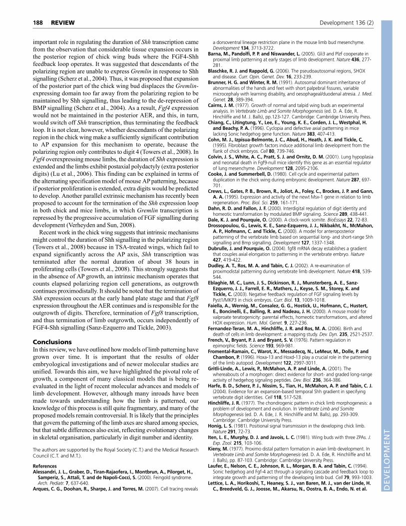
cannot readily be related to cellular behaviour because most cells areproliferating (Fernandez-Teran et al., 2006). There are, however,indications that cell cycle times may be slower in the anterior regionof the chick wing than in the posterior region (Cairns, 1977), thuspotentially contributing to differential expansion. Apoptosis is notthought to influence overall limb bud growth in either mouse orchick, to any large extent, and, where present, is concentrated inrestricted areas. In the early chick wing bud, cell death occurs in theanterior and posterior necrotic zones (Saunders and Gasseling,1962), and might be associated with the relatively narrow hand plateof the chick wing compared with the mouse forelimb (Fernandez-Teran et al., 2006). In mouse forelimb buds, there is also a region ofcell death at the anterior margin but no posterior necrotic zone(Fernandez-Teran et al., 2006).
Models of vertebrate limb patterningIn the 1970s, experiments on chick wing buds produced two classicalmodels of limb development (see Boxes 1 and 2). In the progress zonemodel, growth was suggested to have a direct role in progressivelyspecifying PD positional values (Summerbell et al., 1973) (see Fig.1A; Box 1), whereas in the morphogen gradient model, a morphogengradient was proposed to specify AP values in the early bud, whichare then ‘remembered’ throughout subsequent growth (Tickle et al.,1975) (see Fig. 1C; Box 2). DV patterning involves signals from bothdorsal and ventral ectoderm (MacCabe et al., 1974), but as thereappears to be relatively little growth along this axis, it will not beconsidered further here [for recent insights into DV patterning seeArques et al. (Arques et al., 2007), which reports an unexpected celllineage-restricted compartment boundary that separates dorsal andventral mesenchyme in the mouse limb bud].
Even in simple models of limb development, the relationshipbetween patterning and growth can be complex. Thus, it will taketime for a diffusible morphogen to set up a gradient, and cells willhave to adjust constantly to changing morphogen concentrations.
Furthermore, the fact that the limb bud is continuously growingcomplicates the specification of positional values, and growth mayactually play a key role in determining the size of the field overwhich a morphogen operates (see later). Additionally, another wayof setting up a morphogen concentration gradient is by RNA orprotein decay over time in a growing tissue, thus leading to short-range signals with long-range effects (Dubrulle and Pourquie, 2004).In the following sections, we consider the involvement of growth inPD and AP patterning of the developing limb.
Proximodistal patterningThe progress zone model of chick wing PD patterningThe progress zone model for patterning the PD axis emerged as aresult of many embryological experiments on chick wing buds in the1970s (Box 1; Fig. 3A). It had been known for a long time that theAER is required for limb bud outgrowth and for the accompanyingsequential proximal-to-distal differentiation of skeletal elements (Fig.3A) (Saunders, 1948). It was also known that the removal of the AERat different stages of wing development causes truncations thatprogressively become more distally restricted the later the operationis performed (Fig. 3A). It was, however, experiments in whichtransplanted tips of chick wing buds were shown to developautonomously that led to the idea that the length of time thatundifferentiated mesenchymal cells spend proliferating at the tip ofthe limb – in a region known as the progress zone – specifies PDpositional values (Summerbell et al., 1973). It was suggested thatthese values are generated over time by a ‘clock-like’ mechanism and
REVIEW Development 136 (2)
Fig. 1. French flag models illustrating potential roles of growthin embryonic patterning. The three colours of the French flag depictpositional values that specify different cell fates across an embryonicfield, such as the limb bud, over time (see Wolpert, 1969; Wolpert,1989). (A) Progressive specification. The blue value is specified first,followed by the white and the red as growth occurs. The final positionsthat make up the final flag are attained by the displacement of blue,white and then red cells from the right-hand side of the field over timeby growth. (B) Intercalary specification. The outlying blue and redpositional values are specified first and the disparity between theseextreme values promotes local growth that provides the whiteintermediate positional value. (C) Early specification. The colouredpositional values that make up the flag are specified very early by, forexample, a morphogen gradient, and further growth expands the field.
Box 1. The development of the classical progress zonemodelThe classical progress zone model proposes that, as the limb budgrows out under the influence of signalling from the apicalectodermal ridge (AER), proximodistal (PD) positional values arespecified progressively by the length of time cells spend in anundifferentiated region at the bud tip called the progress zone(Summerbell et al., 1973). Cells that spend a short time in theprogress zone are specified to form proximal structures, whereas cellsthat spend longer there form more-distal structures (see Fig. 1A).The finding that the chick wing is truncated when the AER isremoved (Saunders, 1948; Summerbell et al., 1973) was the key toshowing the importance of AER signalling in limb development. Theextent of the truncation depends on the time at which the AER isremoved: when removed early, only proximal structures develop;when removed later, more distal wing structures form. Another JohnSaunders study showed that when the AER from a late chick wingbud is replaced with the AER from an early wing bud and vice versa,normal limbs still develop, leading to the conclusion that AERsignalling is permissive (Rubin and Saunders, 1972). Saunders alsodiscovered that cells from the proximal region of a chick leg budplaced under the AER of a chick wing bud form toes, thus showingthat proximal cells can be re-specified when placed at the bud tip(Saunders and Gasseling, 1959).In 1973, Dennis Summerbell and colleagues reported thattransplanting the undifferentiated tip of an early chick wing bud tothe stump of a late wing bud, or transplanting a late bud tip to anearly stump, resulted in duplications or deletions, respectively, thusshowing that the limb bud tip behaves autonomously, a key findingfor the progress zone model (Summerbell et al., 1973). LewisWolpert and colleagues then showed that killing cells in the earlywing bud with high doses of X-irradiation led to loss of proximalstructures, whereas distal structures remained relatively unaffected,a result that can be explained by the progress zone model (Wolpertet al., 1979).
DEVELO
PMENT

181REVIEWDevelopment 136 (2)
become fixed when cells are displaced from the progress zone (Fig.3A). It was calculated using data from AER removal experiments thatthis timing mechanism could be linked to the cell cycle because sevencell generations are required to lay down a complete chick wingskeleton, about one cell generation for each element, if one includesthe two carpal bones in the wrist and the three phalanges of digit 3(Lewis, 1975). Thus, in the classical progress zone model,specification of PD pattern depends on growth, timing and length ofexposure of a population of undifferentiated mesenchyme cells to apermissive AER signal (Fig. 3A).
The early specification model of chick wing PD patterningOver the past few years, the progress zone model has beenchallenged by the early specification model (Fig. 3A), whichproposes that the PD pattern is specified very early and thenexpands, so that structures differentiate in the observed proximal-to-distal sequence, as the limb grows out under the influence of theAER (Dudley et al., 2002). The recent data assembled in support ofthe early specification model can, however, be accommodated bythe progress zone model (Tickle and Wolpert, 2002). Thus, forexample, it is a matter of interpretation as to whether the cell deaththat accompanies AER removal leads to the loss of cells that have
already been specified to form distal structures (indicated by crossesin Fig. 3A) or to the loss of the progress zone. Furthermore, theresults of fate-mapping experiments that suggest that cell lineage-restricted compartments might exist along the PD axis, a findingused to support the early specification model, have not beenconfirmed (Pearse et al., 2007; Sato et al., 2007). Therefore, it is notyet clear whether the progress zone model of PD limb patterningshould be abandoned for the early specification model. Indeed, aprogress zone model is currently favoured to explain how somitesare generated along the main body axis (Dale and Pourquie, 2000).One limitation of the early specification model is that it does notexplain how the pattern is set up in the early limb bud.
In summary, embryological approaches have yielded twostrikingly different models of PD patterning. In the followingsections, we discuss how these models stand up in light of recentgenetic and molecular advances in our understanding of AERsignalling.
The molecular/genetic basis of PD patterningA simple experiment in chick wing buds, in which an FGF-soakedbead rescued wing bud outgrowth and PD patterning in the absenceof the AER, showed that AER signalling is mediated by FGFs
Fig. 2. Chick wing and mouse forelimb development. (A) Aschematic of fully developed chick wing (yellow) and mouse forelimb(blue) skeletons with anteroposterior (AP) and proximodistal (PD) axesshown (as applied to all elements except the humerus). (B) Schematicsof equivalently staged chick wing (Hamburger-Hamilton stages, HH)and mouse forelimb buds (embryonic day, E), from early stages to handplate development. Note, mouse hindlimb development is delayed byabout half a day relative to the forelimb (Martin, 1990; Fernandez-Teran et al., 2006). (C) Timeline of Shh, Fgf4 and Fgf8 expression inrelation to embryonic stages shown in B (thin line indicates very lowShh expression in the chick wing). (D) (a) Orientation of AP, PD and DVaxes in early stage limb buds. (b) A schematic of the expression of: Shhin the polarizing region, Fgf8 throughout the apical ectodermal ridge(AER) and Fgf4 in the posterior AER.
Box 2. Experimental evidence for the morphogengradient model of anteroposterior patterningThe morphogen gradient model proposes that the polarizing region,a group of mesenchyme cells at the posterior limb bud margin (seeFig. 2D), produces a diffusible morphogen that establishes aconcentration gradient across the anteroposterior (AP) axis.According to this model, cells nearest the polarizing region will beexposed to high morphogen concentrations and form posteriordigits, whereas cells further away, exposed to increasingly lowerconcentrations, form progressively more anterior digits (Tickle et al.,1975; Wolpert, 1969).Saunders and Gasseling discovered the polarizing region or zone ofpolarizing activity (ZPA) (Saunders and Gasseling, 1968). When tissuefrom the posterior region of a chick wing bud was grafted to theanterior margin of a second bud, a mirror-image symmetrical digitpattern resulted, with an additional set of digits arising from theanterior region of the host wing (Saunders and Gasseling, 1968). Theulna can also be duplicated if the graft is performed early in limbdevelopment, but not the humerus (Wolpert and Hornbruch, 1987),showing that the polarizing region patterns the limb distal to theelbow.Grafts of X-irradiated polarizing regions (Smith et al., 1978), or ofsmall numbers of polarizing region cells (Tickle, 1981), specify onlyadditional anterior digits, showing that polarizing region signalling isdose dependent. A polarizing region must be grafted for at least 16hours to produce an additional digit 2, and for 20 hours to producedigit 3 (Smith, 1980). An early response to a polarizing region graftwas found to be increased S-phase entry in adjacent mesenchymecells (Cooke and Summerbell, 1980). X-irradiating wing buds within2 hours of grafting a polarizing region reduced AP growth and ledto loss of anterior digits (Smith and Wolpert, 1981).In the 1980s, it was suggested that intercalation, involving local cell-cell interactions, could explain digit duplications produced bypolarizing region grafts (Iten et al., 1981). But when two polarizingregions were grafted, the complete mirror-image duplicationspredicted by intercalation were not obtained. Honig showed directlythat polarizing region signalling was long range; a chick wing digit 2could be specified in cells separated from a grafted polarizing regionby a 200 μm wide piece of leg tissue (Honig, 1981).
DEVELO
PMENT

182
(Niswander et al., 1993). It was later found that an FGF-soaked beadapplied to the inter-limb region of an early chick embryo can inducethe development of a complete limb (Cohn et al., 1995).
Several genes that encode FGFs are expressed in the AER atdifferent times and with different spatial patterns, and thus thequality and quantity of FGF signalling in the limb varies over time(Martin, 1998) (Fig. 2C,D). Fgf8 is expressed throughout the entireAP extent of the AER, from the earliest bud stages up until limboutgrowth is completed, whereas three other FGFs – Fgf4, Fgf9,Fgf17 – are expressed for a shorter period of time and are initiallymore posteriorly restricted. Mesenchymal signals control thesepatterns of FGF gene expression in the AER (Laufer et al., 1994;Niswander et al., 1994), and this could account for the fact that,in embryological experiments, old and young AERs areinterchangeable (Rubin and Saunders, 1972) (Box 1).
According to both progress zone and early specification models,FGFs secreted from the AER into the underlying mesenchymemediate outgrowth along the PD axis and maintain the region ofundifferentiated cells at the tip of the limb bud. The function of FGFsignalling in mouse limb outgrowth has been tested directly byconditionally inactivating each of the FGF genes that are expressedin the AER. When Fgf8 is functionally inactivated (Lewandoski etal., 2000; Moon and Cappechi, 2000), bud outgrowth is reduced andsome digits are lost, whereas functional inactivation of the otherFGFs expressed in the AER has no affect on limb development(Colvin et al., 2001; Moon et al., 2000; Sun et al., 2000; Xu et al.,2000). However, when both Fgf4 and Fgf8 are inactivated togetherat the earliest stages of bud outgrowth, limb development fails,although, when inactivated together slightly later, proximalapoptosis occurs, followed by loss of proximal structures (Sun et al.,2002). Taken together with the loss of proximal structures in micelimbs following targeted disruption of Plzf (Promyelocytic zincfinger) and Gli3 (Gli/kruppel family member 3) transcription factor(Barna et al., 2005), these findings were used as support for the earlyspecification model (Sun et al., 2002).
The range over which FGF signalling extends from the AER isunclear because although FGF8 protein can be visualised in theAER, it has not yet been detected in the mesenchyme. Mkp3 (Mapkinase phosphatase 3), a gene encoding a dual specificityphosphatase that is a transcriptional target of FGF (and whichnegatively regulates FGF signalling), is expressed in a gradientalong the PD axis in early wing buds (Eblaghie et al., 2003).Although mRNA decay may contribute to the distribution of Mkp3transcripts in the limb (Pascoal et al., 2007a), the extent of Mkp3expression nevertheless suggests that FGF signalling from the AERcan exert long-range effects on the underlying limb mesenchyme.Other genes expressed at the tip of the limb bud include the Msx1(muscle segment homeobox1) gene, which encodes a transcriptionfactor that, in other contexts, including regenerating newt limbs,keeps cells in an undifferentiated state (Crews et al., 1995). It hasalso recently been shown that the gene Hairy2, which encodes acomponent of the somitogenesis clock, is expressed in an intriguingoscillatory fashion in cells at the tip of the chick wing bud (Pascoalet al., 2007b). The identification of further molecular clock geneswould clearly support the progress zone model, although, asmentioned earlier, the periodicity of the cell cycle still providesanother plausible timing mechanism.
A major question in the field concerns the identities of genes thatare expressed in response to positional cues in different regions ofthe limb bud and then govern the development of that particular partof the pattern. Meis genes, which encode TALE-homeodomainproteins, are candidate factors for proximal limb identity that mightcontrol subsequent humerus development (Mercader et al., 2000),whereas genes that occupy 5� positions in the Hoxa and Hoxdclusters are candidate distal identity factors that govern subsequentdigit development (Zakany and Duboule, 2007). The expressionpatterns of these genes are established in early chick wing buds, withFGF signalling from the AER being required to maintain Hoxa13and Hoxd13 expression distally, and retinoic acid (RA) signalling atthe base of the bud maintaining Meis expression proximally. Theoverexpression of Meis genes in distal areas of chick limb budsinhibits the development of distal structures (Mercader et al., 2000),whereas knocking out Hoxd13 and Hoxa13 together in mice leadsto loss of digits (Fromental-Ramain et al., 1996). Careful cellmarking experiments in chick wing buds have indicated, however,that, as the bud grows out, some cells that express Hoxd13 early onbecome displaced from the tip and cease to express Hoxd13
REVIEW Development 136 (2)
Fig. 3. Three models of proximodistal patterning in chick andmouse limbs. Schematics of how positional values (red, white andblue) are established that specify different proximodistal (PD) structures.(A) Apical ectodermal ridge (AER) removal in the chick wing at differentstages leads to truncations, the extent of which depends on when theoperation is performed (the earliest developmental stage is to the left).These results have been explained by two different models. (a) Progresszone model of chick wing PD patterning (Summerbell et al., 1973).Positional values are specified depending on the length of time cellsremain in the distal progress zone (the right-hand side of the field inthe figure). Cells that are displaced first from the progress zone formthe humerus (blue), followed by forearm (white) and finally digits (red).AER removal terminates progress zone activity (shown by the loss of thewhite and red values). (b) Early specification model of chick wing PDpatterning (Dudley et al., 2002). All three positional values, humerus(blue), forearm (white) and digits (red) are specified in the early chickwing bud prior to outgrowth and are expanded by growth. Analternative explanation for the limb truncations that follow AER removalis that cell death eliminates distal pre-specified positional values(crosses). Outcomes of some genetic experiments in the mouse havealso supported this model (see main text for references and details).(B) Intercalation model of mouse limb PD patterning (Mariani et al.,2008). Reducing FGF activity in the AER of the mouse limb can lead toloss of intermediate structures, while distal and proximal structures stillform (as shown in schematic of the skeleton), suggesting thatpositional values of the humerus (blue) and digits (red) are specified firstin the early mouse limb bud with intermediate positional values (white)generated by intercalation. A loss of digits across the AP limb axis alsooccurs in these experiments.
DEVELO
PMENT

183REVIEWDevelopment 136 (2)
(Vargesson et al., 1997). This behaviour is difficult to reconcile withthe early specification model, but also seems at odds with the ideathat cells become progressively more distal in character over time.One interpretation of these data is that distal cells becomeprogressively proximalised during limb bud outgrowth.
In addition to the gene products mentioned above that areimplicated as determinants of proximal and distal limb differentiation,the product of the Shox (short stature homeobox containing) genemight govern intermediate limb differentiation. Mutations that affectShox gene function are responsible for short stature in individuals withTurner syndrome and for the disproportionate shortening of the arm,particularly involving the radius/ulna, in individuals with Leri-Weilland Langer syndromes (Blaschke and Rappold, 2006). In chick wingbuds, Shox is expressed in an intermediate region where it overlapsMeis expression proximally. This expression pattern could beexplained by the fact that Shox is inhibited by distal FGF and byproximal RA signals (Tiecke et al., 2006).
In summary, significant progress has been made in identifying themolecules involved in PD limb patterning but this has not clarifiedwhether this process is best explained in terms of the progress zoneor early specification model. In fact, in the next section we describehow the dissection of FGF genetics in the mouse limb has led to analternative model that, again, has classical roots.
The intercalation model of mouse PD patterningThe classical model for PD patterning in regenerating limbs involvesintercalary growth (Maden, 1980) (Fig. 1B). In a recent paper, it hasbeen suggested that intercalation might be involved in establishingPD positional values in the early mouse limb bud (Mariani et al.,2008) (Fig. 3B). Gail Martin and colleagues have used conditionalgene targeting in mice to delete painstakingly FGF-encoding genes(Fgf4, Fgf8, Fgf9, Fgf17) specifically in the AER, singly and also indouble and triple combinations (Mariani et al., 2008). This analysisshows that Fgf8 can support normal forelimb development in theabsence of Fgf4, Fgf9 and Fgf17. When FGF signalling was thentitrated genetically, by knocking out Fgf8 function together with, inturn, that of Fgf17, Fgf9 or Fgf4, and by making Fgf4/Fgf8conditional knockouts that were heterozygous for Fgf9 function,mouse embryos were produced that had progressively smaller limbbuds, which developed into forelimbs with correspondingly fewerskeletal elements (Mariani et al., 2008). First, digits were lost, and,in some cases, forearm elements too, although all elements of PDpattern were still represented. With further reduction in FGFfunction in Fgf4–/–/Fgf8–/–/Fgf9+/– conditional knockouts, forelimbsdeveloped that had a reduced humerus and lacked a radius andulna, but still had a digit-like structure at the distal tip. InFgf4–/–/Fgf8–/–/Fgf9–/– conditional knockouts, no limb structuresformed. This series of limb morphologies contrasts with theprogressively more proximal truncations that are predicted by bothearly specification and progress zone models (Fig. 3A). A similarphenotype in which distal structures form in the absence of proximalstructures was obtained when cell death was induced throughoutchick wing buds by X irradiation (Wolpert et al., 1979). This wasinterpreted in terms of the progress-zone model because survivingcells would spend longer in the progress zone in order to repopulateit and thus become distalized. However, in FGF-deficient mouselimbs, this phenotype is not easy to correlate with the distributionand timing of cell death (Mariani et al., 2008). Instead, it is proposedthat loss of forearm structures in Fgf4–/–/Fgf8–/–/Fgf9+/– conditionalknockouts is due to loss of intermediate positional values that arenormally intercalated by local growth, between proximal positionalvalues specified by RA and distal positional values specified by
FGFs. Because the role of FGFs in this model is to specify just thedistal structures, the authors propose that AER signalling should beconsidered to be instructive rather than permissive.
As mentioned above, in the intercalation model for amphibianlimb regeneration, it is the juxtaposition of cells with disparatepositional values that generates local growth to restore the missingintermediate positional values (Maden, 1980). However, anintercalation model does not appear to apply to the chick limbbecause there is little regulation along the PD axis. Thus, forexample, when the distal tip of an early wing bud is grafted to aproximal stump of an older wing bud, the intermediate part of thepattern is not produced (Summerbell et al., 1973) [but see alsoKieny (Kieny, 1977)]. Some regulation can occur at very earlystages of wing development when slices are cut out of the PD axis,but this regulative ability rapidly declines as the bud develops(Summerbell, 1977). The intercalation model for the mouseforelimb, however, predicts that the intermediate pattern isspecified at a later stage of limb development than are the distal andproximal patterns.
In summary, there is still uncertainty as to how the PD limbpattern is specified. There could be differences in patterningmechanisms between different species that could reflect, forexample, their regulative capacities, as well as overlap betweenseemingly opposing models. However, in the next section, wedescribe how such considerations based on seemingly conflictingdata might yield a unified model of AP limb patterning.
Anteroposterior patterningClassical morphogen model of chick wing AP patterningA landmark discovery by Saunders and Gasseling was the discoveryof the polarizing region, a classical organiser located at the posteriormargin of a chick wing bud, which induces a new pattern of digitsin mirror-image symmetry to the normal pattern when grafted to theanterior margin of another wing bud (Saunders and Gasseling, 1968)(see Box 2). The discovery of the polarizing region paved the wayfor a series of embryological experiments, which, over the nextdecade, yielded results consistent with a model in which APpositional values are specified by a gradient of a long-rangemorphogen (Box 2). The number and identity of the induced digitswas shown to depend on both the strength (Smith et al., 1978;Tickle, 1981) and duration (Smith, 1980) of the polarizing signal(Box 2). These studies showed that only an additional digit 2 formswhen the number of polarizing region cells is reduced or if thepolarizing region is removed early. It should be noted that it wasproposed that a morphogen gradient might act on a digit pre-pattern,which is specified by a wave-like distribution of a morphogengenerated by a reaction-diffusion mechanism (Turing, 1952). Oneof the pieces of evidence favouring this is the formation of digits inchick limb reaggregates in which the mesenchymal cells from thelimb buds were disaggregated into single cells and then placed insidea normal ectodermal jacket (Pautou, 1973) (reviewed by Wolpert,1989). The morphogen gradient model was briefly challenged in the1980s by the suggestion that local cell-cell interactions andintercalation might account for the digit duplications produced bypolarizing region grafts (Iten et al., 1981) (Box 2).
The width of the bud and the length of the AER is a good indicatorof the number of digits that form. Several experiments on the chickwing in the 1970s and 1980s indicated that the polarizing regionmight directly control mesenchymal cell proliferation while thedigits are being specified (Cooke and Summerbell, 1980; Smith andWolpert, 1981) (Box 2). Enhanced mesenchymal proliferation wasdetected prior to the extension of the overlying AER following a D
EVELO
PMENT

184
polarizing graft to the anterior margin (Cooke and Summerbell,1980). Thus, growth and specification were considered to becontrolled by the same or by different signals emanating from thepolarizing region (Summerbell, 1981). This proposal gained supportwhen it was found that instead of a fully duplicated digit pattern(432234), anterior digits were lost (4334 or 434) when AP expansionwas inhibited following a polarizing region graft to the anteriormargin, although the mechanism by which this occurred was unclear(Smith and Wolpert, 1981). Thus, a direct role for growth inspecification of AP positional values remained speculative until themolecular basis of AP patterning began to be revealed.
Molecular basis of AP patterningIt is now clear that the sonic hedgehog (Shh) gene, which isexpressed in the polarizing region of mouse and chick limbs (Fig.2D), is pivotal in controlling digit number and pattern (Riddle et al.,1993). RA, however, was the first defined chemical found to havepolarizing activity (Tickle et al., 1982); RA-soaked beads implantedat the anterior margins of chick wings induce full-digit duplicationswith the same characteristics as polarizing region grafts (Tickle etal., 1985). RA is proposed to be involved in proximal limb formationthrough the regulation of Meis gene expression (Mercader et al.,2000), and its effects on AP pattern are possibly mediated via thetranscriptional activation of Shh (Riddle et al., 1993). Consistentwith the classical morphogen model of AP patterning, Shh proteincan be directly detected away from the polarizing region in mouselimb buds (Gritli-Linde et al., 2001) and is indirectly detected viapatched 1 (Ptch1) transcripts (Ptch1 encodes the Shh receptor),which are expressed very rapidly in response to Shh signalling in thechick wing (Marigo and Tabin, 1996). Shh signalling has beenshown to specify AP positional values with the time and dosedependency of polarizing region grafts (Yang et al., 1997). Fate-mapping experiments in chick wing buds have demonstrated that theprospective digit progenitor cells can be ‘promoted’ to more-posterior digit fates following a longer exposure to Shh signalling(Yang et al., 1997). Bone morphogenetic protein (BMP) signallingis implicated in this promotion of AP positional values downstreamof Shh signalling in the chick wing bud (Drossopoulou et al., 2000).
Again, as for the PD axis, the identity of genes that determine thedevelopment of the different digits remains a major question.Evidence suggests that 5� Hoxd genes might play a role in patterningthis axis, especially at hand plate stages (Zakany and Duboule,2007). Other candidates include members of the Tbox (Tbx) familyand the vertebrate Sall orthologues of the Drosophila Spalt genes.The overexpression of either Tbx2 or Tbx3, which are expressed instripes both anteriorly and posteriorly in chick and mouse limb buds,posteriorizes toes in chick legs (Suzuki et al., 2004). Furthermore,mutations that affect the function of TBX3 and SALL1 and SALL4genes underlie congenital digital abnormalities in humans(Sweetman and Munsterberg, 2006).
Integrated growth and specification model of chick wingAP patterningA recent growth/morphogen model of chick wing patterningsuggests that growth plays an essential role in the specification ofpositional values in the early bud and that both processes arecontrolled and integrated by Shh signalling (Towers et al., 2008)(Fig. 4A). This study showed that Shh regulates the high-levelexpression of several genes that encode regulators of S-phase entry,including N-myc and cyclins D1/2 in the digit-forming region of theearly wing bud, both in polarizing region cells, which give rise todigit 4, and in adjacent posterior cells, which give rise to digits 2 and
3. It was already known that posterior digits are lost when Shhsignalling is inhibited by cyclopamine (an inhibitor of smoothened,which activates the Shh signalling pathway) (Scherz et al., 2007),but this more recent study showed that loss of posterior digits wascaused by a combination of reduced AP growth and specification(Fig. 4B) (Towers et al., 2008). Importantly, fate maps ofcyclopamine-treated chick limbs revealed that all prospective digitprogenitors contributed to the anterior elements that formed. Bycontrast, transient inhibition of AP growth, either by overexpressingthe cyclin-dependent kinase inhibitor p21cip1, or by applying mitoticinhibitors, including trichostatin A (TSA), resulted in loss of anteriordigits (Towers et al., 2008) (Fig. 4C). In such wings, posteriorspecification was inhibited during growth arrest but continued forthe normal duration after outgrowth recovered. However, APexpansion of the digit-forming field failed to recover followinggrowth arrest, and fate-mapping showed this entire cell populationcontributed to the remaining posterior structures, often a single digit
REVIEW Development 136 (2)
Fig. 4. Growth-morphogen model of chick wing anteroposteriorpatterning. [See Towers et al. (Towers et al., 2008).] In this figure,squares represent positional values and numbers indicate which digitshave been specified. Timings, shown on the vertical axis, estimate theduration of Shh activity required for the specification of digits. Left isposterior, right is anterior. (A) Normal chick wing development. Shhprotein emanating from the posterior polarizing region (left-hand side)promotes sufficient AP growth of the mesenchymal field for three digitsto form (digits 2, 3 and 4). Simultaneously, the Shh specificationgradient forms over the field and establishes the three AP positionalvalues of each digit identity; low levels, digit 2 (red); intermediate levels,digit 3 (white); and high levels, digit 4 (blue). Specification involves cellsbeing promoted through increasing AP positional values and ispredicted from experimental data to take 16-24 hours (Smith, 1980;Yang et al., 1997). Positional values are remembered; digitdifferentiation occurs later in the sequence digit 4, 3 and 2, as shown inthe skeletons below. (B) Reduced Shh signalling. Cyclopaminetreatment blocks Shh-dependent AP expansion of the mesenchymalfield to the extent that only two digits can form, as depicted in theskeleton (Scherz et al., 2007). Simultaneously, reduced Shh signallingspecifies only anterior digits 2 and 3 (red and white). (C) Reducedproliferation and growth. TSA-treatment irreversibly inhibits APexpansion of the mesenchymal field during the interval when digits arenormally specified. In the most severely affected wings, only one digitcan form, as depicted in the skeleton. The duration of Shh signalling isnot affected, so a single posterior digit 4 (blue) is specified.
DEVELO
PMENT

185REVIEWDevelopment 136 (2)
4. These findings demonstrate that Shh normally promotes APexpansion and specification of the digit-forming field, which togetherdetermine digit number and identity in the chick wing (Fig. 4A).
In the next sections, we discuss how models of AP patterningderived in the chick wing stand up in the light of results derived fromgenetic studies in the mouse.
Genetics of mouse limb AP patterningMany of the fundamental concepts of mouse digit AP patterningoriginate in embryological studies undertaken in the chick wing.For example, grafts of tissue from the posterior of the mouse limbto the anterior of chick wing buds can induce a full set of chickwing digits (Tickle et al., 1976). The simplest model that accountsfor these results is that a gradient of polarizing activity patternsmouse digits, as it does in the chick wing. However, it isbecoming evident that mouse AP limb patterning is much morecomplicated than chick AP wing patterning, not least because themouse has five digits rather than three. The complete inactivationof Shh in the mouse results in the loss of all digits in the forelimband of all but the most anterior digit (digit 1) in the hindlimb,which is therefore considered to be independent of Shh (Chianget al., 1996). The same pattern of digit loss is also seen in thechick mutant, oligozeugodactyly, which lacks Shh function in thewing and leg (Ros et al., 2003). The inactivation of Gli3 alone andof Shh and Gli3 together causes many unpatterned digits to form(Litingtung et al., 2002; te Welscher et al., 2002). Gli3 is one ofthe transcriptional effectors of Shh signalling, and Shh signallingprevents its activator form (Gli3A) from being processed into itsrepressor form (Gli3R). This shows that the function of Shh incontrolling digit number and identity is achieved principally byrepressing Gli3R activity in the posterior part of the limb bud thatforms the digits. The precise balance of Gli3A and Gli3R mayprovide the basis for the graded response to Shh signalling. Thegeneration of many unpatterned digits is also consistent with theproposed digit pre-pattern (see earlier).
The above findings, although contributing highly importantmolecular insights, have not revealed the mechanism by which theAP axis of the mouse limb is patterned. However, recent conditionalgene-targeting approaches have yielded two strikingly differentmodels of AP patterning of the mouse digits, which we discussbelow.
Temporal expansion model of mouse limb AP patterningRecent conditional gene-targeting approaches in mice have beendesigned to follow the descendants of polarizing region cells andto manipulate the duration, dose and range of Shh signalling in themouse limb. In one study (Harfe et al., 2004), which invokes anew timing mechanism for the patterning of the most posteriormouse digits (Fig. 5A), the lineages of Shh-expressing cellswere traced using an inducible lacZ reporter line(ShhGFPCre/+,R26R/+), revealing that these cells progressivelycontribute to part of digit 3 and to the two most-posterior digits (4and 5). This contrasts with the chick wing, in which polarizingregion descendants contribute only to digit 4 (Towers et al., 2008).Furthermore, the formation of posterior mouse digits does notappear to depend on Shh diffusion because only digit 2 was lostwhen long-range Shh signalling was severely reduced followingthe inactivation of the dispatched 1 (Disp1) gene (Disp1 isresponsible for transporting cholesterol-modified Shh) (Harfe etal., 2004) (Fig. 5B). This suggests that the development of thethree most-posterior digits in the mouse limb occurs by amechanism that is related to the proliferation of the polarizing
region cell lineage, rather than being specified by the highestlevels of Shh signalling. Thus, proliferation could provide atiming mechanism by which polarizing cells become committedto different posterior digit fates. Although these fate-mappingstudies do not actually reveal when digit identities are specified,the fact that three digits derive from the same population of cellssuggests a requirement for an extended period of proliferation(Fig. 5A). Shh is expressed for around 60 h in the mouse limb andcould underpin such a mechanism (Fig. 2C). This is considerablylonger than the 24 h exposure to Shh signalling required to specifythe full set of AP values in the chick wing (Yang et al., 1997) (Fig.4A). It has also recently been shown that posterior digits still formin limbs of Shhgfpcre/Shhc mice, in which levels of long-rangeShh signalling are reduced but in which Shh is expressed for thenormal length of time, although in such limbs, digit 2 is absent(Scherz et al., 2007).
Together, these data strongly suggest that, in the mouse limb, aspecification gradient of Shh patterns anterior digits and that thelength of time that proliferating polarizing region cells are exposedto direct Shh signalling patterns the posterior digits. It remains to beseen whether Shh signalling from the polarizing region also controlsthe growth of the adjacent digit-forming field in the mouse limb asin the chick wing (see earlier) and thus whether the growth-morphogen model outlined above for the chick wing applies tomouse anterior digits.
Biphasic model of mouse AP limb patterningThe temporal requirements of Shh signalling for mouse digitpatterning have now been tested in further careful experiments inwhich Shh function has been rapidly inactivated at different stagesusing an inducible Hoxb6CreER line (Zhu et al., 2008). Theoutcome of these experiments forms the basis of a new biphasicmodel, in which Shh specifies digits at the very earliest stages oflimb development (possibly by a concentration gradient) and then isrequired as a mitogen for the progressive formation of individualdigits (Fig. 5C). This study reported that only two digits formfollowing a 3-hour pulse of Shh transcription in the hindlimb (whichprovides 9 hours of Shh activity, as assessed by Ptch1 expression)and following 9 hours in the forelimb (15 hours Shh activity). Insuch limbs, the digits that formed were digit 1 and, unexpectedly,digit 4. A longer period of Shh signalling permitted other digits toform in the alternating sequence, digit 2, digit 5 and then digit 3 (Fig.5C). Strikingly, using Sox9 and Noggin knock-in alleles to drive lacZexpression, the authors observed that the cartilage condensation ofeach digit differentiates in the same order, i.e., digit 4, digit 2, digit5 and finally digit 3. The exception is the condensation of digit 1,which appears last, but forms after a short pulse of Shh expressionin the forelimb. It should be noted, however, that other accounts ofmouse limb development suggest that digit 5 forms after digit 3(Martin, 1990).
This interdigitating pattern of digit condensation does not fitwith the expected anterior-to-posterior sequence of specification,which is pivotal to all other models of AP patterning. Instead, theauthors suggest that digit morphogenesis relies on Shh-dependentproliferation allowing sufficient numbers of specified cells tosurvive to form a condensation. This could explain why cells thatrequire the shortest duration of Shh-dependent proliferation formthe cartilage condensation of digit 4, the digit that differentiatesfirst (Fig. 5C). Thus, in this biphasic model, the authors suggestthat the control of digit identity and number are temporallyuncoupled and that Shh signalling acts first as a morphogen andthen second as a growth-promoting factor. D
EVELO
PMENT

186
Early or late specification of posterior digits in the mouselimb?The temporal and biphasic models of AP mouse limb patterning areclearly at odds with each other; the major issue concerns the durationof Shh expression required to specify the posterior digits. Theidentification of digit 4, which underpins the biphasic model, andthe use of Tbx3 expression to identify posterior digits have beenrecently challenged (Tabin and McMahon, 2008), highlighting theproblems of not being able to identify unequivocally mouse digits.There is clearly an urgent need to identify molecular markers forindividual digits (if they exist) to aid the interpretation of patterningdefects, and to provide insights into digit evolution.
In the biphasic model, if the digit with the shortest requirementfor Shh signalling turns out to be a more-anterior digit than digit 4,then this would agree with the more conventional models of APpatterning. Unfortunately, other independent studies also fail toclarify this issue. For example, three digits form in thePrx1Cre;Shhc/c mouse mutant, in which Shh expression is attenuatedearly. This phenotype is interpreted as representing the loss of digits4 and 5 by the authors (Scherz et al., 2007), but as the loss of digits3 and 5 by others (Zhu et al., 2008). The first interpretation fits withthe temporal model because a much longer time would be requiredto specify digit 4; the second with the biphasic model, because digit4 would have been specified earlier.
REVIEW Development 136 (2)
Fig. 5. Extended flag models of mouse limb anteroposterior patterning. In this figure, the squares represent positional values and numbersindicate which digits have been specified. Timings, shown on the vertical axis, estimate the duration of Shh activity required for the specification ofhindlimb digits. Left is posterior, right is anterior. Digit 1 is considered to form independently of Shh signalling in the hindlimb (black). (A) Temporalexpansion model (Harfe et al., 2004). Positional values of the digits 3 (dark blue) and digit 2 (white) are specified by a morphogen gradientmechanism, involving Shh, although the involvement of mesenchymal expansion is unclear. Posterior digits 4 (purple) and 5 (light blue) are specifiedaccording to the length of time cells remain in the polarizing region, which exclusively contributes to these two digits. This could require the fullduration of Shh activity (~60 hours) but does not require a concentration gradient of Shh protein. Digit 3 (dark blue) can be specified by acombination of the above processes, as depicted. (B) Temporal expansion model in dispatched 1 (Disp1) mutant limbs (Harfe et al., 2004). Top,schematic of mouse Disp1 mutant limb skeleton. Bottom, restricted long-range Shh movement prevents the positional value of digit 2 (white) frombeing established. Posterior digits are still specified by a timing mechanism linked to the proliferation of Shh-expressing polarizing region cells.(C) Biphasic model (Zhu et al., 2008). Positional identities of all five digits require less than 9 hours of Shh activity to be specified by an unknownmechanism that could involve a concentration gradient. A longer duration of Shh activity allows the survival of specified digit progenitor cells(shown by progressive enlargement of boxes representing different digits); digits differentiate in the order shown in the skeletons underneath.(D) Alternating temporal expansion/growth morphogen model. Positional values of digits 2 (white) and 3 (dark blue) are specified in the same wayas digits 2-4 of the chick wing by a growth-morphogen mechanism (see Fig. 4A). At the same time, digits 4 (purple) and 5 (light blue) begin to bespecified by temporal expansion (see Fig. 5A), resulting in an alternating sequence of specification that recapitulates the sequence in which digitsdevelop with increasing lengths of exposure to Shh signalling (see Fig. 5C). (E) Alternating temporal expansion/growth morphogen model invalproate-treated limbs. Top, schematic of valproate-treated mouse limb skeleton (Faiella et al., 2000). Valproate treatment could irreversibly restrictAP expansion of the mouse limb, without affecting the timing of posterior digit specification or the Shh concentration gradient. This results in alldescendant cells of the polarizing region being specified as digit 5 (light blue) and the concentration gradient of Shh establishing the positionalvalues of digit 3 (dark blue) (compare with Fig. 4C).DEVELO
PMENT

187REVIEWDevelopment 136 (2)
The temporal and biphasic models of mouse digit patterning arenot readily reconciled with the growth-morphogen model of chickwing digit patterning. For example, there is no evidence that theposterior digits of the chick wing are specified by a temporalexpansion mechanism controlled only by the duration of polarizingregion signalling because a morphogen gradient still best explainsthe specification of digit 4, the posterior-most digit (see Box 2).Additionally, there appears to be no evidence that a correlation existsbetween the duration of Shh signalling required to specify a digit andthe order in which cartilage condensations appear, as predicted bythe biphasic model. For example, in chick limbs, digit 4 requires thelongest exposure to Shh signalling to be specified (Yang et al., 1997;Scherz et al., 2007), and yet its cartilage condensations appear first(Hinchliffe, 1977). Furthermore, in lizards such as Ambystomamexicanum, digit 4 requires the longest exposure to Shh to bespecified, and yet its cartilage condensations appear last (Stopperand Wagner, 2007).
Alternating specification model of mouse limb APpatterningOne common theme underpinning the biphasic and temporal modelsof mouse digit patterning is the fundamental concept of the anterior-to-posterior sequence of digit specification that is inherited from chicklimb studies. For the biphasic model, it is assumed that because a digit4 can form after only 2-3 hours of Shh transcription, more-anteriorpositional values have already been specified, but are not then realised.Likewise, in the temporal model, as digit 4 is derived from Shh-expressing cells, it is also assumed that more anterior digits have beenspecified much earlier. It is possible, however, that two separateprocesses – a growth-morphogen mechanism for digits 2-3, like thatproposed for the chick wing, and a temporal expansion mechanism fordigits 4-5 – occur simultaneously, leading to an alternating sequenceof mouse digit specification (Fig. 5D). Thus, in the mouse hindlimb,early descendants of Shh-expressing cells might be specified as a digit4 at around the same time that the Shh morphogen concentrationspecifies a positional value for digit 2. Later descendants of Shh-expressing cells might be specified as a digit 5 at around the same timethat the Shh morphogen concentration specifies a positional value ofdigit 3 (Fig. 5D). This interpretation fits with the results that led to thebiphasic model, while avoiding the assumption that all the digits arespecified very early. In addition, the alternating specification modelagrees well with the suggestion that the two digits that are missing inforelimbs of the Prx1Cre;Shhc/c mice are digits 3 and 5 (see earlier).
One test of the idea that two separate interdigitating processes,both involving growth, specify AP pattern would be to challengedeveloping mouse limbs with cell cycle inhibitors, such as TSA. Infact, the anticonvulsant drug, valproic acid (valproate), which is adeacetylase inhibitor like TSA (Phiel et al., 2001) and which causesthe loss of anterior digits in the chick wing (Whitsel et al., 2002), cancause the loss of an anterior digit, that could either be digit 2 or 3,but also the loss of a posterior digit, digit 4 in the mouse forelimb(Faiella et al., 2000). The reduced AP expansion of both the digitfield and the polarizing region at an early stage could result in thesetwo unexpanded cell populations giving rise to the highest possibleposterior values if the normal duration of specification is maintained(digit 3 and digit 5, respectively) (compare Fig. 5E with TSA chickwing model in Fig. 4C). Interestingly, inactivation of the N-myc gene(Mycn) in mouse limbs reduces AP expansion and leads to digitfusions (Ota et al., 2007). It is possible that, in such limbs, cellproliferation was not reduced sufficiently to cause loss of digits.Additionally, the most common clinical affect on the limb followingloss of N-myc function in individuals with Feingolds’s syndrome
(van Bokhoven et al., 2005) is fusion of the second and third, andalso fourth and fifth toes (Brunner and Winter, 1991). This againsuggests that two independent growth processes are affected: oneoperating in the anterior part of the limb bud, the other in theposterior part. Individuals with Feingolds’s syndrome sometimesalso lose the thumb (Alessandri et al., 2000), consistent with theeffects of valproate on the hands of babies whose mothers wereexposed to this drug during pregnancy and also with the effects ofvalproate and TSA on chick wings (as discussed earlier).
Integrating patterning along the proximodistaland anteroposterior axesAlthough we have considered the specification and growth of eachaxis as independent processes, it has long been known that they areintegrated, and the molecular basis of this integration has recentlybeen identified. Early evidence that specification along the AP and PDaxes is integrated came from the finding that the duplicating effectsthat polarizing region grafts have on AP patterning become moredistally restricted the later the operation is performed (Summerbell,1974). The finding that the polarizing region has to be grafted incontact with the AER to induce the formation of additional digitsprovided further evidence that the patterning of the AP and PD axesis integrated. In turn, the polarizing region maintains the AER, via theproduction of a maintenance factor (Zwilling and Hansborough,1956). It is now known that FGF signalling in the posterior AER, inparticular by FGF4, transcriptionally regulates Shh expression in thepolarizing region and that Shh signalling maintains Fgf4 expressionin the AER, thus forming a positive-feedback loop (Niswander et al.,1994; Laufer et al., 1994). However, it should be noted that geneticexperiments in mice show that Shh is still expressed in the absence ofFgf4, Fgf9 and Fgf17 when Fgf8 is present. A landmark finding wasthat the AER maintenance factor is a BMP antagonist, encoded by theGremlin gene (Zuniga et al., 1999). Shh signalling by the polarizingregion maintains mesenchymal Gremlin expression and activity,which in turn prevents BMPs from repressing Fgf4 expression in theposterior AER.
It remains unclear how the AER is involved in promoting the APexpansion of the posterior part of the bud. In the chick wing, recentexperiments show that Shh can induce the expression of cell cyclegenes in the anterior mesenchyme in the absence of the AER(Towers et al., 2008). Indeed, recent work in the mouse limb showsthat loss of individual digits results when the function of posteriorlyexpressed FGFs is progressively deleted in combination with Fgf8function before any PD structures are completely lost (Mariani et al.,2008). Previous cell labelling experiments in the chick wing haveshown that groups of proliferating mesenchymal cells extendtowards an FGF4-soaked bead (Li and Muneoka, 1999). Therefore,one possibility is that FGF4 signalling (and that of other FGFs) inthe posterior AER, as well as being involved in limb outgrowth andmaintaining the progress zone, also acts an external cue that informsmesenchymal cells about the direction in which they shouldproliferate. The ability of cells to proliferate towards sources ofsignals might be under the control of the planar cell polarity (PCP)pathway, which has not been investigated in limb development.Interestingly, Wnt5a and genes that encode other components of thePCP pathway are expressed in mesenchyme cells at the tip of thelimb bud (Yamaguchi et al., 1999).
A recent focus of attention has been how Shh expression isterminated in the developing limb. This is particularly important inlight of recent models of AP digit patterning in the mouse, whichsuggest that posterior digits are specified by the length of time thatcells express Shh. The first hint that growth itself may fulfil an D
EVELO
PMENT
188
important role in regulating the duration of Shh transcription camefrom the observation that considerable tissue expansion occurs inthe posterior region of chick wing buds where the FGF4-Shhfeedback loop operates. It was suggested that descendants of thepolarizing region are unable to express Gremlin in response to Shhsignalling (Scherz et al., 2004). Thus, it was proposed that expansionof the posterior part of the chick wing bud displaces the Gremlin-expressing domain too far away from the polarizing region to bemaintained by Shh signalling, thus leading to the de-repression ofBMP signalling (Scherz et al., 2004). As a result, Fgf4 expressionwould not be maintained in the posterior AER, and this, in turn,would switch off Shh transcription, thus terminating the feedbackloop. It is not clear, however, whether descendants of the polarizingregion in the chick wing make a sufficiently significant contributionto AP expansion for this mechanism to operate, because thepolarizing region only contributes to digit 4 (Towers et al., 2008). InFgf4 overexpressing mouse limbs, the duration of Shh expression isextended and the limbs exhibit postaxial polydactyly (extra posteriordigits) (Lu et al., 2006). This finding can be explained in terms ofthe alternating specification model of mouse AP patterning, becauseif posterior proliferation is extended, extra digits would be predictedto develop. Another parallel extrinsic mechanism has recently beenproposed to account for the termination of the Shh expression loopin both chick and mice limbs, in which Gremlin transcription isrepressed by the progressive accumulation of FGF signalling duringdevelopment (Verheyden and Sun, 2008).
Recent work in the chick wing suggests that intrinsic mechanismsmight control the duration of Shh signalling in the polarizing region(Towers et al., 2008) because in TSA-treated wings, which fail toexpand significantly across the AP axis, Shh transcription wasterminated after the normal duration of about 38 hours inproliferating cells (Towers et al., 2008). This strongly suggests thatin the absence of AP growth, an intrinsic mechanism operates thatcounts elapsed polarizing region cell generations, as outgrowthcontinues proximodistally. It should be noted that the termination ofShh expression occurs at the early hand plate stage and that Fgf8expression throughout the AER continues and is responsible for theoutgrowth of digits. Therefore, termination of Fgf8 transcription,and thus termination of limb outgrowth, occurs independently ofFGF4-Shh signalling (Sanz-Ezquerro and Tickle, 2003).
ConclusionsIn this review, we have outlined how models of limb patterning havegrown over time. It is important that the results of olderembryological investigations and of newer molecular studies areunified. Towards this aim, we have highlighted the pivotal role ofgrowth, a component of many classical models that is being re-evaluated in the light of recent molecular advances and models oflimb development. However, although many inroads have beenmade towards understanding how the limb is patterned, ourknowledge of this process is still quite fragmentary, and many of theproposed models remain controversial. It is likely that the principlesthat govern the patterning of the limb axes are shared among species,but that subtle differences also exist, reflecting evolutionary changesin skeletal organisation, particularly in digit number and identity.
The authors are supported by the Royal Society (C.T.) and the Medical ResearchCouncil (C.T. and M.T.).
ReferencesAlessandri, J. L., Graber, D., Tiran-Rajaofera, I., Montbrun, A., Pilorget, H.,
Samperiz, S., Attali, T. and de Napoli-Cocci, S. (2000). Feingold syndrome.Arch. Pediatr. 7, 637-640.
Arques, C. G., Doohan, R., Sharpe, J. and Torres, M. (2007). Cell tracing reveals
a dorsoventral lineage restriction plane in the mouse limb bud mesenchyme.Development 134, 3713-3722.
Barna, M., Pandolfi, P. P. and Niswander, L. (2005). Gli3 and Plzf cooperate inproximal limb patterning at early stages of limb development. Nature 436, 277-281.
Blaschke, R. J. and Rappold, G. (2006). The pseudoautosomal regions, SHOXand disease. Curr. Opin. Genet. Dev. 16, 233-239.
Brunner, H. G. and Winter, R. M. (1991). Autosomal dominant inheritance ofabnormalities of the hands and feet with short palpebral fissures, variablemicrocephaly with learning disability, and oesophageal/duodenal atresia. J. Med.Genet. 28, 389-394.
Cairns, J. M. (1977). Growth of normal and talpid wing buds an experimentalanalysis. In Vertebrate Limb and Somite Morphogenesis (ed. D. A. Ede, R.Hinchliffe and M. J. Balls), pp.123-127. Cambridge: Cambridge University Press.
Chiang, C., Litingtung, Y., Lee, E., Young, K. E., Corden, J. L., Westphal, H.and Beachy, P. A. (1996). Cyclopia and defective axial patterning in micelacking Sonic hedgehog gene function. Nature 383, 407-413.
Cohn, M. J., Izpisua-Belmonte, J. C., Abud, H., Heath, J. K. and Tickle, C.(1995). Fibroblast growth factors induce additional limb development from theflank of chick embryos. Cell 80, 739-746.
Colvin, J. S., White, A. C., Pratt, S. J. and Ornitz, D. M. (2001). Lung hypoplasiaand neonatal death in Fgf9-null mice identify this gene as an essential regulatorof lung mesenchyme. Development 128, 2095-2106.
Cooke, J. and Summerbell, D. (1980). Cell cycle and experimental patternduplication in the chick wing during embryonic development. Nature 287, 697-701.
Crews, L., Gates, P. B., Brown, R., Joliot, A., Foley, C., Brockes, J. P. and Gann,A. A. (1995). Expression and activity of the newt Msx-1 gene in relation to limbregeneration. Proc. Biol. Sci. 259, 161-171.
Dahn, R. D. and Fallon, J. F. (2000). Interdigital regulation of digit identity andhomeotic transformation by modulated BMP signaling. Science 289, 438-441.
Dale, K. J. and Pourquie, O. (2000). A clock-work somite. BioEssays 22, 72-83.Drossopoulou, G., Lewis, K. E., Sanz-Ezquerro, J. J., Nikbakht, N., McMahon,
A. P., Hofmann, C. and Tickle, C. (2000). A model for anteroposteriorpatterning of the vertebrate limb based on sequential long- and short-range Shhsignalling and Bmp signalling. Development 127, 1337-1348.
Dubrulle, J. and Pourquie, O. (2004). fgf8 mRNA decay establishes a gradientthat couples axial elongation to patterning in the vertebrate embryo. Nature427, 419-422.
Dudley, A. T., Ros, M. A. and Tabin, C. J. (2002). A re-examination ofproximodistal patterning during vertebrate limb development. Nature 418, 539-544.
Eblaghie, M. C., Lunn, J. S., Dickinson, R. J., Munsterberg, A. E., Sanz-Ezquerro, J. J., Farrell, E. R., Mathers, J., Keyse, S. M., Storey, K. andTickle, C. (2003). Negative feedback regulation of FGF signaling levels byPyst1/MKP3 in chick embryos. Curr. Biol. 13, 1009-1018.
Faiella, A., Wernig, M., Consalez, G. G., Hostick, U., Hofmann, C., Hustert,E., Boncinelli, E., Balling, R. and Nadeau, J. H. (2000). A mouse model forvalproate teratogenicity: parental effects, homeotic transformations, and alteredHOX expression. Hum. Mol. Genet. 9, 227-236.
Fernandez-Teran, M. A., Hinchliffe, J. R. and Ros, M. A. (2006). Birth anddeath of cells in limb development: a mapping study. Dev. Dyn. 235, 2521-2537.
French, V., Bryant, P. J. and Bryant, S. V. (1976). Pattern regulation inepimorphic fields. Science 193, 969-981.
Fromental-Ramain, C., Warot, X., Messadecq, N., LeMeur, M., Dolle, P. andChambon, P. (1996). Hoxa-13 and Hoxd-13 play a crucial role in the patterningof the limb autopod. Development 122, 2997-3011.
Gritli-Linde, A., Lewis, P., McMahon, A. P. and Linde, A. (2001). Thewhereabouts of a morphogen: direct evidence for short- and graded long-rangeactivity of hedgehog signaling peptides. Dev. Biol. 236, 364-386.
Harfe, B. D., Scherz, P. J., Nissim, S., Tian, H., McMahon, A. P. and Tabin, C. J.(2004). Evidence for an expansion-based temporal Shh gradient in specifyingvertebrate digit identities. Cell 118, 517-528.
Hinchliffe, J. R. (1977). The chondrogenic pattern in chick limb morphogenesis: aproblem of development and evolution. In Vertebrate Limb and SomiteMorphogenesis (ed. D. A. Ede, J. R. Hinchliffe and M. Balls), pp. 293-309.Cambridge: Cambridge University Press.
Honig, L. S. (1981). Positional signal transmission in the developing chick limb.Nature 291, 72-73.
Iten, L. E., Murphy, D. J. and Javois, L. C. (1981). Wing buds with three ZPAs. J.Exp. Zool. 215, 103-106.
Kieny, M. (1977). Proximo distal pattern formation in avian limb development. InVertebrate Limb and Somite Morphogenesis (ed. D. A. Ede, R. Hinchliffe and M.J. Balls), pp. 87-103. Cambridge: Cambridge University Press.
Laufer, E., Nelson, C. E., Johnson, R. L., Morgan, B. A. and Tabin, C. (1994).Sonic hedgehog and Fgf-4 act through a signaling cascade and feedback loop tointegrate growth and patterning of the developing limb bud. Cell 79, 993-1003.
Lettice, L. A., Horikoshi, T., Heaney, S. J., van Baren, M. J., van der Linde, H.C., Breedveld, G. J., Joosse, M., Akarsu, N., Oostra, B. A., Endo, N. et al.
REVIEW Development 136 (2)
DEVELO
PMENT

189REVIEWDevelopment 136 (2)
(2002). Disruption of a long-range cis-acting regulator for Shh causes preaxialpolydactyly. Proc. Natl. Acad. Sci. USA 99, 7548-7553.
Lewandoski, M., Sun, X. and Martin, G. R. (2000). Fgf8 signalling from the AERis essential for normal limb development. Nat. Genet. 26, 460-463.
Lewis, J. H. (1975). Fate maps and the pattern of cell division: a calculation for thechick wing-bud. J. Embryol. Exp. Morphol. 33, 419-434.
Li, S. and Muneoka, K. (1999). Cell migration and chick limb development:chemotactic action of FGF-4 and the AER. Dev. Biol. 211, 335-347.
Litingtung, Y., Dahn, R. D., Li, Y., Fallon, J. F. and Chiang, C. (2002). Shh andGli3 are dispensable for limb skeleton formation but regulate digit number andidentity. Nature 418, 979-983.
Logan, M., Martin, J. F., Nagy, A., Lobe, C., Olson, E. N. and Tabin, C. J.(2002). Expression of Cre Recombinase in the developing mouse limb bud drivenby a Prxl enhancer. Genesis 33, 77-80.
Lu, P., Minowada, G. and Martin, G. R. (2006). Increasing Fgf4 expression in themouse limb bud causes polysyndactyly and rescues the skeletal defects thatresult from loss of Fgf8 function. Development 133, 33-42.
MacCabe, J. A., Errick, J. and Saunders, J. W., Jr (1974). Ectodermal control ofthe dorsoventral axis in the leg bud of the chick embryo. Dev. Biol. 39, 69-82.
Maden, M. (1980). Intercalary regeneration in the amphibian limb and the rule ofdistal transformation. J. Embryol. Exp. Morphol. 56, 201-209.
Mariani, F. V., Ahn, C. P. and Martin, G. R. (2008). Genetic evidence that FGFshave an instructive role in limb proximal-distal patterning. Nature 453, 401-405.
Marigo, V. and Tabin, C. J. (1996). Conservation in hedgehog signaling:induction of a chicken patched homolog by Sonic hedgehog in the developinglimb. Development 122, 1225-1233.
Martin, G. R. (1998). The roles of FGFs in the early development of vertebratelimbs. Genes Dev. 12, 1571-1586.
Martin, P. (1990). Tissue patterning in the developing mouse limb. Int. J. Dev. Biol.34, 323-336.
Mercader, N., Leonardo, E., Piedra, M. E., Martinez, A. C., Ros, M. A. andTorres, M. (2000). Opposing RA and FGF signals control proximodistalvertebrate limb development through regulation of Meis genes. Development127, 3961-3970.
Moon, A. M. and Capecchi, M. R. (2000). Fgf8 is required for outgrowth andpatterning of the limbs. Nat. Genet. 26, 455-459.
Moon, A. M., Boulet, A. M. and Capecchi, M. R. (2000). Normal limbdevelopment in conditional mutants of Fgf4. Development 127, 989-996.
Muneoka, K., Wanek, N. and Bryant, S. V. (1989). Mammalian limb buddevelopment: in situ fate maps of early hindlimb buds. J. Exp. Zool. 249, 50-54.
Niswander, L., Tickle, C., Vogel, A., Booth, I. and Martin, G. R. (1993). FGF-4replaces the apical ectodermal ridge and directs outgrowth and patterning ofthe limb. Cell 75, 579-587.
Niswander, L., Jeffrey, S., Martin, G. R. and Tickle, C. (1994). A positivefeedback loop coordinates growth and patterning in the vertebrate limb. Nature371, 609-612.
Ota, S., Zhou, Z. Q., Keene, D. R., Knoepfler, P. and Hurlin, P. J. (2007).Activities of N-Myc in the developing limb link control of skeletal size with digitseparation. Development 134, 1583-1592.
Pascoal, S., Andrade, R. P., Bajanca, F. and Palmeirim, I. (2007a). ProgressivemRNA decay establishes an mkp3 expression gradient in the chick limb bud.Biochem. Biophys. Res. Commun. 352, 153-157.
Pascoal, S., Carvalho, C. R., Rodriguez-Leon, J., Delfini, M. C., Duprez, D.,Thorsteinsdottir, S. and Palmeirim, I. (2007b). A molecular clock operatesduring chick autopod proximal-distal outgrowth. J. Mol. Biol. 368, 303-309.
Pautou, M. P. (1973). Morphogenesis of the feet of birds using interspecificcellular mixtures. I. Morphological study. J. Embryol. Exp. Morphol. 29, 175-196.
Pearse, R. V., 2nd, Scherz, P. J., Campbell, J. K. and Tabin, C. J. (2007). Acellular lineage analysis of the chick limb bud. Dev. Biol. 310, 388-400.
Phiel, C. J., Zhang, F., Huang, E. Y., Guenther, M. G., Lazar, M. A. and Klein,P. S. (2001). Histone deacetylase is a direct target of valproic acid, a potentanticonvulsant, mood stabilizer, and teratogen. J. Biol. Chem. 276, 36734-36741.
Riddle, R. D., Johnson, R. L., Laufer, E. and Tabin, C. (1993). Sonic hedgehogmediates the polarizing activity of the ZPA. Cell 75, 1401-1416.
Ros, M. A., Dahn, R. D., Fernandez-Teran, M., Rashka, K., Caruccio, N. C.,Hasso, S. M., Bitgood, J. J., Lancman, J. J. and Fallon, J. F. (2003). The chickoligozeugodactyly (ozd) mutant lacks sonic hedgehog function in the limb.Development 130, 527-537.
Rubin, L. and Saunders, J. W., Jr (1972). Ectodermal-mesodermal interactions inthe growth of limb buds in the chick embryo: constancy and temporal limits ofthe ectodermal induction. Dev. Biol. 28, 94-112.
Sanz-Ezquerro, J. J. and Tickle, C. (2003). Fgf signaling controls the number ofphalanges and tip formation in developing digits. Curr. Biol. 13, 1830-1836.
Sato, K., Koizumi, Y., Takahashi, M., Kuroiwa, A. and Tamura, K. (2007).Specification of cell fate along the proximal-distal axis in the developing chicklimb bud. Development 134, 1397-1406.
Saunders, J. W. and Gasseling, M. T. (1968). Ectodermal-mesenchymalinteractions in the origin of limb symmetry. In Mesenchymal-Epithelial
Interactions (ed. R. Fleichmeyer and R. E. Billingham), pp. 78-97. Baltimore:Williams and Wilkin.
Saunders, J. W., Jr (1948). The proximo-distal sequence of origin of the parts ofthe chick wing and the role of the ectoderm. J. Exp. Zool. 282, 628-668.
Saunders, J. W., Jr and Gasseling, M. T. (1959). Effects of reorienting the wing-bud apex in the chick embryo. J. Exp. Zool. 142, 553-569.
Saunders, J. W., Jr and Gasseling, M. T. (1962). Cellular death in morphogenesisof the avian wing. Dev. Biol. 5, 147-178.
Scherz, P. J., Harfe, B. D., McMahon, A. P. and Tabin, C. J. (2004). The limb budShh-Fgf feedback loop is terminated by expansion of former ZPA cells. Science305, 396-399.
Scherz, P. J., McGlinn, E., Nissim, S. and Tabin, C. J. (2007). Extended exposureto Sonic hedgehog is required for patterning the posterior digits of thevertebrate limb. Dev. Biol. 308, 343-354.
Smith, J. C. (1980). The time required for positional signalling in the chick wingbud. J. Embryol. Exp. Morphol. 60, 321-328.
Smith, J. C. and Wolpert, L. (1981). Pattern formation along the anteroposterioraxis of the chick wing: the increase in width following a polarizing region graftand the effect of X-irradiation. J. Embryol. Exp. Morphol. 63, 127-144.
Smith, J. C., Tickle, C. and Wolpert, L. (1978). Attenuation of positionalsignalling in the chick limb by high doses of gamma-radiation. Nature 272, 612-613.
Stopper, G. F. and Wagner, G. P. (2007). Inhibition of Sonic hedgehog signalingleads to posterior digit loss in Ambystoma mexicanum: parallels to natural digitreduction in urodeles. Dev. Dyn. 236, 321-331.
Summerbell, D. (1974). Interaction between the proximo-distal and antero-posterior co-ordinates of positional value during the specification of positionalinformation in the early development of the chick limb-bud. J. Embryol. Exp.Morphol. 32, 227-237.
Summerbell, D. (1977). Regulation of the deficiencies along the proximal distalaxis of the chick wing-bud: a quantitative analysis. J. Embryol. Exp. Morphol. 41,137-159.
Summerbell, D. (1981). The control of growth and the development of patternacross the anteroposterior axis of the chick limb bud. J. Embryol. Exp. Morphol.63, 161-180.
Summerbell, D., Lewis, J. H. and Wolpert, L. (1973). Positional information inchick limb morphogenesis. Nature 244, 492-496.
Sun, X., Lewandoski, M., Meyers, E. N., Liu, Y. H., Maxson, R. E., Jr andMartin, G. R. (2000). Conditional inactivation of Fgf4 reveals complexity ofsignalling during limb bud development. Nat. Genet. 25, 83-86.
Sun, X., Mariani, F. V. and Martin, G. R. (2002). Functions of FGF signalling fromthe apical ectodermal ridge in limb development. Nature 418, 501-508.
Suzuki, T., Takeuchi, J., Koshiba-Takeuchi, K. and Ogura, T. (2004). Tbx genesspecify posterior digit identity through Shh and BMP signaling. Dev. Cell 6, 43-53.
Sweetman, D. and Munsterberg, A. (2006). The vertebrate spalt genes indevelopment and disease. Dev. Biol. 293, 285-293.
Tabin, C. J. and McMahon, A. P. (2008). Developmental biology. Grasping limbpatterning. Science 321, 350-352.
te Welscher, P., Zuniga, A., Kuijper, S., Drenth, T., Goedemans, H. J.,Meijlink, F. and Zeller, R. (2002). Progression of vertebrate limb developmentthrough SHH-mediated counteraction of GLI3. Science 298, 827-830.
Tickle, C. (1981). The number of polarizing region cells required to specifyadditional digits in the developing chick wing. Nature 289, 295-298.
Tickle, C. and Wolpert, L. (2002). The progress zone – alive or dead? Nat. CellBiol. 4, E216-E217.
Tickle, C., Summerbell, D. and Wolpert, L. (1975). Positional signalling andspecification of digits in chick limb morphogenesis. Nature 254, 199-202.
Tickle, C., Shellswell, G., Crawley, A. and Wolpert, L. (1976). Positionalsignalling by mouse limb polarising region in the chick wing bud. Nature 259,396-397.
Tickle, C., Alberts, B., Wolpert, L. and Lee, J. (1982). Local application ofretinoic acid to the limb bud mimics the action of the polarizing region. Nature296, 564-566.
Tickle, C., Lee, J. and Eichele, G. (1985). A quantitative analysis of the effect ofall-trans-retinoic acid on the pattern of chick wing development. Dev. Biol. 109,82-95.
Tiecke, E., Bangs, F., Blaschke, R., Farrell, E. R., Rappold, G. and Tickle, C.(2006). Expression of the short stature homeobox gene Shox is restricted byproximal and distal signals in chick limb buds and affects the length of skeletalelements. Dev. Biol. 298, 585-596.
Towers, M., Mahood, R., Yin, Y. and Tickle, C. (2008). Integration of growthand specification in chick wing digit-patterning. Nature 452, 882-886.
Turing, A. M. (1952). The chemical basis of morphogenesis. Philos. Trans. R. Soc.Lond., B, Biol. Sci. 237, 37-72.
van Bokhoven, H., Celli, J., van Reeuwijk, J., Rinne, T., Glaudemans, B., vanBeusekom, E., Rieu, P., Newbury-Ecob, R. A., Chiang, C. and Brunner, H. G.(2005). MYCN haploinsufficiency is associated with reduced brain size andintestinal atresias in Feingold syndrome. Nat. Genet. 37, 465-467.
Vargesson, N., Clarke, J. D., Vincent, K., Coles, C., Wolpert, L. and Tickle, C. DEVELO
PMENT

190
(1997). Cell fate in the chick limb bud and relationship to gene expression.Development 124, 1909-1918.
Verheyden, J. M. and Sun, X. (2008). An Fgf/Gremlin inhibitory feedback looptriggers termination of limb bud outgrowth. Nature 454, 638-641.
Whitsel, A. I., Johnson, C. B. and Forehand, C. J. (2002). An in ovo chickenmodel to study the systemic and localized teratogenic effects of valproic acid.Teratology 66, 153-163.
Wilkie, A. O., Slaney, S. F., Oldridge, M., Poole, M. D., Ashworth, G. J.,Hockley, A. D., Hayward, R. D., David, D. J., Pulleyn, L. J., Rutland, P. et al.(1995). Apert syndrome results from localized mutations of FGFR2 and is allelicwith Crouzon syndrome. Nat. Genet. 9, 165-172.
Wolpert, L. (1969). Positional information and the spatial pattern of cellularformation. J. Theor. Biol. 25, 1-47.
Wolpert, L. (1989). Positional information revisited. Development 107 Suppl., 3-12.
Wolpert, L. and Hornbruch, A. (1987). Positional signalling and the developmentof the humerus in the chick limb bud. Development 100, 333-338.
Wolpert, L., Tickle, C. and Sampford, M. (1979). The effect of cell killing by x-irradiation on pattern formation in the chick limb. J. Embryol. Exp. Morphol. 50,175-193.
Xu, J., Liu, Z. and Ornitz, D. M. (2000). Temporal and spatial gradients of Fgf8
and Fgf17 regulate proliferation and differentiation of midline cerebellarstructures. Development 127, 1833-1843.
Yamaguchi, T. P., Bradley, A., McMahon, A. P. and Jones, S. (1999). A Wnt5apathway underlies outgrowth of multiple structures in the vertebrate embryo.Development 126, 1211-1223.
Yang, Y., Drossopoulou, G., Chuang, P. T., Duprez, D., Marti, E., Bumcrot, D.,Vargesson, N., Clarke, J., Niswander, L., McMahon, A. et al. (1997).Relationship between dose, distance and time in Sonic Hedgehog-mediatedregulation of anteroposterior polarity in the chick limb. Development 124, 4393-4404.
Zakany, J. and Duboule, D. (2007). The role of Hox genes during vertebrate limbdevelopment. Curr. Opin. Genet. Dev. 17, 359-366.
Zhu, J., Nakamura, E., Nguyen, M. T., Bao, X., Akiyama, H. and Mackem, S.(2008). Uncoupling Sonic hedgehog control of pattern and expansion of thedeveloping limb bud. Dev. Cell 14, 624-632.
Zuniga, A., Haramis, A. P., McMahon, A. P. and Zeller, R. (1999). Signal relay byBMP antagonism controls the SHH/FGF4 feedback loop in vertebrate limb buds.Nature 401, 598-602.
Zwilling, E. and Hansborough, L. (1956). Interactions between limb budectoderm and mesoderm in the chick embryo. III. Experiments withpolydactylous limbs. J. Exp. Zool. 132, 219-239.
REVIEW Development 136 (2)REVIEW
DEVELO
PMENT