GROUNDWATER POLLUTION BY HUMAN AND ANIMAL …
18
GROUNDWATER POLLUTION BY HUMAN AND ANIMAL ENTERIC VIRUSES Sagar M. Goyal and Robert A. Robinson Bulletin 127 WATER RESOURCES RESEARCH CENTER October 1988 UNIVERSITY OF MINNESOTA ST. PAUL, MN 55108 ,.-. \ [
Transcript of GROUNDWATER POLLUTION BY HUMAN AND ANIMAL …

GROUNDWATER POLLUTION BY HUMAN AND ANIMAL ENTERIC VIRUSES
Sagar M. Goyal and Robert A. Robinson
Bulletin 127 WATER RESOURCES RESEARCH CENTER October 1988 UNIVERSITY OF MINNESOTA
ST. PAUL, MN 55108
,.-. \
[

Bulletin 127
Completion Report
GROUNDWATER POLLUTION BY HUMAN AND ANIMAL ENTERIC VIRUSES
Sagar M. Goyal and Robert A. Robinson College of Veterinary Medicine
St. Paul, MN 55108
October 1988
The research described in this report was financed in part by the United States Department of the Interior, Geological Survey. through the Minnesota Water Resources Research Center.
Results presented in this report also have been published in the following journal paper: Amundson, D. C. Lindholm, S.M. Goyal, and R.A. Robinson, "Microbial Pollution of Well Water in Southeastern Minnesota", J. Enviorn. Sci. Health, A23(5), 453-468 (1988).
Contents of the publication do not necessarily reflect the views and policies of the United States Department of the Interior, nor does mention of trade names or commercial products constitute their endorsement by the United States Government.

Contents
Abstract ..
Introduction
Materials and Methods
Results
Discussion
References
I I I I
1
2 I 3
4 I 6
13 I I I I I I I I I I I I I
f

I I ABSTRACT
I Groundwater contamination is a growing problem in southeastern Minnesota.
This region is characterized by karst topography which is responsible for
I the formation of sinkholes, subsurface cracks, and underground rivers. These features enhance transportation of surface contaminants into groundwater. The present study was conducted to determine the presence of viruses, coliforms, fecal coliforms and coliphages in private rural wells
I situated in this region. Another purpose was to study the occurrence of drug resistance in bacteria isolated from groundwater. A total of 268 water samples was collected from 26 different sites over a period of 34
I months. Coliform bacteria were detected at least once from 22 of the 26 sites. HUman enteric viruses were detected in 9 groundwater samples collected from 7 different sites, some in the absence of fecal coliforms. A total of 161 samples was tested for the presence of coliphages. Of these, 13 samples from 7 sites were found positive. On two occasions, coliphages were isolated from samples in which coliforms were undetectable.
I Water from 10 sites yielded drug resistant indicator bacteria. Twenty-five of 38 (65.8$) total coliforms and 9 of 27 (33.3~) fecal coliforms tested were found to carry drug resistance.
I I I I I I I I I I I
1
I

INTRODUCTION
Groundwater constitutes 95 percent of the nation's freshwater reserves and has historically been considered to be a safe and reliable source of water. However, a number of well documented outbreaks of hepatitis A and viral gastroenteritis traced to groundwater contamination has destroyed the widely held misconception that groundwater is always safe from pollution (Gerba and Goyal, 1985). At least 673 water-related outbreaks of these Idiseases occurred in the U.S. between 1946 and 1980, of which 295 (44~) were attributed to contamination of groundwater. Septic tanks, animal feedlots, land application of wastewater, and leakage from municipal sewer Isystems and treatment lagoons all may contribute to pollution of subsurface waters. Although the fate of pathogenic bacteria in soil has been studied since the early part of this century, it has only been during the last decade that serious effort has been made to understand the fate and migra I tion of human enteric viruses in the subsurface environment. It is known that viruses are more resistant than bacteria to natural inactivation and sewage treatment processes, including disinfection. Application of treated Idomestic sewage and/or animal manure to soil thus may result in groundwater contamination because viruses can travel long distances in soil (depending upon the type of soil, environment, and virus) (Goyal and Gerba, 1979). Most of the research on pollution of water has focused on municipal water I systems and semipublic water systems, which number 40,000 and 200,000, respectively. Much less attention has been paid to individual water systems, however, and these number about 10,000,000. Individual water I supplies account for only 13-14~ of the total number of waterborne outbreaks reported annually probably because only one family gets sick and the illness usually is not reported (Brewer and Lucas, 1982). I
Groundwater serves as a water source for 93~ of the communities in Minnesota, the most extensive groundwater resources being located in the southeastern part of the state (Ross, 1979). This area is underlain by I carbonate rocks in which karst topography has developed. This terrain is characterized by subsurface cracks, sinkholes and underground rivers, and these features allow rapid percolation of surface water and run-off to Iunderground aquifers. The rapid transport of water may not allow soil filtration of pathogens to take place which may result in groundwater contamination. I
Although groundwater is disinfected before distribution via public water supplies, water from private rural wells is often used for human and animal consumption without any treatment or disinfection. The present I study was undertaken to determine the extent to which rural groundwater supplies in southeastern Minnesota are contaminated with human pathogenic viruses. Another purpose was to determine the presence of fecal indicator Ibacteria and coliphages in groundwater and their relationships, if any, to the presence of viruses. The presence of drug resistance in indicator bacteria was also studied. Drug resistance determinants in coliforms are believed to be of public health significance (Goyal and Hoadley, 1979). I
MATERIALS AND METHODS ISampling Sites: Initially, wells were selected for sampling on the basis of information obtained from the Olmsted County Health Department on wells that had been tested at the request of individual owners for the presence I
2 I

I of nitrate and total coliform contamination. Based on the results obtained
I by the Health Department on these single samples, we selected some wells that showed coliform contamination and some that did not (Fig. 1). After field sampling began, other wells in close proximity were added to the
I study. A total of 35 wells were included in this study but 9 wells were
I dropped due to logistical problems in sampling. The location of these wells is shown in Figs. 1-3. The sampling was done between September 1984 and June 1987.
Sample Collection for Bacteria: A 1.0 L sample of water was collected in a sterile screw cap bottle after passing several liters of water through the
I point of collection. Samples were placed on ice and transported back to the laboratory for processing. Three sites were sampled on each sampling day. When samples could not be processed the same day, they were stored at
I 4°C and were processed within 24 hours of collection.
Indicator Bacteria: Standard methods were employed for the isolation of coliform bacteria (APHA, 1980). Selective broths were made immediately
I prior to sample processing. Two broths were used, m-Endo broth (Difco Lab,
I Detroit MI) for total coliforms and m-FC broth (Difco) for fecal coliforms. The raw water samples were shaken vigorously and 100 mL was poured into each of two filtration funnels in a filtration apparatus (Gelman SCientific, Ann Arbor, MI) and passed through a 0.45 ~ Gelman membrane filter by vacuum filtration. The filter was placed in a 50 mm diameter
I Petri dish containing an absorbent pad with 2 mL of either m-FC broth or m-Endo broth. The m-FC plates were placed inside a plastic bag and incubated in a water bath at 44.5°C. The m-Endo plates were placed in an incubator at 35°C. Colonies of bacteria were counted after 24 hours.
I Drug Resistance: Colonies of fecal and total coliforms were selected and inoculated into 3 mL of Mueller Hinton broth (Difco). Two colonies were
I picked from plates containing more than one colony. The broths were incubated for 6 hours at 35°C. After incubation, a sterile swab was dipped into the broth and swabbed onto a 150 mm Petri dish containing Mueller Hinton agar to form a confluent lawn of bacteria. After the lawn was
I allowed to dry, 12 different antibiotic discs were placed on each lawn.
I Following incubation, zones of inhibition were measured, and sensitivity or resistance was determined by comparison to standard charts (Schering, Kenilworth, NJ). The antibiotics tested were: ampicillin, cephalothin, gentamicin, kanamycin, neomycin, nitrofurantoin, polymyxin B, spectinomycin, streptomycin, tetracycline, trimethoprim-sulfamethoxazole, and
I triple sulfa.
Virus Concentration: For concentration of human enteric viruses, 400 L of water were passed through an electropositive filter (1MDSj AMF-Cuno,
I Meriden, CT). The adsorbed viruses were eluted with 1500 mL of 3% beef extract (pH 10.5). Later in the study, we used the method of Payment et al.,(1985), which calls for elution of virus with 0.5% beef extract (p~
I ~8). During summer, elution was completed in the field immediately after filtration. In winter, elution was done in the laboratory 3 to 24 hours after filtration. The eluate was neutralized to pH 7.4 and held at 4°C until reconcentration. To reduce the volume of the eluate the pH was
I adjusted to 3.5 (Goyal and Gerba, 1983; Katzenelson et al, 1976), resulting in the formation of a virus-containing floc. Flocculation in 0.5% BE eluate was increased by adding 0.5 ~ ferric chloride (Payment et al.,
I 3
I

I 1985). At pH 3.5 the eluate was allowed to flocculate for 10-30 minutes Iand then was centrifuged at 1500X g for 10 min at 4°C. The supernatant was discarded and the floc was resuspended in 10-20 mL of 0.5 Mglycine (pH 11.5). After neutralizing the concentrate to pH 7.5, we removed 0.5 mL and stored it in cryotubes at -20°C for phage assay. The remaining eluate was I treated with antibiotics and assayed for human viruses.
Virus Isolation: Isolation of hUman enteric viruses from concentrated Iwater samples was achieved by two procedures e.g., the plaque-forming unit (PFU) procedure and the cytopathic effects (CPE) procedure (Goyal et al, 1978). For plaque assay, Buffalo Green Monkey (BGM) kidney cells were-grown to confluence in 75 cm2 cell culture flasks. After sample inocu I lation, the cells were incubated for 90 min at 37°C. The cultures were then overlaid with agar, incubated at 37°C, and examined daily for 14 days for the appearance of plaques. Plaques were plucked, passed once in BGM I cells under liquid medium, and progeny viruses were identified by using pools of enterovirus antisera (Melnick et al., 1973). Approximately 60~ of the total eluate volume of each sample was assayed for viruses. For CPE Iassay, BGM cells in 24-well tissue culture plates were inoculated at 4 wells per sample. After virus adsorption, the inoculated cultures were incubated at 37°C for 7-10 days. Two blind passages were given to each sample before considering it negative for viruses. Samples showing CPE I were considered positive. Porcine kidney (PK15) and bovine turbinate cells under liquid overlay were used for the isolation of porcine and bovine enteroviruses, respectively (Onyekaba et al., 1987 a, b). I Virus Detection by DNA Hybridization: The method of Margolin et al. (1987) was used. Briefly, the samples were extracted with phenol/chloroform Ifollowed by two extractions with chloroform and three extractions with water-saturated ether. The latter was removed by bubbling air through the sample. The samples then were spotted onto Gene Screen Plus (DuPont NEN, Wilmington, DE) using a BioRad dot-blotter. The hybridization membrane was I baked at 80°C in a vacuum oven for 2 hours. Prehybridization was done at 42°C for 6 hr with constant agitation, and hybridization was carried out for 36-48 hr. The hybridization membrane was then ~ashed, and the results Iwere visualized by autoradiography.
Coliphage Assay: Coliphages were detected by using Escherichia coli B, ATCC 11303-1 as a host (Bell and Bole, 1976). Phage assay was done as I described previously (Goyal et al. 1980). In brief, 1 mL of a 4-hour culture of E. coli was added to 3 mL of melted top agar (3 g NaCl, 30 g trypticase soy broth, 1 mL of 1 ~ CaC12, 1.0 L distilled water, and 8 g I Agar) followed by the addition of 0.25 mL of the sample. The mixture was poured on a 100 mm Petri dish containing trypticase soy agar. The plate were incubated for 24 hours at 35°C PFU's were counted, and plates were Ireincubated for 24 hours. PFU's were again counted, and the total number of PFU's in each sample was calculated.
RESULTS I A total of 268 samples were collected during a 34 month period. All
samples were examined for the presence of human pathogenic viruses and Ifecal indicator bacteria. As shown in Table 1, 145 (54~) samples were positive for total coliforms and 67 (25~) had fecal coliforms. Coli forms were isolated at least once from 22 of the 26 sites and fecal coliforms I
4 I
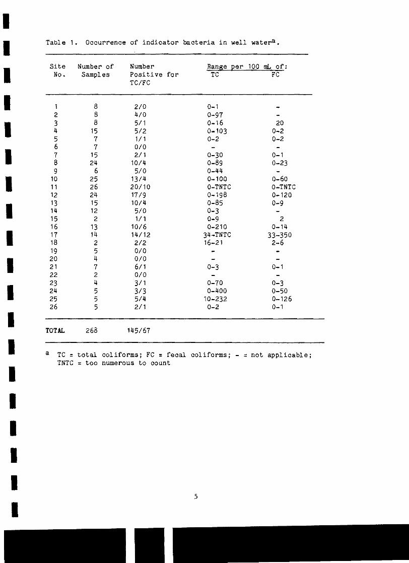
I I Table 1. Occurrence of indicator bacteria in well watera •
Site Number of Number Ranse Eer 100 mL of:
I No. Samples Positive for TC FC TC/FC
I 1 8 2/0 0-1 2 8 4/0 0-97 3 8 5/1 0-16 20 4 15 5/2 0-103 0-2
I I 5 7 1/1 0-2 0-2
6 7 0/0 7 15 2/1 0-30 0-1 8 24 10/4 0-89 0-23
I 9 6 5/0 0-44
10 25 13/4 0-100 0-60 11 26 20/10 O-TNTC O-TNTC 12 24 17/9 0-198 0-120 13 15 10/4 0-85 0-9
I 14 12 5/0 0-3
I 15 2 1/1 0-9 2 16 13 10/6 0-210 0-14 17 14 14/12 34-TNTC 33-350
I 18 2 2/2 16-21 2-6 19 5 0/0 20 4 0/0 21 7 6/1 0-3 0-1
I 22 2 0/0 23 4 3/1 0-70 0-3 24 5 3/3 0-400 0-50 25 5 5/4 10-232 0-126 26 5 2/1 0-2 0-1
I TOTAL 268 145/67
I a TC = total coliformsj FC = fecal coliformsj - =not applicable; TNTC = too numerous to count
I I I I I
5
I

II were present in 18 of them. At 21 sites, total coliforms (Table 1) Ioccurred in levels exceeding the limits of 1 total coliform per 100 mL of potable drinking water suggested by the National Technical Advisory Committee (1968). A total of 38 isolates was tested for drug resistance using the 12 antibiotics listed in Materials and Methods. Of these, 25 I (661) showed resistance to at least one antibiotic and 17 (451) showed multiple resistance. The most frequent drug resistances among total coliforms were to ampicillin, cephalothin, nitrofurantoin and tetracycline (Tabl e 2).
Fecal coliforms were found in 18 of the 26 sites sampled (Table 1). According to current standards (NTAC, 1968), drinking water should be free of fecal coliform contamination in a 100 mL sample. Of the 22 sites showing total and/or fecal coliform contamination, 18 are used actively as sources of drinking water. We tested 27 fecal coliform isolates for drug resistance, and 9 (331) showed resistance to one or more of the 12 antibiotics. Four (161) were multiply resistant. Resistance to tetracycline and cephalothin was found to be the most prevalent in fecal coliforms (Table 2).
Highest levels of indicator bacteria were detected immediately after rainfalls of 0.6 cm (0.25 inches) or greater. These levels dropped off I rapidly within 2-days after rain stopped. Several wells were negative for indicator bacteria on repeated sampling, were positive after a rainfall event, and were negative again on subsequent sampling. I
A total of 161 samples were tested for coliphages, and 13 (171) were found to be positive, with numbers ranging from 60-2820 coliphages per 400 IL of groundwater (Table 3). Coliphages appeared in 7 of the 18 sites sampled, with multiple occurrences at 4 of the 7 sites. It is interesting to note that two of the samples positive for coliphages were from water free of both total and fecal coliforms and six coliphage-positive samples I had total coliforms but no fecal coliforms.
Viruses were isolated from only two sites: site 11 yielded echovirus Itype 25 and site 17 yielded coxsackievirus type B-2 (Table 4). The former site is a private well, whereas the latter is a spring near the city of Fountain. Approximately half of all the samples (n = 137) were tested by the cDNA dot-blot hybridization technique (Margolin et al., 1987). Eight I samples gave positive indication for viruses, and one-sample (from site 11) also was positive for virus isolation. Of the samples positive for viruses, all 9 had total coliforms, but only 5 had fecal coliforms. None of I the samples was positive for viruses of veterinary importance.
DISCUSSION I Microbial contamination of groundwater represents a major source of
waterborne disease outbreaks (Yates et al., 1984). According to current standards for potable water, finished water should contain no fecal coli I forms and no more than one total coliform/100 ml of water(NTAC, 1968). Groundwater contamination may occur when surface water enters deposits of underground water without being thoroughly filtered by the soil. IGroundwater contamination in southeastern Minnesota may be attributed to the karst topography which is characterized by the presence of sinkholes, subsurface cracks, and disappearing streams. These structures can act as I
6 I

I Table 2. Drug Resistance in Indicator Bacteriaa •
I Site Number Tested Number Resistant Drug Resistance Pattern in b : No. TC FC TC FC TC FC
I 7 2 SSS,TE SSS,TE
8 4 3 3 o Am,CF,SI
I Am ,FD ,SSS Am
I 10 4 3 2 CF Am,CF ,FD Am,CF
11 5 3 3 o Am,SI Am,CF,KI Am
I 12 6 3 4 3 CF TE CF ,FD CF CF GM,K
I Am ,CF ,FD,TMP
13 5 2 4 Am,CF CF ,SI Am ,CF ,FD,TE CF ,FDI CF
I 15 o Am,CF,FD,PB,SSS,TE
16 3 3 2 o Am,K
I CF
17 6 6 3 3 Am,CF TE Am,CF,TE TE
I Am,CF,TE TE
I 18 2 2 2 o Am
Am,CF,SSS
Total 38 27 25 9
I a TC = Total coliforms; FC = Fecal coliforms.
I b AM = ampicillin, CR = cephalothin, FD = nitrofurantoin,
GM =gentamicin, K = kanamycin, PB = polymyxin B, SI = spectinomycin, SSS = triple sulfa, TE = tetracycline, TMP = trimethoprim-sulfamethoxazole.
I c No resistance found.
I I I
7

I Table 3. Coliphages in Groundwater. I Site Number of Number (I) Range (mean) of coliphages per
No. Samples Positive 400 L of groundwater I 8 13 (8) 720 I
10 14 2 (14) 60-130 (95)
11 14 4 (29 ) 60-2820 (930) I 13 13 (8) 180
14 10 2 (20) 120-600 (360) I 17 9 2 (22) 65-140 (102) I18 2 (50) 150
ITotal 75 13 (17) 60-2820 (453)
I I
Table 4. Occurrence of viruses in well watera.
ISite Virus Detection Total Coli forms Fecal Col iforms Samples Positive
No. by DNA Hybridi per 100 mL per 100 mL by Virus Isolation zation and Virus
Identification I 7 + 30 0 I 8 + 30 23
10 + 100 60 11 + TNTC TNTC I11 + 41 0 11 + 25 0 Echovirus type 25 12 + 70 57 16 + 21 14 I 17 TNTC 0 Coxsackie Virus
Type B-2 I a TNTC = too numerous to count; + = positive; - = negative.
I I
8
I

I pathways by which contaminants can reach groundwater. Surface soils
I generally act as a natural filter for water as it passes downward to the aquifer. However, in a karst area, the direct connection between surface water and groundwater eliminates the filtration function of the soil. The
I random occurrence of karst features in this region may account for the
I variable quality of well water (Ross, 1979). The transient and recurring nature of detectable levels of indicator bacteria observed in this study may indicate that potable water in this region is continually being invaded by surface pollutants.
I In this study, bacterial contamination was widespread, which is not
surprising because this region is characterized by active karst topography
I and sinkhole development over widespread areas. The groundwater was also positive for human enteric viruses albeit at a low level. One of the reasons for this could be the use of positively charged filters for virus concentration. Although these filters have been used in several studies and have been shown to recover viruses, it is possible that viruses adsorbed and subsequently eluted from these filters somehow get in
I activated. Some evidence that this may have occurred derives from the fact that viruses were detected by dot-hybridization in eight samples but viruses could be isolated in cell culture from only one sample (Table 4).
I Late in the study, we discovered that negatively charged filters detected
I more viruses in raw sewage than did the positively charged filters (Goyal, unpublished data). We are continuing these studies to determine the relative efficiency of virus recovery by these two filter types and the role of positively charged filters in virus inactivation.
I Farm animals are known to excrete a variety of viruses in their feces
(Derbyshire and Brown, 1978). Many of these viruses are pathogenic to farm livestock, and some may be transmissible to man. Adult animals excrete fewer viruses than the recently weaned animals. Information on the
I occurrence of animal viruses in groundwater is non-existent. In the present study, no animal virus could be demonstrated in any of the 268 samples. This may reflect the real absence of such viruses in groundwater, but another reason may be that the methods used in this study were origi
I nally designed to concentrate human enteric viruses (Goyal and Gerba, 1983; Sobsey and Jones, 1979), and they may not be suitable for the concentration and detection of animal enteric viruses.
I
A major concern following the exposure of humans and animals to fecally contaminated water is the potential introduction of foreign drug-resistant bacteria (Goyal et al., 1979; Goyal and Hoadley, 1979). If drug-resistant bacteria colonize the gastrointestinal tract of animals or humans, the potential exists for diseases to develop that are resistant to therapeutic levels of commonly employed antibiotics. Thirty-three percent of the fecal coliforms tested in this study showed some level of drug resistance. The source of these drug resistant bacteria is considered to be animal or human carriers that have had repeated or long term exposure to these drugs. These bacteria may constitute a threat by themselves by colonizing the human gastrointestinal tract and also may transfer their drug resistance to other bacterial pathogens (Goyal and Hoadley, 1979).
I In our study the highest and most frequent levels of fecal and total coliforms occurred in two of the shallowest wells: one 19 feet deep and the other 20 feet deep. Deeper wells (70 feet and over) showed little or
I 9
I

I no detectable contamination. The wells that were cased, i.e. drilled and lined with concrete, showed extremely low levels of total coliforms or none I at all.
Although a high correlation has been shown between the presence of Icoliforms and the occurrence of pathogens in surface water and water distribution systems several studies indicate that the lack of demonstrable coliform levels in groundwater does not preclude the presence of all pathogens (Yates et al., 1984; Geldreich, 1972; Boring et al., 1971). I Human viruses have been detected in situations where indicator bacteria were absent (Deetz et al., 1984; Goyal, 1983; Keswick et al., 1984; Marzouk et al., 1979), and the present study substantiates these data. I
Hallberg et al (1983) assessed regional groundwater quality problem in a karst carbonate aquifer - The Big Spring in Clayton County, Iowa. They found the bacterial contamination of the groundwater aquifer to be related to peak I runoff periods. Persistent bacterial problems were suspected to be associated with faulty domestic water systems rather than the karst-groundwater system. I
They separated groundwater discharge into two principle components e.g., 1) a base flood or infiltration component; and 2) a peak conduit flow related to surface water run-in to sink holes. The run-in component was believed to con Itribute to the peak bacteria problems in groundwater.
According to Gupta (1988), however, the infiltration component should be better defined. He contends that one or two inches of water may not be enough I to saturate soil so that water drips out over the restricting layer (or shale) and flows to the conduit. However, if the same one or two inches of water from a watershed concentrates downslope in a small area, it will saturate the soil and Iwill carry the contaminants with the infiltered water. For the purposes of the present report, however, it is suffice to say that if the infiltering water does not come into contact with adequate depth and right type of soil, viruses may Inot be removed (Gerba et al, 1981).
The detection of coliforms is not the only indicator of contamination. In several studies phages have been used because of their greater longevity I and better correlation with enteric viruses (Stetler, 1984). Phages were found in 7 of the 18 sites sampled and 2 out of 13 times in the absence of detectable levels of fecal and total coliforms. Some investigators have Ishown coliphage to be better indicators of enteric viruses than total and fecal coliforms (Stetler, 1984; Vaughn et al., 1983). In the present study, no such correlation was found. However, more work needs to be done on this topic. I
In karst regions where there is a potential for groundwater contamination, well construction plays a crucial role in preventing or I encouraging contaminant invasion. Subsurface cracks not only reach downward to subsurface aquifers but also travel laterally often passing through wells. A high degree of variability was encountered with regard to Iconstruction of wells sampled in this study. Well depths varied from 19 to over 250 feet. Most wells were not cased, and for wells that were cased the casing usually covered only half of the well depth. I
This study indicates bacterial and viral contamination of groundwater in parts of southeastern Minnesota. When people continually consume contaminated
I 10
I

I I water they increase their risk of exposure to hazardous substances. It is
essential, therefore, to increase public awareness of the health risks associated with groundwater contamination. Plan for better watershed
I management and improved well construction are essential if the purity of groundwater in southeastern Minnesota is to be improved. Attempts should be made to decrease the interaction of surface waters with groundwater. Sinkholes can be lined with plastic and filled with cement and soil. To pro
I tect wells from contamination, deeper wells should be dug and they should be
I cased by two or more linings. The transient nature of contamination at several sites in this study indicates the importance of repeated sampling to determin the sanitary quality of well water. It should be realized that this study was conducted in a small portion of southeastern Minnesota. To determine if these results reflect the overall situation in this whole region, further studies should be done throughout southeastern Minnesota.
I I I I I I I I I I I
Mpls.lSt. Psul •
I Fig. 1 Map of the State of Minnesota showing the two counties where this study was carried out.
I 11
I

I
5 3
19 to .~~)~ 7 t "11) i) \J
20...,:.J' 29f t4 28
OLMSTED COUNTY
__------------~90'~-----
StewlifViHe_._._._._._._._._._._._._._._.
I I I I I I I
Fig. 2 Location of wells sampled - Olmsted County
I I I I I I I I I
Fig. 3
I I
FILLMORE COUNTY
21
Location of well sampled - Fillmore County
12

I REFERENCES
I American Public Health Association. 1980. Standard methods for the examination of water and wastewater, 15th Ed. Amer. Public Health Assn.,
I Washington, D.C.
Bell, R.G. and Bole, J.B. 1976. Elimination of fecal coliform bacteria from reed canarygrass irrigated with municipal sewage lagoon effluent. J.
I Environ. Quality, 2:417-418.
I Boring, J.R., Martin, W.T. and Elliot, L.M. 1971. Isolation of Salmonella typhimurium from municipal water, Riverside, California. Amer. J. Epidemiol. 93:49-54.
I Brewer, W.S. and Lucas, J. 1982. Examination of probable causes of individual water supply contamination. J. Env. Hlth. 44:305-307.
Deetz, T.R., Smith, E.M., Goyal, S.M., Gerba, C.P., Vollet, J.J., Tsai, L.,
I DuPont, H.L. and Keswick, B.H. 1984. Occurrence of rota- and enteroviruses in drinking and environmental water in a developing nation. Water Res. ~:567-571.
I Derbyshire, J.B. and Brown, E.G. 1978. Isolation of animal viruses from farm livestock waste, soil and water. J. Hyg. ~:295-302.
I Geldreich, E.E. 1972. Water-borne pathogens. In: R. Mitchell (Ed.), Water Pollution Microbiology. John Wiley and Sons, New York.
I Gerba, C.P. and Goyal, S.M. 1985. Pathogen removal from wastewater during groundwater recharge. In: T. Asano (Ed.), Artificial Recharge of Groundwater, pp. 283-317. Butterworths, Boston, MA.
I Gerba, C.P., Goyal, S.M., Cech, 1., and Bogdan, GF. 198,. Quantitative assessement of the adsorptive behavior of virus to soils. Environ. Sci. Technol. 12:940-944.
I I Goyal, S.M. 1983. Indicators of viruses. In: G. Berg (Ed.), Viral
Pollution of the Environment, vol. 1, pp. 211-230. C.R.C. Press, Boca Raton, FL.
I Goyal, S.M. and Gerba, C.P. 1979. Comparative adsorption of human enteroviruses, simian rotavirus, and selected bacteriophages to soils. Appl. Environ. Microbiol. 38:241-247.
I Goyal, S.M. and Gerba, C.P. 1982. Concentration of viruses from water by membrane filters. In: C.P. Gerba and S.M. Goyal, (Eds.), Methods in Environmental Virology, pp. 59-116. Marcel Dekker, New York, NY.
I Goyal, S.M. and Gerba, C.P. 1983. Viradel method for detection of rotavirus from seawater. J. Virol. Methods. 1:279-285.
Goyal, S.M., Gerba, C.P. and Melnick, J.L. 1978. Preval ence of human
I enteric viruses in coastal canal communities. J. Water Pollute Control Fed. 50:2247-2256.
I 13
I

I Goyal, S.M., Gerba, C.P. and Melnick, J.L. 1919. Transferable drug resistance in bacteria of coastal canal water and sediment. Water Res., I.11:349-356.
Goyal, S.M. and Hoadley, A.W. 1919. Salmonellae and their associated Rplasmids in poultry processing wastes. Rev. Microbiol. (S. Paulo, I Brazil) t lQ.:50-58.
Goyal, S.M., Zerda, K.S. and Gerba, C.P. 1982. Concentration of coli Iphages from large volumes of water and wastewater. Appl. Environ. Microbiol. 39:85-91.
IGupta, S. 1988. Personal communication.
Hallberg, G.R., Hoyer, B.E., Bettis, E.A. and Libra, R.D. 1983. Hydrogeology, water quality, and land management in the Big Spring Basin, Clayton County, IIowa. Iowa Geological Survey Report 83-3, Iowa City, Iowa.
Katzenelson, E., Fattal, B. and Hostovesky, I. 1916. Organic flocu Ilation: A second step concentration method for the detection of viruses in tapwater. Appl. Environ. Microbiol. 1916; ~:638-639.
Keswick, B.H., Gerba, C.P., DuPont, H.L. and Rose, J.D. 1984. Detection I of enteric viruses in treated drinking water. Appl. Environ. Microbiol. 41:1290-1294.
IMargolin, A.B., Hewlett, M.J. and Gerba, C.P. 1985. Use of a cDNA dotblot hybridization technique for detection of enteroviruses in water. Proc. Water Qual. Technol. Conf., Houston, TX, December 8-11, 1985; pp 81-95. I Marzouk, Y., Goyal, S.M. and Gerba, C.P. 1919. Prevalence of enteroviruses in groundwater of Israel. Groundwater 11:481-491. I Melnick, J.L., Rennick, V., Hampil, B., Schmidt, N.J. and Ho, H.H. 1913. Lyophilized combination pools of enterovirus equine antisera: Preparation Iand test procedures for the identification of field strains of 42 enteroviruses. Bull. World Hlth. Org. 48:263-268.
National Technical Advisory Committee. 1968. Water Quality Criteria. I Federal Water Pollution Control Administration, Washington, D.C.
Onyekaba, C., Bueon, L., King, P., Fahrmann, J. and Goyal, S.M. 1981a. ISusceptibility of various cell culture systems to pseudorabies virus. Compo Immunol. Microbiol. Infect. Dis. lQ.:163-166.
IOnyekaba, C., Bueon, L., King, P., Fahrmann, J. and Goyal, S.M. 1981b. Comparison of five cell lines for the propagation of bovine viral diarrhea (BVD) and infectious bovine rhinotracheitis viruses. Microbiologica lQ.:3 11 -3 15. I Payment, P., Trudel, M. and R. Plante. 1985. Elimination of viruses and indicator bacteria at each step of treatment during preparation of drinking Iwater seven water treatment plants. Appl. Environ. Microbiol. 49: 1418-1428.
I 14
I

I I
Ross, E.H. 1979. State groundwater protection programs - adequate? p. 94-101 Proc. 4th Nat. Groundwater Qual. Symp., Minneapolis, MN.
I Sobsey, M.D. and Jones, B.L. 1979. Concentration of poliovirus from tap water using positively charged microporous filters. Appl. Environ. Microbiol. 37:588-595.
I Stetler, R.E. Coliphages as Indicators of Enteroviruses. 1984. Appl. Environ. Microbiol. 48:668-670.
Vaughn, J.M., Landry, E.F. and Thomas, M.Z. 1983. Entrainment of viruses
I from septic tank leach fields through a shallow, sandy soil aquifer. Appl. Environ. Microbiol. 45:1474-1480.
I Yates, M.V., Gerba, C.P., Kelley, L.M. 1984. Virus persistence in groundwater. Appl. Environ. Microbol. 49:778-781.
I I I I I I I I I I I I
15
I