Gross anatomy & histology of ileum, jejunum
17
Practical demonstration of gross anatomy & histology of Jejunum, Ileum,Appendix and Colon By Dr. Abdul Waheed Ansari Chairperson &Prof. Anatomy, RAKCOMS. RAKMHSU. 12/18/2014 1
-
Upload
abdul-ansari -
Category
Health & Medicine
-
view
1.334 -
download
1
Transcript of Gross anatomy & histology of ileum, jejunum
Practical demonstration of gross anatomy & histology of Jejunum,
Ileum,Appendix and Colon
By Dr. Abdul Waheed Ansari
Chairperson &Prof. Anatomy, RAKCOMS. RAKMHSU.
12/18/2014 1
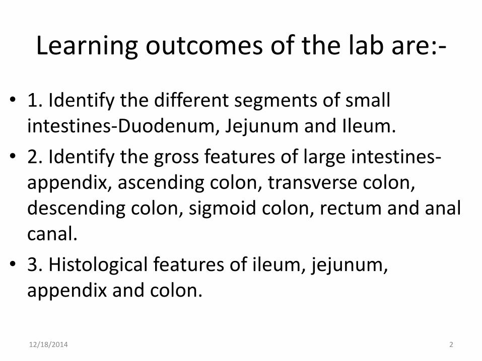
Learning outcomes of the lab are:-
• 1. Identify the different segments of small intestines-Duodenum, Jejunum and Ileum.
• 2. Identify the gross features of large intestines-appendix, ascending colon, transverse colon, descending colon, sigmoid colon, rectum and anal canal.
• 3. Histological features of ileum, jejunum, appendix and colon.
12/18/2014 2

Features of duodenum
• It is the first part of small intestines, 25 cms long.
• It is retroperitoneal except first inch of first part of duodenum.
• It has four parts, C shaped.
• Second part has opening of CBD and pancreatic ducts, major duodenal papilla and minor duodenal papilla.
12/18/2014 3

The Jejunum is the second portion of small intestines
• It starts from duodenojejunal flexure to ileocaecal junction.
• The proximal 1/3rd are the jejunum loops and distal 2/3rd
are the loops of ileum.• The jejunum loops are more
muscular and more vascular where as ileal loops are thin walled and less vascular, interior shows plicae circularis.
• Internally ileum have Payer’s patches.
• The plicae circularis are few in the ileum.
12/18/2014 4

The arterial supply of jejunum and ileum
• The superior mesenteric artery is the artery of midgut.
• It gives jejunal branches and ileal branches and terminates as ileocaecal artery.
• The accompanying vein joins the splenic vein to form the portal vein.
12/18/2014 5

Histology of Jejunum• The mucosa of the small
intestine is lined by simple columnar epithelium which evaginates into villi.
• The epithelium of villi is continuous with that of adjacent crypts.
• Lamina propria of the small intestine forms the core of villi and surrounds the crypts.
• The muscularis mucosa of the small intestine forms a thin layer.
12/18/2014 6

12/18/2014 7
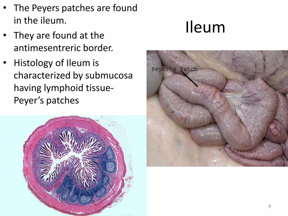
Ileum • The Peyers patches are found
in the ileum.
• They are found at the antimesentreric border.
• Histology of Ileum is characterized by submucosa having lymphoid tissue-Peyer’s patches
12/18/2014 8

Appendix • The human's appendix averages
11 cm in length but can range from 2 to 20 cm.
• The diameter of the appendix is usually between 7 and 8 mm
• It has no haustrations, no taenia coli and no plicae circularis and no Peyer’s patches and no appendices epiploicae.
• It has a mesentery of its own, carrying the artery to appendix.
• The tip of the appendix can have a variable position within the abdominal cavity :
• retro-caecal (65-70%)
• pelvic (25-30%)
• pre- or post-ileal (5%)
• Inflammation is called as appendicitis.
• Pain will be felt around umbilicus because of referred pain-T10 segment dermatome.
• The arterial supply- appendicular artery, a branch of the ileocolic artery which is a branch of the superior mesenteric artery.
12/18/2014 9
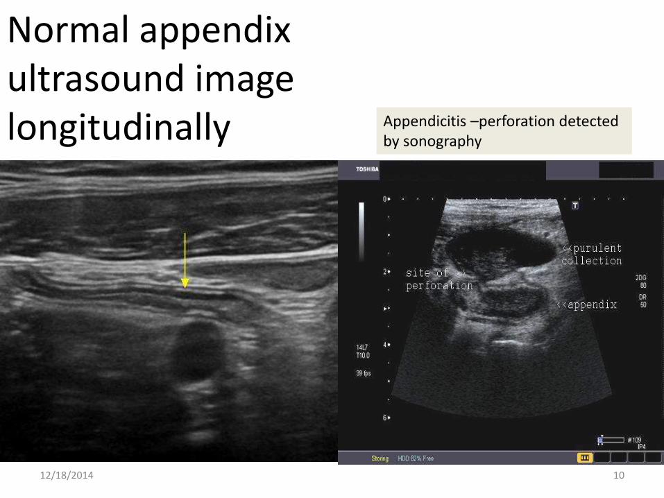
Normal appendix ultrasound image longitudinally
12/18/2014 10
Appendicitis –perforation detected by sonography

Sonography of swollen and inflamed appendix longitudinal section
• Appendicitis shows up as a tubular structure that does not push out of the way or compress, often with changes in the adjacent fat from the inflammation.
• This will often cause the patient to say, “Ouch, that is where it hurts.”
12/18/2014 11

Histology of appendix
12/18/2014 12

Colon histology
• The mucosa is having simple columnar epithelium with lots of mucus glands-goblet cells. Simple tubular intestinal glands.
• Submucosa having neurovascular bundles.
• Muscularis external having both circular and longitudinal fibers- taenia coli are longitudinal bundles
12/18/2014 13

The large intestines
• The caecum, appendix , ascending colon, transverse colon, descending colon, sigmoid colon, rectum and anal canal are parts of large intestines.
• It is supplied by branches from superior and inferior mesenteric arteries.
• At the anal canal there is anastomosis between systemic veins and portal veins, hence one of the sites for anal hemorrhoids formation.
• It shows haustrations, taenia coli, appendices epiploicae, all typical features of large intestines, except appendix rectum and anal canal.
• The anal canal has dual epithelial lining, upper part simple columnar and lower part stratified squamous non keratinized.
• The anal canal has two sphincters, external and internal. External anal sphincter is voluntary where as internal is involuntary.
12/18/2014 14
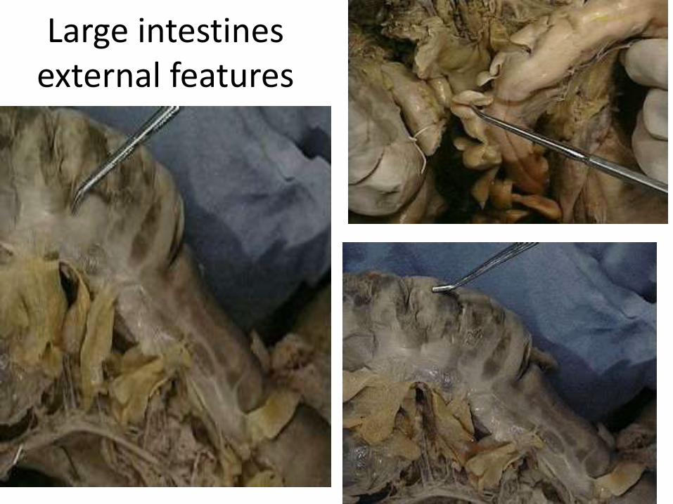
Large intestines external features
12/18/2014 15
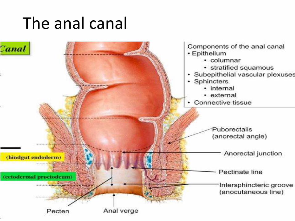
The anal canal
12/18/2014 16

References
• http://www.studyblue.com/notes/note/n/anal-triangle/deck/8502291
• http://ect.downstate.edu/courseware/haonline/labs/L39/130202.htm
• http://histologyatlas.wisc.edu/slides/398
• http://quizlet.com/17115737/27-digestive-system-flash-cards/
• http://www.ultrasoundcases.info/Cases-Home.aspx
12/18/2014 17