27 filling defects in the jejunum and ileum
19
27 Filling Defects in the Jejunum and Ileum
-
Upload
muhammad-bin-zulfiqar -
Category
Education
-
view
69 -
download
1
Transcript of 27 filling defects in the jejunum and ileum

27 Filling Defects in the Jejunum and Ileum

CLINICAL IMAGAGINGAN ATLAS OF DIFFERENTIAL DAIGNOSIS
EISENBERG
DR. Muhammad Bin Zulfiqar PGR-FCPS III SIMS/SHL

• Fig GI 27-1 Leiomyoma of the jejunum (arrow).31

• Fig GI 27-2 Hemangiomatosis of the small bowel and mesentery. Characteristic phleboliths are associated with multiple filling defects in the small bowel.

• Fig GI 27-3 Peutz-Jeghers syndrome. Multiple small bowel hamartomas are present in a patient with mucocutaneous pigmentation.32

• Fig GI 27-4 Primary adenocarcinoma of the ileum (arrow) appearing as an annular constricting lesion.

• Fig GI 27-5 Lymphoma. Note the large, bulky, irregular lesion (arrow).

• Fig GI 27-6 Lymphoma. Multiple large irregular masses.
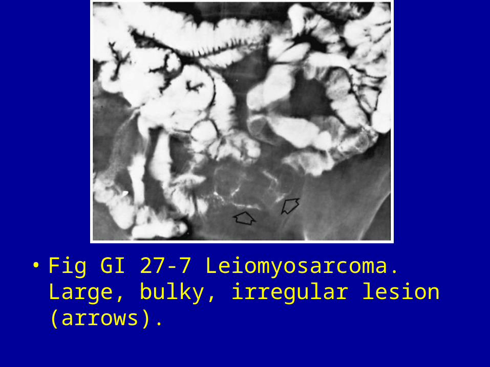
• Fig GI 27-7 Leiomyosarcoma. Large, bulky, irregular lesion (arrows).

• Fig GI 27-8 Metastatic hypernephroma. Multilobulated nodular mass in the proximal jejunum.

• Fig GI 27-9 Carcinoid tumor (arrow).

• Fig GI 27-10 Gallstone ileus. The obstructing stone (white arrows) in the jejunum is associated with evidence of barium in the biliary tree (black arrow).

• Fig GI 27-11 Ascaris. The linear intestinal tract of the roundworm is filled with barium (arrow).32

• Fig GI 27-12 Nodular lymphoid hyperplasia. Large filling defects suggest multiple polypoid masses.

• Fig GI 27-13 Phytobezoar. Large, irregular, proximal jejunal filling defect containing barium within the interstices of the lesion. Note the second bezoar in the stomach.33

• Fig GI 27-14 Crohn's disease. Multiple polypoid lesions in the distal jejunum and proximal ileum show both smooth and lobulated contours.34
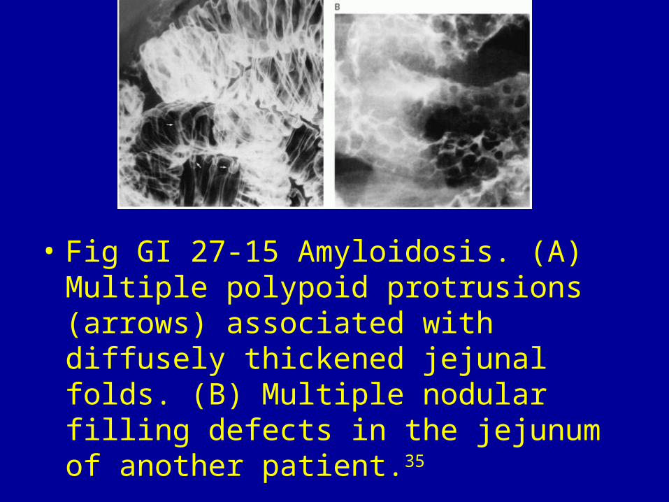
• Fig GI 27-15 Amyloidosis. (A) Multiple polypoid protrusions (arrows) associated with diffusely thickened jejunal folds. (B) Multiple nodular filling defects in the jejunum of another patient.35