Granulomas in the Liver- - UCSF Departments of …labmed.ucsf.edu/uploads/517/285... · Granulomas...
96
Granulomas in the Liver- with an emphasis on infectious etiologies Laura W. Lamps, M.D. University of Arkansas for Medical Sciences
Transcript of Granulomas in the Liver- - UCSF Departments of …labmed.ucsf.edu/uploads/517/285... · Granulomas...

Granulomas in the Liver-with an emphasis on infectious etiologies
Laura W. Lamps, M.D.University of Arkansas for Medical Sciences
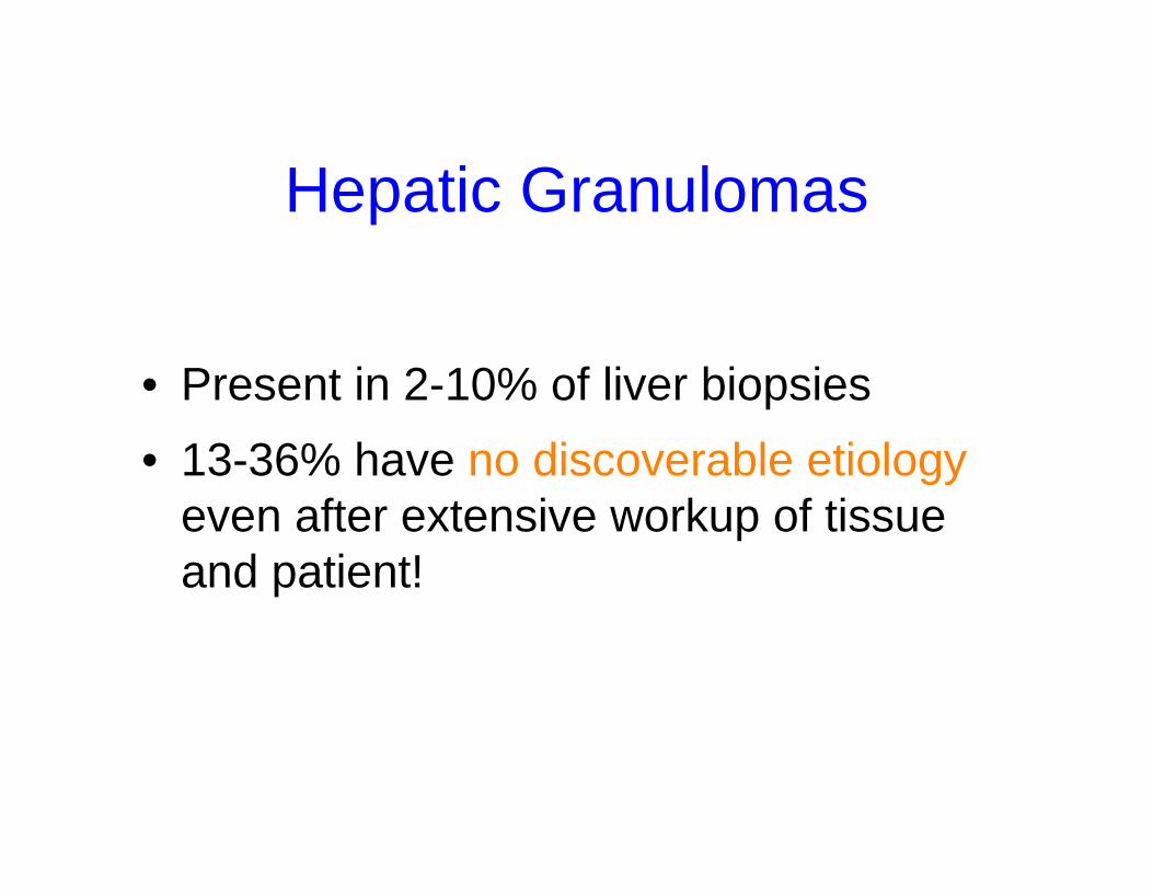
Hepatic Granulomas
• Present in 2-10% of liver biopsies• 13-36% have no discoverable etiology
even after extensive workup of tissue and patient!
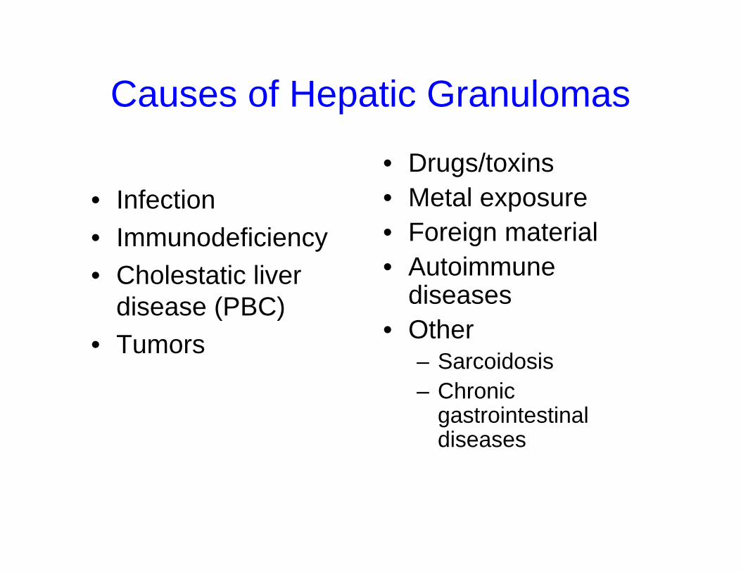
Causes of Hepatic Granulomas
• Infection• Immunodeficiency• Cholestatic liver
disease (PBC)• Tumors
• Drugs/toxins• Metal exposure• Foreign material• Autoimmune
diseases• Other
– Sarcoidosis– Chronic
gastrointestinal diseases

Morphological Classification of Granulomas
• Epithelioid (+/-) necrosis• Lipogranulomas• Microgranulomas• Fibrin ring granulomas• Foamy macrophage aggregates• Granulomatous inflammation• Stellate abscess with granulomatous
inflammation

Morphological Classification of Granulomas
• Epithelioid (+/-) necrosis– Discrete with distinct edges– Necrosis, lack of respect for architecture are often
associated with infection– TB, sarcoidosis
• Lipogranulomas– Contain lipid– Mineral oil
• Microgranulomas– Some define as 3-7 cells in cross-section– Very nonspecific; often associated with other
inflammatory cells– Drug reaction, Listeria
Morphological Classification of Granulomas
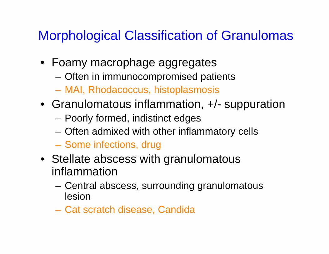
• Foamy macrophage aggregates– Often in immunocompromised patients– MAI, Rhodacoccus, histoplasmosis
• Granulomatous inflammation, +/- suppuration– Poorly formed, indistinct edges– Often admixed with other inflammatory cells– Some infections, drug
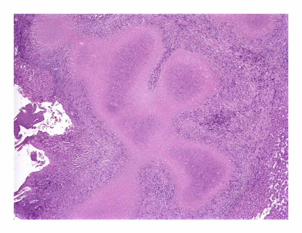
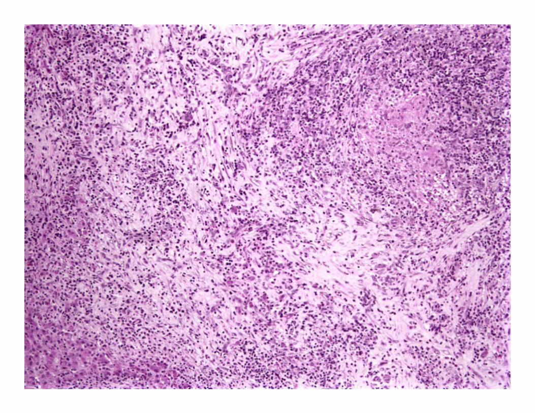
• Stellate abscess with granulomatous inflammation– Central abscess, surrounding granulomatous
lesion– Cat scratch disease, Candida

Helpful questions to ask:
• Morphology of granuloma• Accompanying inflammatory infiltrate• Location of granulomas• Nature of necrosis, if present• Is there anything in the granuloma• Other associated morphologic changes• Need for special stains

Helpful clinical questions to ask:
• Immune status of patient
• Exposure to animals
• Foreign travel
• Medication/drug history

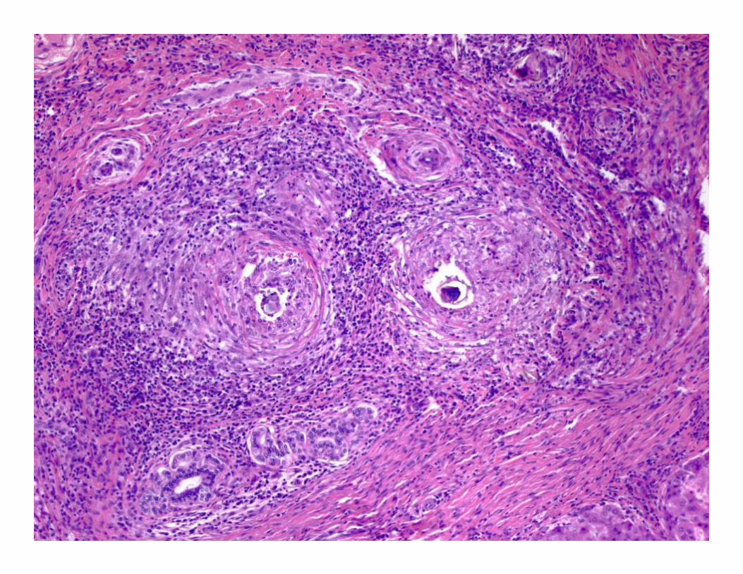
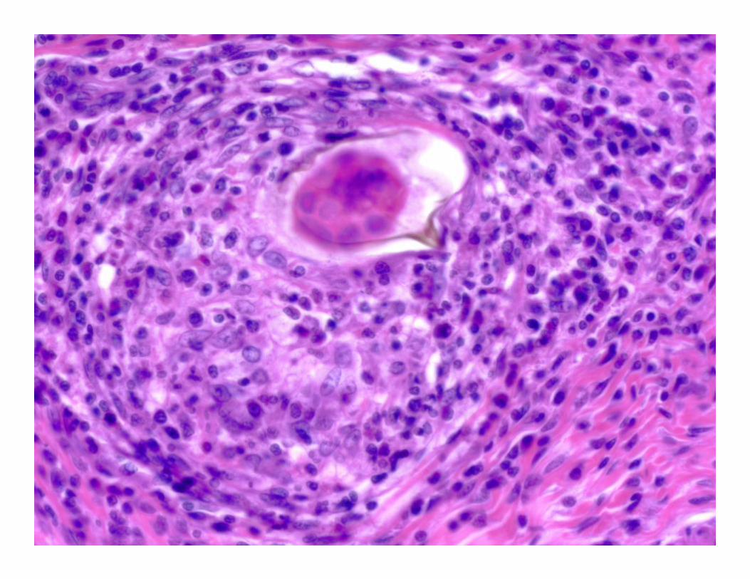
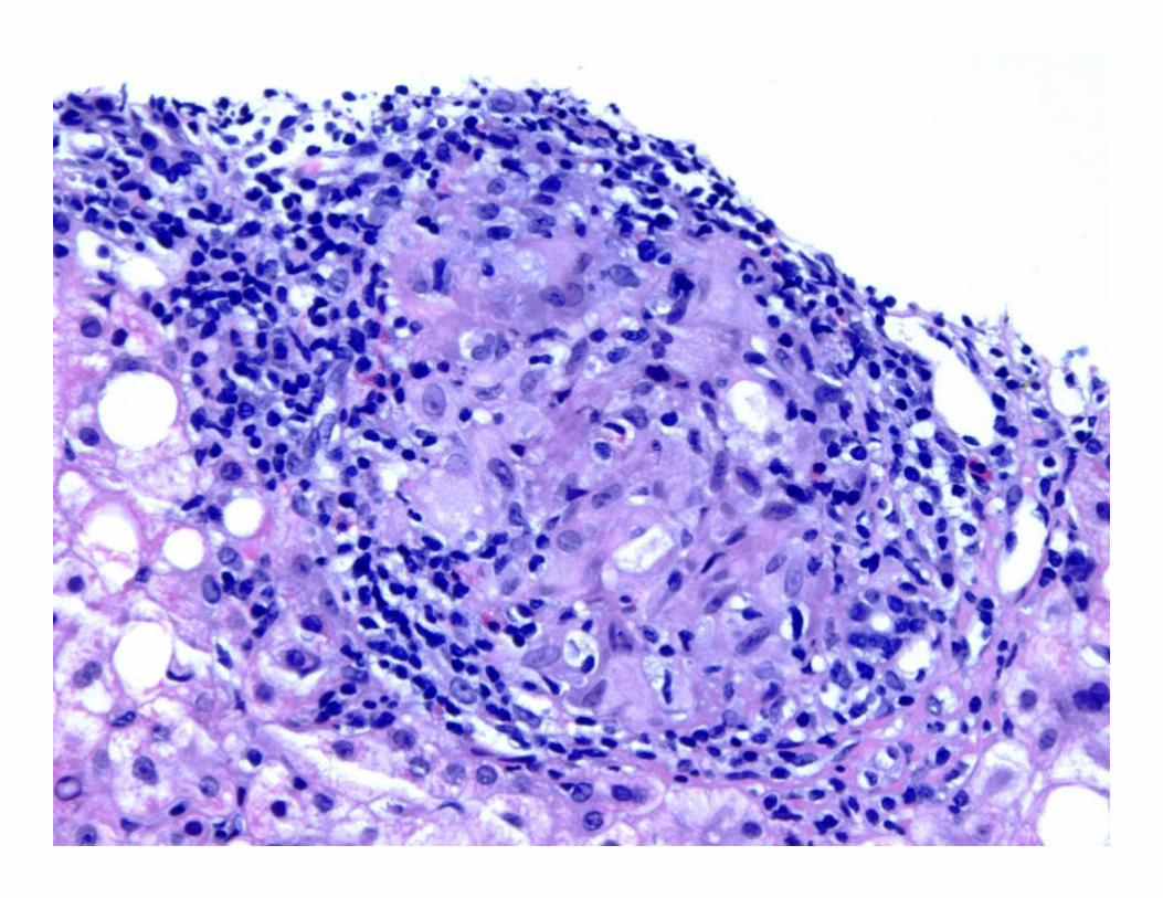
Fibrin Ring Granuloma
• Epithelioid granuloma composed of lipid vacuole surrounded by fibrin ring
• Classically described in association with Q-fever
• Also associated with CMV, EBV, MAI, typhoid, drug reaction, Hodgkin’s disease

Courtesy Dr Dhanpat Jain


Is it time for wine and cheese yet?

Infectious Causes of Hepatic Granulomas
• Viral– CMV, EBV, HCV
• Bacterial– Cat scratch disease– Mycobacteria– Lyme disease– Brucella– Tularemia– Rickettsia– Whipple’s disease
• Fungal– Histoplasmosis– Candida
• Parasitic– Schistosomiasis– Ascaris– Pinworms– Toxoplasma– Fasciola hepatica

Mycobacterium tuberculosis• Granulomas present in virtually all
cases of miliary TB• Signs/symptoms of liver disease may be
dominant presenting feature• Presentation ranges from asymptomatic
to fever/RUQ pain/hepatomegaly• Helpful tests: special stains, PCR,
culture


Courtesy Dr. Joe Misdraji


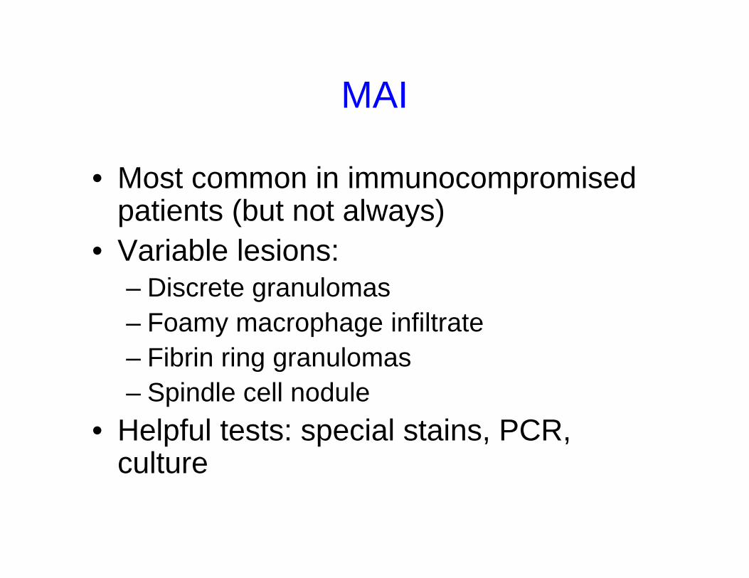
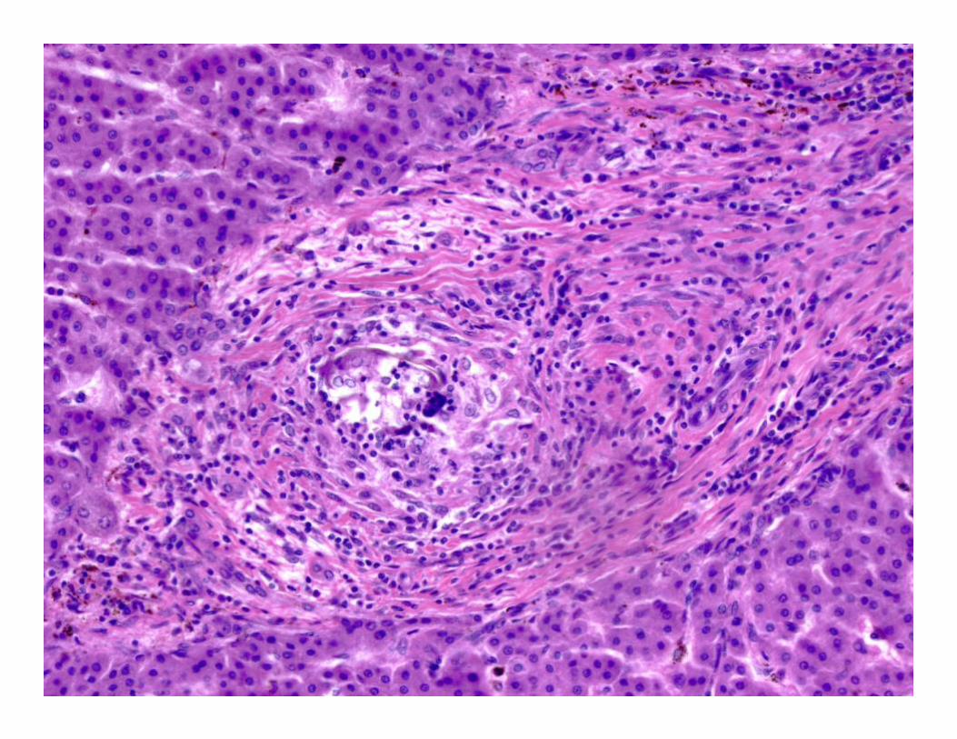
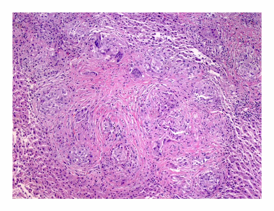
MAI
• Most common in immunocompromised patients (but not always)
• Variable lesions: – Discrete granulomas– Foamy macrophage infiltrate– Fibrin ring granulomas– Spindle cell nodule
• Helpful tests: special stains, PCR, culture

MAI with epithelioid granulomas
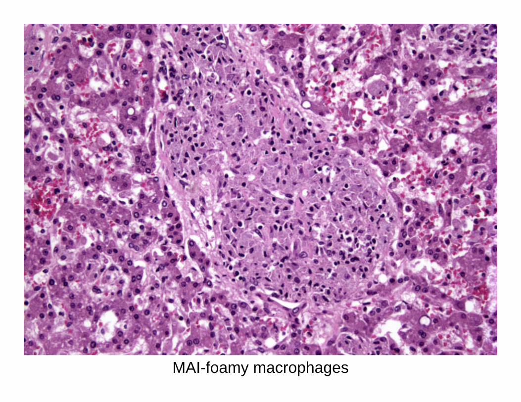
MAI-foamy macrophages


MAI-fibrin ring granulomas

MAI-spindle cell nodule


Leprosy
• Both lepromatous and tuberculoid leprosy involve the liver; often subclinical
• Lesions depend on type of leprosy, but may be “in-between” the classic types
• Bacilli common in lepromatous leprosy, rare in tuberculoid
• Liver may harbor bacilli even when skin is clear
• Helpful tests: special stains, culture, PCR


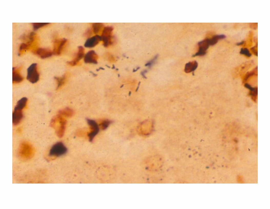
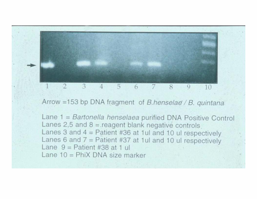
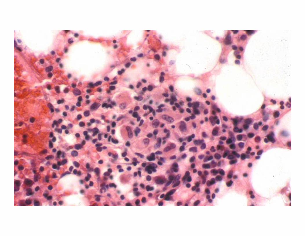
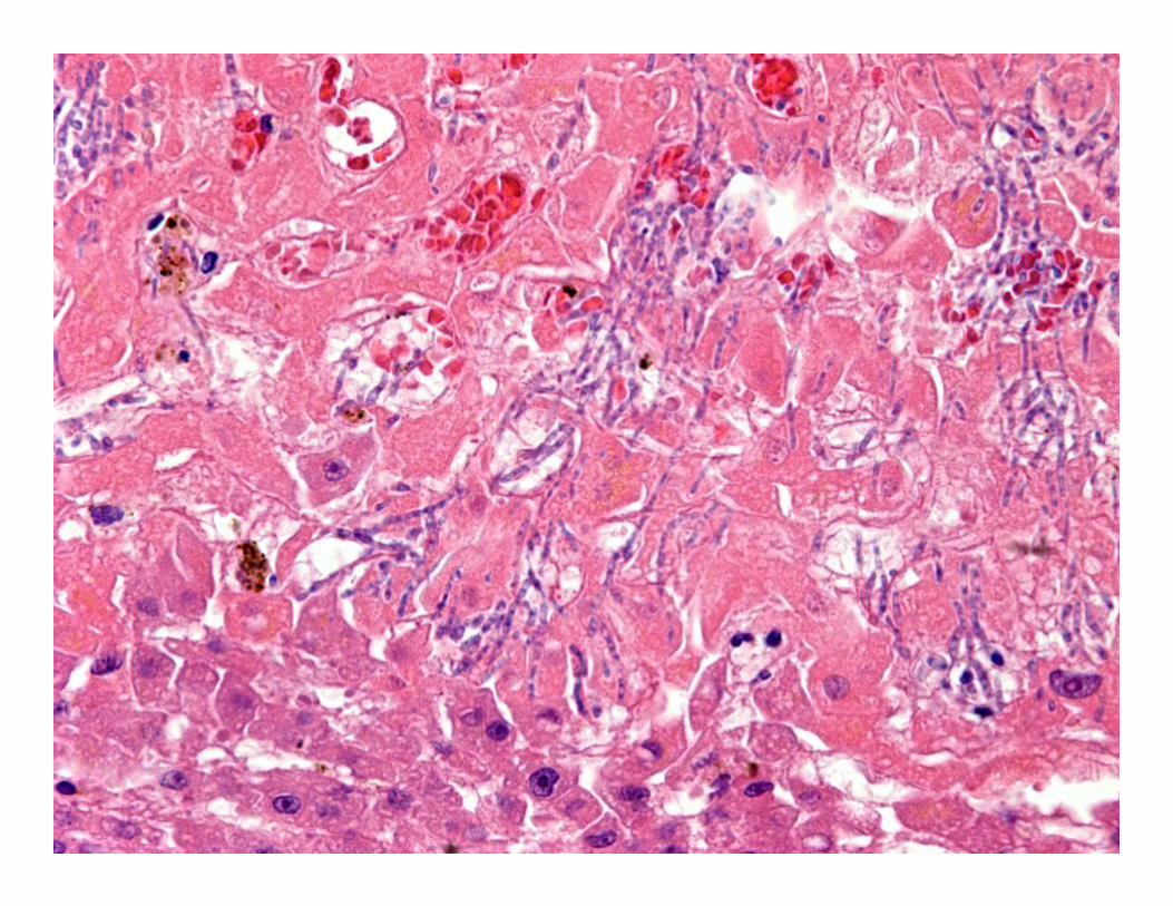
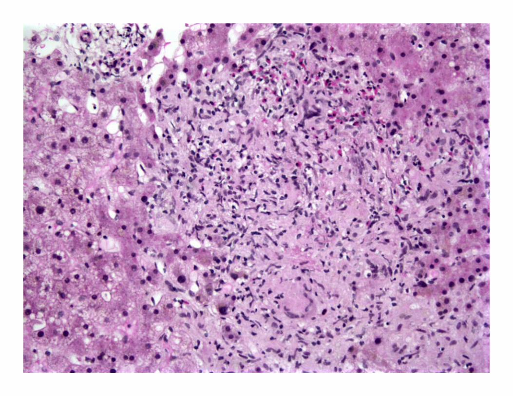
Cat Scratch Disease
• Small percentage of patients have disseminated disease
• Lack inoculation site• Usually not immunocompromised• Helpful tests: special stains, PCR,
ELISA, history
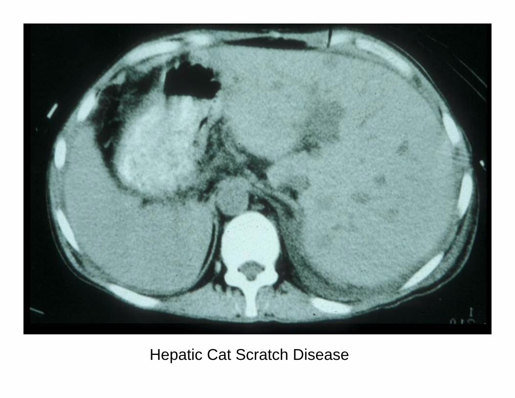
Hepatic Cat Scratch Disease


Brucellosis
• Exposure to farm animals, contaminated food
• Dominant systemic symptoms; liver involved in about half of cases
• Helpful tests: history, serologies; special stains and culture not helpful

Courtesy Dr. David Walker

Tularemia
• Transmitted through contact with rodents/rabbits
• Patients often systemically ill, +/-hepatomegaly and elevated transaminases
• Helpful tests: serologies, PCR, culture; special stains not helpful


I think we need more beds.

Hepatic Fungal Infections
• Usually part of disseminated disease• Patients usually immunocompromised• Liver involvement manifests with
hepatomegaly, abdominal pain, elevated transaminases and bilirubin
• Helpful tests: special stains, culture

Hepatic Fungal Infections
• Candida-granulomas with central suppuration
• Histoplasma-lymphohistiocytic nodules• Aspergillus, Zygomycetes-suppurative;
rare granulomas• Cryptococcus-very variable, can involve
biliary tree


H&E/methenamine silver


Cryptococcus


Fontana-Masson


Schistososmiasis
• Most common worldwide cause of portal hypertension
• Granulomatous reaction is usually to the eggs; eggs harder to find as disease progresses
• Helpful tests: finding eggs in urine, feces, or tissue (shells and spines variably acid-fast); serologies


Schistosomal haemozoin pigment


Viral Infections
• Both epithelioid and fibrin ring granulomas associated with EBV, CMV
• Also in a minority of HCV and HBV patients
• Must try and rule out other causes of hepatic granulomas, however


Important Non-infectious Causes of Liver Granulomas
• Primary cholestatic disorders
• Chronic GI disease• Vasculitides• Adverse drug
reaction
• Metal toxicity• Foreign material• Inherited disorders• Reaction to
neoplasms• Sarcoidosis

Sarcoidosis
• Liver involved in majority of cases, second only to lung and nodes
• May cause fibrosis, cirrhosis, and cholestatic liver disease
• Helpful tests: chest xray, ACE assay; must rule out other causes of granulomas


Adverse Drug Reaction• Many different granuloma
morphologies; necrosis within granulomas is rare
• Look for associated inflammation, duct injury, vascular injury
• Combination of granulomatous inflammation + hepatocellular damage very suggestive of drug reaction

Allopurinol

Coneflower Tea

Propylthiouracil


Other Noninfectious Etiologies
• Vasculitis/collagen vascular diseases (polyarteritis nodosa, Churg-Strauss, Lupus)
• Chronic biliary disease (PBC, PSC)• Chronic GI diseases
– not clear if granulomas are primary or associated with drugs, PSC, other in cases of UC, Crohn’s with granulomas
– Idiopathic eosinophilic enteritis may cause granulomas in biliary tree, liver


In Summary
• Morphology of granuloma can be clue to diagnosis
• Portal lymph node pathology may be helpful• Low threshold for special stains• Culture, molecular testing, and serologic
studies are very useful diagnostic tools• Clinical history may be the diagnostic tool that
is most helpful, cheapest, but not always easiest to get

Thanks!
• Dr. Joe Misdraji• Dr. Lucas Campbell• Dr. David Walker• Dr. George F. Gray, Jr.• Dr. Margie Scott