
Going beyond the surface of your retina - visioncare.biz OCT...1. START • Auto gnmentali • Auto...
8
Going beyond the surface of your retina OCT-HS100 OPTICAL COHERENCE TOMOGRAPHY
Transcript of Going beyond the surface of your retina - visioncare.biz OCT...1. START • Auto gnmentali • Auto...

Going beyond the surface of your retina
OCT-HS100OPTICAL COHERENCE TOMOGRAPHY
your retina
1. START
• Auto alignment• Auto focus• Auto C-gate
2. CAPTURE
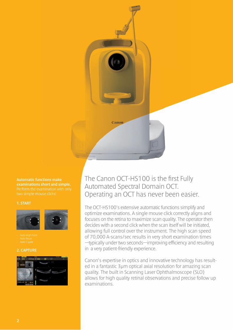
The Canon OCT-HS100 is the fi rst Fully Automated Spectral Domain OCT. Operating an OCT has never been easier.
The OCT-HS100’s extensive automatic functions simplify and optimize examinations. A single mouse click correctly aligns and focuses on the retina to maximize scan quality. The operator then decides with a second click when the scan itself will be initiated, allowing full control over the instrument. The high scan speed of 70,000 A-scans/sec results in very short examination times—typically under two seconds—improving effi ciency and resulting in a very patient-friendly experience.
Canon’s expertise in optics and innovative technology has result-ed in a fantastic 3µm optical axial resolution for amazing scan quality. The built in Scanning Laser Ophthalmoscope (SLO) allows for high quality retinal observations and precise follow up examinations.
Automatic functions make examinations short and simple. Perform the examination with only two simple mouse clicks!
2

Full Auto The OCT-HS100’s Full Auto feature signifi cantly simplifi es operation; standard procedures can easily be delegated and results are operator independent.
High Image Quality.3µm axial optical resolution
Auto anterior eye alignment and trackingThe OCT-HS100 will automatically maintain the exact alignment on the center of the eye .
Auto Fundus tracking by SLOBy detecting the amount of movement in fundus images, the unit can automatically com-pensate for small involuntary movements of the eye.
Auto C-gate control (Coherence Gate)Scan depth is automatically adjusted.
Auto FocusAutomatic compensation of patient refraction.
10 mm scan widthIncreased effectiveness of examinations by capturing large areas with just one scan.
Easy examinationsFive stored examination sets: Macula Disease, Glaucoma, Choroid, Anterior and General. Examinations for other retinal diseases can be easily created in the initial settings menu.
Extensive connectivityThe unit can be used stand-alone or in a network. Tomograms or reports can be output as JPEG, BMP or DICOM fi le.
High scanning Speed70,000 A-scans per second allow two-second examinations for reduced risk of motion artefacts and increased patient comfort.
Full Auto OCTHigh specifi cations in a very compact design
Built in SLOFor high quality retinal observation and precise follow up examinations
3
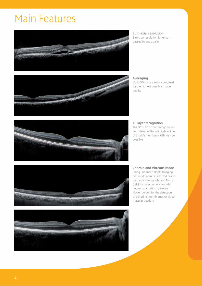
3µm axial resolution3 micron resolution for unsur-passed image quality
AveragingUp to 50 scans can be combined for the highest possible image quality
10 layer recognitionThe OCT-HS100 can recognize ten boundaries of the retina; detection of Bruch’s membrane (BM) is now possible
Choroid and Vitreous modeUsing Enhanced Depth Imaging, two modes can be selected based on the pathology: Choroid Mode (left) for detection of choroidal neovascularization. Vitreous Mode (below) for the detection of epiretinal membranes or vitreo macular traction;
4
Main Features
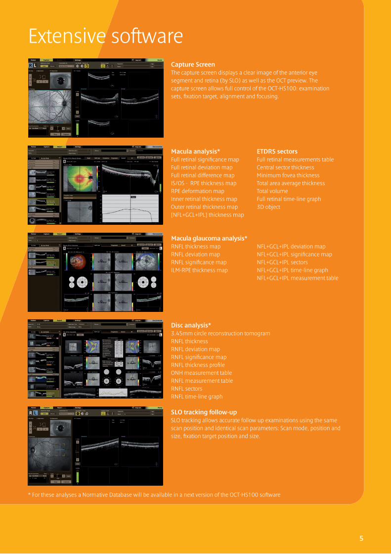
Extensive softwareCapture ScreenThe capture screen displays a clear image of the anterior eye segment and retina (by SLO) as well as the OCT preview. The capture screen allows full control of the OCT-HS100: examination sets, fi xation target, alignment and focusing.
Macula analysis* Full retinal signifi cance mapFull retinal deviation mapFull retinal difference mapIS/OS - RPE thickness mapRPE deformation mapInner retinal thickness mapOuter retinal thickness map[NFL+GCL+IPL] thickness map
ETDRS sectorsFull retinal measurements tableCentral sector thicknessMinimum fovea thicknessTotal area average thicknessTotal volumeFull retinal time-line graph3D object
Macula glaucoma analysis* RNFL thickness map RNFL deviation mapRNFL signifi cance mapILM-RPE thickness map
NFL+GCL+IPL deviation mapNFL+GCL+IPL signifi cance mapNFL+GCL+IPL sectorsNFL+GCL+IPL time-line graphNFL+GCL+IPL measurement table
Disc analysis*3.45mm circle reconstruction tomogramRNFL thicknessRNFL deviation mapRNFL signifi cance mapRNFL thickness profi leONH measurement tableRNFL measurement tableRNFL sectorsRNFL time-line graph
SLO tracking follow-up SLO tracking allows accurate follow up examinations using the same scan position and identical scan parameters: Scan mode, position and size, fi xation target position and size.
5
* For these analyses a Normative Database will be available in a next version of the OCT-HS100 software
A-scans/sec Max 70,000 Fundus Preview Fs SLO
Axial resolution 3 µm Observation light source 780 ± 5nm
Transversal Resolution 20 µm Internal Eye Fixation 2 mm or 6mm , 590nm (orange)
Pupil size requirement Min 3.0 mm Field of view 10 x 10 mm, OCT 33O x 33O, SLO 44O x 33O
Scanning width 2 ~10 mm Dimensions (WxDxH) 387 x 499 x 474 (mm)
Scan depth 2 mm Weight 29 (kg)
OCT light source 855 nm ±5 nm Optional Accessory Anterior segment adapter (ASA-1)
Working Distance 35 mm
Specifications
Macula 3DAn area scan is done centering on the macula., for examination of the macula and the deeper layersScan size 10 x 10 mm
Extensive scan modesCrossHigh resolution scan with up to 50 times averaging for highest image qualityScan size 3 x 3 to 10 x 10 mm
Glaucoma 3D A vertical area scan of the macula for ex-tensive analysis of the NFL+GCL+IPLScan size 10 x 10mm
Multi CrossMult purpose high resolution scans with up to 10 times averaging.Scan size 3 x 3 to 10 x 10 mm
Disc 3DArea scan for disc analysisScan size 6 x 6 mm
Anterior 3D*Area scan is perfromed on the anterior segment.Scan size 6 x 6 mm
CustomGeneral application, for analysis of any diseaseScan size 3 x 3 to 10 x 10 mm
Anterior cross*High resolution cross scan, for imaging cornea and angle.Scan size 3 x 3 to 10 x 10 mm
* With optional adapter
6
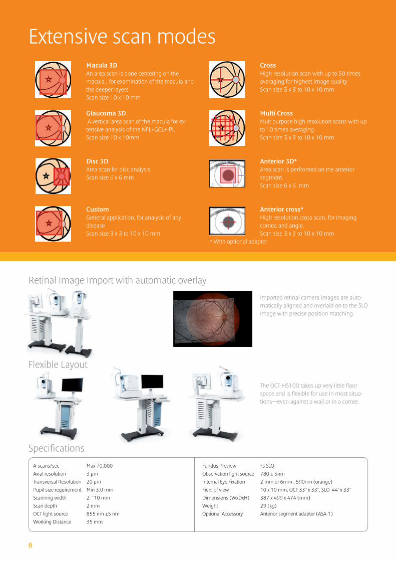
Retinal Image Import with automatic overlay
Flexible Layout
Imported retinal camera images are auto-matically aligned and overlaid on to the SLO image with precise position matching.
The OCT-HS100 takes up very little fl oor space and is fl exible for use in most situa-tions—even against a wall or in a corner.
7
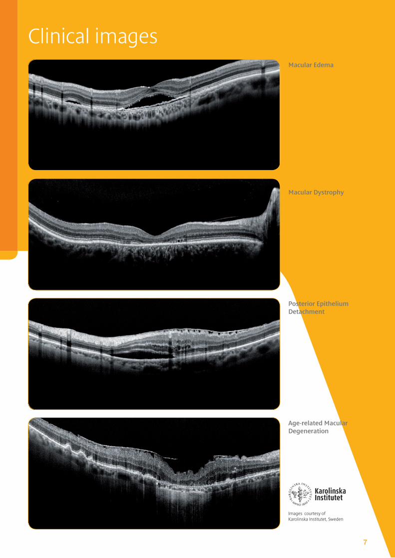
Clinical images
Posterior Epithelium Detachment
Age-related Macular Degeneration
Macular Edema
Macular Dystrophy
Images courtesy of Karolinska Institutet, Sweden
Canon Europa N.V. Medical Systems Division
Bovenkerkerweg 59 – 61 1185 XB Amstelveen The Netherlands Phone: +31(0)20-5 45-89 26 Fax: +31(0)20-5 45-82 20 www.canon-europe.com/medical
OCT-HS100English-NL Edition 2166V375© Canon Europa N.V. 2013
Canon Eco Canon Quality Canon Flexibility
Canon has been defining the future with innovative solutions for more than 70 years. In all that time we’ve constantly strived to improve medical diagnostics in healthcare. Perhaps that’s what made us a leading global provider of eye care solutions.
Our actions are based on honesty and sustainability.
Safety and quality are an integral component of our actions.
Everything we do has to have a superior customer advantage.
Choose the eye care system of the future and let our local, authorized Canon dealer advise you:


![GSK Part 1 Oct 2010_compressed.ppt Auto Saved]](https://static.fdocuments.net/doc/165x107/577d202c1a28ab4e1e9228c2/gsk-part-1-oct-2010compressedppt-auto-saved.jpg)


