GI Bleeding for the PCP - cmetracker.netcmetracker.net/PPMC/Files/EventMaterials/35-GIBleeding.pdfGI...
24
GI Bleeding for the PCP Viju P. Deenadayalu, M.D. The Oregon Clinic Gastroenterology East Outline • Acute upper GI bleeding - Variceal - Non-variceal • Acute lower GI bleeding • Occult/Obscure GI bleeding - 35a -
Transcript of GI Bleeding for the PCP - cmetracker.netcmetracker.net/PPMC/Files/EventMaterials/35-GIBleeding.pdfGI...
GI Bleeding for the PCP
Viju P. Deenadayalu, M.D.The Oregon Clinic
Gastroenterology East
Outline
• Acute upper GI bleeding
- Variceal
- Non-variceal
• Acute lower GI bleeding
• Occult/Obscure GI bleeding
- 35a -

Etiology of Upper GI Bleeding
• Peptic ulcer disease (PUD) – 35%
• Esophagitis
• Gastric erosions
• Arteriovenous malformations (AVMs)
• Mallory Weiss tear
• Dieulafoy’s lesion
• Varices – 9%
• Malignancy
Clinical Management of UGI Bleeding
• Assess hemodynamic status• Resuscitation
– ABC’s (intubation, large bore IV) • CBC; type and cross-match; fluids• Monitoring of vital signs and urine output• Nasogastric tube• Empiric therapy (IV PPI, octreotide)• Endoscopy - Diagnosis & therapy
– Indications for urgent endoscopy • Active bleeding• Shock• Elderly patients
- 35b -

Clinical Presentation
• Hematemesis – 30%
• Melena – 26%
• Both 41%
• Hematochezia – 3%
Prognostic Indicators
• Etiology
• Severity of initial bleed
• Age (> 60 worse prognosis)
• Comorbid disease(s)
• Ulcer size (> 2 cm increased mortality)
• Hospitalization
- 35c -

Variceal Bleeding
Esophageal Varices
• Prevalence: 60 to 80% of cirrhotics• Incidence: 4 to 12% per year
– 225 pts, surveillance EGD q1-3 yrs, followed 10 years
– 41% developed varices, 55% did not develop varices– Progression in size: 5% per year– Bleeding rate: 7% per year
D’Amico, Gastroenterology, 2001
• Predictors for bleeding– High Child-Pugh Score– Large varices– Stigmata (red wale, hemocystic spots, etc.)
- 35d -

Esophageal Varices Endoscopic Grading: Size and Stigmata
• Size– Absent– Small– Medium– Large
• Stigmata– Red wale marking– Hemocystic spot– Cherry red spots
Jensen, Gastroenterology, 2001
Gastric Varices
• Prevalence: 4 to 10%
• Risk of bleeding
– 16% at 1 year
– 36% at 3 years
– 44% at 5 yearsKim, Hepatology, 1997
• Predictors for bleeding
– High Child-Pugh
– Red markings
– Size
- 35e -

Primary Prophylaxis: Summary
• Large varices, high Child-Pugh Score, cherry red spot or red wale signs
• Propranolol 40 mg bid or nadolol 40 mg daily
– 25% reduction in resting pulse rate or pulse rate of 60
• Endoscopic band ligation
– As effective as beta-blocker therapy
– Considered for cirrhotics who are not candidates for drug therapy (COPD, IDDM)
– Typically do not band small varices
Octreotide
• Longer acting analog of somatostatin
• Reduces azygos blood flow (a measure of collateral blood flow including variceal flow)
• Bolus of 50 mcg, 50 mcg/hour infusion for 3 -5 days
• No effects on cardiac function, systemic blood pressure, or extra splanchnic tissue perfusion
• No special monitoring required
- 35f -

Endoscopic Band Ligation
• Limited field of vision
• Slightly more difficult to perform for acute bleeding
• Place 1 to 2 bands per varix
• Start distally and work proximally
ACG Algorithm for bleeding cirrhotics
Rebleeding
ResuscitationOctreotide x 5d
Early EGD
Non portal hypertensive
Portal hypertensivegastropathy
EsophagealVarices
Gastric Varices
Treatappropriately
Beta-blockerBand ligationSclerotherapyBeta-blocker
TIPSShunts
Liver Transplant (B/C)
Variceal ablationBand ligation
SclerotherapyBeta-blocker
TIPS
- 35g -
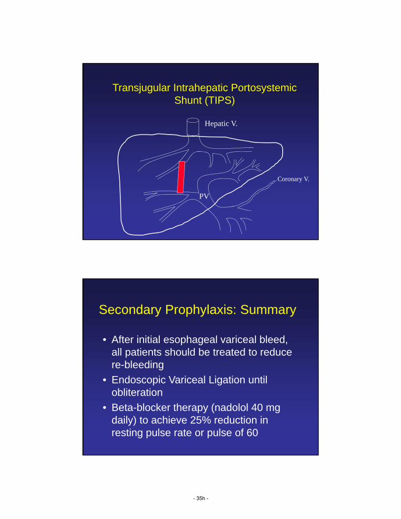
Transjugular Intrahepatic Portosystemic Shunt (TIPS)
Hepatic V.
PV
Coronary V.
Secondary Prophylaxis: Summary
• After initial esophageal variceal bleed, all patients should be treated to reduce re-bleeding
• Endoscopic Variceal Ligation until obliteration
• Beta-blocker therapy (nadolol 40 mg daily) to achieve 25% reduction in resting pulse rate or pulse of 60
- 35h -

Non-variceal UGI Bleeding
Stigmata of Recent Hemorrhage
• Spurting
• Oozing
• Adherent clot
• Visible vessel
• Flat pigmented spot
• Clean base ulcer
- 35i -

Endoscopic risk stratification
Endoscopic appearance Rebleeding
(%)
Surgery
(%)
Mortality
(%)
Clean based 5 0.5 2
Flat spot 10 6 3
Adherent clot 22 10 7
Non-bleeding visible vessel 43 34 11
Active bleeding 55 35 11
Laine and Peterson, N Engl J Med 1994; 331:717
Treatment
• Epinephrine injection (1:10,000)
• Electrocoagulation (BICAP)
• Thermal therapy (Argon Plasma Coagulation)
• Hemostasis clips
- 35j -

Management of Re-bleeding
• Signs of re-bleeding– Hematemesis, melena, orthostasis, fall in Hgb >2 g/dL
within 24 hours
• Repeat endoscopy
• Perform 2nd endoscopic hemostasis
• If bleeding stops, close observation
• Uncontrolled bleeding or 2nd re-bleed, then surgery or embolization (interventional radiology)
Pharmacotherapy for Prevention of Ulcer Re-bleeding
Agent BenefitsH2-receptor antagonists no
Anti-fibrinolytics no
Vasoconstrictors no
Proton pump inhibitors yes
- 35k -

Influence of intragastric pH on hemostasis and proteolysis
• pH<5
– Pepsin accelerates clot lysis
• pH>6
– Effective platelet aggregation
– Irreversible inactivation of pepsin
– Optimal maintenance of hemostasis
Summary: Criteria of IV PPI use
• In treatment of bleeding ulcer, randomized trials of high dose PPI by continuous infusion is effective for prevention re-bleeding
• Dose must maintain intragastric pH>6• No evidence of mortality reduction• Use limited to high-risk patients with active
bleeding or non-bleeding visible vessels at time of EGD
- 35l -

Effects of Hp Eradication
• Reduce recurrent ulcer & ulcer complications
• Reduce risk of aspirin-induced ulcer bleeding
• Is insufficient to protect against NSAID-induced ulcer bleeding
Chan et al. N Engl J Med 2001
ACG recommended first-line H. pylori eradication regimens
PPI* + clarithromycin + amoxicillin 1 gm bid or
bid 500 mg bid metronidazole 500 mg bid
All for 10 to 14 days
PPI for 1 month
Howden & Hunt. Am J Gastro 1998
- 35m -

Summary: Management of Bleeding Ulcers
• Team approach
• Identify high risk patients & resuscitation
• Early endoscopy + hemostasis
• Combined therapy (Epinephrine + Heat Probe or Mechanical metal clips)
• IV and then oral PPI, monitor for re-bleeding
• Eradicate H. pylori infection
• Stop NSAIDs or use of COX-2 selective inhibitors or use prophylactic therapy
Other causes of UGI Bleeding
Mallory-Weiss Tear (MWT)
• Linear, longitudinal fissure-like tear at or below GE junction
• Lower power setting vs. PUD bleeding
• Injection therapy for cirrhotics with MWT
• High dose PPI for non-active bleeding
- 35n -

Other causes of UGI Bleeding
Dieulafoy lesion
• High body of stomach or fundus location
• Arterial bleeding in absence of underlying ulcer
• Similar hemostasis methods used for bleeding ulcer disease
Lower GI Bleeding (LGIB)• Bleeding distal to the ligament of Treitz
• Annual incidence ~ 20-27 cases per 100,000
• Compared to UGI bleed: 100-200 cases per 100,000
• More common in men
• Incidence increases with age with 200-fold increase from 3rd to 9th decade
• 80-85% cease spontaneously
• Overall mortality 10%
Longstreth, Am J Gastroenterol, 1997; 92:419Longstreth, Am J Gastroenterol, 1995; 90:206
Zuckerman, Gastrointest Endosc, 1999; 49:227
- 35o -

Differential diagnosis: LGIB
Diverticular (40%)
Inflammatory disease (21%)- IBD, infectious, ischemia
Neoplasia (14%)-Cancer, polyps,-Polypectomy sites
Coagulopathy(12%)
Anorectal disease (11%)- Hemorrhoids, fissures
Other (2%): AVM, radiation, small bowel lesions
Vernava et al, Dis Colon Rectum, 1997; 40: 846
Initial Clinical Management : LGIB
• Assess hemodynamic status• Resuscitation• CBC, type and cross-match, fluids• Monitoring of vital signs and urine
output• Nasogastric tube
- 35p -

LGIB: Diagnostic tests• Digital rectal examination
– Anorectal pathology– Up to 40% rectal carcinomas palpable
• Nasogastric aspiration– If positive (gross blood or strongly guaiac
positive), 93% had UGI source • Anoscopy/Sigmoidoscopy
– Diagnostic yield ~ 10%– Role uncertain in era of emergent
colonoscopy
Richter et al, Gastrointest Endosc 1995
LGIB: Colonoscopy
• Urgent colonoscopy after rapid purge– After resuscitation
– Within 8-24 hours upon presentation
• Diagnositic yield: 48-90%– Active bleeding site, non-bleeding visible vessel,
adherent clot, fresh blood localized to colonic segment, ulceration of diverticulum with blood, absence of blood in ileum
• Endoscopic therapy– Hemostasis with injection, coagulation, clipping, APC
Richter et al, Gastrointest Endosc 1995; 41: 93Colacchio et al, Am J Surg 1982; 143: 607
- 35q -

LGIB: Tagged red blood scan
• Technetium labeling– Detects bleeding rates 0.1 mL/min
– Remains in circulation for 48 hours
• ~ 45 % detection rate
• Typically used prior to angiography
• Problems– Reliability of scan to direct surgery
– Overall ~ 22% inaccuracy rate for localization
Hunter et al, Am J Surg 1990; 159: 504Ryan et al, Dis Colon Rectum, 1992; 35: 219
LGIB: Angiography
• Arterial bleeding rate 0.5 mL/min
• After positive TRBC scan
• 47% sensitivity, 100% specificity
• Complication rate 9.3%– Hematoma, thrombosis, renal failure, contrast allergy
• Therapeutic benefit– Localization prior to surgery (methylene blue, catheter or coils
at site, etc.)
– Selective embolization
– Vasopressin infusion
Fiorito et al, Am J Gastroenterol 1998; 143: 569
- 35r -

LGIB: diagnostic studies
• EGD– Up to 11% yield of gastric or duodenal lesions
• Barium Enema/Enteroclysis– Seldom used
• Meckel scan– younger patients
• Capsule endoscopy• Push Enteroscopy
– Single balloon enteroscope, double balloon enteroscopy
LGIB: Diverticular Hemorrhage
• ~80% spontaneously stop
• Transfusion requirement– < 4 U RBC, near 100% stopped bleeding
– > 4 U RBC, 60% required surgery
• Re-bleeding– 25% risk of re-bleeding
– >50% risk of continued bleeding after 2nd bleed
McGuire, Ann Surg 1994; 220: 653
- 35s -

Acute Lower GI Bleeding
Resuscitate
ProctoscopyElective colonoscopy
EGD
Capsule endoscopy
Enteroscopy
NG aspirate/EGD
Urgent colonoscopy
Angiography
SurgeryAngiography
MeckelsEnteroclysis
Bleeding continuesBleeding ceases
Bleeding recurs
Obscure GI Bleeding
• Recurrent or persistent bleeding of the GI tract without obvious source despite endoscopic evaluation and imaging
• Accounts for 5% of patients with GI bleed
• 75% of bleeds involve the small bowel
• Remainder of bleeds may be missed lesions in upper lower GI tract
Davies et al, Gut 1995; 37:346
- 35t -

Repeat EGD and Colonoscopy
• Greatest value in patients with obscure GI bleeding and iron deficiency anemia or overt bleeding with melena or maroon blood
• Colonoscopy yield is lower (consider if inadequate prep)
Occult bleeding
• Initial presentation of + fecal occult blood test and/or iron deficiency anemia without evidence of blood loss– Testing for occult blood loss (FOBT, FIT)
– Evaluation of + FOBT with EGD, colonoscopy, and or small bowel study
- 35u -

Wireless Video Capsule Endoscopy (VCE)
• Equally or more sensitive than other methods of small bowel evaluation
• Revealed additional findings in 25 – 55% of cases
• Wireless VCE may be more effective if performed prior to push enteroscopy
Raju et al., Gastro 2007;133:1694
Deep Small Bowel Enteroscopy
• Double/Single balloon enteroscopy
• Allows visualization and treatment
• DBE detected bleeding sources in 78% of patients with obscure GI bleeding
• Capsule directed DBE may provide better long-term outcomes and cost effectiveness
Shinozaki et al., Gastroenterol Hepatol 2010;8:151Gerson et al., Gastrointest Endosc 2008:68:920
- 35v -

Double Balloon Enteroscopy
Conclusions
• Monitoring vitals and aggressive volume replacement are the sine qua non for patients with severe GI bleeding
• Variceal bleeding has the highest risk of recurrence/death
• Early endoscopy in patients with ongoing upper GI bleeding
• Colonoscopy for patients with acute lower GI bleeding with hemodynamic stability
- 35w -

Questions ?
- 35x -