
Genus Mycobacterium by Dr. Mesola
79
Mycobacterium Mycobacterium Virginia P. Mesola, MD Virginia P. Mesola, MD Cebu Institute of Cebu Institute of Medicine Medicine
-
Upload
degee-gonzales -
Category
Documents
-
view
38 -
download
0
description
Mycobacterium lecture
Transcript of Genus Mycobacterium by Dr. Mesola

MycobacteriumMycobacterium
Virginia P. Mesola, MDVirginia P. Mesola, MD
Cebu Institute of MedicineCebu Institute of Medicine

MycobacteriumMycobacterium
• Most distinctive property – acid fastnessMost distinctive property – acid fastness
• Some are saprophytes in soil, others are Some are saprophytes in soil, others are parasitesparasites
• 2 species causing 2 most dreaded 2 species causing 2 most dreaded diseases – tuberculosis and leprosydiseases – tuberculosis and leprosy
• Some species of environmental Some species of environmental saprophytes – cause human infectionssaprophytes – cause human infections
• Aerobic, slightly curved or straight rodsAerobic, slightly curved or straight rods

MycobacteriumMycobacterium
• Most distinctive structure of cell – Most distinctive structure of cell – multi-layered structure with multi-layered structure with abundance of complex lipids some abundance of complex lipids some unique to mycobacteriaunique to mycobacteria
• Have important characteristics in Have important characteristics in common with Corynebacterium and common with Corynebacterium and Nocardia – called CNM groupNocardia – called CNM group
• All produce mycolic acid and similar All produce mycolic acid and similar guanine-plus-cytosine (G+C) contentguanine-plus-cytosine (G+C) content

TuberculosisTuberculosis
• Pulmonary tuberculosis – most Pulmonary tuberculosis – most common manifestationcommon manifestation
• Primary tuberculosis – primary Primary tuberculosis – primary complexcomplex
• Secondary tuberculosisSecondary tuberculosis


Morphology – M. Morphology – M. tuberculosistuberculosis• Slender, straight or slightly curved rods Slender, straight or slightly curved rods
with rounded endswith rounded ends
• True branching in old culturesTrue branching in old cultures
• Acid fast, no spores, no capsuleAcid fast, no spores, no capsule
• Appear beaded in tissue and sputum due Appear beaded in tissue and sputum due to vacuoles and polyphosphate contentto vacuoles and polyphosphate content
• Acid fastness due to mycolic acid and Acid fastness due to mycolic acid and physical integrity of the cellphysical integrity of the cell


Morphology – M. Morphology – M. tuberculosistuberculosis• Acid fastness explained by lipid barrier Acid fastness explained by lipid barrier
principle – increased hydrophobicity of the principle – increased hydrophobicity of the surface layers follows the complexing of dye surface layers follows the complexing of dye with mycolic acid residues present in cell wall with mycolic acid residues present in cell wall –> prevents exit of carbol fuchsin trapped in –> prevents exit of carbol fuchsin trapped in the cell the cell
• Difficult to stain with gram stain -> failure of Difficult to stain with gram stain -> failure of dye to penetrate the cell wall considered dye to penetrate the cell wall considered gram positivegram positive
• Gram stains of clinical materials – invalid for Gram stains of clinical materials – invalid for identification of mycobacteriaidentification of mycobacteria

M. Tuberculosis – cell wallM. Tuberculosis – cell wall
• Thick wall - 3 layers enclosing a 3 Thick wall - 3 layers enclosing a 3 layered structure plasma membranelayered structure plasma membrane
• Wall is very complex with complex Wall is very complex with complex lipophilic macromoleculeslipophilic macromolecules
• 60% of dry weight of cell wall is lipids 60% of dry weight of cell wall is lipids which enable organisms to resist which enable organisms to resist adverse environmental conditionsadverse environmental conditions

Cultural characteristics Cultural characteristics M. tuberculosisM. tuberculosis
• Obligate aerobeObligate aerobe• Grow on ordinary simple synthetic med., Grow on ordinary simple synthetic med.,
but for 1but for 1oo isolation from clinical material – isolation from clinical material –requires a more complex mediumrequires a more complex medium
• Grow very slowly – colonies seen in 11-12 Grow very slowly – colonies seen in 11-12 days of incubation at 37days of incubation at 37ooCC
• Colonies – small, dry, scaly (cauliflower)Colonies – small, dry, scaly (cauliflower)• Liquid media – pellicle on surface of med.Liquid media – pellicle on surface of med.• Generation time 14-15 hoursGeneration time 14-15 hours


Resistance– M. tuberculosisResistance– M. tuberculosis
• Highly resistant to dryingHighly resistant to drying• Cultures kept for 12 yrs at 37Cultures kept for 12 yrs at 37ooC still viable C still viable
and virulentand virulent• When exposed to direct sunlight:When exposed to direct sunlight:
– Organism from cultures – killed in 2 hoursOrganism from cultures – killed in 2 hours– Organisms in sputum – killed in 20-30 hoursOrganisms in sputum – killed in 20-30 hours
• Bacilli in dried sputum protected from Bacilli in dried sputum protected from sunlight – viable for 6-8 months sunlight – viable for 6-8 months
• More resistant to chemical agents More resistant to chemical agents • Killed by pasteurizationKilled by pasteurization

Determinants of Determinants of pathogenicitypathogenicity• No exotoxin or endotoxinNo exotoxin or endotoxin
• No single structure, antigen or No single structure, antigen or mechanism can explain the virulence of mechanism can explain the virulence of the bacteriathe bacteria
• No simple in-vitro test based on colonial No simple in-vitro test based on colonial morphology & serologic differences – morphology & serologic differences – distinguish virulent from avirulent distinguish virulent from avirulent strains. Possible only with virulence strains. Possible only with virulence testing in animalstesting in animals

Determinants of Determinants of pathogenicitypathogenicity
• Has properties associated with Has properties associated with virulent strains -> progressive virulent strains -> progressive disease, none can account disease, none can account completely for virulence, each plays completely for virulence, each plays a role in pathogenesis of diseasea role in pathogenesis of disease– Cord factor – serpentine cordsCord factor – serpentine cords– Sulfatides – responsible for neutral red Sulfatides – responsible for neutral red
reactivity associated with virulent strainsreactivity associated with virulent strains


Laboratory diagnosisLaboratory diagnosis
• Many species of mycobacteria both Many species of mycobacteria both saprophytes and potential pathogens saprophytes and potential pathogens may be isolated from humansmay be isolated from humans
• If the isolate is mycobacteria other If the isolate is mycobacteria other than M. tuberculosis, the lab should than M. tuberculosis, the lab should be able to identify the species in by be able to identify the species in by in vitro tests.in vitro tests.

Collection of specimenCollection of specimen
• Collect before anti-TB drug therapy is Collect before anti-TB drug therapy is startedstarted
• Use sterile containerUse sterile container• Send specimen to the lab immediatelySend specimen to the lab immediately• A series of 3-5 single early morning A series of 3-5 single early morning
samples of sputum of 5-10 ml is collectedsamples of sputum of 5-10 ml is collected• Other specimens – pus, CSF, urine, Other specimens – pus, CSF, urine,
gastric lavage. Fluids from inflammed gastric lavage. Fluids from inflammed serous cavitiesserous cavities

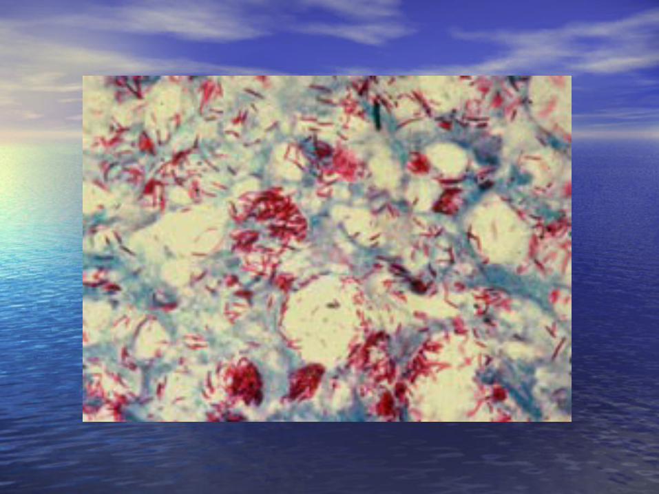
Microscopic examinationMicroscopic examination
• Detection of AFB in stained smears – Detection of AFB in stained smears – easiest and most rapid procedure for easiest and most rapid procedure for evaluating a clinical specimenevaluating a clinical specimen
• Most symptomatic TB patients – sputum Most symptomatic TB patients – sputum is positive for AFB. Sputum exam – has is positive for AFB. Sputum exam – has important role in TB control programimportant role in TB control program
• Making smear –> get caseous areas in Making smear –> get caseous areas in sputum –> spread thin –> stain with AFSsputum –> spread thin –> stain with AFS

Microscopic examinationMicroscopic examination
•Reading smear – make 3 Reading smear – make 3 longitudinal sweeps of the longitudinal sweeps of the stained area parallel to the stained area parallel to the length of the slidelength of the slide


Microscopic examinationMicroscopic examination
• Reading recommended by American Lung Reading recommended by American Lung AssociationAssociation– Number of bacilliNumber of bacilli ReportReport
•00 No AFB seenNo AFB seen
•1-2/slide1-2/slide report # found and report # found and
request repeat specimenrequest repeat specimen
•3-9/slide3-9/slide rare or +rare or +
•10 or more per slide10 or more per slide few or ++few or ++
•1 or more per field1 or more per field Numerous or ++++Numerous or ++++

Digestion and Digestion and DecontaminationDecontamination• Decontamination before culture – Decontamination before culture –
destroy contaminants which grow destroy contaminants which grow faster than the Mycobacteriafaster than the Mycobacteria
• Digestion –since bacteria are usually Digestion –since bacteria are usually trapped in cellular & organic debris, trapped in cellular & organic debris, exudates must be liquified before exudates must be liquified before cultureculture
• N-acetyl cysteine for digestion N-acetyl cysteine for digestion and NaOH for decontamination are usedand NaOH for decontamination are used

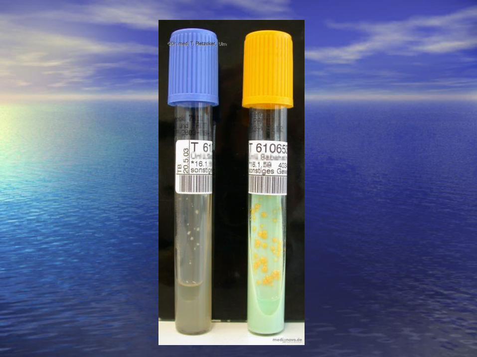
CultureCulture
• Some culture media can detect as few as 10 Some culture media can detect as few as 10 bacteria/ml of digested concentrated materialbacteria/ml of digested concentrated material
• Two types of media:Two types of media:– Egg-potato based media (Lowenstein Jensen)Egg-potato based media (Lowenstein Jensen)– Agar based media (Middlebrook 7H-10)Agar based media (Middlebrook 7H-10)
• Incubate inoculated media at 37Incubate inoculated media at 37ooC in 5-10% C in 5-10% COCO22
• Test for M. tuberculosis – positive niacin testTest for M. tuberculosis – positive niacin test
• Report a negative culture after 2 monthsReport a negative culture after 2 months


M. TB complexM. TB complex
• M. tuberculosisM. tuberculosis
• M. bovis – seen in countries where raw M. bovis – seen in countries where raw milk is ingested. Almost completely milk is ingested. Almost completely eliminated with pasteurization of milkeliminated with pasteurization of milk
• M. africanum – isolated in AfricaM. africanum – isolated in Africa
• Bacillus of Calmette and Guerin (BCG) – Bacillus of Calmette and Guerin (BCG) – attenuated mutant of M. bovisattenuated mutant of M. bovis

Mycobacteria associated with Mycobacteria associated with non-tuberculous infection non-tuberculous infection (MOTT)(MOTT)• Initially considered as strictly Initially considered as strictly
saprophytessaprophytes
• Last 40 years – recognized clinical Last 40 years – recognized clinical significancesignificance
• Also called “atypical” mycobacteriaAlso called “atypical” mycobacteria
• Can produce severe and even fatal Can produce severe and even fatal disease in humans disease in humans

Runyon classification Runyon classification 4 groups of mycobacteria 4 groups of mycobacteria
• Group I – photochromogensGroup I – photochromogens
• Group II – scotochromogensGroup II – scotochromogens
• Group III – nonphotochromogensGroup III – nonphotochromogens
• Group IV – rapid growers – visible Group IV – rapid growers – visible growth in less than 7 daysgrowth in less than 7 days
• Groups I,II,III – slow growers requiring Groups I,II,III – slow growers requiring 7 days or more to yield visible colonies7 days or more to yield visible colonies

Slowly growing Slowly growing MycobacteriaMycobacteria
• PhotochromgenPhotochromgen– M. kansasiiM. kansasii– M. marinumM. marinum
• ScotochromogenScotochromogen– M. scrofulaceumM. scrofulaceum
• Non-photochromogenNon-photochromogen– M. avium-intracellulareM. avium-intracellulare


Rapidly growing Rapidly growing mycobacteriamycobacteria• Most species are purely environmental Most species are purely environmental
saprophytessaprophytes• M. fortuitum and M. chelonei – occasional M. fortuitum and M. chelonei – occasional
pathogens of humans, birds and animalspathogens of humans, birds and animals• M. fortuitum-M. chelonei complex – share M. fortuitum-M. chelonei complex – share
similar characteristics and seen in same similar characteristics and seen in same type of infectiontype of infection
• Colonies appear in 72 hours of incubation Colonies appear in 72 hours of incubation at 37at 37ooCC
• M. smegmatis and M. phlei – do not cause M. smegmatis and M. phlei – do not cause diseasedisease


Laboratory diagnosisLaboratory diagnosis
• Many species of mycobacteria both Many species of mycobacteria both saprophytes and potential pathogens saprophytes and potential pathogens may be isolated from humansmay be isolated from humans
• If the isolate is mycobacteria other If the isolate is mycobacteria other than M. tuberculosis, the lab should than M. tuberculosis, the lab should be able to identify the species in by be able to identify the species in by in vitro tests.in vitro tests.

Laboratory diagnosisLaboratory diagnosis
• Diagnosis of specific organism – crucial in Diagnosis of specific organism – crucial in determining therapydetermining therapy
• ID by Runyon classification is inadequateID by Runyon classification is inadequate
• Do bacteriologic ID of organism and Do bacteriologic ID of organism and repeated demonstration in patient secretions repeated demonstration in patient secretions in the absence of other potential pathogensin the absence of other potential pathogens
• Tuberculin test is not helpfulTuberculin test is not helpful
• A + AF smear and a (–) tuberculin test may A + AF smear and a (–) tuberculin test may lead one to suspect the disease.lead one to suspect the disease.

IdentificationIdentification
• Grow well on Lowenstein-Jensen (LJ), Grow well on Lowenstein-Jensen (LJ), Middlebrook 7H10 and other media Middlebrook 7H10 and other media used for culturing M. TBused for culturing M. TB
• Niacin test negativeNiacin test negative
• Only a few of these are known Only a few of these are known human pathogenshuman pathogens


EpidemiologyEpidemiology
• Ubiquitous and found in all parts of the worldUbiquitous and found in all parts of the world• Endemic in certain geographic areasEndemic in certain geographic areas• No known 1No known 1oo animal host but apparently exist animal host but apparently exist
in the soilin the soil• No evidence of direct transmission of No evidence of direct transmission of
organisms from man to manorganisms from man to man• Disease usually occurs in man with previous Disease usually occurs in man with previous
lung damagelung damage• Disease results from 2 coinciding events:Disease results from 2 coinciding events:
– Colonization of a large # of mycobacteriaColonization of a large # of mycobacteria– Impairment of body’s defense mechanismImpairment of body’s defense mechanism

Disease is group according to Disease is group according to organ involvementorgan involvement
• Pulmonary diseasePulmonary disease– Most common manifestation in adults in Most common manifestation in adults in
USAUSA– Most common cause: M. kansasii; M. Most common cause: M. kansasii; M.
fortuitum, M. avium-intracellulare fortuitum, M. avium-intracellulare complex (MAC)complex (MAC)
• Lymphadentis – usually seen in Lymphadentis – usually seen in childrenchildren– M. scrofulaceumM. scrofulaceum

Disease is group according to Disease is group according to organ involvementorgan involvement
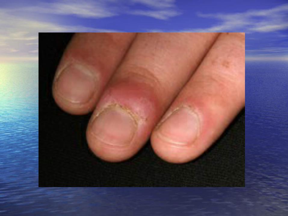
• Cutaneous lesions (skin lesions)Cutaneous lesions (skin lesions)– USA – M. marinumUSA – M. marinum– Africa and Southeast Pacific – M. ulceransAfrica and Southeast Pacific – M. ulcerans
• Disseminated infections Disseminated infections – M. kansasii; M. fortuitum; MACM. kansasii; M. fortuitum; MAC– AIDS - disseminated infection usually AIDS - disseminated infection usually
caused by M. avium-intracellulare complexcaused by M. avium-intracellulare complex ((MAC)MAC)

Mycobacterium lepraeMycobacterium leprae
• Leprosy is an ancient disease.Leprosy is an ancient disease.• Hansen in 1874 described the myriads Hansen in 1874 described the myriads
of bacilli in the lesions of leprosy of bacilli in the lesions of leprosy patients.patients.
• Cannot be cultured in vitro.Cannot be cultured in vitro.• Difficult to propagate and transmit to Difficult to propagate and transmit to
experimental animals.experimental animals.• Slow growth in both animals and in Slow growth in both animals and in
patients.patients.

Mycobacterium lepraeMycobacterium leprae
• Leprosy is a disease that continues to Leprosy is a disease that continues to threaten the quality of life of more than 12 threaten the quality of life of more than 12 million people world wide.million people world wide.
• With increase in population and rapid With increase in population and rapid means of transportation, there is means of transportation, there is increased contact between susceptible increased contact between susceptible travelers and millions of patients who have travelers and millions of patients who have leprosy.leprosy.
• Leprosy is of global importance.Leprosy is of global importance.

Mycobacterium lepraeMycobacterium lepraeMorphologyMorphology
• Acid fast in modified mononuclear or Acid fast in modified mononuclear or epitheloid cells called lepra cells arranged epitheloid cells called lepra cells arranged like packets of cigars.like packets of cigars.
• Organisms are found singly or in large Organisms are found singly or in large masses called globi.masses called globi.
• Bacilli are usually straight or curved and may Bacilli are usually straight or curved and may stain uniformly or show granular beads.stain uniformly or show granular beads.
• Uniformly stained bacilli are healthy bacilli; Uniformly stained bacilli are healthy bacilli; beaded bacilli are probably non viable.beaded bacilli are probably non viable.


Mycobacterium lepraeMycobacterium lepraeMorphologyMorphology• Acid fastness can be removed by Acid fastness can be removed by
extraction with pyridine – distinguishes M. extraction with pyridine – distinguishes M. leprae from other mycobacterialeprae from other mycobacteria
• Structure resembles that of M. TBStructure resembles that of M. TB• Not grown in tissue cultures of various Not grown in tissue cultures of various
types of human cellstypes of human cells• Grown experimentally only in animalsGrown experimentally only in animals• Phenolase in M. leprae has been obtained Phenolase in M. leprae has been obtained
from lepromatous nodules – separates M. from lepromatous nodules – separates M. leprae from other mycobacteria and leprae from other mycobacteria and nocardias.nocardias.

M. leprae – experimental M. leprae – experimental disease in animalsdisease in animals• Animal models – extensively used for study Animal models – extensively used for study
of the organisms and experimental leprosyof the organisms and experimental leprosy• Footpads of normal mice – injected with Footpads of normal mice – injected with
materials from leprosy patientsmaterials from leprosy patients• In mouse, infection can be initiated with as In mouse, infection can be initiated with as
low as 1-10 bacillilow as 1-10 bacilli• Footpad temp. is 30Footpad temp. is 30ooC – maintained by C – maintained by
keeping room tempt at 20-30keeping room tempt at 20-30ooC, the secret C, the secret in the footpad successin the footpad success
• Footpads of mice – generation time is 12 Footpads of mice – generation time is 12 daysdays

M. leprae – experimental M. leprae – experimental disease in animals – footpads disease in animals – footpads of miceof mice• Multiplication continues for 150-180 days -> until Multiplication continues for 150-180 days -> until
# of bacilli reaches 1 X 10# of bacilli reaches 1 X 1066 bacteria. Multiplication bacteria. Multiplication stops because of CMI.stops because of CMI.
• Used for drug screening and vaccination Used for drug screening and vaccination experimentsexperiments
• Nine-banded armadillos has been usedNine-banded armadillos has been used• Armadillos has disseminated leprosyArmadillos has disseminated leprosy• Used to study immunologic factors that control Used to study immunologic factors that control
development of disease.development of disease.• Provide large #s of M. leprae for vaccination study.Provide large #s of M. leprae for vaccination study.• Man is highly resistant to experimental infection.Man is highly resistant to experimental infection.


M. Leprae - epidemiologyM. Leprae - epidemiology
• 12 million persons have leprosy worldwide12 million persons have leprosy worldwide• Man is only natural host.Man is only natural host.• Infection thru contact with patients with LL who Infection thru contact with patients with LL who
shed organisms in nasal secretions.shed organisms in nasal secretions.• Major portal of entry – respiratory tractMajor portal of entry – respiratory tract• Potential source of infection – insect bites and Potential source of infection – insect bites and
breast milkbreast milk• Cutaneous route thru excoriations – no significant Cutaneous route thru excoriations – no significant
rolerole• Genetic factors – contributes to susceptibility and Genetic factors – contributes to susceptibility and
type of response to infection with M. lepraetype of response to infection with M. leprae

Ridley and Jopling Ridley and Jopling classificationclassification• Tuberculoid leprosy (TT)Tuberculoid leprosy (TT)• Borderline tuberculoid leprosy (BT)Borderline tuberculoid leprosy (BT)• Borderline leprosy (BB)Borderline leprosy (BB)• Borderline lepromatous leprosy (BL)Borderline lepromatous leprosy (BL)• Lepromatous leprosy (LL)Lepromatous leprosy (LL)• Only TT and LL are stable, other forms are Only TT and LL are stable, other forms are
unstableunstable• Spectrum of disease characterized by pronounced Spectrum of disease characterized by pronounced
variations in clinical, histopathologic and variations in clinical, histopathologic and immunologic findingsimmunologic findings

Clinical manifestationsClinical manifestations
• Long incubation period – 2-3 yrs (40 yrs)Long incubation period – 2-3 yrs (40 yrs)• Earliest symptoms – asymptomatic, Earliest symptoms – asymptomatic,
slightly hypopigmented macule usually slightly hypopigmented macule usually in trunk or distal portion of extremitiesin trunk or distal portion of extremities
• ¾ of patients – has solitary lesion and ¾ of patients – has solitary lesion and heal spontaneouslyheal spontaneously
• Disease progress from 1 form to form to Disease progress from 1 form to form to anotheranother

Clinical manifestationsClinical manifestations
• Immunologic status of patient – Immunologic status of patient – determines the prognosisdetermines the prognosis
• TT – benign, few skin lesionsTT – benign, few skin lesions
• BB – intermediate in positionBB – intermediate in position
• LL – most severe, extensive form of LL – most severe, extensive form of disease – leonine facies, trauma and disease – leonine facies, trauma and 22oo infection infection


M. leprae – nerve M. leprae – nerve involvementinvolvement• M. leprae ia an obligate intracellular parasite M. leprae ia an obligate intracellular parasite
that multiplies very slowly within the that multiplies very slowly within the mononuclear phagocytes, especially the mononuclear phagocytes, especially the histiocytes of the skin and Schwann cells of histiocytes of the skin and Schwann cells of the nerves.the nerves.
• It has strong predilection for the nerves.It has strong predilection for the nerves.• TT – nerve damage is non specific due to CMITT – nerve damage is non specific due to CMI• LL – nerves are infected with numerous LL – nerves are infected with numerous
bacteriabacteria in Schwann cells.in Schwann cells.• LL – nerve damage is less than in TTLL – nerve damage is less than in TT


M. leprae - immunityM. leprae - immunity
• Leprosy is a disease of low infectivity.Leprosy is a disease of low infectivity.• Lepers are immunologically defective Lepers are immunologically defective
with respect to M. lepraewith respect to M. leprae• CMI and clinical forms:CMI and clinical forms:
– TT strong DTH to lepromin -> LL loss of CMITT strong DTH to lepromin -> LL loss of CMI
• Antibodies to M. leprae - no protective Antibodies to M. leprae - no protective rolerole
• Antibodies to M. leprae cross react with Antibodies to M. leprae cross react with other mycobacteriaother mycobacteria

Lepromin testLepromin test
• Not diagnostically useful – of value in determining Not diagnostically useful – of value in determining the position of the patient on the immunologic the position of the patient on the immunologic spectrumspectrum
• Lepromin – suspension of heat killed M. lepraeLepromin – suspension of heat killed M. leprae• Early reaction – Fernandez reactionEarly reaction – Fernandez reaction• Late reaction – Mitsuda reactionLate reaction – Mitsuda reaction• TT - + early and late reactionsTT - + early and late reactions• LL – (-)LL – (-)• Lacks specificityLacks specificity• + reaction – persons with TB, healthy children + reaction – persons with TB, healthy children
with BCGwith BCG


Laboratory diagnosisLaboratory diagnosis
• Suspect leprosy – from symptoms, type and Suspect leprosy – from symptoms, type and distribution of lesion, history of living in endemic distribution of lesion, history of living in endemic areaarea
• Demonstration of AFB in smears of skin lesion, Demonstration of AFB in smears of skin lesion, nasal scrapings, early lobes, tissue secretionsnasal scrapings, early lobes, tissue secretions
• LL – bacilli numerous; TT – very difficult to LL – bacilli numerous; TT – very difficult to impossible to detectimpossible to detect
• Histopathologic response in biopsy material – Histopathologic response in biopsy material – helpful in TThelpful in TT
• Wade Fite technique – used in tissue secretions to Wade Fite technique – used in tissue secretions to demonstrate the AFBdemonstrate the AFB


Thank youThank you