Genomic Features of Environmental and Clinical Vibrio ...aem.asm.org/content/82/4/1102.full.pdf ·...
12
Genomic Features of Environmental and Clinical Vibrio parahaemolyticus Isolates Lacking Recognized Virulence Factors Are Dissimilar J. Ronholm, a N. Petronella, b C. Chew Leung, a * A. W. Pightling, a S. K. Banerjee a Microbiology Research Division, Bureau of Microbial Hazards, Food Directorate, Health Products and Food Branch, Health Canada, Ottawa, Ontario, Canada a ; Biostatistics and Modelling Division, Bureau of Food Surveillance and Science Integration, Food Directorate, Health Products and Food Branch, Health Canada, Ottawa, Ontario, Canada b Vibrio parahaemolyticus is a bacterial pathogen that can cause illness after the consumption or handling of contaminated sea- food. The primary virulence factors associated with V. parahaemolyticus illness are thermostable direct hemolysin (TDH) and Tdh-related hemolysin (TRH). However, clinical strains lacking tdh and trh have recently been isolated, and these clinical iso- lates are poorly understood. To help understand the emergence of clinical tdh- and trh-negative isolates, a genomic approach was used to comprehensively compare 4 clinical tdh- and trh-negative isolates with 16 environmental tdh- and trh-negative iso- lates and 34 clinical isolates positive for tdh or trh, or both, with the objective of identifying genomic features that are unique to clinical tdh- and trh-negative isolates. The prevalence of pathogenicity islands (PAIs) common to clinical isolates was thoroughly examined in each of the clinical tdh- and trh-negative isolates. The tdh PAI was not present in any clinical or environmental tdh- and trh-negative isolates. The trh PAI was not present in any environmental isolates; however, in clinical tdh- and trh-negative isolate 10-4238, the majority of the trh PAI including a partial trh1 gene was present, which resulted in reclassification of this isolate as a tdh-negative and trh-positive isolate. In the other clinical tdh- and trh-negative isolates, neither the trh gene nor the trh PAI was present. We identified 862 genes in clinical tdh- and trh-negative isolates but not in environmental tdh- and trh-neg- ative isolates. Many of these genes are highly homologous to genes found in common enteric bacteria and included genes encod- ing a number of chemotaxis proteins and a novel putative type VI secretion system (T6SS) effector and immunity protein (T6SS1). The availability of genome sequences from clinical V. parahaemolyticus tdh- and trh-negative isolates and the compara- tive analysis may help provide an understanding of how this pathotype is able to survive in vivo during clinical illness. V ibrio parahaemolyticus is a Gram-negative, halophilic bacte- rium that is ubiquitous in marine and estuarine environments and is often found colonizing shellfish or shrimp. While most strains are nonpathogenic, many have acquired virulence factors that result in illness when individuals are exposed to V. parahae- molyticus strains carrying these virulence factors (1). V. parahae- molyticus is recognized to be a leading cause of foodborne illness worldwide and is transmitted via the handling and consumption of raw or undercooked contaminated seafood (1). Infections oc- cur both sporadically and in very large outbreaks. The most com- mon manifestation of V. parahaemolyticus infection is acute, wa- tery diarrhea accompanied by abdominal pain and nausea, although symptoms can also be severe and include a dysentery- like illness or septicemia (2). Since most cases of illness caused by V. parahaemolyticus are self-limiting, rates of infection are prob- ably underestimated due to underreporting. Clinical V. parahaemolyticus isolates generally have at least one of two major toxigenic virulence factors, thermostable direct he- molysin (TDH) (3) and TDH-related hemolysin (TRH) (4). TDH has hemolytic activity on a blood-containing medium, Wagat- suma agar, and the process is referred to as the Kanagawa phe- nomenon (KP) (3). During infection, TDH is involved in cytotox- icity and hemolytic activity, and on the basis of the sequence similarity between TDH and TRH, TRH is believed to act similarly (5–7). The presence of tdh and/or trh is common in pathogenic isolates but relatively rare in environmental strains; therefore, the presence of these genes is used to assess the virulence potential of V. parahaemolyticus isolates (8, 9). In addition to tdh and trh, whole-genome sequencing (WGS) of V. parahaemolyticus led to the identification of two nonredundant type III secretion system (T3SS) gene clusters, dubbed T3SS1 and T3SS2, on chromosome 1 and chromosome 2, respectively, which are also involved in vir- ulence (5, 10). T3SSs are needle-like apparatuses that inject bac- terial effector proteins, such as toxins or hemolysins, directly through the membrane and into the cytoplasm of eukaryotic cells (11). On the basis of its GC content and its high degree of se- quence identity with the T3SSs of other Vibrio species, T3SS1 ap- pears to have been ancestrally acquired and is present in all V. parahaemolyticus isolates, even nonpathogenic strains (10). Once it is in vivo, T3SS1 appears to inject effectors, such as VopQ, VopR, VopS, and VPA0450, directly into eukaryotic cells, resulting in Received 22 October 2015 Accepted 25 November 2015 Accepted manuscript posted online 4 December 2015 Citation Ronholm J, Petronella N, Chew Leung C, Pightling AW, Banerjee SK. 2016. Genomic features of environmental and clinical Vibrio parahaemolyticus isolates lacking recognized virulence factors are dissimilar. Appl Environ Microbiol 82:1102–1113. doi:10.1128/AEM.03465-15. Editor: H. L. Drake, University of Bayreuth Address correspondence to J. Ronholm, [email protected]. * Present address: C. Chew Leung, Health Evaluation Directorate, Pest Management Regulatory Agency, Ottawa, Ontario, Canada. Supplemental material for this article may be found at http://dx.doi.org/10.1128 /AEM.03465-15. Copyright © 2016, American Society for Microbiology. All Rights Reserved. crossmark 1102 aem.asm.org February 2016 Volume 82 Number 4 Applied and Environmental Microbiology on August 26, 2018 by guest http://aem.asm.org/ Downloaded from
Transcript of Genomic Features of Environmental and Clinical Vibrio ...aem.asm.org/content/82/4/1102.full.pdf ·...

Genomic Features of Environmental and Clinical Vibrioparahaemolyticus Isolates Lacking Recognized Virulence FactorsAre Dissimilar
J. Ronholm,a N. Petronella,b C. Chew Leung,a* A. W. Pightling,a S. K. Banerjeea
Microbiology Research Division, Bureau of Microbial Hazards, Food Directorate, Health Products and Food Branch, Health Canada, Ottawa, Ontario, Canadaa; Biostatisticsand Modelling Division, Bureau of Food Surveillance and Science Integration, Food Directorate, Health Products and Food Branch, Health Canada, Ottawa, Ontario,Canadab
Vibrio parahaemolyticus is a bacterial pathogen that can cause illness after the consumption or handling of contaminated sea-food. The primary virulence factors associated with V. parahaemolyticus illness are thermostable direct hemolysin (TDH) andTdh-related hemolysin (TRH). However, clinical strains lacking tdh and trh have recently been isolated, and these clinical iso-lates are poorly understood. To help understand the emergence of clinical tdh- and trh-negative isolates, a genomic approachwas used to comprehensively compare 4 clinical tdh- and trh-negative isolates with 16 environmental tdh- and trh-negative iso-lates and 34 clinical isolates positive for tdh or trh, or both, with the objective of identifying genomic features that are unique toclinical tdh- and trh-negative isolates. The prevalence of pathogenicity islands (PAIs) common to clinical isolates was thoroughlyexamined in each of the clinical tdh- and trh-negative isolates. The tdh PAI was not present in any clinical or environmental tdh-and trh-negative isolates. The trh PAI was not present in any environmental isolates; however, in clinical tdh- and trh-negativeisolate 10-4238, the majority of the trh PAI including a partial trh1 gene was present, which resulted in reclassification of thisisolate as a tdh-negative and trh-positive isolate. In the other clinical tdh- and trh-negative isolates, neither the trh gene nor thetrh PAI was present. We identified 862 genes in clinical tdh- and trh-negative isolates but not in environmental tdh- and trh-neg-ative isolates. Many of these genes are highly homologous to genes found in common enteric bacteria and included genes encod-ing a number of chemotaxis proteins and a novel putative type VI secretion system (T6SS) effector and immunity protein(T6SS1). The availability of genome sequences from clinical V. parahaemolyticus tdh- and trh-negative isolates and the compara-tive analysis may help provide an understanding of how this pathotype is able to survive in vivo during clinical illness.
Vibrio parahaemolyticus is a Gram-negative, halophilic bacte-rium that is ubiquitous in marine and estuarine environments
and is often found colonizing shellfish or shrimp. While moststrains are nonpathogenic, many have acquired virulence factorsthat result in illness when individuals are exposed to V. parahae-molyticus strains carrying these virulence factors (1). V. parahae-molyticus is recognized to be a leading cause of foodborne illnessworldwide and is transmitted via the handling and consumptionof raw or undercooked contaminated seafood (1). Infections oc-cur both sporadically and in very large outbreaks. The most com-mon manifestation of V. parahaemolyticus infection is acute, wa-tery diarrhea accompanied by abdominal pain and nausea,although symptoms can also be severe and include a dysentery-like illness or septicemia (2). Since most cases of illness caused byV. parahaemolyticus are self-limiting, rates of infection are prob-ably underestimated due to underreporting.
Clinical V. parahaemolyticus isolates generally have at least oneof two major toxigenic virulence factors, thermostable direct he-molysin (TDH) (3) and TDH-related hemolysin (TRH) (4). TDHhas hemolytic activity on a blood-containing medium, Wagat-suma agar, and the process is referred to as the Kanagawa phe-nomenon (KP) (3). During infection, TDH is involved in cytotox-icity and hemolytic activity, and on the basis of the sequencesimilarity between TDH and TRH, TRH is believed to act similarly(5–7). The presence of tdh and/or trh is common in pathogenicisolates but relatively rare in environmental strains; therefore, thepresence of these genes is used to assess the virulence potential ofV. parahaemolyticus isolates (8, 9). In addition to tdh and trh,
whole-genome sequencing (WGS) of V. parahaemolyticus led tothe identification of two nonredundant type III secretion system(T3SS) gene clusters, dubbed T3SS1 and T3SS2, on chromosome1 and chromosome 2, respectively, which are also involved in vir-ulence (5, 10). T3SSs are needle-like apparatuses that inject bac-terial effector proteins, such as toxins or hemolysins, directlythrough the membrane and into the cytoplasm of eukaryotic cells(11). On the basis of its G�C content and its high degree of se-quence identity with the T3SSs of other Vibrio species, T3SS1 ap-pears to have been ancestrally acquired and is present in all V.parahaemolyticus isolates, even nonpathogenic strains (10). Onceit is in vivo, T3SS1 appears to inject effectors, such as VopQ, VopR,VopS, and VPA0450, directly into eukaryotic cells, resulting in
Received 22 October 2015 Accepted 25 November 2015
Accepted manuscript posted online 4 December 2015
Citation Ronholm J, Petronella N, Chew Leung C, Pightling AW, Banerjee SK. 2016.Genomic features of environmental and clinical Vibrio parahaemolyticus isolateslacking recognized virulence factors are dissimilar. Appl Environ Microbiol82:1102–1113. doi:10.1128/AEM.03465-15.
Editor: H. L. Drake, University of Bayreuth
Address correspondence to J. Ronholm, [email protected].
* Present address: C. Chew Leung, Health Evaluation Directorate, PestManagement Regulatory Agency, Ottawa, Ontario, Canada.
Supplemental material for this article may be found at http://dx.doi.org/10.1128/AEM.03465-15.
Copyright © 2016, American Society for Microbiology. All Rights Reserved.
crossmark
1102 aem.asm.org February 2016 Volume 82 Number 4Applied and Environmental Microbiology
on August 26, 2018 by guest
http://aem.asm
.org/D
ownloaded from

cytotoxicity (5, 10). T3SS2 derives from two separate lineages:T3SS2� is typically found on a pathogenicity island (PAI) with thetdh gene, and T3SS2� is found with the trh gene. T3SS2 appears toinject VopA, VopC, VopL, and VopT into eukaryotic cells, result-ing in both cytotoxicity and enterotoxicity (5, 12–14). T3SS2 has alower G�C content than the genomic average, which is indicativeof a recent acquisition of this region through lateral gene transfer(15).
Some V. parahaemolyticus strains also have two type VI secre-tion systems (T6SSs) (16). The T6SS was recently defined func-tionally in V. cholerae and is structurally similar to a contractilephage tail, but in the reverse orientation, that is fully assembledinside the bacterial cell and injects effectors directly into the recip-ient cell (17, 18). It is composed of 13 essential genes and a variablenumber of nonessential genes, including various effectors (19). InV. parahaemolyticus, T6SS2 is found in all strains, while T6SS1 ismostly associated with clinical isolates (20) and may play a role invirulence (21), although this has not yet been demonstrated con-clusively. The T6SS1 also appears to have antibacterial activity inthe environment, which may give isolates containing this system acompetitive advantage (16).
Recent studies have reported the isolation of strains that lackboth tdh and trh (tdh- and trh-negative strains) from clinical sam-ples (22–27). However, the ability of tdh- and trh-negative strainsto independently cause clinical illness is still controversial. Forexample, coinfection with multiple V. parahaemolyticus strains isknown to occur, and if at least one infecting strain carries tdh ortrh, it is possible that nonpathogenic tdh- and trh-negative strainscould be isolated from a sick individual without having a directinvolvement in illness (24). The coinfection model is supported bythe finding that a single seafood sample is often contaminated byseveral different V. parahaemolyticus strains, some of which ap-pear to be nonpathogenic (24). Alternatively, there are severallines of evidence that support the opinion that some tdh- andtrh-negative isolates are able to induce clinical infection. During acoinfection study, three sick patients produced 30 tdh- and trh-negative isolates, and despite multiple culturing attempts, noother enteric pathogen or tdh- and trh-positive V. parahaemolyti-cus strain could be isolated from these patients (24). However,regardless of their independent pathogenicity, clinical tdh- andtrh-negative isolates have a demonstrated ability to survive in vivoduring illness, and we do not understand if the role of these iso-lates in human illness is as a causative agent, an innocent by-stander, or an active participant in a multistrain infection. There-fore, this investigation was undertaken to better understandclinical tdh- and trh-negative isolates and how they compare on agenomic level to traditional pathogenic V. parahaemolyticus iso-lates and environmental tdh- and trh-negative isolates. Here wepresent a thorough comparative genomic analysis of multipleclinical tdh- and trh-negative isolates. This comprehensive ap-proach has provided several insights into the pangenomics of clin-ical V. parahaemolyticus isolates and led to the identification of anovel putative T6SS effector.
MATERIALS AND METHODSGenome sequencing, assembly, and annotation. Each of the clinical iso-lates sequenced in this study were Canadian clinical strains originatingfrom provincial public health laboratories and submitted to the NationalMicrobiology Laboratory (Public Health Agency of Canada), the BritishColumbia Centre for Disease Control (BCCDC), or the Bureau of Micro-
bial Hazards (BMH) (Health Canada). All isolates were routinely propa-gated on TSA-2N agar (Difco BD, NJ, USA). The environmental isolateswere each isolated from seafood by the Vibrio Reference Laboratory inCanada. DNA for whole-genome sequencing was extracted using a Max-well 16 SEV cell DNA purification kit (Promega, Madison, WI). Theshort-read sequence data were generated by preparing a paired-end li-brary with a Nextera XT DNA sample preparation kit (Illumina, San Di-ego, CA) and sequencing the library on a MiSeq benchtop sequencer(Illumina) for 500 or 600 cycles. Previously, Banerjee et al. (26) performedPCR amplification of the tdh and trh genes, and this allowed us to selectisolates for WGS and group isolates according to clinical pathotype (tdhand trh positive, tdh negative and trh positive, tdh positive and trh nega-tive, or tdh and trh negative). This grouping scheme was used during theanalysis in our study.
Genomes were assembled de novo using the SPAdes (version 3.1.1)program, and quality was assessed using QUAST, as outlined by Ronholmet al. (Table 1) (47). De novo assembly resulted in the PAIs of interest,including the tdh PAI and the trh PAI, being split between contigs. Foranalysis of pathogenicity islands, raw MiSeq reads were reassembled byreference-guided assembly using the Burrows-Wheeler aligner (BWA;version 07.05a) (29) and then visualized and inspected using the Tablet(version 1.14.10.20) program (30).
Annotation of the function of the protein-coding sequences was per-formed using the Basic Local Alignment Search Tool (BLAST), which wasused to compare the sequences obtained against the sequences in the COG(Clusters of Orthologous Groups) of proteins databases and the NCBI nrprotein database.
Comparative genomics. An in-house Perl script was used to identifyall orthologous, accessory, and unique genes between Vibrio genomes (thesource code can be found at https://github.com/bfssi-nicholas-petronella/HARDCore.git.) Using the BLAST program, genes having 60% sequenceidentity over 80% of their length were considered orthologs (31, 32). Theset of orthologous genes shared by all genomes was defined as the coregenome, total genes identified within all genomes were defined as thepangenome, accessory genomes were defined as the set of genes possessedby a subset of genomes (pathotypes), and unique genomes were the subsetof genes possessed only by a single strain’s genome. To construct whole-genome rarefaction curves, an in-house script was also used in aniterative manner to obtain the number of genes contained in thepangenome relative to the number of genomes analyzed. For example, forthe data points for the 16 genomes, the size of the pangenome wascalculated for 16 randomly selected genomes (from the 38 possibilities).This was repeated 1,000 times using 16 genomes randomly selected from38 genomes, and the average was taken. The purpose was to determine ifV. parahaemolyticus had an open or closed pangenome and verify that thegenomes from the number of strains selected for the experiment couldadequately represent the core genome.
Phylogenetics. To construct phylogenetic trees for the clinical iso-lates, the largest representative sequence of each core gene was retrievedfrom the pangenome. The homologue for each representative sequencewas then retrieved from the whole genome of each strain. The Prokkasoftware tool was used to generate the corresponding amino acid se-quence (33). Trees were constructed from the core genome amino acidsequence. Each gene was aligned using the Muscle (version 3.8.31) pro-gram with default parameters (34), and then RAxML software was used toconstruct a maximum likelihood phylogeny from concatenated align-ments with 100 bootstrap values (35).
To construct phylogenetic trees for each isolate used in the study, themultilocus sequence typing (MLST) sequences were retrieved from theWGS. Trees were constructed from the MLST alleles. Each gene wasaligned using the Muscle (version 3.8.31) program with default parame-ters (34), and then RAxML was used to construct a maximum likelihoodphylogeny from concatenated alignments with 1,000 bootstrap values(35).
Clinical and Environmental V. parahaemolyticus Isolates
February 2016 Volume 82 Number 4 aem.asm.org 1103Applied and Environmental Microbiology
on August 26, 2018 by guest
http://aem.asm
.org/D
ownloaded from
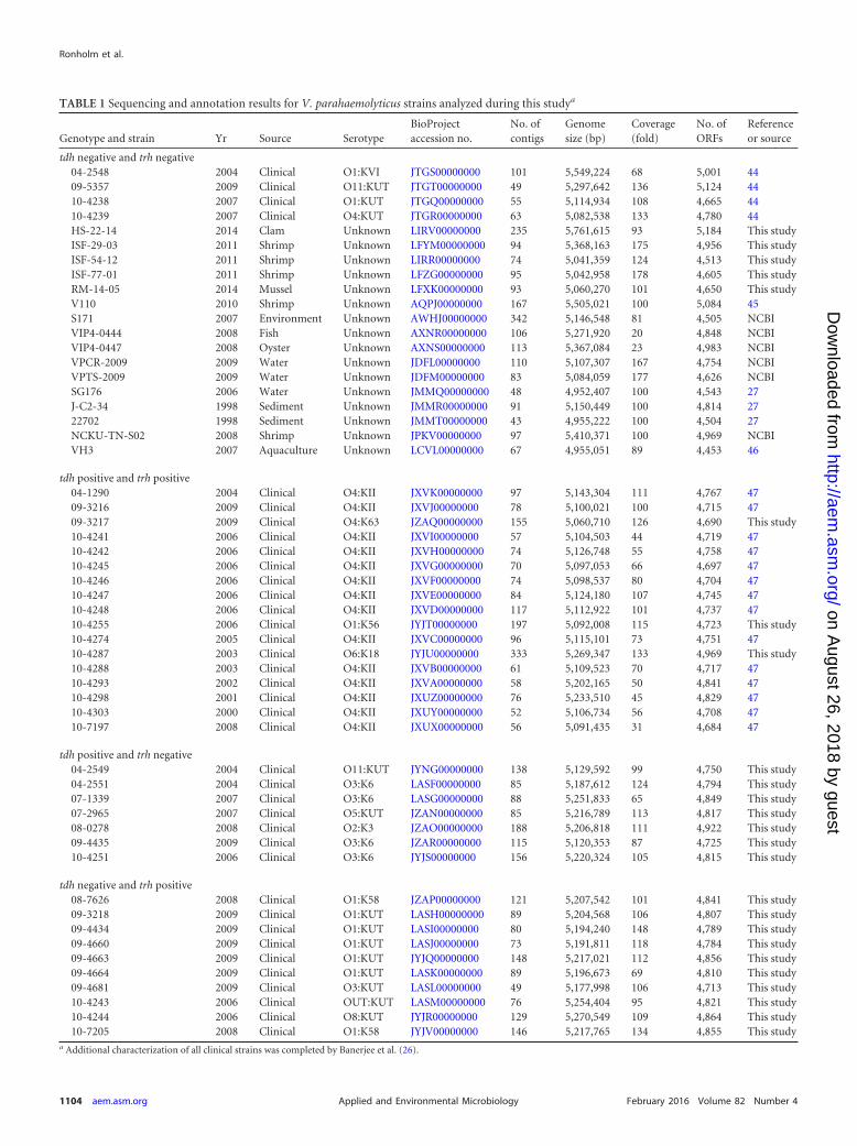
TABLE 1 Sequencing and annotation results for V. parahaemolyticus strains analyzed during this studya
Genotype and strain Yr Source SerotypeBioProjectaccession no.
No. ofcontigs
Genomesize (bp)
Coverage(fold)
No. ofORFs
Referenceor source
tdh negative and trh negative04-2548 2004 Clinical O1:KVI JTGS00000000 101 5,549,224 68 5,001 4409-5357 2009 Clinical O11:KUT JTGT00000000 49 5,297,642 136 5,124 4410-4238 2007 Clinical O1:KUT JTGQ00000000 55 5,114,934 108 4,665 4410-4239 2007 Clinical O4:KUT JTGR00000000 63 5,082,538 133 4,780 44HS-22-14 2014 Clam Unknown LIRV00000000 235 5,761,615 93 5,184 This studyISF-29-03 2011 Shrimp Unknown LFYM00000000 94 5,368,163 175 4,956 This studyISF-54-12 2011 Shrimp Unknown LIRR00000000 74 5,041,359 124 4,513 This studyISF-77-01 2011 Shrimp Unknown LFZG00000000 95 5,042,958 178 4,605 This studyRM-14-05 2014 Mussel Unknown LFXK00000000 93 5,060,270 101 4,650 This studyV110 2010 Shrimp Unknown AQPJ00000000 167 5,505,021 100 5,084 45S171 2007 Environment Unknown AWHJ00000000 342 5,146,548 81 4,505 NCBIVIP4-0444 2008 Fish Unknown AXNR00000000 106 5,271,920 20 4,848 NCBIVIP4-0447 2008 Oyster Unknown AXNS00000000 113 5,367,084 23 4,983 NCBIVPCR-2009 2009 Water Unknown JDFL00000000 110 5,107,307 167 4,754 NCBIVPTS-2009 2009 Water Unknown JDFM00000000 83 5,084,059 177 4,626 NCBISG176 2006 Water Unknown JMMQ00000000 48 4,952,407 100 4,543 27J-C2-34 1998 Sediment Unknown JMMR00000000 91 5,150,449 100 4,814 2722702 1998 Sediment Unknown JMMT00000000 43 4,955,222 100 4,504 27NCKU-TN-S02 2008 Shrimp Unknown JPKV00000000 97 5,410,371 100 4,969 NCBIVH3 2007 Aquaculture Unknown LCVL00000000 67 4,955,051 89 4,453 46
tdh positive and trh positive04-1290 2004 Clinical O4:KII JXVK00000000 97 5,143,304 111 4,767 4709-3216 2009 Clinical O4:KII JXVJ00000000 78 5,100,021 100 4,715 4709-3217 2009 Clinical O4:K63 JZAQ00000000 155 5,060,710 126 4,690 This study10-4241 2006 Clinical O4:KII JXVI00000000 57 5,104,503 44 4,719 4710-4242 2006 Clinical O4:KII JXVH00000000 74 5,126,748 55 4,758 4710-4245 2006 Clinical O4:KII JXVG00000000 70 5,097,053 66 4,697 4710-4246 2006 Clinical O4:KII JXVF00000000 74 5,098,537 80 4,704 4710-4247 2006 Clinical O4:KII JXVE00000000 84 5,124,180 107 4,745 4710-4248 2006 Clinical O4:KII JXVD00000000 117 5,112,922 101 4,737 4710-4255 2006 Clinical O1:K56 JYJT00000000 197 5,092,008 115 4,723 This study10-4274 2005 Clinical O4:KII JXVC00000000 96 5,115,101 73 4,751 4710-4287 2003 Clinical O6:K18 JYJU00000000 333 5,269,347 133 4,969 This study10-4288 2003 Clinical O4:KII JXVB00000000 61 5,109,523 70 4,717 4710-4293 2002 Clinical O4:KII JXVA00000000 58 5,202,165 50 4,841 4710-4298 2001 Clinical O4:KII JXUZ00000000 76 5,233,510 45 4,829 4710-4303 2000 Clinical O4:KII JXUY00000000 52 5,106,734 56 4,708 4710-7197 2008 Clinical O4:KII JXUX00000000 56 5,091,435 31 4,684 47
tdh positive and trh negative04-2549 2004 Clinical O11:KUT JYNG00000000 138 5,129,592 99 4,750 This study04-2551 2004 Clinical O3:K6 LASF00000000 85 5,187,612 124 4,794 This study07-1339 2007 Clinical O3:K6 LASG00000000 88 5,251,833 65 4,849 This study07-2965 2007 Clinical O5:KUT JZAN00000000 85 5,216,789 113 4,817 This study08-0278 2008 Clinical O2:K3 JZAO00000000 188 5,206,818 111 4,922 This study09-4435 2009 Clinical O3:K6 JZAR00000000 115 5,120,353 87 4,725 This study10-4251 2006 Clinical O3:K6 JYJS00000000 156 5,220,324 105 4,815 This study
tdh negative and trh positive08-7626 2008 Clinical O1:K58 JZAP00000000 121 5,207,542 101 4,841 This study09-3218 2009 Clinical O1:KUT LASH00000000 89 5,204,568 106 4,807 This study09-4434 2009 Clinical O1:KUT LASI00000000 80 5,194,240 148 4,789 This study09-4660 2009 Clinical O1:KUT LASJ00000000 73 5,191,811 118 4,784 This study09-4663 2009 Clinical O1:KUT JYJQ00000000 148 5,217,021 112 4,856 This study09-4664 2009 Clinical O1:KUT LASK00000000 89 5,196,673 69 4,810 This study09-4681 2009 Clinical O3:KUT LASL00000000 49 5,177,998 106 4,713 This study10-4243 2006 Clinical OUT:KUT LASM00000000 76 5,254,404 95 4,821 This study10-4244 2006 Clinical O8:KUT JYJR00000000 129 5,270,549 109 4,864 This study10-7205 2008 Clinical O1:K58 JYJV00000000 146 5,217,765 134 4,855 This study
a Additional characterization of all clinical strains was completed by Banerjee et al. (26).
Ronholm et al.
1104 aem.asm.org February 2016 Volume 82 Number 4Applied and Environmental Microbiology
on August 26, 2018 by guest
http://aem.asm
.org/D
ownloaded from

Genetic islands and secretion systems. The presence and absence ofthe tdh and trh pathogenicity islands and various secretion systems wereinvestigated using BLASTn. The EasyFig (version 2.1) program was usedfor visualization of genomic islands (36). For larger genomic islands (tdhPAI and trh PAI), a Burrows-Wheeler transform reference-guided assem-bly was used on raw fastq data with the canonical sequence of the genomicisland (29). The assembly was inspected for accuracy and validity usingTablet, prior to visualization (30). To possibly identify novel pathogenic-ity islands in the tdh- and trh-negative pathotype, the completed results ofan IslandViewer analysis on strain CDC_K4557 were downloaded fromthe IslandViewer3 website (37). BLASTn was used to search each genomeincluded in this study (Table 1) for each island that was identified.
Nucleotide sequence accession numbers. All nucleotide sequencedata referred to in this article have been deposited in the DDBJ/EMBL/GenBank database under the various BioProject accession numbers pro-vided in Table 1. Additional important data are included in the supple-mental material.
RESULTS AND DISCUSSION
Type III secretion systems and tdh and trh pathogenicity is-lands. Traditionally, a pathogenic V. parahaemolyticus strain hasbeen defined by the presence of tdh or trh, or both (5). These twovirulence factors generally occur near a T3SS. Two T3SSs havebeen reported in V. parahaemolyticus, and two variants of T3SS2(T3SS2� and T3SS2�) have been described (12). T3SS2� is asso-ciated with tdh, while T3SS2� is typically found with trh (14),though exceptions exist (27). T3SS1 is composed of 42 genes(VPA1656 to VAP1696 and VPA0450 in V. parahaemolyticusRIMD2210633 [15]). T3SS1 was present in each of the clinical andenvironmental isolates in this investigation. The tdh PAI (alsoknown as VPAI-7) is composed of 87 coding sequences (in V.parahaemolyticus RIMD2210633) and includes tdhA, tdhS, andthe T3SS2� genes (38). In our current work, due to multiple ho-
FIG 1 Pathogenicity islands and T3SS2. tdh PAI (A) and trh PAI (B) are the two canonical genomic islands present in clinical isolates of V. parahaemolyticus. Thepresence or absence of these islands is strongly correlated with the presence of the tdh and trh genes. In three of the four tdh- and trh-negative isolates, these genesare entirely absent. (C) In the fourth tdh- and trh-negative isolate, 10-4328, a trh PAI including a partial trh1 gene is mostly present. Abbreviations: neg, negative;pos, positive.
Clinical and Environmental V. parahaemolyticus Isolates
February 2016 Volume 82 Number 4 aem.asm.org 1105Applied and Environmental Microbiology
on August 26, 2018 by guest
http://aem.asm
.org/D
ownloaded from

mologous areas in the tdh PAI and trh PAI areas, the use of de novoassembly led to this large PAI being split between multiple contigs.Therefore, to analyze this region properly, a reference-guided as-sembly based on the tdh PAI sequence (VPA1310 to VPA1396)from V. parahaemolyticus RIMD2210663 was used (39). Usingthis method, a complete tdh PAI including T3SS2� was consis-tently observed in all clinical tdh-positive and trh-negative isolates(Fig. 1A). Clinical tdh- and trh-positive isolates contained homo-logues to some coding sequences typically found in this island(VPA1310, VPA1311, VPA1313 to VPA1318, VPA1320,VPA1321, VPA1329, VPA1342, VPA1347, and VPA1382 toVPA1393), and their presence was consistent among all clinicaltdh- and trh-positive isolates (see Fig. 3A). Isolates categorized astdh negative and trh positive or tdh and trh negative did not con-tain a PAI with homology to tdh PAI genes (Fig. 1A).
The trh PAI, composed of 81 coding sequences, was also inde-pendently assembled using reference-guided assembly and V.parahaemolyticus VIPARAQ4037 residues 1748 to 1830 as a refer-ence and was consistently observed in isolates of both the tdh-negative and trh-positive pathotype and the tdh- and trh-positivepathotype and in all instances included trh1 and the T3SS2� genes(Fig. 1B). The association between the trh PAI and T3SS2� withthe tdh-negative and trh-positive pathotype and the tdh- and trh-positive pathotype agrees with the findings of previous studies (12,27). Three clinical tdh- and trh-negative isolates, isolates 04-2548,09-5357, and 10-4239, did not contain the tdh PAI, the trh PAI, ora T3SS2; however, an almost complete trh PAI including a partialtrh gene, urease gene cluster, and the T3SS2� genes was identifiedin isolate 10-4238, which was categorized as tdh and trh negativeby PCR analysis by Banerjee et al. (26) (Fig. 1C). In addition to theTRH hemolysins, the T3SS� present in this PAI also contains ef-fectors thought to be involved in enterotoxicity and cytotoxicity(5). T3SS2� appears to be a recent acquisition by V. parahaemo-lyticus and is sometimes found in non-O1, non-O139, and CTX V.cholerae strains (12). The presence of the trh PAI in 10-4238 madeus question whether this strain should indeed be classified as aclinical tdh- and trh-negative isolate or if it would be more accu-rate, on the basis of its WGS, to classify it as a tdh-negative andtrh-positive strain. Therefore, this strain was removed from theremaining analysis specific to clinical tdh- and trh-negative iso-lates.
General genomic features of V. parahaemolyticus clinicalisolates. To depict the genetic diversity of pathogenic V. parahae-molyticus strains, 38 clinical isolates (Table 1) representing each ofthe four previously described genotypes (tdh and trh positive, tdhnegative and trh positive, tdh positive and trh negative, and tdhand trh negative) (40, 41) were extensively compared. The pange-nome of clinical V. parahaemolyticus isolates was calculated andconsisted of 8,399 protein-coding genes (Fig. 2A). To assess theaccuracy of computing of a pangenome using draft genomes,three clinical isolates with closed genomes (see Table S1 in thesupplemental material) were added to our data set and the pange-nome size was recalculated. The addition of three closed genomescaused the size of the pangenome of the clinical isolates to increaseto 8,609 genes. This increase would also have been expected afterthe addition of three draft genomes, and therefore, we concludedthat our calculations based on the draft genomes were accurate.
A gene intersection analysis of the accessory genomes was per-formed. From this analysis, we defined a core genome of ortholo-
gous genes that were shared by all clinical V. parahaemolyticusisolates. The core genome contained 3,807 protein-coding genes,which represented between 76 to 81% of each isolate’s genome(Fig. 2A). The size of the core genome remained relatively stablefor each additional genome added after the first four. When theclosed genomes of three additional clinical isolates (see Table S1 inthe supplemental material) were added to the data set and the coregenome was recalculated, it decreased to a size of 3,803 protein-coding genes. This indicated that the 38 draft genomes sequencedhere provided an excellent estimation of the true core genome ofclinical V. parahaemolyticus isolates.
FIG 2 General genomic features of clinical V. parahaemolyticus isolates. (A)The pangenome of clinical V. parahaemolyticus isolates was constructed usingthe de novo assembly of 38 clinical isolates and contained 8,399 genes. Pange-nomes were also assembled for each pathotype and compared. (B) A phyloge-netic tree, constructed from concatenated core genes, shows the phylogeny ofclinical isolates and demonstrates that each of the pathotypes is polyphyletic.
Ronholm et al.
1106 aem.asm.org February 2016 Volume 82 Number 4Applied and Environmental Microbiology
on August 26, 2018 by guest
http://aem.asm
.org/D
ownloaded from

Accessory genes that were unique to each pathotype were alsoidentified, and 654, 520, 918, and 1,097 genes were specific to thetdh- and trh-positive, tdh-negative and trh-positive, tdh-positiveand trh-negative, and tdh- and trh-negative pathotypes of clinicalisolates, respectively (Fig. 2A). Strain-specific unique genes werealso identified, and the sizes of the unique genome varied betweenstrains. For example, seven strains (09-3216, 10-4303, 10-7197,09-3218, 09-4434, 09-4660, and 09-4664) possessed no uniquegenes, while one strain, 04-2548 (tdh and trh negative), had 405unique genes. Clinical isolates of the tdh- and trh-negative patho-type consistently had large unique genomes.
To determine if clinical tdh- and trh-negative isolates aremonophyletic and possibly the result of a single loss of a pathoge-nicity island, an unrooted phylogenetic tree was constructed fromthe concatenated amino acid sequences of the 3,899 core genes(Fig. 2B). Use of the core genes provides a high-resolution view ofphylogeny. This revealed that isolates of each of the pathotypes,tdh- and trh-negative isolates, are polyphyletic. This may indicatea high degree of mobility of pathogenicity elements between V.parahaemolyticus isolates, rather than a single PAI deletion event.
The distribution of clusters of orthologous groups (COGs) ofproteins was determined for the core genome, the accessory ge-nome of each pathotype, and the unique genome of each of thetdh- and trh-negative isolates, to determine if there were differ-
ences in the proportion of the genome attributable to particularcellular processes in clinical isolates (see Table S2 in the supple-mental material). Almost 20% of the core genome was classified ashaving an unknown function; this proportion dropped to lessthan 10% in each of the pathotype-specific unique genomes (Fig.3A and B). The tdh- and trh-negative isolates had a large propor-tion of genes involved in cell motility relative to the proportion forthe other pathotypes (Fig. 3B). Individual tdh- and trh-negativeisolates also had large and functionally consistent unique genomes(Fig. 3C). Within this functional category of cell motility, each ofthe three clinical tdh- and trh-negative isolates contained a meth-yl-accepting chemotaxis protein not observed in the other patho-types or in environmental isolates. Cell motility is generally con-sidered to be a factor associated with the ability of V.parahaemolyticus to survive in vivo (5); therefore, the finding of anincrease in the number of genes involved in motility in clinicalisolates is logical.
General genomic features of V. parahaemolyticus environ-mental isolates. To provide a basis for comparison of the clinicaltdh- and trh-negative isolates, we sequenced the genomes of 5environmental tdh- and trh-negative isolates and collected the ge-nomes of an additional 11 from the NCBI database (Table 1). Todemonstrate the wide diversity and the relationships of the strainsused in this study, a rooted phylogenetic tree was constructed
FIG 3 COG profiles of the core (A), accessory (B), and unique (C) genomes of clinical tdh- and trh-negative isolates. The numbers at the top of each columndenote the number of genes in the unique genome of the corresponding strain listed at the bottom of the column. Abbreviations: neg, negative; pos, positive.
Clinical and Environmental V. parahaemolyticus Isolates
February 2016 Volume 82 Number 4 aem.asm.org 1107Applied and Environmental Microbiology
on August 26, 2018 by guest
http://aem.asm
.org/D
ownloaded from

from the concatenated nucleotide sequences of seven housekeep-ing genes (recA, gyrB, dnaE, dtdS, pntA, pyrC, and tnaA) tradition-ally used in V. parahaemolyticus MLST analysis (Fig. 4A). Clinicaland environmental isolates shared several common lineages,again demonstrating the dynamic nature of virulence factors inthis species.
The sizes of the core genome and pangenome were calculatedfor the 16 environmental isolates. The core genome of the envi-ronmental tdh- and trh-negative isolates was composed of 2,773protein-coding genes, and though the pangenome was con-structed from fewer genomes, it was much larger than that of theclinical isolates at 11,669 protein-coding genes (Fig. 4B). For both
FIG 4 General genomic features of environmental V. parahaemolyticus isolates. (A) A phylogenetic tree, constructed from concatenated MLST sequences,demonstrates the diversity of the strains used in this study, as well as the relationships between each clinical and environmental strain included in this study. (B)A rarefaction curve of the genetic diversity of clinical and environmental V. parahaemolyticus strains was created. Environmental isolates have a much greatergenetic diversity than clinical isolates. In addition, these curves demonstrate that the pangenome of V. parahaemolyticus is open.
FIG 5 Genomic comparison of environmental and clinical tdh- and trh-neg-ative isolates. On the basis of a comparison of protein-coding genes, 862protein-coding genes that are unique to clinical tdh- and trh-negative isolateswere identified. neg, negative.
Ronholm et al.
1108 aem.asm.org February 2016 Volume 82 Number 4Applied and Environmental Microbiology
on August 26, 2018 by guest
http://aem.asm
.org/D
ownloaded from

TA
BLE
2D
istr
ibu
tion
ofpa
thog
enic
ity
isla
nds
incl
inic
alis
olat
es
Gen
otyp
ean
dst
rain
Gen
e(s)
inth
efo
llow
ing
V.p
arah
aem
olyt
icus
PA
Ior
pres
ence
orab
sen
ceof
aP
AI:
VP
AI-
1(V
P03
80to
VP
0403
)V
PA
I-2
(VP
0635
toV
P06
43)
VP
AI-
3(V
P10
71to
VP
1094
)V
PA
I-4
(VP
2131
toV
P21
44)
VP
AI-
5(V
P29
00to
VP
2910
)V
PA
I-6
(VP
A12
53to
VP
A12
70)
tdh
neg
ativ
ean
dtr
hn
egat
ive
04-2
548
�V
P06
35,V
P06
36V
P10
88to
VP
1094
��
�
09-5
357
VP
0380
,VP
0398
,VP
0400
VP
0635
,VP
0636
VP
1088
toV
P10
94�
��
(T12
739)
10-4
238
��
VP
1077
toV
P10
94�
��
(T91
09)
10-4
239
VP
0380
,VP
0397
toV
P04
03V
P06
35,V
P06
36V
P10
71,V
P10
88to
VP
1094
��
�
tdh
posi
tive
and
trh
posi
tive
04-1
290
�V
P06
35,V
P06
36V
P10
88to
VP
1094
��
�
09-3
216
�V
P06
35,V
P06
36V
P10
88to
VP
1094
��
�
09-3
217
�V
P06
35,V
P06
36V
P10
88to
VP
1094
VP
2144
��
10-4
241
�V
P06
35,V
P06
36V
P10
88to
VP
1094
��
�
10-4
242
�V
P06
35,V
P06
36V
P10
88to
VP
1094
��
�
10-4
245
�V
P06
35,V
P06
36V
P10
88to
VP
1094
��
�
10-4
246
�V
P06
35,V
P06
36V
P10
88to
VP
1094
��
�
10-4
247
�V
P06
35,V
P06
36V
P10
88to
VP
1094
��
�
10-4
248
�V
P06
35,V
P06
36V
P10
88to
VP
1094
��
�
10-4
255
VP
0380
toV
P03
84,V
P03
86to
VP
0392
VP
0635
,VP
0636
VP
1088
toV
P10
94V
P21
44�
�
VP
0396
toV
P04
00,V
P04
02,V
P04
0310
-427
4�
VP
0635
,VP
0636
VP
1088
toV
P10
94�
��
10-4
287
VP
0380
,VP
0397
toV
P04
00V
P04
02,V
P04
03V
P06
35,V
P06
36V
P10
71,V
P10
76,V
P10
88to
VP
1094
VP
2144
��
10-4
288
�V
P06
35,V
P06
36V
P10
88to
VP
1094
��
�
10-4
293
�V
P06
35,V
P06
36V
P10
88to
VP
1094
��
�
10-4
298
VP
0380
,VP
0382
,VP
0386
,VP
0395
,VP
0402
VP
0635
,VP
0636
VP
1088
toV
P10
94�
��
10-4
303
�V
P06
35,V
P06
36V
P10
88to
VP
1094
��
�
10-7
197
�V
P06
35,V
P06
36V
P10
88�
VP
1094
��
�
tdh
posi
tive
and
trh
neg
ativ
e04
-254
9�
��
��
�
04-2
551
��
��
��
07-1
339
��
��
��
07-2
965
�V
P06
35,V
P06
36V
P10
88to
VP
1094
��
�
08-0
278
VP
0380
,VP
0398
,VP
0400
,VP
0402
,VP
0403
VP
0635
,VP
0636
VP
1088
toV
P10
94�
��
09-4
435
��
��
��
10-4
251
��
��
��
tdh
neg
ativ
ean
dtr
hpo
siti
ve08
-762
6V
P03
80,V
P03
98to
VP
0400
,VP
0402
,VP
0403
�V
P10
88to
VP
1094
��
�
09-3
218
VP
0380
,VP
0398
toV
P04
00,V
P04
02,V
P04
03�
VP
1088
toV
P10
94�
��
09-4
434
VP
0380
,VP
0398
toV
P04
00,V
P04
02,V
P04
03�
VP
1088
toV
P10
94�
��
09-4
660
VP
0380
,VP
0398
toV
P04
00,V
P04
02,V
P04
03�
VP
1088
toV
P10
94�
��
09-4
663
VP
0380
,VP
0398
toV
P04
00,V
P04
02,V
P04
03�
VP
1088
toV
P10
94�
��
09-4
664
VP
0380
,VP
0398
toV
P04
00,V
P04
02,V
P04
03�
VP
1088
toV
P10
94�
��
09-4
681
VP
0380
toV
P03
88,V
P03
95to
VP
0400
VP
0402
,VP
0403
VP
0635
,VP
0636
VP
1088
toV
P10
94�
��
10-4
243
�V
P06
35,V
P06
36V
P10
88to
VP
1094
��
�
10-4
244
�V
P06
35,V
P06
36V
P10
88to
VP
1094
��
�
10-7
205
VP
0380
,VP
0398
toV
P04
00,V
P04
02,V
P04
03�
VP
1088
toV
P10
94�
��
Clinical and Environmental V. parahaemolyticus Isolates
February 2016 Volume 82 Number 4 aem.asm.org 1109Applied and Environmental Microbiology
on August 26, 2018 by guest
http://aem.asm
.org/D
ownloaded from

the clinical and environmental isolates, the pangenome size in-creased, in terms of protein-coding gene number, after the addi-tion of each genome, which indicates an open pangenome (42),and this is in agreement with the findings of earlier studies (43)
FIG 6 Genomic islands identified in CDC_K4557. A closed genome is re-quired to identify novel genomic islands. To determine if a novel pathogenicityisland is responsible for virulence in the tdh- and trh-negative pathotype,the closed genome of V. parahaemolyticus CDC_K4557, another clinicaltdh- and trh-negative isolate, was searched for genomic islands using Is-landViewer3, and the images shown above were modified from the Island-Viewer3 (37) website (http://www.pathogenomics.sfu.ca/islandviewer/accession/NC_021848.1/ [top panel] and http://www.pathogenomics.sfu.ca/islandviewer/accession/NC_021822.1/ [bottom panel]). BLASTn wasused to search for each of the islands in the clinical and environmental isolatesfrom Table 1. IslandViewer3 uses multiple algorithms (IslandPick,SIGI-HMM, and IslandPath-DIMOB) to predict the presence of genomicislands. Red, islands identified by an integrative algorithm incorporatingmultiple mechanisms of island prediction; blue, islands predicted by the IslandPath algorithm, yellow, islands predicted by the SIGI-HMM algorithm; green,islands predicted by the IslandPick algorithm. M, millions; k, thousands.T
AB
LE3
Dis
trib
uti
onof
path
ogen
icit
yis
lan
dsin
envi
ron
men
tali
sola
tes
tdh-
and
trh-
neg
ativ
est
rain
Gen
e(s)
inth
efo
llow
ing
V.p
arah
aem
olyt
icus
PA
Ior
pres
ence
orab
sen
ceof
aP
AI:
VP
AI-
1(V
P03
80to
VP
0403
)V
PA
I-2
(VP
0635
toV
P06
43)
VP
AI3
(VP
1071
toV
P10
94)
VP
AI-
4(V
P21
31to
VP
2144
)V
PA
I-5
(VP
2900
toV
P29
10)
VP
AI-
6(V
PA
1253
toV
PA
1270
)
HS-
22-1
4V
P03
87to
VP
0395
��
��
VP
A12
54,V
PA
1256
,VP
A12
58to
VP
A12
59,V
PA
1262
toV
PA
1265
,V
PA
1267
ISF-
29-3
VP
0380
,VP
0398
toV
P04
00,
VP
0402
,VP
0403
VP
0635
,VP
0636
VP
1088
toV
P10
94�
��
ISF-
54-1
2�
VP
0635
toV
P06
36,
VP
0638
toV
P06
43V
P10
88to
VP
1094
��
�
ISF-
77-0
1�
VP
0635
,VP
0636
VP
1088
toV
P10
94�
��
RM
-14-
5�
VP
0635
,VP
0636
VP
1088
toV
P10
94V
P21
31to
VP
2133
VP
2136
toV
P21
44
��
V11
0�
VP
0635
,VP
0636
VP
1088
toV
P10
94�
��
S171
VP
0381
toV
P03
84V
P06
35,V
P06
36V
P10
88to
VP
1094
VP
2144
��
VIP
4-04
44�
�V
P10
71,V
P10
88to
VP
1094
��
�
VIP
4-04
47�
VP
0635
,VP
0636
VP
1088
toV
P10
94V
P21
31to
VP
2133
VP
2136
toV
P21
44
��
VP
CR
-200
9�
�V
P10
71,V
P10
73,V
P10
74,V
P10
75,
VP
1088
toV
P10
94�
��
VP
TS-
2009
�V
P06
35,V
P06
36V
P10
88to
VP
1094
��
�
SG17
6V
P03
81to
VP
0384
VP
0635
,VP
0636
VP
1088
toV
P10
94�
��
J-C
2-34
�V
P06
35,V
P06
36V
P10
88to
VP
1094
��
�
2270
2�
VP
0635
,VP
0636
VP
1088
toV
P10
94�
��
NC
KU
-TN
-S02
VP
0381
toV
P03
83,V
P03
88,V
P03
95V
P06
35,V
P06
36V
P10
88to
VP
1094
��
�
VH
3V
P06
35,V
P06
36V
P10
71,V
P10
76,V
P10
88to
VP
1094
��
�
Ronholm et al.
1110 aem.asm.org February 2016 Volume 82 Number 4Applied and Environmental Microbiology
on August 26, 2018 by guest
http://aem.asm
.org/D
ownloaded from
(Fig. 4B). The increased size of the pangenome in environmentalisolates is likely due to their ability to survive in diverse niches insitu. For example, environmental isolates were collected from col-onized shrimp, muscle, oyster, and clams as well as from marinewater and sediments, while clinical isolates have been preselectedby use of a very narrow criterion, which is the ability to colonizehumans and cause illness. This likely leads to the low level ofgenetic diversity observed in clinical isolates.
Whole-genome comparison of clinical and environmentaltdh- and trh-negative isolates. The genomes of the 3 clinical tdh-and trh-negative isolates were compared with the genomes of 16environmental tdh- and trh-negative isolates, resulting in theidentification of 862 protein-coding genes unique to clinical iso-lates (Fig. 5; see also Table S3 in the supplemental material). Fromthese genes, 529 were annotated as hypothetical proteins. A largeportion of both hypothetical proteins and annotated proteins hadhigh sequence similarity with other genes from other enteric bac-teria, such as V. cholerae, Listeria monocytogenes, Campylobacterjejuni, Salmonella enterica, Escherichia coli, and Enterobacter.While this finding is not conclusive, it may indicate that the ac-quisition of genes from other enteric bacteria may contribute tothe ability of clinical tdh- and trh-negative isolates to colonizehumans during a gastrointestinal illness.
Pathogenicity islands. By examining the complete genomeof V. parahaemolyticus RIMD2210633, Hurley et al. (2006)identified seven genomic islands which occur in pathogenic V.parahaemolyticus isolates (38). VPAI-1, VPAI-4, VPAI-5, andVPAI-6 appeared to represent DNA acquired by pandemic V.parahaemolyticus isolates (38). The presence of these islands wasvariable in our clinical and environmental isolates (Tables 2 and3). VPAI-1 was present in most clinical tdh-positive and trh-neg-
ative isolates but was largely absent from isolates of the otherpathotypes. The entire VPAI-2 was present in most tdh-positiveand trh-negative isolates and tdh-negative and trh-positive isolatesbut was only partially present in tdh- and trh-positive and tdh- andtrh-negative strains. VPAI-6 was consistently present in each ofthe clinical isolates. Strains 07-2965 and 08-0278 did not typicallyhave the same PAI profile as the other tdh-positive and trh-nega-tive strains, and this was also reflected in the phylogenetic tree(Fig. 2B). The 10-4238 tdh- and trh-negative strain carried VPAIsmore similar to those carried by the tdh-negative and trh-positiveisolates, agreeing with findings presented earlier in this paper thatthis strain is likely a tdh-negative and trh-positive strain and wasmisidentified by PCR analysis. The environmental tdh- andtrh-negative isolates carried pathogenicity island profiles similarto those of the clinical isolates (Table 3), this observation raisesseveral questions about the true roles of these islands and whethertheir inclusion in pandemic strains is associated more with fitnessin the environment than with pathogenicity.
IslandViewer3 was used to search the closed genome ofCDC_K4557, which is also a clinical V. parahaemolyticus tdh- andtrh-negative isolate, for genomic islands that are common to clin-ical isolates but that are not found in environmental tdh- andtrh-negative isolates (37). We reasoned that if an island was pres-ent in at least some of the clinical tdh- and trh-negative isolates butabsent from all of the environmental tdh- and trh-negative iso-lates, it would be a reasonable candidate for evaluation as a novelpathogenicity island. IslandViewer3 was used to identify 29genomic islands on chromosome 1 and 8 genomic islands onchromosome 2 (Fig. 6). These genomic islands were assessed fortheir presence or absence across our 43 genomes (see Table S4 inthe supplemental material). We found that islands 1, 3, 33, 35, and
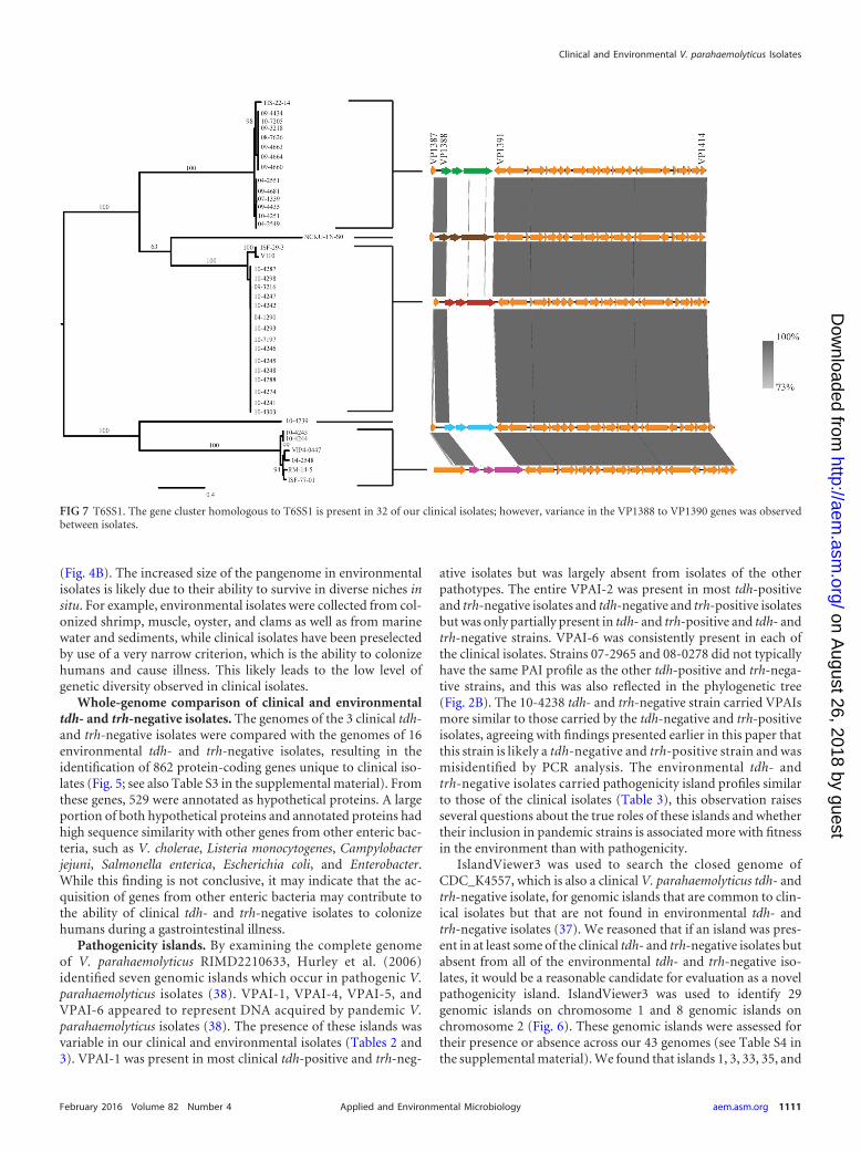
FIG 7 T6SS1. The gene cluster homologous to T6SS1 is present in 32 of our clinical isolates; however, variance in the VP1388 to VP1390 genes was observedbetween isolates.
Clinical and Environmental V. parahaemolyticus Isolates
February 2016 Volume 82 Number 4 aem.asm.org 1111Applied and Environmental Microbiology
on August 26, 2018 by guest
http://aem.asm
.org/D
ownloaded from
36 (as denoted in Fig. 6) were present in almost every strain of V.parahaemolyticus. An island that was present in clinical tdh- andtrh-negative strains but not in environmental tdh- and trh-nega-tive strains was not identified.
Type VI secretion systems. Two T6SSs have previously beenfound in the pangenome of V. parahaemolyticus. T6SS1 (VP1387to VP1414) is found on chromosome 1 and is commonly associ-ated with clinical isolates, while T6SS2 (VPA1025 to VPA1046) isfound on chromosome 2 and has been found in all tested strains(16, 20). In the current study, six clinical isolates (10-4238, 08-0278, 09-5357, 07-2965, 09-3217, and 10-4255) and eight envi-ronmental isolates (ISF-54-12, S171, VPCR-2009, VPTS-2009,SG176, 22720, J-C2-34, and VH3) did not have a T6SS1. Of theisolates that had a T6SS1, variance in this gene cluster was ob-served between isolates, although variance occurred only in theVP1388, VP1389, and VP1390 genes (Fig. 7). There were five dif-ferent alleles of these genes, and a phylogenetic tree is shown todemonstrate which isolates contained which alleles (Fig. 7). The10-4239 isolate had unique alleles for each of these genes whichwere not observed in any of the other isolates. The N terminus ofVP1388 was conserved in all isolates that had a T6SS1, while vari-ance was observed in the C terminus. There is biological signifi-cance underlying the variation observed in VP1388 to VP1390.VP1388 has previously been identified to be an antibacterial effec-tor, and VP1389 is its associated immunity protein (21). Changesin the effector must be accompanied by changes in the immunityprotein to maintain self-protection. The putative functionality ofthe third larger gene downstream (VP1390) is still unknown, buton the basis of its association with VP1388 and VP1390, it mayhave a role in antimicrobial activity. The finding of a novel puta-tive effector in a V. parahaemolyticus clinical tdh- and trh-negativeisolate indicates that this protein should be further investigatedfor roles during infection.
Conclusion. The ability of tdh- and trh-negative strains tocause clinical illness is still controversial, and several theories havebeen proposed to explain why tdh- and trh-negative strains aresometimes isolated from clinical cases, including coinfection withpathogenic V. parahaemolyticus strains, the loss of virulence genesduring infection, the presence of novel and uncharacterized viru-lence factors, or the fact that they play a role in a multistraininfection. However, in this investigation we have identified 862genes that are present in clinical tdh- and trh-negative isolates butthat are not present in environmental isolates. Several of thesegenes are highly homologous to genes from other enteric bacteria,indicating that horizontal gene transfer may play an importantrole in the ability of tdh- and trh-negative isolates to survive in thehuman gastrointestinal tract. In addition, tdh- and trh-negativeisolate 10-4239 contains a unique T6SS1 effector/immunity genecombination that should be investigated further.
ACKNOWLEDGMENTS
We are very grateful to Robyn Kenwell for technical assistance with thesequencing of the genomes analyzed in this paper. We also thank JohnAustin and Franco Pagotto of the BMH Research Division of Health Can-ada for reviewing the manuscript and offering helpful comments.
This work was funded (A-base) by Health Canada in support of Can-ada’s Food Safety Programs. J.R. is supported by the Visiting Fellow in aGovernment Laboratory program.
J.R., N.P., C.C.L., and S.K.B. conceived of and designed the experi-ments. J.R. and C.C.L. performed all wet laboratory experimental, molec-ular, and next-generation sequencing work. J.R. and N.P. conducted the
in silico analysis, including the use of novel tools contributed by A.W.P.;J.R. wrote the paper with input from all other authors.
FUNDING INFORMATIONGouvernement du Canada | Health Canada (Santé, Canada) providedfunding to Swapan K. Banerjee.
REFERENCES1. Ceccarelli D, Hasan NA, Huq A, Colwell RR. 2013. Distribution and
dynamics of epidemic and pandemic Vibrio parahaemolyticus virulencefactors. Front Cell Infect Microbiol 3:97. http://dx.doi.org/10.3389/fcimb.2013.00097.
2. Joseph SW, Colwell RR, Kaper JB. 1982. Vibrio parahaemolyticus andrelated halophilic vibrios. Crit Rev Microbiol 10:77–124. http://dx.doi.org/10.3109/10408418209113506.
3. Nishibuchi M, Fasano A, Russell RG, Kaper JB. 1992. Enterotoxigenicityof Vibrio parahaemolyticus with and without genes encoding thermostabledirect hemolysin. Infect Immun 60:3539 –3545.
4. Honda T, Ni Y, Miwatani T, Adachi T, Kim J. 1992. The thermostabledirect hemolysin of Vibrio parahaemolyticus is a pore-forming toxin. CanJ Microbiol 38:1175–1180. http://dx.doi.org/10.1139/m92-192.
5. Broberg CA, Calder TJ, Orth K. 2011. Vibrio parahaemolyticus cell biol-ogy and pathogenicity determinants. Microbes Infect 13:992–1001. http://dx.doi.org/10.1016/j.micinf.2011.06.013.
6. Hiyoshi H, Kodama T, Iida T, Honda T. 2010. Contribution of Vibrioparahaemolyticus virulence factors to cytotoxicity, enterotoxicity, and le-thality in mice. Infect Immun 78:1772–1780. http://dx.doi.org/10.1128/IAI.01051-09.
7. Matsuda S, Kodama T, Okada N, Okayama K, Honda T, Iida T. 2010.Association of Vibrio parahaemolyticus thermostable direct hemolysinwith lipid rafts is essential for cytotoxicity but not hemolytic activity. In-fect Immun 78:603– 610. http://dx.doi.org/10.1128/IAI.00946-09.
8. Letchumanan V, Chan K-G, Lee L-H. 2014. Vibrio parahaemolyticus: areview on the pathogenesis, prevalence, and advance molecular identifi-cation techniques. Front Microbiol 5:705. http://dx.doi.org/10.3389/fmicb.2014.00705.
9. Miyamoto Y, Kato T, Obara Y, Akiyama S, Takizawa K, Yamai S. 1969.In vitro hemolytic characteristic of Vibrio parahaemolyticus: its close cor-relation with human pathogenicity. J Bacteriol 100:1147–1149.
10. Makino K, Oshima K, Kurokawa K, Yokoyama K, Uda T, Tagomori K,Iijima Y, Najima M, Nakano M, Yamashita A, Kubota Y, Kimura S,Yasunaga T, Honda T, Shinagawa H, Hattori M, Iida T. 2003. Genomesequence of Vibrio parahaemolyticus: a pathogenic mechanism distinctfrom that of V. cholerae. Lancet 361:743–749. http://dx.doi.org/10.1016/S0140-6736(03)12659-1.
11. Izoré T, Perdu C, Job V, Attree I, Faudry E, Dessen A. 2011. Structuralcharacterization and membrane localization of ExsB from the type IIIsecretion system (T3SS) of Pseudomonas aeruginosa. J Mol Biol 413:236 –246. http://dx.doi.org/10.1016/j.jmb.2011.07.043.
12. Okada N, Iida T, Park K-S, Goto N, Yasunaga T, Hiyoshi H, MatsudaS, Kodama T, Honda T. 2009. Identification and characterization of anovel type III secretion system in trh-positive Vibrio parahaemolyticusstrain TH3996 reveal genetic lineage and diversity of pathogenic machin-ery beyond the species level. Infect Immun 77:904 –913. http://dx.doi.org/10.1128/IAI.01184-08.
13. Park K-S, Ono T, Rokuda M, Jang M-H, Okada K, Iida T, Honda T.2004. Functional characterization of two type III secretion systems ofVibrio parahaemolyticus. Infect Immun 72:6659 – 6665. http://dx.doi.org/10.1128/IAI.72.11.6659-6665.2004.
14. Noriea NF, III, Johnson CN, Griffitt KJ, Grimes DJ. 2010. Distributionof type III secretion systems in Vibrio parahaemolyticus from the northernGulf of Mexico. J Appl Microbiol 109:953–962. http://dx.doi.org/10.1111/j.1365-2672.2010.04722.x.
15. Ham H, Orth K. 2012. The role of type III secretion system 2 in Vibrioparahaemolyticus pathogenicity. J Microbiol 50:719 –725. http://dx.doi.org/10.1007/s12275-012-2550-2.
16. Salomon D, Gonzalez H, Updegraff BL, Orth K. 2013. Vibrio parahae-molyticus type VI secretion system 1 is activated in marine conditions totarget bacteria, and is differentially regulated from system 2. PLoS One8:e61086. http://dx.doi.org/10.1371/journal.pone.0061086.
17. Salomon D, Orth K. 2015. Type VI secretion system. Curr Biol 25:R265–R266. http://dx.doi.org/10.1016/j.cub.2015.02.031.
Ronholm et al.
1112 aem.asm.org February 2016 Volume 82 Number 4Applied and Environmental Microbiology
on August 26, 2018 by guest
http://aem.asm
.org/D
ownloaded from
18. Pukatzki S, McAuley SB, Miyata ST. 2009. The type VI secretion system:translocation of effectors and effector-domains. Curr Opin Microbiol 12:11–17. http://dx.doi.org/10.1016/j.mib.2008.11.010.
19. Boyer F, Fichant G, Berthod J, Vandenbrouck Y, Attree I. 2009. Dis-secting the bacterial type VI secretion system by a genome wide in silicoanalysis: what can be learned from available microbial genomic resources?BMC Genomics 10:104. http://dx.doi.org/10.1186/1471-2164-10-104.
20. Yu Y, Yang H, Li J, Zhang P, Wu B, Zhu B, Zhang Y, Fang W. 2012.Putative type VI secretion systems of Vibrio parahaemolyticus contributeto adhesion to cultured cell monolayers. Arch Microbiol 194:827– 835.http://dx.doi.org/10.1007/s00203-012-0816-z.
21. Salomon D, Kinch LN, Trudgian DC, Guo X, Klimko JA, Grishin NV,Mirzaei H, Orth K. 2014. Marker for type VI secretion system effectors.Proc Natl Acad Sci U S A 111:9271–9276. http://dx.doi.org/10.1073/pnas.1406110111.
22. Lüdeke CHM, Kong N, Weimer BC, Fischer M, Jones JL. 2015. Com-plete genome sequences of a clinical isolate and an environmental isolateof Vibrio parahaemolyticus. Genome Announc 3(2):e00216-15. http://dx.doi.org/10.1128/genomeA.00216-15.
23. Meador CE, Parsons MM, Bopp CA, Gerner-Smidt P, Painter JA, VoraGJ. 2007. Virulence gene- and pandemic group-specific marker profilingof clinical Vibrio parahaemolyticus isolates. J Clin Microbiol 45:1133–1139. http://dx.doi.org/10.1128/JCM.00042-07.
24. Bhoopong P, Palittapongarnpim P, Pomwised R, Kiatkittipong A, Ka-mruzzaman M, Nakaguchi Y, Nishibuchi M, Ishibashi M, VuddhakulV. 2007. Variability of properties of Vibrio parahaemolyticus strains iso-lated from individual patients. J Clin Microbiol 45:1544 –1550. http://dx.doi.org/10.1128/JCM.02371-06.
25. Jones JL, Lüdeke CHM, Bowers JC, Garrett N, Fischer M, Parsons MB,Bopp CA, DePaola A. 2012. Biochemical, serological, and virulence char-acterization of clinical and oyster Vibrio parahaemolyticus isolates. J ClinMicrobiol 50:2343–2352. http://dx.doi.org/10.1128/JCM.00196-12.
26. Banerjee SK, Kearney AK, Nadon CA, Peterson C-L, Tyler K, BakoucheL, Clark CG, Hoang L, Gilmour MW, Farber JM. 2014. Phenotypic andgenotypic characterization of Canadian clinical isolates of Vibrio parahae-molyticus collected from 2000 to 2009. J Clin Microbiol 52:1081–1088.http://dx.doi.org/10.1128/JCM.03047-13.
27. Hazen TH, Lafon PC, Garrett NM, Lowe TM, Silberger DJ, Rowe LA,Frace M, Parsons MB, Bopp CA, Rasko DA, Sobecky PA. 2015. Insightsinto the environmental reservoir of pathogenic Vibrio parahaemolyticususing comparative genomics. Front Microbiol 6:204. http://dx.doi.org/10.3389/fmicb.2015.00204.
28. Bankevich A, Nurk S, Antipov D, Gurevich AA, Dvorkin M, KulikovAS, Lesin VM, Nikolenko SI, Pham S, Prjibelski AD, Pyshkin AV,Sirotkin AV, Vyahhi N, Tesler G, Alekseyev MA, Pevzner PA. 2012.SPAdes: a new genome assembly algorithm and its applications to single-cell sequencing. J Comput Biol 19:455– 477. http://dx.doi.org/10.1089/cmb.2012.0021.
29. Li H, Durbin R. 2009. Fast and accurate short read alignment with Bur-rows-Wheeler transform. Bioinformatics 25:1754 –1760. http://dx.doi.org/10.1093/bioinformatics/btp324.
30. Milne I, Stephen G, Bayer M, Cock PJA, Pritchard L, Cardle L, ShawPD, Marshall D. 2013. Using Tablet for visual exploration of second-generation sequencing data. Brief Bioinform 14:193–202. http://dx.doi.org/10.1093/bib/bbs012.
31. Marchesi JR. 2012. Metagenomics: current innovations and future trends.Future Microbiol 7:813– 814. http://dx.doi.org/10.2217/fmb.12.41.
32. Chen JC, Sun S, Li W, Wooley JC. 2011. A community cyberinfra-structure for metagenomics: CAMERA 2.0. In Metagenomics: currentinnovations and future trends. Caister Academic Press, Norfolk,United Kingdom.
33. Seemann T. 2014. Prokka: rapid prokaryotic genome annotation. Bioinfor-matics 30:2068–2069. http://dx.doi.org/10.1093/bioinformatics/btu153.
34. Edgar RC. 2004. MUSCLE: multiple sequence alignment with high accu-racy and high throughput. Nucleic Acids Res 32:1792–1797. http://dx.doi.org/10.1093/nar/gkh340.
35. Stamatakis A. 2014. RAxML version 8: a tool for phylogenetic analysis andpost-analysis of large phylogenies. Bioinformatics 30:1312–1313. http://dx.doi.org/10.1093/bioinformatics/btu033.
36. Sullivan MJ, Petty NK, Beatson SA. 2011. EasyFig: a genome comparisonvisualizer. Bioinformatics 27:1009 –1010. http://dx.doi.org/10.1093/bioinformatics/btr039.
37. Dhillon BK, Laird MR, Shay JA, Winsor GL, Lo R, Nizam F, Pereira SK,Waglechner N, McArthur AG, Langille MGI, Brinkman FSL. 2015.IslandViewer 3: more flexible, interactive genomic island discovery, visu-alization and analysis. Nucleic Acids Res 43(W1):W104 –W108. http://dx.doi.org/10.1093/nar/gkv401.
38. Hurley CC, Quirke A, Reen FJ, Boyd EF. 2006. Four genomic islands thatmark post-1995 pandemic Vibrio parahaemolyticus isolates. BMCGenomics 7:104. http://dx.doi.org/10.1186/1471-2164-7-104.
39. Boyd EF, Cohen ALV, Naughton LM, Ussery DW, Binnewies TT, StineOC, Parent MA. 2008. Molecular analysis of the emergence of pandemicVibrio parahaemolyticus. BMC Microbiol 8:110. http://dx.doi.org/10.1186/1471-2180-8-110.
40. Nishibuchi M, Kaper JB. 1995. Thermostable direct hemolysin gene ofVibrio parahaemolyticus: a virulence gene acquired by a marine bacterium.Infect Immun 63:2093–2099.
41. Honda T, Ni Y, Miwatani T. 1988. Purification and characterization of ahemolysin produced by a clinical isolate of Kanagawa phenomenon-negative Vibrio parahaemolyticus and related to the thermostable directhemolysin. Infect Immun 56:961–965.
42. Tettelin H, Riley D, Cattuto C, Medini D. 2008. Comparative genomics:the bacterial pan-genome. Curr Opin Microbiol 11:472– 477. http://dx.doi.org/10.1016/j.mib.2008.09.006.
43. Li L, Wong H, Nong W, Cheung MK, Law PTW, Kam KM, Kwan HS.2014. Comparative genomic analysis of clinical and environmental strainsprovides insight into the pathogenicity and evolution of Vibrio parahae-molyticus. BMC Genomics 15:1135. http://dx.doi.org/10.1186/1471-2164-15-1135.
44. Banerjee S, Petronella N, Chew Leung C, Farber J. 2015. Draft genomesequences of four Vibrio parahaemolyticus isolates from clinical cases inCanada. Genome Announc 3:e01482-14. http://dx.doi.org/10.1128/genomeA.01482-14.
45. Liu M, Chen S. 2013. Draft genome sequence of Vibrio parahaemolyticusV110, isolated from shrimp in Hong Kong. Genome Announc 1:e00300-13. http://dx.doi.org/10.1128/genomeA.00300-13.
46. Castillo D, Jun JW, D’Alvise P, Middelboe M, Gram L, Liu S, KathariosP. 2015. Draft genome sequence of Vibrio parahaemolyticus VH3, isolatedfrom an aquaculture environment in Greece. Genome Announc3:e00731-15. http://dx.doi.org/10.1128/genomeA.00731-15.
47. Ronholm J, Petronella N, Kenwell R, Banerjee S. 2015. Draft whole-genome sequences of 14 Vibrio parahaemolyticus clinical isolates with anambiguous K serogroup. Genome Announc 3:e00217-15. http://dx.doi.org/10.1128/genomeA.00217-15.
Clinical and Environmental V. parahaemolyticus Isolates
February 2016 Volume 82 Number 4 aem.asm.org 1113Applied and Environmental Microbiology
on August 26, 2018 by guest
http://aem.asm
.org/D
ownloaded from