Genipin-modified dentin collagen: Biochemical and ...
64
Genipin-modified dentin collagen: Biochemical and biomechanical characterization Hiroko Nagaoka A thesis submitted to the faculty of the University of North Carolina at Chapel Hill in partial fulfillment of the requirements for the degree of Master of Sciences in the School of Dentistry. Chapel Hill 2013 Approved by Mitsuo Yamauchi, DDS, PhD André V. Ritter, DDS, MS, MBA Lee W. Boushell, DMD, MS Ricardo Walter, DDS, MS
Transcript of Genipin-modified dentin collagen: Biochemical and ...

Genipin-modified dentin collagen:
Biochemical and biomechanical characterization
Hiroko Nagaoka
A thesis submitted to the faculty of the University of North Carolina at Chapel Hill in partial
fulfillment of the requirements for the degree of Master of Sciences in the School of
Dentistry.
Chapel Hill
2013
Approved by
Mitsuo Yamauchi, DDS, PhD
André V. Ritter, DDS, MS, MBA
Lee W. Boushell, DMD, MS
Ricardo Walter, DDS, MS

ii
©2013
Hiroko Nagaoka
ALL RIGHTS RESERVED
iii
ABSTRACT
HIROKO NAGAOKA: Genipin-modified dentin collagen:
Biochemical and mechanical characterization
(Under the direction of Dr. Mitsuo Yamauchi)
The objectives of this study were to: 1) characterize genipin (GE)-modified bovine
dentin collagen treated with three different doses of GE (0.01%, 0.1% and 0.5%) for several
treatment durations as related to changes in its biochemical properties, 2) evaluate changes
in the biomechanical properties, as assessed by microtensile bond strength (MTBS), of
human dentin collagen that had been modified by four different treatment durations of 0.5%
GE. The collagen stability and collagen discoloration assays indicated that the treatment of
bovine dentin collagen with GE resulted in a dose- and time-dependent decrease of collagen
digestibility and increase of discoloration. Amino acid analysis indicated that the GE-
induced cross-links involved the lysine and hydroxylysine residues in dentin collagen.
Cross-link analysis showed that GE treatment did not affect the mature cross-link,
pyrodinoline; however, dihydroxylysinonorleucine was slightly but significantly decreased
(p<0.05) after 12 h of treatment with GE. MTBS analysis showed that MTBS significantly
increased in samples treated with GE for 4 h, 12 h and 24 h when compared to control
(p<0.0001). In addition, MTBS of samples treated with GE for 4 h was significantly higher
than those of 12 h and 24 h groups (p<0.0001). Taken together, these results demonstrate
that GE treatment improves the stability of dentin collagen and MTBS, and its potential use
for caries prevention and restorative dentistry.

iv
To my parents Masako and Yasuhisa Nakata,
for their support, extraordinary courage and unconditional love.
To my husband Hideaki,
who constantly encouraged and supported me to make my dream reality.
To my daughter Sakura,
who has made my life wonderful, fascinating and extraordinary.
I would not have done anything without you.
I love you.

v
ACKNOWLEDGEMENTS
I would like to express my gratitude to and recognition of many people who have
supported me to accomplish this work.
Great appreciation, respect and admiration are given to my mentor, Dr. Mitsuo
Yamauchi for his relentless support through all aspects of this research. His mentorship was
essential for this thesis to be completed. I am grateful to my thesis committee members, Dr.
André V. Ritter, Dr. Lee W. Boushell and Dr. Ricardo Walter for their input, advice, support
and patience.
I am also grateful to Dr. Edward J. Swift, Jr. (Chairman, Department of Operative
Dentistry), Dr. André V. Ritter (Director, Graduate Program in Operative Dentistry) and Dr.
Harald O. Heymann for giving me opportunity to be in Graduate Program in Operative
Dentistry. I would also like to thank all faculty members and in the Department of Operative
Dentistry. Special thanks go to Shannon Tate, Jamie Desoto, Dayna McNaught and Barbara
Walton for their encouragement and support.
My heartfelt thanks to Hideaki for his advice, example, patience and time spent. I
would not have achieved this goal without you.
My appreciation and gratefulness to Dr. Shizuko Yamauchi and Dr. Nozomu
Yamauchi, who inspired and guided me to this life.

vi
Thanks to my co-residents, who have been always present and supportive: Dr.
Fernando Astorga, Dr. Kristi Erickson, Dr. Alejandro Delgado, Dr. Silvia Amaya Pajares, Dr.
Sumitha Ahmed, Dr. Clayton Rau, Dr. Vilhelm Olafsson, Dr. Upoma Guha. My best wishes
to you all.
I also would like to thank my parents Masako and Yasuhisa, my sister Yoshiko and
my parents-in-law Shikuko and Yoshinori for their support and constant encouragement to
make my dream reality.

vii
TABLE OF CONTENTS
Page
LIST OF TABLES ------------------------------------------------------------------------------------viii
LIST OF FIGURES-------------------------------------------------------------------------------------ix
LIST OF ABBREVIATIONS--------------------------------------------------------------------------x
INTRODUCTION---------------------------------------------------------------------------------------1
Chapter
I. Characterization of Genipin-modified Dentin Collagen----------------------------------13
A. Introduction-------------------------------------------------------------------------------------13
B. Material and Methods-------------------------------------------------------------------------15
C. Results-------------------------------------------------------------------------------------------18
D. Discussion---------------------------------------------------------------------------------------19
II. Genipin-induced Cross-linking and Dentin Bonding-------------------------------------31
A. Introduction-------------------------------------------------------------------------------------31
B. Material and Method--------------------------------------------------------------------------32
C. Results-------------------------------------------------------------------------------------------36
D. Discussion--------------------------------------------------------------------------------------38
SUMMARY AND CONCLUSION-----------------------------------------------------------------45
REFERENCES-----------------------------------------------------------------------------------------46

viii
LIST OF TABLES
Table Page
Table 1.1 Contents of histidine, hydroxylysine, lysine--------------------------------------23
Table 1.2 Contents of enzymatic cross-links and GE-induced
cross-links on bovine dentin--------------------------------------------------------24
Table 2.1 Amino acid composition of GE-treated human dentin--------------------------41
Table 2.2 Contents of enzymatic cross-linking of GE-treated
human dentin--------------------------------------------------------------------------42

ix
LIST OF FIGURES
Figure Page
Figure A Collagen Biosynthesis------------------------------------------------------------------8
Figure B Major cross-linking pathways of collagen-------------------------------------------9
Figure C Dehydrodihydroxylysinonorleucine (deH-DHLNL)------------------------------10
Figure D Pyridinoline (Pyr)----------------------------------------------------------------------11
Figure E Structure of Proanthocyanidine (PA)------------------------------------------------12
Figure F Structure of Genipin (GE)-------------------------------------------------------------12
Figure 1.1 Chemical structure of GE-------------------------------------------------------------25
Figure 1.2 Discoloration of GE-modified bovine dentin collagen---------------------------26
Figure 1.3 Collagen stability of GE-modified bovine dentin collagen----------------------27
Figure 1.4 Representative chromatographs of reducible collagen
cross-links in dentin collagen at 24h treatment of PBS
or three concentrations of GE (0.01%, 0.1% and 0.5%)-------------------------28
Figure 1.5 Representative chromatographs of non-reducible collagen
cross-links in dentin collagen at 24h treatment of PBS or
three concentrations of GE (0.01%, 0.1% and 0.5%)----------------------------29
Figure 1.6 Chemical structure of iminium (A) and keto (B) form of
dehydrodihydroxylysinonorleucine (deH-DHLNL), and
pyridinoline (Pyr) (C) among the type l collagen molecules-------------------30
Figure 2.1 Collagen stability of 0.5% GE-modified human dentin collagen--------------43
Figure 2.2 Microtensile bond strength on the samples treated with PBS
or 0.5% GE for 1h, 4 h, 12 h and 24 h---------------------------------------------44

x
LIST OF ABBREVIATIONS
ANOVA: analysis of variance
Arg: arginine
DDW: deionized distilled water
deH-DHLNL: dehydro-dihydroxylysinonorleucine
deH-HLNL: dehydro-hydroxylysinonorleucine
DHLNL: dihydroxylysinonorleucine
DHNL: dihydroxynorleucine
d-Pyr: deoxy-pyridinoline
EDTA: ethylenediamine-tetraacetic acid
PLSD: protected least significant difference
GA: glutaraldehyde
GE: genipin
HCL: hydrochloric acid
His: histidine
HLNL: hydroxylysinonorleucine
HPLC: high performance liquid chromatography
Hyl: hydroxylysine
Hylald
: hydroxylysine-aldehyde
Hyp: hydroxyproline
LH: lysine-hydroxylase
LOX: lysyl oxidase
Lys: lysine
Lysald
: lysine-aldehyde

xi
MPa: megapascal
MTBS: microtensile bond strength
NaB3H4: sodium borohydride
PBS: phosphate buffered saline
PA: proanthocyanidin
Pro proline
Pyr: pyridinoline

1
Introduction
Resin bonding to enamel substrate is considered a stable, predictable and reliable
clinical procedure. Enamel bonding (Buonocore, 1955) relies mainly on the creation of
micro-porosities by an acidic etchant on the enamel surface and subsequent penetration and
micro-mechanical interlocking by a fluid adhesive resin into the prismatic and interprismatic
spaces to form macro and micro-resin tags, (Swift et al., 1995; Inoue et al., 2002),
respectively.
Bonding to dentin is more complex due to its wet tubular ultrastructure and
heterogeneous, partially organic composition (Pashley, 1989; 1991). Fusayama proposed the
use of phosphoric acid to remove the smear layer and demineralize the underlying dentin in
order to improve adhesion (Fusayama et al., 1979). Acid etching exposes superficial collagen
fibrils and associated non-collagenous proteins. Dentin bonding is affected by saturating the
demineralized surface with hydrophilic monomer resins, present in the adhesive systems, so
as to permeate into the dentinal tubules and intertubular collagen network. The resulting
combination of collagen and resin results in a mixed structure described by Nakabayashi in
1982 as the “hybrid layer” or a “resin-impregnated layer” (Nakabayashi et al., 1982).
Since bonding is created by the impregnation of the dentin substrate by blends of
resin monomers, the stability of the bonded interface relies on the creation of a compact and
homogenous hybrid layer (Breschi et al., 2008). Ideally, the adhesive monomers occupy all
the space that remains following removal of the mineral by acid-etching and envelop the

2
exposed collagen fibrils (Pashley et al., 1993). Recent studies have shown that this objective
is frequently not achieved (Sano et al., 1995; Spencer & Swafford, 1999; Spencer et al.,
2000; Wang & Spencer, 2002). The discrepancy between depth of dentin demineralization
after the acid-etching procedure and depth of resin infiltration allows the presence of
microporous zones underneath and in the hybrid layer detectable by silver nitrate (Sano et al.,
1999). These spaces may result in a pathway for degradation of the interface by hydrolysis of
collagen fibrils and/or unpolymerized resins within the hybrid layer, and/or demineralized
dentin (Sano et al., 1999; Pereira et al., 2001; Bedran-de-Castro et al., 2004). Tay et al. has
also proposed that nanoleakage can be attributed to microvoids in which water may be
incompletely removed from the resin-dentin interfaces in etch-and-rinse and self-etch
adhesive systems (Tay et al., 2002). It has been demonstrated that drying dentin after the
acid-etching step causes architectural changes of the collagen fibrils, such as collapse and
shrinkage (Kanca, 1992; Gwinnett, 1994; Van Meerbeek et al., 1998). The structural changes
impair the penetration of the adhesive resulting in reduction of bond strength (Gwinnett,
1994). The introduction of the wet or moist bonding technique made maintenance of the
spatial architecture, or re-expansion of the collapsed demineralized collagen matrix possible
(Kanca, 1992).
Although significant improvements of adhesive systems have been achieved, the
bonded dentin interface remains the weakest area of tooth colored restorations (Breschi et al.,
2008). The durability and stability of the bonded interface remains questionable (Carrilho et
al., 2005; De Munck et al., 2005; Tay et al., 2005; Breschi et al., 2008). Therefore,
improvement of dentin properties is important for effective dentin bonding.

3
Dentin and collagen structure
Dentin is a specialized mineralized tissue composed of 50% vol. mineral, 25% vol.
organic matrix and 25% vol. fluid (Ten Cate, 1998). Dentin mineral is basically composed of
carbonate-substituted hydroxyapatite. The organic matrix of dentin is composed mostly of
fibrillar type I collagen (90% of the organic component) which functions as a 3-dimensional
stable template for mineralization (Ten Cate, 1998; Yamauchi, 2002). The collagen
molecules in the fibril are packed in parallel and staggered with respect to one another by a
period of ~67 nm that creates a unique periodic pattern of the fibril at the ultrastructural level
(Yamauchi, 2002). The period may vary according to the degree of hydration of the tissue,
decreasing from 67 to 64 nm in dehydrated samples (Baer et al., 1988; Bella et al., 1995;
Wess & Orgel, 2000). Type I collagen molecule is a heterotrimeric molecule composed of
two α1(I) chains and one α2(I) chain. The molecule contains three structural domains: N-
nontriple helical (N-telopeptide), triple helical, and C-nontriple helical domains (C-
telopeptide) (Yamauchi, 2002). The triple helical domain represents more than 95% of the
molecule composed of repetitive amino acid sequence of glycine (Gly)-X-Y where X is
frequently proline (Pro) and Y hydroxyproline (Hyp). Both N- and C- terminal nontriple
helical domains contain lysyl/hydroxylysyl residues which can be enzymatically converted to
the respective aldehydes to initiate the process of covalent intermolecular cross-linking
(Yamauchi, 2002). The collagen biosynthesis is a long complicated process (Figure A). After
gene transcription and translation of mRNA, the pro α chains (pro α1 and pro α2) acquire a
number of post-translational modifications that take place in- and outside of cells. Those
modifications include hydroxylation of specific Pro and lysine (Lys) residues to form Hyp
and hydroxylysine (Hyl). The content of Hyp in type I collagen is relatively high,

4
representing about 10% of the total amino acids. The lysine hydroxylation occurs at the X-
Lys-Gly sequence that is located in the triple helical domain and also at both N- and C-
telopeptide domains of the collagen molecule (Gerriets et al., 1993). Another intracellular
post-translational modification is the glycosylation of specific Hyl residues to form
galactosyl- or glucosyl-galactosyl Hyl. The formation of Hyp is important for stabilization of
the triple helix structure and hydroxylysine for glycosylation and cross-linking. The
glycosylation of hydroxylysine is thought to regulate cross-linking maturation (Yamauchi et
al., 1986; Sricholpech et al., 2012; Yamauchi & Sricholpech, 2012), collagen fibrillogenesis
and remodeling (Notbohm et al., 1999). The covalent intermolecular cross-links that tie
collagen molecules are responsible for the maintenance of the integrity and stability of
collagen fibrils. Post-translational modifications, particularly Lys hydroxylation and cross-
linking, are tissue-specific and vary significantly depending on the tissue’s
physiological/pathological conditions (Brodsky et al., 1980; Yamauchi & Katz, 1993)
Type I collagen provides tissues and organs with tensile strength, form and
connectivity (Sung et al., 1999; Ritter et al., 2001; Bedran-Russo et al., 2007). The collagen
molecules in a fibril are stabilized by lysyl oxidase-mediated covalent intermolecular cross-
linking (Rivera & Yamauchi, 1993; Yamauchi & Shiiba, 2002) and this process is critical for
the formation of stable, functional fibrils.
Collagen cross-linking
The major cross-linking pathway of dentin collagen is the Hyl aldehyde (Hyl ald
)-
derived pathway (Figure B). In the Hyl ald
pathway, the Hyl ald
may pair with Hyl, forming an
aldimine cross-link, dehydroxylysinonoleucine (deH-DHLNL) or with Lys forming dehydro-
hydroxylysinonoleucine (deH-HLNL) (Figure C). These cross-links are reducible with

5
sodium borohydride (NaBH4) cross-links and may mature into more complex, non-reducible
trivalent cross-links, pyridinoline (Pyr) (Figure D) and pyrrolic compounds. Pyr is a naturally
fluorescent cross-link and likely a maturational product of deH-DHLNL The deH-DHLNL is
relatively stable in the tissue and this is due likely to a spontaneous Amandori rearrangement
of the aldimine bond to ketoamine, thus accounting for the large extent of the insolubility of
dentin collagen (Cagidiaco & Ferrari, 1995).
The patterns of cross-linking, including the pathways, the concentration of the
individual cross-links, and their molecular distributions, are tissue specific and are probably
related to the physiological functions of each tissue, but not to the particular genetic types of
collagen (Eyre et al., 1984; Yamauchi & Mechanic, 1988). Even within the same tissue, the
cross-linking to some extent varies depending on sites within the tissue (Yamauchi et al.,
1991). The tissue-specific cross-linking pattern is likely regulated by expression and activity
of various lysyl hydroxylase (LH) and lysyl oxidase isoforms in specific cell types.
Intra- and inter-tooth variations of the amounts of collagen cross-links in dentin
have been reported (Rivera & Yamauchi, 1993; Miguez et al., 2004). Rivera and Yamauchi
(Rivera & Yamauchi, 1993) reported significant increase in the content of Pyr, a small
increase in the content of DHLNL, and minute increases in the content of deoxy-pyridinoline
(d-Pyr) or HLNL from incisors to canine-premolars, and to molar, respectively. Miguez et al
(Miguez et al., 2004) reported significant differences in the amount of cross-links between
crown and root dentin of human molars. Collagen content in crown and root was comparable
to each other, however, the amounts of DHLNL and Pyr cross-links were found significantly
higher in root dentin than those of crown dentin.

6
Cross-linking agents
It has now been well documented that the stability of the collagen fibrils can be
further enhanced by the treatment with chemical- as well as natural cross-linkers (Walter et
al., 2008; Macedo et al., 2009).
One of the most widely used chemical cross-linkers in dentistry is glutaraldehyde
(GA); however, its use in clinical settings is limited due to its toxicity (Lohre et al., 1992;
Lohre et al., 1993; Nishi et al., 1995). Considering this limitation, the potential use of natural
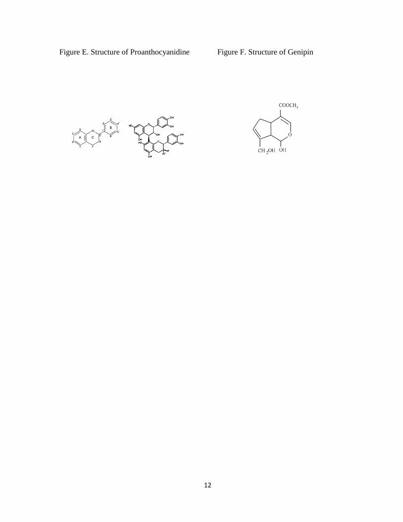
plant and fruit-derived cross-linking agents such as proanthocyanidin (PA) (Fig. E) and
genipin (GE) (Fig. F) has recently been explored (Bedran-Russo et al., 2007; Walter et al.,
2008; Macedo et al., 2009). These studies demonstrated that both compounds effectively
stabilize dentin collagen and improve dentin bond strength.
Of these two compounds, PA has been characterized for its effects on fibroblasts
(Han et al., 2003) and chemical stability of dentin collagen (Walter et al., 2008; Macedo et
al., 2009). It has been proposed that PA-treatment could enhance the dentin collagen stability,
and improve the ultimate tensile strength in dentin collagen (Bedran-Russo et al., 2007).
However, it has been reported that the treatment with PA caused discoloration on
demineralized dentin samples (Walter et al., 2008).
GE is a compound derived from genoposide, which is isolated from the fruits of
Gardenia jasminoides (Sung et al., 1999; Sung et al., 1999; Fujikawa et al., 1976). It was
found that GE-fixed tissue had a resistance against enzymatic degradation comparable to or
better than GA-fixed tissue and GE is 1000 times less cytotoxic than GA (Sung et al., 1999;
Sung et al., 2001). Sung et al, found that GE can react with the amino groups of amino acids
such as Lys, Hyl, and Arg residues within biological tissues (Sung et al., 1998); however, in

7
comparison to PA, the effects of GE on dentin are still poorly characterized and the
mechanism of GE-induced cross-linking is still not well understood.
The aims of the current study are to characterize GE-modified dentin collagen by
evaluating the collagen stability, amino acid composition, lysyl oxidase (LOX)-mediated
collagen cross-links, and microtensile bond strength (MTBS). A better understanding of the
mechanism of GE modification on dentin collagen may provide insight into a development of
new approaches to prevent dentin caries, and to improve dentin bond strength and longevity
of the resin-dentin bond established during restorative procedures.

8
Figure A.
Collagen Biosynthesis.
Schematic for the biosynthesis of type I collagen. The upper part (above the cell membrane)
summarizes the intracellular and the lower (below the cell membrane) the extracellular
events. The intracellular events include extensive post-translational modifications such as
hydroxylation, glycosylation (both O- and N-linked), association of pro α chains and folding
into a triple helical molecule from the C- to N-terminus. The extracellular events involve the
removal of both N- and C-propeptide extensions, self-assembly of collagen molecules into a
fibril, enzymatic oxidative deamination of Lys and Hyl residues by LOX and subsequent
intra- and intermolecular covalent cross-linking. The collagen molecules are packed in
parallel and are longitudinally staggered with respect to one another by some multiple of
axial repeat distance, D (~67nm).

9
Figure B.
Major cross-linking pathways of collagen.

10
Figure C.
Structure of dehydrodihydroxylysinonorleucine (deH-DHLNL).
When Hylald
in the C- or the N-telopeptides pairs with helical Hyl, an iminium cross-link,
deH-DHLNL is formed.

11
Figure D.
Structure of pyridinoline (Pyr).
Pyr is derived from a helical Hyl and two Hylald
in the C- or the N-telopeptides.
12
Figure E. Structure of Proanthocyanidine Figure F. Structure of Genipin

13
Chapter I
Characterization of genipin-modified dentin collagen Introduction
Introduction
Fibrillar type I collagen is the predominant component in dentin matrix, and regulate
the spatial aspect of dentin formation. For instance, it functions as a stable, three-dimensional
template for mineral deposition and growth and it also defines the space for organized
mineralization (Parsons & Glimcher, 1976). Cross-linking is the final post-translational
modification in collagen biosynthesis and is crucial for the stability, tensile strength,
viscoelasticity and appropriate physiological functions of the collagen fibrils (Nimni
& Harkness, 1988; Yamauchi & Mechanic, 1988; Kagan, 2000; Yamauchi, 2002). Collagen
cross-linking can be divided into two categories: enzymatic lysyl oxidase (LOX) mediated
cross-links, and non-enzymatic cross-links induced by chemical and natural agents, UV light,
and heat.
LOX-mediated collagen cross-linking has been extensively reviewed (Tanzer, 1976;
Eyre et al., 1984; Yamauchi & Mechanic, 1988; Yamauchi, 1996; Bailey, 2001). The
chemical structure and the quantity of these cross-links are primarily determined by the
extent of hydroxylation of lysine (Lys) residues in the collagen molecule and the extent of
oxidative deamination of the Lys and hydroxylysine (Hyl) residues present in the telopeptide

14
domains of the molecule. These modifications are catalyzed by lysyl hydroxylases (LHs) and
LOX/LOX-like proteins, The glycosylation pattern of specific Hyl residues that are involved
in cross-linking may regulate the maturation of collagen cross-links (Sricholpech et al., 2012).
The cross-linking pattern can also be determined by the maturation/turnover rate of tissues
(Bailey & Shimokomaki, 1971; Moriguchi & Fujimoto, 1978; Eyre et al., 1988; Yamauchi et
al., 1988), the details of molecular packing structure (Yamauchi et al., 1986; Mechanic et al.,
1987; Katz & David, 1992), and the physical force exerted on the tissue (Otsubo et al., 1992).
Collagen cross-links can also be induced non-enzymatically by the treatment with
chemicals and natural plant/fruit extracts (Sung et al., 1999). These non-enzymatic cross-
linking has been shown to facilitate preservation of substrate shape as a proper scaffolds
(Sung et al., 2001), improve a tissue’s physiological function (Sung et al., 1999; Chang et al.,
2002; Liang et al., 2004), and increase the mechanical properties of collagen (Huang et al.,
1998; Sung et al., 1999; Han et al., 2003) (Walter et al., 2008). Glutaraldehyde (GA), a
synthetic cross-linking agent, has been widely used as a fixative agent and it is well
documented that GA treatment improves the stability of various collagen-based tissues
(Chang et al., 2002; Bedran-Russo et al., 2008; Bedran-Russo et al., 2011; Braile et al.,
2011). However, the utilization of GA on biological tissues in vivo has been limited because
of its toxicity and/or instability over time (Sung et al., 1998; Sung et al., 1999).
Genipin (GE), a traditional Chinese herbal medicine extracted from gardenia fruit, has
been found to be an effective collagen cross-linking agent (Fig. 1.1). GE has favorable in
vitro cytotoxicity and the GE-modified collagenous tissues have increased toughness more
than that of GA-treated collagenous tissues (Cheung et al., 1985; Tsai et al., 2000).

15
While several studies on the effects of GE on dentin have been published (Bedran-
Russo et al., 2007; Walter et al., 2008; Al-Ammar et al., 2009), conditions of GE treatment
are not well established and the nature of GE-induced cross-linking is still not well
understood. A better understanding of GE-induced cross-links in dentin collagen could
provide insight into a strategy to develop a novel biomodification technology in restorative
dentistry. As the first step toward this goal, the present study was designed to characterize
GE-modified dentin collagen matrices using various treatment conditions. Specifically,
discoloration of dentin, collagen stability against bacterial collagenase and changes in amino
acid composition and LOX-mediated collagen cross-links were analyzed.
Materials and methods
Sample collection and demineralization
Extracted intact bovine molars (≤1 year old) were purchased (Aries Scientific, Texas,
USA) and used for this study. Enamel, cementum and pulp were removed using high-speed
diamond burs under water cooling. Dentin was pulverized in liquid N2 by a Spex Freezer
Mill (SPEX CertiPrep, Inc., Metuchen, NJ, USA), washed with distilled water. In order to
obtain sufficient quantities of collagen for biochemical and statistical analyses, three teeth
were combined as one group. The samples were demineralized with 0.5 M
ethylenediaminetetraacetic acid (pH 7.4) (EDTA) with several changes of the solution for 14
days at 4 °C, extensively washed with distilled water and lyophilized. Eighty four 2 mg
aliquots of the demineralized dentin collagen were randomly allocated to the following four
treatment groups based on the GE (Wako Pure Chemical Industries, Ltd., Osaka, Japan)
concentrations (n=21):

16
1. Control group. In the control group, specimens were treated with phosphate buffered
saline (PBS);
2. 0.01% GE. In this group, specimens were treated with 0.01% GE in PBS;
3. 0.1% GE. In this group, specimens were treated with 0.1% GE in PBS; and
4. 0.5% GE. In this group, specimens were treated with 0.5% GE in PBS.
Discoloration and collagen stability of GE-modified dentin collagen
Seventy two 2 mg aliquots of demineralized dentin collagen were treated with 1 mL of
three concentrations of GE solution or PBS and incubated for 30 min, 1 h, 4 h, 8 h, 12 h and
24 h at 37 °C with agitation (n=3 in each time point, n=18 in each group). After treatment,
the samples were extensively washed with distilled water, lyophilized and observed for their
discoloration.
Collagen stability was assessed by enzymatic degradation as reported (Walter et al.,
2008). Samples were suspended in 1 mL of ammonium bicarbonate and digested with 5%
w/w bacterial collagenase derived from Clostridium histolyticum (Worthington Biochemical
Corp., Lakewood, NJ, USA, 1,075 U/ mg) for 24 h at 37 °C. After digestion, the samples
were centrifuged for 15 min at 15,000 g, the residues (undigested collagen) were washed
with distilled water by repeated centrifugation and lyophilized. The residues were
hydrolyzed with 300 μL of 6 N HCl (Pierce, Rockford, IL, USA) in vacuo after flushing with
N2 gas for 22 h at 105°C. The hydrolysates were dried, reconstituted in 300 μL of distilled
water, filtered and subjected to hydroxyproline (Hyp) analysis using a high-performance
liquid chromatography (HPLC) system (Prostar 240/310, Varian, Walnut Creek, CA, USA)

17
fitted with a cation exchange column (AA911; Transgenomic, Inc., San Jose, CA, USA)
(Yamauchi & Shiiba, 2008).Amino acid analysis and collagen cross-link analysis
Twelve 2 mg aliquots of demineralized dentin collagen were treated with three
concentrations of GE solution or PBS and incubated for 24 h at 37 °C (n=3, Total sample
n=12). After treatment, the samples were extensively washed with distilled water, lyophilized,
reduced with standard NaB3H4 and hydrolyzed and an aliquot of each hydrolysate was
subjected to amino acid analysis as described above. The amount of each amino acid was
calculated as residues per 1,000 total amino acids. The hydrolysates with known amounts of
Hyp were then analyzed for cross-links as we reported (Yamauchi & Shiiba, 2008). The
reducible immature cross-link, dehydrodihydroxylysinonorleucine (deH-DHLNL) (Fig.
1.2A)/its ketoamine (Fig. 1.2B), was analyzed as its reduced form, i.e.
dihydroxylysinonorleucine (DHLNL). The non-reducible mature cross-link, pyridinoline
(Pyr) (Fig. 1.2C.), was also analyzed simultaneously. The DHLNL and Pyr were quantified
as moles/mole of collagen based on the value of 300 residues of Hyp per collagen. All
analyses were done in triplicate in independent experiments.
Statistical analysis
The statistical evaluations were performed using Stat View software (SAS Institute Inc.,
Cary, NC, USA). Values were expressed as mean +/- standard deviation, and the difference
between the control group and GE groups was compared by two-way ANOVA and Fisher's
PLSD. A p value of less than 0.05 was considered significant.

18
Results
Discoloration and collagen stability of GE-modified dentin collagen
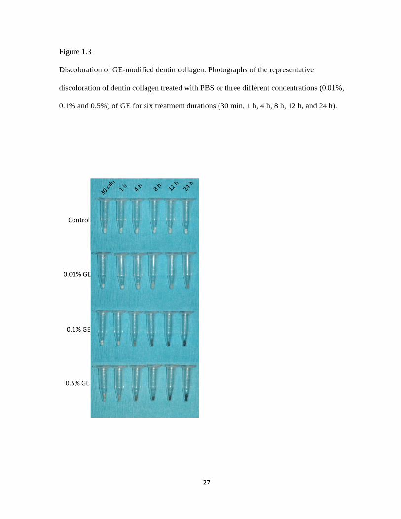
The demineralized dentin collagen treated with GE exhibited distinct features. While
dentin collagen in the control group presented a white color, a dark blue pigmentation was
observed in the GE-treated groups (Fig. 1.3). Hyp analysis showed that dentin collagen in all
of the control groups was almost completely digested. However, when treated with GE, the
digestibility markedly decreased in a dose- and time-dependent manner. In the 0.5% GE,
81.9 ± 4.9 nM, 166.4 ± 3.8 nM, 926.6 ± 37.6 nM, 1,243.5 ± 50.6 nM, 1,259.1 ± 48.3 nM and
1,259.3 ± 61.4 nM Hyp at 30 min, 1 h, 4 h, 8 h, 12 h and 24 h were recovered in the
undigested residue respectively. In the 24 h treatment of PBS, 0.01% GE and 0.1% GE, 2.0 ±
1.7 nM, 166.0 ± 19.3 nM and 1,054.3 ± 47.3 nM Hyp were recovered in the undigested
residues (Fig.1.4).
Amino acid Analysis
Overall amino acid compositions in the control and three GE groups at all time points
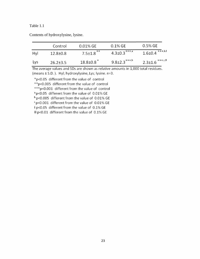
examined were essentially identical to one another (data not shown) except Hyl and Lys. The
amounts of amino acids, Hyl and Lys in control and three GE groups treated for 24 h are
shown in Table 1. Hyl and Lys in three GE groups were significantly decreased in a dose-
dependent manner when compared to the control (p<0.05). No significant changes were
observed for other amino acids including arginine, which is different from the report by Sung
and co-workers (Sung et al., 1998).

19
Collagen cross-link analysis
Typical chromatographic profiles of collagen cross-links from control and three GE
groups treated for 24 h are shown in Fig. 1.5 & 1.6 Two major cross-links, DHLNL
(reducible) and Pyr (non-reducible) were identified in all groups at all time points examined.
In addition, unknown newly formed NaB3H4-reducible compunds were identified in three GE
groups (peaks at 22 min: unk 1, and at 64 min: unk 2) (Fig.1.5). The results of the
quantitative cross-link analysis comparing control group with GE groups are summarized in
Table 1.2.
The amount of DHLNL in 0.01% and 0.1% GE for 24 h (1.35 ± 0.23 M and 1.23 ± 0.19
M, respectively) were not significantly different from those of control (1.36 ± 0.17 M), but
DHLNL in 0.5% GE for 24 h (1.13 ± 0.07 M) was slightly but significantly decreased when
compared to that of control (p< 0.05) (Fig. 1.5 and Table 1.2). The amount of Pyr in all three
GE groups for 24 h showed no significant difference when compared with control (Fig. 1.6,
and Table 1.2).
The amounts of newly formed reducible compounds, unk 1 and unk 2, in GE groups for 24 h
were significantly increased in a dose-dependent manner (Fig.1.5 and Table 1.2).
Discussion
Fibrillar type I collagen is the most abundant protein and the major structural
component in dentin and the molecules in the fibril are stabilized by covalent intermolecular
cross-linking
To further stabilize collagen fibrils in biological tissues, various cross-linking agents have
been used to induce additional intra- and intermolecular cross-links (Cheung et al., 1985;
Huang et al., 1998; Sung et al., 1998; Sung et al., 1999; 1999; Sung et al., 1999; Tsai et al.,

20
2000; Sung et al., 2001; Chang et al., 2002; Han et al., 2003; Liang et al., 2004; Bedran-
Russo et al., 2007; Bedran-Russo et al., 2008; Walter et al., 2008; Al-Ammar et al., 2009;
Bedran-Russo et al., 2011; Braile et al., 2011). GE, a naturally occurring cross-linking agent,
is as efficient as GA but possesses significantly less toxicity (Sung et al., 1998; Sung et al.,
1999) and has been proven to be biocompatible and stable for a long period of time in
animals (Sung et al., 1999). Although several cross-linking agents including GE have been
proposed to improve the mechanical and chemical properties of collagenous tissues, the
mechanisms of cross-linking and the nature of the cross-links are still not well defined.
The present study was undertaken to evaluate the effects of GE on dentin collagen using
biochemical approaches such as Hyp analysis, amino acid analysis and quantitative cross-link
analysis. GE-treatment of collagen resulted in a blue pigmentation as previously reported
(Sung et al., 1998; Lee et al., 2003; Walter et al., 2008). The blue discoloration of
demineralized dentin collagen by GE increases in a dose- and time-dependent manner. The
mechanism of the blue pigmentation is not yet clear but possibly associated with GE-induced
cross-link compounds as the intensity of the color well corresponded with the collagen
stability against enzymatic digestion. Collagen stability was evaluated by the digestibility
with bacterial collagenase which hydrolyzes the peptide bond on the amino-terminal side of
Gly in –X–Gly–Pro. This results in the breakdown of collagen molecules into small peptides
(Watanabe, 2004). Based on the mean values of Hyp in 2 mg demineralized dentin matrix in
this study (1,319.4 ± 46.9 nM) (n= 3), the rate of collagen digestion was 93.8%, 87.4%,
27.3%, 7.6%, 1.1% and 1.0% in 0.5% GE at 30 min, 1 h, 4 h, 12 h and 24 h respectively, and
99.7%, 89.0% and 16.0% in PBS, 0.01% GE and 0.1% GE for 24 h respectively. Thus, the
rate of collagen digestion with GE treatment cleary decreased in a dose- and time-dependent
manner (Fig. 1.4). When collagen was treated with 0.5% GE for 12 h and 24 h, almost no

21
collagen was digested. Most likely GE-induced cross-linking hinders the enzyme
accessibility to collagen and/or it generates a large cross-linked collagen complex so that
collagenase cleavage no longer solubilizes the complex.
The amino acid analysis was conducted to determine which, if any, amino acid residues
in the dentin collagen reacted with GE. The results demonstrated that Hyl and Lys residues
were the only amino acids that decreased with GE treatment in a dose- and time-dependent
manner. It is likely that the ɛ-amino groups on Hyl and Lys in a collagen molecule react with
GE to form cross-links that are stable in acid hydrolysis. When the values of Lys and Hyl
were calculated on a residues per collagen molecule basis (based on the value of 300 residues
of Hyp per collagen molecule), the mean number of Hyl residues in GE groups for 24 h per
collagen molecule decreased from 47.7 (± 3.2) in control to 5.3 (± 1.4) in 0.5% GE group
respectively. In case of Lys, it decreased from 97.9 (± 13.2) in control to 7.6 (± 5.1) in 0.5%
GE group, respectively. It is estimated that approximately 90 Lys and 42 Hyl in a collagen
molecule are utilized for GE-induced cross-links in 0.5% GE treatment for 24 h. Since there
are approximately 98 Lys and 48 Hyl residues per collagen molecule in controls (note: some
of the Lys residues are derived from non-collagenous proteins), ~92% of the Lys and 88% of
the Hyl residues of collagen were likely involved in GE-derived cross-linking with 0.5% GE
treatment for 24 h. Under the conditions used, no other amino acids were significantly
changed by GE treatment. Sung et al reported that Lys, Hyl and Arg were used in the
reaction with GE in porcine pericardia (Sung et al., 1998). The discrepancy between the
current study and the one by Sung et al., could be due to the different treatment conditions
and/or tissues.
Cross-link analysis revealed that several unidentified, reducible compounds were
detected in GE-treated groups; two major ones eluted at 22 and 64 min in our HPLC system,

22
respectively, and they increased in a dose- and time-dependent manner. These newly formed
two reducible compounds are most likely GE-induced cross-links that involve Lys and Hyl
residues in collagen. Further studies are warranted to identify the structures of these cross-
links by isolating and analyzing mass spectrometry and NMR. There was a significant
decrease of DHLNL in 0.5% GE group for 24 h when compared to that of control. The cause
of this slight decrease is not clear but, possibly, the aldimine bond of deH-DHLNL could be
modified when dentin collagen is treated with GE for a longer period of time. Pyr showed no
significant changes at all time points examined and any dose of GE used. This is likely due to
the stability of this cross-link.
Conclusions
The treatment of bovine dentin collagen with GE resulted in a dose- and time-
dependent increase in collagen stability and the intensity of the discoloration. The GE-
induced, reducible cross-links involving Lys and Hyl residues also increase in a time- dose-
dependent manner. The GE treatment does not affect LOX-mediated collagen cross-link, Pyr,
but could modify some of DHLNL cross-link.
23
Table 1.1
Contents of hydroxylysine, lysine.

24
Table 1.2
Contents of enzymatic cross-links and GE-induced cross-links on bovine dentin

25
Figure 1.1
Chemical structure of GE in the study
O
COOCH3
OH OH

26
Figure 1.2A.B.C
Chemical structure of iminium (A) and keto (B) form deH-DHLNL, and Pyr (C) among the
type l collagen molecules. Solid line: α1 chains of type l collagen. Dotted line: α2 chain of
type l collagen. Black dot: Hylald
. White dot: Hyl. glc: glucose. gal: galactose.
27
Figure 1.3
Discoloration of GE-modified dentin collagen. Photographs of the representative
discoloration of dentin collagen treated with PBS or three different concentrations (0.01%,
0.1% and 0.5%) of GE for six treatment durations (30 min, 1 h, 4 h, 8 h, 12 h, and 24 h).

28
Figure 1.4
Collagen stability of GE-modified dentin collagen. The mean amounts of undigested collagen
from 2mg of dentin collagen treated with PBS or three concentrations of GE (0.01%, 0.1%
and 0.5%) for six treatment durations (30 min, 1 h, 4 h, 8 h, 12 h, and 24 h) as showing
hydroxyproline residue amounts in residue after collagenase digestion for 24 h (n=3).

29
Figure 1.5
Representative chromatographs of reducible collagen cross-links in dentin collagen at 24h
treatment of PBS or three concentrations of GE (0.01%, 0.1% and 0.5%).

30
Figure 1.6
Representative chromatographs of non-reducible collagen cross-links in dentin collagen at
24h treatment of PBS or three concentrations of GE (0.01%, 0.1% and 0.5%).

31
Chapter II
Genipin-induced Cross-linking and Dentin Bonding in Human Dentin
Introduction
While composite resin bonding to enamel substrate has been shown to be reliable,
bonding to dentin substrate remains a challenge (Swift et al., 1995). Current adhesive
systems bond to dentin through a micromechanical mechanism relies on the formation of a
hybrid layer (Burrow et al., 1994). Hybrid layer formation can be summarized in the
following steps: 1) dentin is etched prior to or simultaneous with the application of a
primer/adhesive system which results in the exposure of superficial collagen fibrils, 2)
adhesive resin monomers are then polymerized among and around the exposed collagen
fibrils forming a hybrid layer that is believed to be essential for dentin bonding. Therefore,
for effective bonding, the stability and maintenance of dentin collagen is very important
(Hashimoto et al., 2003; Breschi et al., 2008).
Dentin is composed of an extracellular matrix embedded in a predominant inorganic
mineral phase. Type I collagen is the most abundant organic component in dentin,
comprising approximately 90% of the total organic matrix (Butler, 1995; Beateman et al.,
1996). Collagen molecules are stabilized in fibrils by the extensive formation of covalent

32
intermolecular cross-links. Collagen cross-linking is crucial for the stability, tensile strength,
viscoelasticity and appropriate physiological functions of the collagen fibrils (Eyre et al.,
1984; Yamauchi & Mechanic, 1988; Yamauchi, 2002) and can be divided into two types:
enzymatic cross-linking, i.e., lysyl oxidase (LOX) mediated cross-links and non-enzymatic
cross-linking, e.g., cross-linking induced by sources such as chemical agents, UV light and
heat. Several cross-linking agents have been demonstrated to increase the mechanical
properties of collagenous tissues and scaffolds (Sung et al., 1999; 1999; Sung et al., 2003;
Yao et al., 2005; Castellan et al., 2010). Recently, a natural cross-linking agent, genipin (GE)
has been reported to improve the mechanical properties of dentin including ultimate tensile
strength and microtensile bond strength (MTBS) between dentin and resin (Bedran-Russo et
al., 2007; Bedran-Russo et al., 2008). However, the effects of the GE-induced cross-links on
the enzymatic cross-linking and MTBS have not been well documented. In the previous
study (chapter 1), by utilizing bovine dentin, we established that 0.5% GE concentration was
a sufficient concentration to effectively cross-link dentin collagen. The objectives of this
study were to evaluate the effect of 0.5% GE treatment on human dentin collagen and MTBS
between composite resin to dentin. We hypothesized that 0.5% GE treatment exerts the
effects on human dentin collagen similar to those in bovine dentin and the treatment
increases MTBS.
Materials & Methods
Sample Collection and Experimental Treatment for Biochemical Analyses
Extracted intact human molars were used for this study. All samples were stored in
distilled water with 0.5% thymol crystals solution at 4°C until use. The pulp, periodontal
33
ligament and pre-dentin were manually removed, and enamel and cementum were removed
using high-speed diamond burs under water cooling. The remaining dentin was pulverized in
liquid N2 by a Spex Freezer Mill (SPEX CertiPrep, Inc., Metuchen, NJ, USA), washed with
cold distilled water by repeated centrifugation and lyophilized. In order to obtain sufficient
quantities of collagen for biochemical and statistical analyses, two teeth were pooled as a
sample and 48 samples (thus, a total of 96 teeth) were generated. After pulverization, the
samples were demineralized with 0.5 M ethylenediaminetetraacetic acid (pH 7.4) (EDTA)
for 14 days at 4 °C with several changes of EDTA solution, extensively washed with cold
distilled water and lyophilized. Approximately two mg aliquots of demineralized dentin
(n=48) were incubated in 1 mL of either PBS or 0.5% GE for 1 h, 4 h, 12 h and 24 h at 37 °C
with agitation. The pH in the solutions was adjusted to 7.4 with 0.5M NaOH. After treatment,
the specimens were extensively washed with cold distilled water by centrifugation and
lyophilized.
Collagen Stability
Either PBS or 0.5% GE treated-2 mg of demineralized collagen were digested with
bacterial collagenase as described in chapter 1. After digestion, the samples were centrifuged
for 15 min at 15,000 g, the residues (undigested collagen) washed with distilled water and the
pooled residues was lyophilized (n=3 in each time point, total sample number=24). The
residues were hydrolyzed with 6 N HCl (Pierce, Rockford, IL, USA) and subjected to
hydroxyproline (Hyp) analysis as described in chapter 1 (Yamauchi & Shiiba, 2008).

34
Amino acid Analysis and Collagen Cross-link Analysis
Two mg aliquots of dentin collagen treated with PBS or 0.5% GE were reduced with
standard NaB3H4, hydrolyzed with 300 μL of 6N HCl (Pierce, Rockford, IL, USA) in vacuo
(see above), after flushing with N2 gas, for 22 h at 105°C (n=3 in each time point, total
sample number=24). The hydrolysates were dried, reconstituted in 300 μl of distilled water,
filtered, and then an aliquot of each hydrolysate was subjected to amino acid analysis (see
above). The amounts of each amino acid were calculated as residues per 1,000 total amino
acids. The hydrolysates with known amounts of Hyp were analyzed for cross-links on a
HPLC system as described (Yamauchi & Shiiba, 2008). The reducible immature cross-link,
i.e., dehydrodihydroxylysinonorleucine (deH-DHLNL)/its ketoamine was analyzed as the
reduced form, i.e. DHLNL. The non-reducible mature cross-link, i.e. pyridinoline (Pyr) was
also analyzed simultaneously, and these two cross-links were quantified as moles/mole of
collagen. The analyses were done in triplicate in independent experiments.
Sample Collection for MTBS
A total of twenty four sound freshly extracted human molars were selected, cleaned
from debris and immediately stored in distilled water with 0.5% thymol crystals solution at 4
C°. The teeth were ground flat and perpendicular to their long axis using 120-grit silicon
carbide abrasive paper (Buehler Ltd, Lake Bluff, IL, USA) under running water and mild
pressure to expose middle depth dentin. The exposed dentin surfaces were confirmed with a
dissecting microscope to ensure that no enamel remained. Dentin surfaces were polished with

35
wet 240-, 400, and 600-grit silicon carbide abrasive paper (Buehler Ltd, Lake Bluff, IL,
USA).
Dentin Surface Pretreatment and Restorative Procedures for MTBS
Specimens were randomly divided according to the dentin treatment and treatment
duration): PBS treated (control) groups (treated with PBS for 1 h, 4 h, 12 h and 24 h) and GE
treated groups (treated with 0.5% GE in PBS for 1 h, 4 h, 12 h and 24 h). The pH of the
solutions was adjusted to 7.4 with NaOH pellets. Prior to treatment, the dentin surface was
etched using a 37% phosphoric acid gel (Bisco, Schaumburg, IL, USA) for 15 second (s),
then thoroughly rinsed with water for 15 s and kept moist. After etching, the teeth were
further divided into four subgroups (n= 3 in each subgroup), according to the treatment
duration. Teeth were immersed in PBS or 0.5 %GE solution and incubated at 37°С for 1 h, 4
h, 12 h and 24 h. After each time point, specimens were thoroughly rinsed with distilled
water for 60 s to remove excess cross-linking agents and buffer, and kept moist. The
adhesive system (OptiBond Solo Plus, Kerr, Orange, CA, USA) was used following
manufacturer’s instructions. A hybrid resin composite restorative material (Z250, 3M ESPE,
Saint Paul, Minnesota, USA) was placed over the bonded surfaces incrementally (6 mm total
thickness) to allow for gripping during the tensile testing. Increment thickness was limited to
2 mm, and curing was accomplished for 20 s per increment (Ultra-Lume LED5
ULTRADENT INC, South Jordan, UT, USA). Teeth were then stored in distilled water at
37 °С for 24 h. All specimens were sectioned perpendicular to the bonded interface into 0.7 ±
0.2-mm-thick slabs using a slow speed diamond saw (Buehler-Series 15LC Diamond, Lake

36
Bluff, IL, USA) under cooling water. The specimens were glued on a jig placed on a MTBS
machine (Bisco, Schaumburg, IL, USA) and subjected to tensile force at a crosshead speed of
1 mm min−1
. Means and standard deviations were calculated and expressed in MPa.
Statistical Analysis
The statistical evaluations were performed using Stat View software (SAS Institute Inc.,
Cary, NC, USA). Values were expressed as mean +/- standard deviation, and the difference
between the control group and GE groups was compared by two-way ANOVA and Fisher's
PLSD. A p value of less than 0.05 was considered significant.
Results
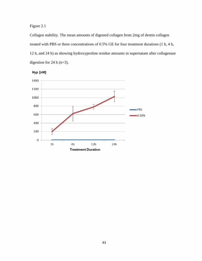
Collagen Stability
The mean values of Hyp in ~2 mg demineralized dentin matrix in this study was
1,198.7 ± 79.8 nM (n= 3). Hyp analysis showed that while samples in control group for all
time points were almost all digested, in the 0.5% GE, the digestibility markedly decreased in
a time-dependent manner. In 0.5 %GE treatment for 1, 4, 12 and 24 h, 198.5 ± 68.3 nM,
624.6 ± 175.3 nM, 782.0 ± 51.2 nM and 1029.4 ± 127.7 nM of Hyp were recovered,
respectively. Thus, the rate of collagen digestion in the GE treated groups were 80.1% (1 h),
47.9% (4 h), 34.5% (12 h) and 14.1% (24h).

37
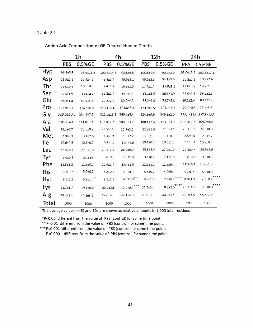
Amino Acid Analysis
Although overall amino acid compositions among the control and GE-treated groups
were similar to one another (Table 2.1), as in the case of bovine dentin, significant
differences were identified in the values of Hyl and Lys residues. Hyl in 0.5% GE treated for
1 h, 4 h, 12 h and 24 h were significantly decreased in comparison to those of PBS treated
groups (p<0.05 for 1 h, p<0.01 for 4 h, p<0.0001 for 12 h and 24 h), and Lys, p<0.001 for 4h,
p<0.0001 for 12h and 24h, respectively. The decreases of Hyl and Lys in 0.5% GE treatment
occurred in a time-dependent manner.
Collagen Cross-link Analysis
Two major enzymatic cross-links in dentin collagen, a reducible immature cross-link,
DHLNL, and a non-reducible mature cross-link, Pyr, were identified together with other
minor cross-links. In addition, unknown reducible compounds that are likely GE-induced
reducible cross-links were identified by HPLC analysis in the GE-treated collagen. The
quantitative results of the collagen cross-link analyses comparing PBS and 0.5% GE
treatment at all time points examined are summarized in Table 2.2. DHLNL in 0.5% GE for
24 h was significant decreased compared with those of controls and 0.5% GE treatment for 1
h and 4 h. (p< 0.05). Pyr showed no significant difference between PBS and 0.5% GE
treatment groups at all examined time points.

38
MTBS
The results of MTBS of PBS and 0.5% GE treated groups are summarized in Fig. 2.2.
While there was no significant difference in MTBS between PBS and 0.5% GE treated
groups for 1 h, those in the GE treated groups for 4 h, 12 h and 24 h were all significantly
increased (p< 0.0001). The highest bond strength was observed for 0.5% GE treatment for 4
h (58.5 ± 17.7 MPa) which was significantly higher than those of 12 h and 24 h GE
treatments (p< 0.0001).
Discussion
In the present study, a natural cross-linking agent, GE, was examined to assess its
potential effects on human dentin collagen and MTBS. It was found that 0.5%GE treatment
for 4 h is sufficient to stabilize dentin collagen matrix and to enhance MTBS.
Although several studies have been performed to assess the effect of GE treatment on
collagenous tissues(Touyama et al., 1994; Lee et al., 2003; Fujikawa et al., 1976), the
mechanism by which collagen is stabilized by GE is not well understood. As shown in Table
1, only two amino acid residues (Hyl and Lys) were diminished with GE treatment in a time-
dependent manner indicating their involvement in GE-induced cross-linking. This is not full
agreement with the study conducted by Sung and co-workers (Sung et al., 1998) which
reported that Lys, Hyl and arginine residues were involved in GE-induced cross-links. In our
study, however, arginine did not change with GE treatment. The difference may be attributed
to the treatment duration (~24 h/our study, 3 days/Sung) and/or tissue specific reaction.
Normalizing the number of Hyl and Lys residues to 300 residues of Hyp (thus, one collagen
molecule) and considering that there are approximately 26 Hyl and 67 Lys residues per

39
collagen molecule in controls (note: some of the Lys residues are derived from non-
collagenous proteins), it is estimated that approximately 31% of the Hyl and 9% of the Lys
for 1h, 46% of the Hyl and 40% of the Lys for 4h, 69% of the Hyl and 57% of the Lys for
12h, and 73% of Hyl and 70% of Lys residues of collagen for 24 h were utilized in GE-
induced cross-links. The MTBS results of 0.5% GE for 1 h did not significantly alter when
compared to that of PBS treatment for 1h and this is consistent with the study conducted by
Al-Ammar et al. However, MTBS in 0.5% GE treatment for 4 h, 12 h and 24 h were
significantly increased as compared to those treated PBS alone. Interestingly, MTBS slightly
but significantly decreased in the samples treated with GE for 12 and 24 h when compared to
those treated for 4 h. The reason is not clear at present but one potential explanation would be
that when collagen is cross-linked by GE more than certain levels, while collagen stability
keeps increasing, the formation of hybrid layer could be compromised. This would lower
MTBS. However, the results need to be confirmed and the potential mechanisms should be
investigated using a larger sample size combined with careful characterization of the GE-
treated hybrid layer.
By cross-link analysis, DHLNL and Pyr, and several unidentified reducible
compounds were detected in the GE-treated collagen (data not shown). These unidentified
reducible compounds induced by GE increased in a time-dependent manner, thus, they are
most likely associated with GE-induced cross-links involving Hyl and Lys residues of
collagen. GE treatment for 1 h and 4 h did not significantly alter the enzymatic cross-links,
DHLNL and Pyr, when compared to the PBS-treated group (Table 2.2). However DHLNL
was significantly decreased at 12 h and 24 h GE treatment when compared to controls. It is
possible, therefore, that the GE reacts with not only free Lys and Hyl of collagen but also

40
with some of the immature cross-link, deH-DHLNL, possibly its aldimine form with a longer
period of treatment. The fate of such modifications needs to be pursued in the future studies.
It is concluded that MTBS increased after dentin collagen was treated with GE for 4 h. Thus
this cannot be applied in clinical settings yet. However, it could be possible that the time can
be shortened by application methods, e.g. changing temperature (note that all the experiments
in this study was performed at 37 °C), pH, or combining with other cross-linking agents. By
improving the application time and discoloration, the use of natural collagen cross-linking
agents could be a safe and promising approach to improve adhesive restorative procedures
and dentin bond strength.
41
Table 2.1

42
Table 2.2
43
Figure 2.1
Collagen stability. The mean amounts of digested collagen from 2mg of dentin collagen
treated with PBS or three concentrations of 0.5% GE for four treatment durations (1 h, 4 h,
12 h, and 24 h) as showing hydroxyproline residue amounts in supernatant after collagenase
digestion for 24 h (n=3).

44
Figure 2.2
MTBS on the samples treated with PBS or 0.5% GE for 1h, 4 h, 12 h and 24 h (tooth n=3,
beams n=45).

45
Summary and conclusion
The two studies demonstrate that:
1. The treatment with GE resulted in discoloration and an increase of collagen stability
in a dose- and time-dependent manner.
2. The GE-induced cross-links utilize Lys or Hyl residues of collagen and the cross-
links increase in a time- and dose-dependent manner
3. Cross-linking induced by 0.5% GE treatment for 4 h is sufficient to improve the
mechanical properties of dentin collagen and bond strength.
4. 0.5% GE may modify some of the LOX-mediated immature divalent cross-links
when treated for a longer period of time.
5. GE-induced cross-linking for a long period of time (12 and 24 h) may result in slight
decreases in MTBS.
Thus, while it requires more studies and modifications, GE treatment could be an efficient
approach to improve mechanical properties of dentin collagen matrix and the longevity of
resin-dentin bond.

46
References
Al-Ammar A., Drummond J. L., Bedran-Russo A. K. The use of collagen cross-linking
agents to enhance dentin bond strength. J Biomed Mater Res B Appl Biomater
2009;91: 419-424.
Baer E. , Cassidy JJ. , Hiltner A. , Collagen Biochemistry and Biomechanics. In: Hierarchical
structure of collagen and its relationship to the physical properties of tendon, Nimni
ME. (ed). Vol. 2. Boca Raton, Florida: CRC Press, 1988,pp. 177-199.
Bailey A. J. Molecular mechanisms of ageing in connective tissues. Mech Ageing Dev
2001;122: 735-755.
Beateman WT., Lamandé SR., Ramshaw AM., Extracellular Matrix. In: Molecular
Components and Interactions., WD Comper (ed). Vol. 2. Amsterdam, the Netherlands
Harwood Academic Publishers, 1996,pp. 22-67.
Bedran-de-Castro A. K., Pereira P. N., Pimenta L. A., Thompson J. Y. Effect of thermal and
mechanical load cycling on nanoleakage of Class II restorations. J Adhes Dent
2004;6: 221-226.
Bedran-Russo A. K., Castellan C. S., Shinohara M. S., Hassan L., Antunes A.
Characterization of biomodified dentin matrices for potential preventive and
reparative therapies. Acta Biomater 2011;7: 1735-1741.
Bedran-Russo A. K., Pashley D. H., Agee K., Drummond J. L., Miescke K. J. Changes in
stiffness of demineralized dentin following application of collagen crosslinkers. J
Biomed Mater Res B Appl Biomater 2008;86: 330-334.
Bedran-Russo A. K., Pereira P. N., Duarte W. R., Drummond J. L., Yamauchi M.
Application of crosslinkers to dentin collagen enhances the ultimate tensile strength. J
Biomed Mater Res B Appl Biomater 2007;80: 268-272.
Bella J., Brodsky B., Berman H. M. Hydration structure of a collagen peptide. Structure
1995;3: 893-906.

47
Braile M. C., Carnevalli N. C., Goissis G., Ramirez V. A., Braile D. M. In vitro properties
and performance of glutaraldehyde-crosslinked bovine pericardial bioprostheses
treated with glutamic acid. Artif Organs 2011;35: 497-501.
Breschi L., Mazzoni A., Ruggeri A., Cadenaro M., Di Lenarda R., De Stefano Dorigo E.
Dental adhesion review: aging and stability of the bonded interface. Dent Mater
2008;24: 90-101.
Brodsky B., Eikenberry E. F., Cassidy K. An unusual collagen periodicity in skin. Biochim
Biophys Acta 1980;621: 162-166.
Buonocore M. G. A simple method of increasing the adhesion of acrylic filling materials to
enamel surfaces. J Dent Res 1955;34: 849-853.
Burrow M. F., Takakura H., Nakajima M., Inai N., Tagami J., Takatsu T. The influence of
age and depth of dentin on bonding. Dent Mater 1994;10: 241-246.
Butler W. T. Dentin matrix proteins and dentinogenesis. Connect Tissue Res 1995;33: 59-65.
Cagidiaco M.C., Ferrari M.A.G., Bonding to Dentin: Mechanism, Morphology and Efficacy
of Bonding Resin Composites to Dentin in Vitro and in Vivo. In: (ed): Casa Editrice O.
Debatte & F., 1995.
Carrilho M. R., Carvalho R. M., Tay F. R., Yiu C., Pashley D. H. Durability of resin-dentin
bonds related to water and oil storage. Am J Dent 2005;18: 315-319.
Castellan C. S., Pereira P. N., Viana G., Chen S. N., Pauli G. F., Bedran-Russo A. K.
Solubility study of phytochemical cross-linking agents on dentin stiffness. J Dent
2010;38: 431-436.
Chang Y., Tsai C. C., Liang H. C., Sung H. W. In vivo evaluation of cellular and acellular
bovine pericardia fixed with a naturally occurring crosslinking agent (genipin).
Biomaterials 2002;23: 2447-2457.
Cheung D. T., Perelman N., Ko E. C., Nimni M. E. Mechanism of crosslinking of proteins by
glutaraldehyde III. Reaction with collagen in tissues. Connect Tissue Res 1985;13:
109-115.

48
De Munck J., Van Landuyt K., Peumans M., Poitevin A., Lambrechts P., Braem M., Van
Meerbeek B. A critical review of the durability of adhesion to tooth tissue: methods
and results. J Dent Res 2005;84: 118-132.
Eyre D. R., Koob T. J., Van Ness K. P. Quantitation of hydroxypyridinium crosslinks in
collagen by high-performance liquid chromatography. Anal Biochem 1984;137: 380-
388.
Eyre D. R., Paz M. A., Gallop P. M. Cross-linking in collagen and elastin. Annu Rev
Biochem 1984;53: 717-748.
Fujikawa S. , Yokota T. , Koga K. , Kumada J. The continuous hydrolysis of geniposide to
genipin using immobilized β-glucosidase on calcium alginate gel. Biotechnol Lett
1976;697-702.
Fusayama T., Nakamura M., Kurosaki N., Iwaku M. Non-pressure adhesion of a new
adhesive restorative resin. J Dent Res 1979;58: 1364-1370.
Gerriets J. E., Curwin S. L., Last J. A. Tendon hypertrophy is associated with increased
hydroxylation of nonhelical lysine residues at two specific cross-linking sites in type I
collagen. J Biol Chem 1993;268: 25553-25560.
Gwinnett A. J. Altered tissue contribution to interfacial bond strength with acid conditioned
dentin. Am J Dent 1994;7: 243-246.
Han B., Jaurequi J., Tang B. W., Nimni M. E. Proanthocyanidin: a natural crosslinking
reagent for stabilizing collagen matrices. J Biomed Mater Res A 2003;65: 118-124.
Hashimoto M., Ohno H., Sano H., Kaga M., Oguchi H. In vitro degradation of resin-dentin
bonds analyzed by microtensile bond test, scanning and transmission electron
microscopy. Biomaterials 2003;24: 3795-3803.
Huang L. L., Sung H. W., Tsai C. C., Huang D. M. Biocompatibility study of a biological
tissue fixed with a naturally occurring crosslinking reagent. J Biomed Mater Res
1998;42: 568-576.

49
Inoue S., Murata Y., Sano H., Kashiwada T. Effect of NaOCl treatment on bond strength
between indirect resin core-buildup and dentin. Dent Mater J 2002;21: 343-354.
Kagan H. M. Intra- and extracellular enzymes of collagen biosynthesis as biological and
chemical targets in the control of fibrosis. Acta Trop 2000;77: 147-152.
Kanca J., 3rd. Resin bonding to wet substrate. 1. Bonding to dentin. Quintessence Int
1992;23: 39-41.
Lee Sang-Won, Lim Jong-Min, Bhoo Seong-Hee, Paik Young-Sook, Hahn Tae-Ryong.
Colorimetric determination of amino acids using genipin from Gardenia jasminoides.
Analytica Chimica Acta 2003;480: 267-274.
Liang H. C., Chang Y., Hsu C. K., Lee M. H., Sung H. W. Effects of crosslinking degree of
an acellular biological tissue on its tissue regeneration pattern. Biomaterials 2004;25:
3541-3552.
Lohre J. M., Baclig L., Sagartz J., Guida S., Thyagarajan K., Tu R. Evaluation of two epoxy
ether compounds for biocompatible potential. Artif Organs 1992;16: 630-633.
Lohre J. M., Baclig L., Wickham E., Guida S., Farley J., Thyagarajan K., Tu R., Quijano R.
C. Evaluation of epoxy ether fixed bovine arterial grafts for mutagenic potential.
ASAIO J 1993;39: 106-113.
Macedo G. V., Yamauchi M., Bedran-Russo A. K. Effects of chemical cross-linkers on
caries-affected dentin bonding. J Dent Res 2009;88: 1096-1100.
Miguez P. A., Pereira P. N., Atsawasuwan P., Yamauchi M. Collagen cross-linking and
ultimate tensile strength in dentin. J Dent Res 2004;83: 807-810.
Nakabayashi N., Kojima K., Masuhara E. The promotion of adhesion by the infiltration of
monomers into tooth substrates. J Biomed Mater Res 1982;16: 265-273.
Nimni ME., Harkness RD., Molecular structures and functions of collagen. In: Collagen,
Nimni ME. (ed). Vol. 1. Boca Raton, Florida: CRC Press, 1988,pp. 1-77.

50
Nishi C., Nakajima N., Ikada Y. In vitro evaluation of cytotoxicity of diepoxy compounds
used for biomaterial modification. J Biomed Mater Res 1995;29: 829-834.
Notbohm H., Nokelainen M., Myllyharju J., Fietzek P. P., Muller P. K., Kivirikko K. I.
Recombinant human type II collagens with low and high levels of hydroxylysine and
its glycosylated forms show marked differences in fibrillogenesis in vitro. J Biol
Chem 1999;274: 8988-8992.
Parsons D. B., Glimcher M. J. Reducible crosslinks of type I and type II collagens of chicken
cartilage. FEBS Lett 1976;65: 373-376.
Pashley D. H. Dentin: a dynamic substrate--a review. Scanning Microsc 1989;3: 161-174;
discussion 174-166.
Pashley D. H. Dentin bonding: overview of the substrate with respect to adhesive material. J
Esthet Dent 1991;3: 46-50.
Pashley D. H., Ciucchi B., Sano H., Horner J. A. Permeability of dentin to adhesive agents.
Quintessence Int 1993;24: 618-631.
Pereira P. N., Okuda M., Nakajima M., Sano H., Tagami J., Pashley D. H. Relationship
between bond strengths and nanoleakage: evaluation of a new assessment method.
Am J Dent 2001;14: 100-104.
Ritter A. V., Swift E. J., Jr., Yamauchi M. Effects of phosphoric acid and glutaraldehyde-
HEMA on dentin collagen. Eur J Oral Sci 2001;109: 348-353.
Rivera E. M., Yamauchi M. Site comparisons of dentine collagen cross-links from extracted
human teeth. Arch Oral Biol 1993;38: 541-546.
Sano H., Takatsu T., Ciucchi B., Horner J. A., Matthews W. G., Pashley D. H. Nanoleakage:
leakage within the hybrid layer. Oper Dent 1995;20: 18-25.
Sano H., Yoshikawa T., Pereira P. N., Kanemura N., Morigami M., Tagami J., Pashley D. H.
Long-term durability of dentin bonds made with a self-etching primer, in vivo. J Dent
Res 1999;78: 906-911.

51
Spencer P., Swafford J. R. Unprotected protein at the dentin-adhesive interface. Quintessence
Int 1999;30: 501-507.
Spencer P., Wang Y., Walker M. P., Wieliczka D. M., Swafford J. R. Interfacial chemistry of
the dentin/adhesive bond. J Dent Res 2000;79: 1458-1463.
Sricholpech M., Perdivara I., Yokoyama M., Nagaoka H., Terajima M., Tomer K.
B., Yamauchi M. Lysyl hydroxylase 3-mediated glucosylation in type I collagen:
molecular loci and biological significance. J Biol Chem 2012;287: 22998-23009.
Sung H. W., Chang W. H., Ma C. Y., Lee M. H. Crosslinking of biological tissues using
genipin and/or carbodiimide. J Biomed Mater Res A 2003;64: 427-438.
Sung H. W., Chang Y., Chiu C. T., Chen C. N., Liang H. C. Crosslinking characteristics and
mechanical properties of a bovine pericardium fixed with a naturally occurring
crosslinking agent. J Biomed Mater Res 1999;47: 116-126.
Sung H. W., Chang Y., Chiu C. T., Chen C. N., Liang H. C. Mechanical properties of a
porcine aortic valve fixed with a naturally occurring crosslinking agent. Biomaterials
1999;20: 1759-1772.
Sung H. W., Huang D. M., Chang W. H., Huang R. N., Hsu J. C. Evaluation of gelatin
hydrogel crosslinked with various crosslinking agents as bioadhesives: in vitro study.
J Biomed Mater Res 1999;46: 520-530.
Sung H. W., Huang R. N., Huang L. L., Tsai C. C. In vitro evaluation of cytotoxicity of a
naturally occurring cross-linking reagent for biological tissue fixation. J Biomater Sci
Polym Ed 1999;10: 63-78.
Sung H. W., Huang R. N., Huang L. L., Tsai C. C., Chiu C. T. Feasibility study of a natural
crosslinking reagent for biological tissue fixation. J Biomed Mater Res 1998;42: 560-
567.
Sung H. W., Liang I. L., Chen C. N., Huang R. N., Liang H. F. Stability of a biological tissue
fixed with a naturally occurring crosslinking agent (genipin). J Biomed Mater Res
2001;55: 538-546.

52
Swift E. J., Jr., Perdigao J., Heymann H. O. Bonding to enamel and dentin: a brief history
and state of the art, 1995. Quintessence Int 1995;26: 95-110.
Tanzer ML., Cross-linking. In: Biochemistry of collagen, Ramachandran GN. ,Reddi AH.
(ed). New York: Plenum Press, 1976,pp. 137-162.
Tay F. R., Pashley D. H., Suh B. I., Hiraishi N., Yiu C. K. Water treeing in simplified dentin
adhesives--deja vu? Oper Dent 2005;30: 561-579.
Tay F. R., Pashley D. H., Yoshiyama M. Two modes of nanoleakage expression in single-
step adhesives. J Dent Res 2002;81: 472-476.
Ten Cate AR., Oral histology : development, structure, and function In: (ed). St. Louis:
Mosby, 1998,pp. 150.
Touyama R. , Inoue K., Takeda Y. , Yatsuzuka M. , Ikumoto T., Moritome N. , Shingu
T., Yokoi T., Inoue H. Studies on the Blue Pigments Produced from Genipin and
Methylamine. I. Structures of the Brownish-Red Pigments, Intermediates Leading to
the Blue Pigments. Chem Pharm Bull 1994;1571-1578.
Tsai C. C., Huang R. N., Sung H. W., Liang H. C. In vitro evaluation of the genotoxicity of a
naturally occurring crosslinking agent (genipin) for biologic tissue fixation. J Biomed
Mater Res 2000;52: 58-65.
Van Meerbeek B., Perdigao J., Lambrechts P., Vanherle G. The clinical performance of
adhesives. J Dent 1998;26: 1-20.
Walter R., Miguez P. A., Arnold R. R., Pereira P. N., Duarte W. R., Yamauchi M. Effects of
natural cross-linkers on the stability of dentin collagen and the inhibition of root
caries in vitro. Caries Res 2008;42: 263-268.
Wang Y., Spencer P. Quantifying adhesive penetration in adhesive/dentin interface using
confocal Raman microspectroscopy. J Biomed Mater Res 2002;59: 46-55.
Watanabe K. Collagenolytic proteases from bacteria. Appl Microbiol Biotechnol 2004;63:
520-526.
53
Wess Tim J., Orgel Joseph P. Changes in collagen structure: drying, dehydrothermal
treatment and relation to long term deterioration. Thermochimica Acta 2000;365:
119-128.
Yamauchi M., Collagen; The major matrix molecule in mineralized tissues. . In: Calcium and
phosphorus in health and disease. , Anderson John J. B.,Garner Sanford C. (ed). Boca
Raton: CRC Press, 1996,pp. 127-145.
Yamauchi M., Collagen biochemistry: an overview. In: Bone Morphogenetic Protein and
Collagen, Phillips GO. (ed). Vol. 2. River Edge, New Jersey: World Scientific
Publishing, 2002,pp. 93-148.
Yamauchi M. , Mechanic GL., Cross-linking of collagen. In: (ed). Boca Raton, Florida:
CRC Press, 1988,pp. 157-172.
Yamauchi M., Katz E. P. The post-translational chemistry and molecular packing of
mineralizing tendon collagens. Connect Tissue Res 1993;29: 81-98.
Yamauchi M., Katz E. P., Mechanic G. L. Intermolecular cross-linking and stereospecific
molecular packing in type I collagen fibrils of the periodontal ligament. Biochemistry
1986;25: 4907-4913.
Yamauchi M., Prisayanh P., Haque Z., Woodley D. T. Collagen cross-linking in sun-exposed
and unexposed sites of aged human skin. J Invest Dermatol 1991;97: 938-941.
Yamauchi M., Shiiba M. Lysine hydroxylation and crosslinking of collagen. Methods Mol
Biol 2002;194: 277-290.
Yamauchi M., Shiiba M. Lysine hydroxylation and cross-linking of collagen. Methods Mol
Biol 2008;446: 95-108.
Yamauchi M., Sricholpech M. Lysine post-translational modifications of collagen. Essays
Biochem 2012;52: 113-133.
Yao C. H., Liu B. S., Hsu S. H., Chen Y. S. Calvarial bone response to a tricalcium
phosphate-genipin crosslinked gelatin composite. Biomaterials 2005;26: 3065-3074.