Development and characterization of genipin crosslinked ...
60
1 Development and characterization of genipin crosslinked gelatin emulsion hydrogels and gelatin-starch inclusion physical hydrogels A THESIS SUBMITTED IN PARTIAL FULFILLMENT OF THE REQUIREMENTS FOR THE DEGREE Of Master of Technology In Biotechnology By SARADA PRASANNA MALLICK Roll No. 211BM2224 Under the Guidance of Dr. Kunal Pal DEPARTMENT OF BIOTECHNOLOGY & MEDICAL ENGINEERING NATIONAL INSTITUTE OF TECHNOLOGY ROURKELA, ODISHA-769008
Transcript of Development and characterization of genipin crosslinked ...

1
Development and characterization of genipin crosslinked gelatin
emulsion hydrogels and gelatin-starch inclusion physical
hydrogels
A THESIS SUBMITTED IN PARTIAL FULFILLMENT
OF THE REQUIREMENTS FOR THE DEGREE
Of
Master of Technology
In
Biotechnology
By
SARADA PRASANNA MALLICK
Roll No. 211BM2224
Under the Guidance of
Dr. Kunal Pal
DEPARTMENT OF BIOTECHNOLOGY & MEDICAL ENGINEERING
NATIONAL INSTITUTE OF TECHNOLOGY
ROURKELA, ODISHA-769008

2
CERTIFICATE
National Institute Of Technology
Rourkela
Date: 30.05.2013
This is to certify that the thesis entitled “Development and characterization of
genipin crosslinked gelatin emulsion hydrogels and gelatin-starch inclusion
physical hydrogels” submitted by Mr. Sarada Prasanna Mallick in partial
fulfillment of the requirements for the award of Master of Technology Degree in
“Biotechnology” at the National Institute of Technology, Rourkela, Odisha is an
authentic work carried out by him under my supervision and guidance. To the best
of my knowledge, the matter embodied in the thesis has not been submitted to any
other University / Institute for the award of any Degree or Diploma.
Dr. Kunal Pal
Project supervisor
Department of Biotechnology and Medical engineering
National Institute of Technology
Rourkela, Odisha.

3
ACKNOWLEDGEMENTS
My deepest and sincere thanks to Dr. Kunal Pal, Assistant Professor, Department
of Biotechnology & Medical Engineering, National Institute of Technology,
Rourkela for giving me an opportunity to carry out this project under his
supervision. He has been very kind and patient to me while suggesting the outlines
of the project and has also been very helpful in the successful completion of the
same. I thank him for his overall support.
I would like to extend my heartfelt gratitude to research scholars Mr. Sai sateesh
Sagiri, Ms. Beauty Behera, Mr. Vinay Singh and Mr. Biswajeet Champaty
whose ever helping nature and suggestions has made my work easier by many
folds.
I would like to thank all my friends, my classmates especially Shankar, Krishan,
Niraj, chandrakamal and Uttam Kumar for their constant moral support,
suggestions, ideas, and the thoughtful discussions we had. I have enjoyed their
presence so much during my stay at NIT.
Lastly I express my abysmal adoration and heartfelt devotion to my beloved
parents for their countless blessings, unmatchable love, affection and incessant
inspiration that has given me strength to fight all odds and has shaped my life and
career till today.
In the end I must record my special appreciation to my almighty who has always
been source of my strength, inspiration and my achievements.
Date: Sarada Prasanna Mallick

4
CONTENTS Page No.
List of Figures 5
List of Tables 6
Abbreviations 7
Review of Literature
9-20
Chapter 1 ABSTARCT 22
1. Introduction 23-24
MATERIALS & METHODS 24-27
2.1 Materials 24
2.2 Preparation of EHs 24-25
2.3 Microscopic evaluation of the gels 25
2.4 Thermal studies 25
2.5 Swelling behavior 25-26
2.6 pH measurement 26
2.7 Hemocompatibility test 26
2.8 In vitro drug release studies 26-27
RESULTS AND DISCUSSIONS 28-37
3.1 Preparation of EHs 28-31
3.2 Microscopic evaluation of the emulsion 31-33
3.3 Swelling studies 34
3.4 pH measurement 34-35
3.5 Hemocompatibility studies 35-36
3.6 In vitro drug release studies 36-37
CONCLUSION 37
Chapter 2 ABSTARCT 39
1. Introduction 40-41
MATERIALS & METHODS 41-45
2.1 Materials 41
2.2 Preparation of the hydrogels 42-44
2.3 Microscopic studies 44
2.4 Thermal studies 44
2.5 pH measurement 44
2.6 Hemocompatibility test 44-45
2.7 In vitro drug release 45
RESULTS AND DISCUSSIONS 45-53
3.1 Preparation of physical hydrogels 45-47
3.2 Hydrogel morphology 48-49
3.3 Thermal studies 49-50
3.4 pH measurements 50-51
3.5 Hemocompatibility studies 51-52
3.6 In vitro drug release studies 52-53
CONCLUSION 54
References 56-60

5
LIST OF FIGURES
Chapter-1
Figure
no.
Title/description
1 uEHs of different compositions
2 cEHs of different compositions
3 Light micrographs of MO-in-gelatin sol emulsion.
4 Droplets size distribution of uEHs
5 Swelling behavior of cEHs
6 In vitro CPDR profile of CF from (a) uEHs and (b) cEHs
Chapter-2
Figure
no. Title/description
1 The stable physical hydrogels 2 Phase contrast micrographs of hydrogels 3 In vitro drug release profiles of gels
4 Higuchian kinetics of gels
6
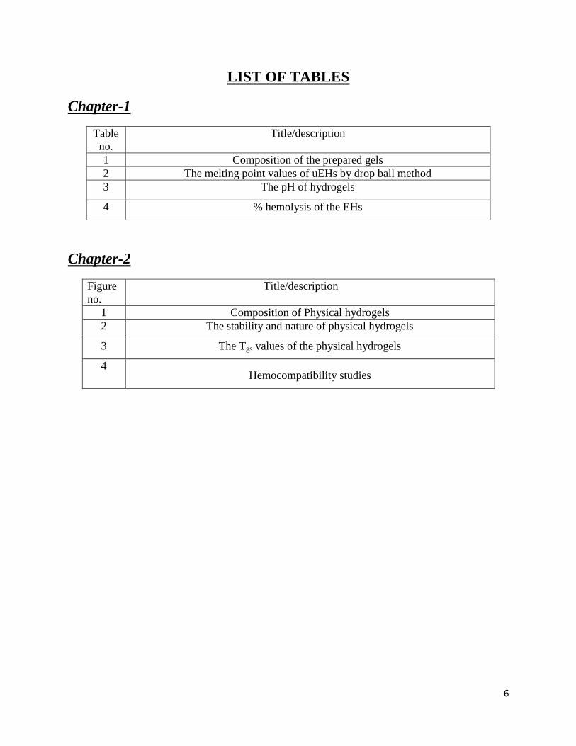
LIST OF TABLES
Chapter-1
Table
no.
Title/description
1 Composition of the prepared gels
2 The melting point values of uEHs by drop ball method
3 The pH of hydrogels
4 % hemolysis of the EHs
Chapter-2
Figure
no. Title/description
1 Composition of Physical hydrogels 2 The stability and nature of physical hydrogels
3 The Tgs values of the physical hydrogels
4 Hemocompatibility studies

7
ABBREVIATIONS
Abbreviation Definitions
EHs Emulsion Hydrogels
uEHs Uncrosslinked Emulsion Hydrogels
cEHs Crosslinked Emulsion Hydrogels
PH Physical hydrogels
CF Ciprofloxacin
MZ Metronidazole
CPDR Cumulative percent drug release
MO Mustard oil
GS Gelatin Solution
DW Distilled Water
w/w Weight by Weight
w/v Weight by Volume
Tm Melting Point
SS Stainless Steel
µm Micrometer
R2 Regression coefficients
SR Swelling Ratio
OD Optical Density
CS Corn Starch
SS Soluble Starch
BS Boiled Starch

8
REVIEW OF LITERATURE
9
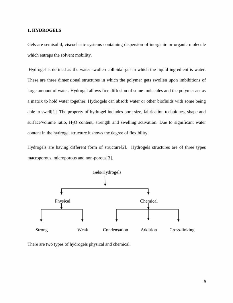
1. HYDROGELS
Gels are semisolid, viscoelastic systems containing dispersion of inorganic or organic molecule
which entraps the solvent mobility.
Hydrogel is defined as the water swollen colloidal gel in which the liquid ingredient is water.
These are three dimensional structures in which the polymer gets swollen upon imbibitions of
large amount of water. Hydrogel allows free diffusion of some molecules and the polymer act as
a matrix to hold water together. Hydrogels can absorb water or other biofluids with some being
able to swell[1]. The property of hydrogel includes pore size, fabrication techniques, shape and
surface/volume ratio, H2O content, strength and swelling activation. Due to significant water
content in the hydrogel structure it shows the degree of flexibility.
Hydrogels are having different form of structure[2]. Hydrogels structures are of three types
macroporous, microporous and non-porous[3].
Gels/Hydrogels
Physical Chemical
Strong Weak Condensation Addition Cross-linking
There are two types of hydrogels physical and chemical.
10
1.1. Physical Hydrogel
Physical hydrogels (PHs) are thermoreversible gel networks made of molecular rearrangement
and secondary forces including hydrophobic interactions. It is categorized in to two types strong
and weak. Strong gel includes glassy nodules, lamellar microcrystals and double/triple helices.
Example includes elastomers / block copolymers and gelatin. Weak gels are due to hydrogen
bonds, ionic and hydrophobic associations. Example includes xanthan, paint and matured acacia
gum etc[4-5]
1.2. Chemical Hydrogel
When the gels are covalently cross-linked it is called as chemical hydrogel. They are also termed
as permanent hydrogel. The crosslinking density and the polymer-water interface are responsible
for maintaining the swelling state symmetry of the hydrogels. Chemical hydrogel can be formed
by the addition of critical percolation for example polyester gel, also from condensation for
example polyester gel and form crosslinking[6-7].
2. CLASSIFICATION OF HYDROGELS
There are generally two types of hydrogels.
a. Natural hydrogels
b. Synthetic hydrogels
2. a. Natural hydrogels
Natural hydrogels are existing naturally in the environment. These are the material being
inspected for articular tissue engineering. The polymer used in the natural hydrogels are natural

11
hydrogels are gelatin, methyl cellulose, alginate, agarose/agar, fibrin, chitosan, hyaluronan,
chondroitin sulfate and other naturally derived polymers.
Advantages
Due to the excellent biocompatibility natural hydrogels used for tissue engineering application.
Natural hydrogels are also having different application like they are low poisonous byproducts,
intrinsic cellular interactions and biodegradable in nature.
Disadvantages
The negative aspect of natural hydrogel includes variation of batch, low mechanical strength and
the material derived from animal may pass on viruses.
2. b. Synthetic hydrogels
Synthetic polymer hydrogels constitute a group of materials used in numerous biomedical
disciplines and developing for new promising applications. These hydrogels are synthesized
artificially. Manufacture microarrays and soft contact lens are produce from synthetic hydrogel
polymers. Synthetic hydrogels have excellent mechanical properties. Synthetic hydrogels are
made from protein-polymer adducts. The synthesis of hydrogels was performed through radical
copolymerization. For some cases synthetic hydrogels can perform the task of natural
hydrogel[8].The polymer used in the synthetic hydrogels are polyanhydrides, poly(aldehyde
guluronate), Poly ethylene glycol etc.
Advantages
Synthetic hydrogels shows various biomedical applications like coating and in devices. These
hydrogels are having very low immunogenicity and minimize the risk of biological impurities

12
Disadvantages
They are having poisonous substances and low degradability.
3. Application of Hydrogels
Hydrogels are utilized naturally by the human body, for example cartilage, mucin, blood
clots and vitreous humor of the eye. There is various of application Hydrogels like soft
contact lenses, Pills/capsules, Bioadhesive carriers, Implant coatings, etc.
3.1. Application of hydrogels in Drug Delivery Delivery
Hydrogels are having various helpful applications in drug delivery and pharmaceutical sciences
due to their large amount of water content. Hydrogels are mainly utilized for conventional
controlled drug release system, bioactive materials etc. Hydrogel based drug delivery system can
be used for various types like oral, ocular, conventional, epidermal and subcutaneous application.
Hydrogels is the suitable medium for the drug delivery due to its biocompatibility, network
structure. Hydrogels are also applicable gene delivery and subcutaneous delivery.
3.2. Application of hydrogels in Tissue Engineering
Hydrogels are three dimensional water swollen structures which is insoluble networks of
crosslinked hydrophilic polymers. Hydrogels plays an important role in different tissue
engineering application. Hydrogels have been used as scaffold materials for various purposes
like tissue replacement, drug delivery, call and tissue delivery, bioactive molecule delivery,
space filling agent and various other applications[9].

13
3.3. Application of hydrogels in Biomedical Engineering
Hydrogels have been effectively used in various biomedical applications due to its biocompatible
and biodegradable nature[2, 10]. For the biomedical application most of the polymer used for
cytotoxicity and in-vivo toxicity tests. The application of hydrogel in biomedical area contains
Phospholipids bilayer, energy conversion system, mass transport properties etc.
3.4. Application of hydrogels in Biomaterials
Hydrogels have been used in various applications in biomaterials due to the biodegradable and
bioadhesive nature. The example includes soft contact lenses, wound dressing and
superabsorbent.
3.5. Application of hydrogels in Agriculture
Hydrogel have been used in various agricultural applications. The water holding capability of the
soil increased when the hydrogels are added to the surface. It also decreases the nutrient loss
from the soil. Hydrogels are less efficient in the saline soil. Hydrogels can be applied directly in
the soil or by spraying.
4. Limitations
The limitations of hydrogels includes high cost, Low mechanical strength, Difficult to load,
Difficult to sterilize, can be hard to handle and Non adherent in nature. The limitation of using
hydrogels for engineering tissue is poor in mechanical characteristics. Due to the low tensile
potency many hydrogels limit their use in load bearing application and can result in flow away
of hydrogel from a targeted local site[11-12].

14
5. Gelatin
Gelatin can be obtained from collagen of bones, ligaments and tendons. Due to the gelling agent
gelatin can be used in various purposes like in pharmaceutical industry, food industry etc.
Gelatin is having various application in food, biomedical and nutritional properties. Gelatin can
be extracted from two processes acid process and alkaline process.
5.1. Use of Gelatin
The functional uses of gelatin include Stabilizer, gelling agent, emulsifying agent and
crystallization inhibitor. Gelatin in a highly purified form is a fascinating substance for food and
recognized to have thickening, jellifying, foaming, viscosity enhancing, binding, emulsifying,
filming etc. It is used for its pharmaceutical, photographic and technical applications.
The pharmaceutical industry uses very large quantities of gelatin for making hard and soft gel
capsules.
There are two types of gelatin Edible (e.g. Jello (gelatin + sucrose + flavor) & Knox (plain
gelatin)) and Inedible (e.g. Glue, paste)
5.2. Technical use
Gelatin are having various technical use like in coating and sizing, Paper Manufacture, Printing
Processes, Protective colloidal applications, matches, Coated Abrasives, Adhesives, Gelatin
Films and Light Filters, Microencapsulation etc.
15
6. Application of Gelatin
6.1. Food Industry
It plays an important role in the food industry.Gelatin is also used as a binding and glazing agent
in meat and aspics[13].
5.2. Pharmaceutical Health Industry
In the pharmaceutical health industry, gelatin is highly digestible.
5.3. Cosmeceuticals
Gelatin has been used for many years in the cosmetics industry as in shampoos, conditioners and
lipsticks etc.
6. Manufacture of empty gelatin capsules
Steps involving in making gelatin capsules are Dipping, Spinning, Drying, Stripping, Trimming
and joining and Polishing.
Crosslinkers are either homo or hetero bifunctional reagents with identical or non identical
reactive groups. They can be covalent bonds or ionic bonds.
7. Genipin
Genipin can be obtained from an iridoid glucoside, geniposide abundantly present in the fruit of
Genipa Americana and Gadenia jasminoides. Gadenia jasminoides is an evergreen flowering
plant of the family Rubiaceae. Iridioid compound is a large class of natural products generally
present in plants. They are the source of yellow pigments traditionally used in East Asia for

16
dyeing textiles also edible blue pigment used in the food industry. The blue pigments are
important because they are highly stable to heat, pH and light. Genipin was reacted with beta-LG
to produce a new class of modified molecules[15]. The structure of the genipin was discovered in
1960‟s by Djerassi and his colleagues. The molecular formula of genipin is C11H14O5. The molar
mass of the genipin is 226.226 g/mol. Genipin and its derivatives have been used as an herbal
medicine, biomedical products, tissue engineering application and a natural colorant in the food
industry. Genipin has been widely used as antiphlogistic and a cholagogue in herbal medicine.
Genipin is less toxic and degrades more slowly as compared to formaldehyde and
glutaraldehyde. It has been reported that genipin bind with biopolymer like chitosan and gelatin.
A dark blue color look was found when genipin crosslinked with gelatin. Time and
polymer/genipin concentrations are the main two factors in genipin crosslinking reaction.
Genipin also used in Cell encapsulation. Genipin is sparingly soluble in aqueous buffers. Genipin
is being examined as new way of latent fingerprints on paper products forensic science. Recently
genipin has come to attention in biomaterial industry. Chitosan and gelatin crosslinked with
genipin has been reported to increase neuroblastoma cell adhesion and its proliferation. Genipin
can be used as regulating agent in drug delivery. For the stability of genipin thin layer
chromatography was used. Genipin exhibited significant topical anti-inflammatory effect and
anti-angiogenic properties.
Glutaraldehyde (GA) is an organic compound having chemical formula CH2(CH2CHO)2. GA is a
pungent colorless oily liquid. GA is an aliphatic dialdehyde that undergoes most of the typical
aldehyde reactions to form acetals, cyanohydrins, oximes, hydrazones and bisulfite complex.GA
has found spread use for enzyme immobilization[16].

17
Fig 3.(a) Genipin (b) Glutaraldehyde
8.Emulsion
Emulsion are defined as a thermodynamically unstable system which consist of two immiscible
liquid phases which are converted in to a single liquid phase in the help of emulsifying agent.
Emulsion can be delivered by oral, topical and parenteral routes.
8.1.Water in oil emulsion (w/o)
In the water in oil emulsion oil is the dispersion medium where the water is dispersed phase. In
this process it is not depends on the water and oil ration rather depends on the type of
emulsifier.Due to better water resistant it is used as the sun protective factor. It is generally
choose for external use like cream.
8.2. Oil in water emulsion (o/w)
In the oil in water emulsion water is the dispersion medium where the oil is the dispersed
phase.It is generally oily and not water soluble. Oil in water emulsion are having various use like

18
they are used in the adjuvants for the influenza vaccines. It is usually prefered because because it
provide cooling when used externally for e.g. vanishing cream.
8.3. Dilution Test
It can be of two type‟s o/w emulsion diluted with water and w/o emulsion diluted with oil.
8.4. Conductivity Test
Continuous phase of water is greater than continuous phase in oil.
8.5. Dye-Solubility Test
Water soluble dye will dissolve in the aqueous phase. The oil soluble dye will dissolve in the oil
phase.
9. Application of Emulsion
There are several of applications are there for emulsion like oily drugs are prepared in form of
emulsion. In the metallurgical method the concentration of ore by forth floatation process is
based upon the treatment of the powdered ore in the emulsion. Milk is an emulsion of liquid fats
in water. The cleaning action is based upon the formation of oil-in-water emulsion.
10. Starch
Starch is a natural, low-priced, obtainable, renewable, and biodegradable polymer produced by
many plants as a source of stored energy. It is the most rich storage polysaccharide in plants and

19
described as, pulse and tubers. A glycosidic bond joins the glucose moieties of starch. Amylase
and amylopectin are the two main composition of starch [17].
10.1. Amylose
Amylase is a linear polymer generally made of D-glucose unit. This polysaccharide is one of the
two main components of starch. Which make up 20-30% of the structure. Amylose normally
contributes to gelling characterstics.
10.2. Amylopectin
Amylopectin is a soluble polysaccharide and highly branched polymer of glucose found in
plants. Glucose units are linked with α glycosidic bond. It formulates 70-80% of the structure.
Amylopectin normally contributes to thickening agent.
Corn Starch
Corn starch is obtained from the corn (maize grain). The corn plant converted large amount of
radiant energy in to stable chemical energy. Corn starch is having various uses like thickening
agent in foods for e.g. soup and sauces etc.
Soluble starch
Soluble starch is defined as a high molecular weight water soluble dextrin manufactured by
partial hydrolysis of starch. It is white amorphous powder. It is quite dispersible in hot water.

20
Use of Starch
Starch is having several of use like in Beverages, Confectionery, Bakery products, Chocolate,
Processed foods, Desserts & Dairy, Paper & Board, Pharmaceutical & Cosmetics, Aqua feed,
Animal feed, Pet food and in various industrial applications.
16. Application of starch
Starch and its modified counter parts have found its wider applicability in food and non-food
modified starches have a wide variety of use both in the food and non food vicinities. The major
application of starches lies in pharmaceuticals, food and cosmetics industry. Starch plays an
increasing role in the field of biodegradable plastics, packaging material, printed circuit boards,
dry cell batteries and moulds. Starch is used as an adhesive for example hot-melt glues, stamps,
bookbinding, envelopes, wood adhesive, lamination, automotive, corrugation and paper
sacks[18].

21
Development and characterization of genipin crosslinked gelatin
emulsion hydrogels
CHAPTER 1

22
Abstract
The present study discusses about the development and characterization of genipin-crosslinked
gelatin based emulsion hydrogels (EHs). EHs were prepared by varying the proportions of
gelatin solution and mustard oil. Both the uncrosslinked (uEHs) and the crosslinked (cEHs) gels
were characterized thoroughly by microscopy. The microscopic results suggested that the
internal phase droplet size distribution was broader for gels with higher proportions of oil.
Thermal properties of the gels were found to be affected by both gelatin and mustard oil
proportions. The gels were loaded with ciprofloxacin (CF, model drug). The pHs of the gels were
within the limits of the pH of the human skin. The gels were hemocompatible and tried as a
carrier for controlled drug delivery.

23
1. Introduction
Gels are defined as 3D polymeric networked structures having the ability to undergo extensive
swelling when immersed in proper solvent [19-22]. If the absorbed solvent is aqueous in nature,
the gel is regarded as hydrogel. The swelling properties of the hydrogels may be tailored by
changing the crosslinking density of the polymeric matrix. Since the diffusion of the drug
molecules are dependent on the amount of physiological solution present within the matrices,
altering the crosslinking density of the matrix has been found to modulate the release properties
of the bioactive agents [23]. Various biopolymers (e.g. gelatin, chitosan, alginate, celluloses and
collagen) have been used for the designing of the hydrogels for pharmaceutical applications [23-
30]. Gelatin is a protein based biopolymer, which is derived from animal collagen. It is a highly
biocompatible biopolymer and hence has been used in the development of various
pharmaceutical products [23, 31-33].
Mustard oil (MO) MO has been traditionally extensively used in the southern Asian countries
(India, Pakistan and Bangladesh) for the skin massages of infants and adults [34-36]. Apart from
this, MO containing emulsions have been developed for the topical ocular delivery of non-
steroidal anti-inflammatory drugs (NSAIDs) viz., diclofenac [37]. In recent years, MO has been
used as an adjuvant for the development of novel new nutritive-immune enhancing delivery
system [38-39]. Taking inspiration from the above studies, MO was used as the representative
oil.
In the current study, attempts were made to develop genipin crosslinked mucoadhesive gelatin
EHs as a matrix for controlled delivery of antimicrobials. EHs may be defined as the hydrogel
based matrix system in which an oil has been distributed uniformly [40-41]. Genipin is a
naturally occurring crosslinker obtained from the fruits of Gardenia jasminoides. It has been

24
gaining importance in biomedical industries due to its far less cytotoxic nature as compared to
glutaraldehyde [42-44]. The developed gelatin based EHs were studied for their suitability as
dermal/transdermal drug delivery systems.
2. Materials and methods
2.1. Materials
Tween 80 (polyxyethylene sorbitan monooleate), gelatin and glycine were procured from
Himedia, Mumbai, India. Ciprofloxacin (CF) was procured from Fluka Biochemica, China.
Sodium citrate was procured from Loba Chemie, Mumbai, India. Genipin was procured from
Challenge Bioproduct Company Limited, China. Mustard oil (MO) was purchased from local
market. Goat intestine and blood were collected from the local butcher shop. Double distilled
water (DW) was used throughout the study.
2.2. Preparation of EHs
The EHs were prepared as per the method reported earlier with slight modifications in the
procedure [40]. In short, twenty percent (w/w) gelatin solution was prepared by dissolving 20 g
of gelatin in 80 g of DW, kept on stirring at 50 °C (GS). To this clear homogenous solution, 2 ml
of Tween 80 was added. MO, maintained at 50 °C, was added to GS and homogenized at 800
rpm for 15 min to form primary emulsion (PE). 0.1 g of genipin was added to the PE and further
homogenized for 30 sec. The emulsion was subsequently poured into petri-plates and incubated
at 40 oC for 30 min to promote crosslinking of the gelatin matrix. Excess genipin was neutralized
by 1% (w/w) glycine solution [22]. The crosslinked gels were washed thoroughly with DW and
stored under refrigerated conditions (5 °C). These gels were regarded as cEHs. CF loaded EHs
were prepared in a similar manner by using 1% (w/v) CF solution in MO as the internal phase. In
25
the similar manner, uncrosslinked EHs (uEHs) were prepared by pouring the PEs into perti-
plates and subsequent incubation at 4 °C for 30 min. The gels were stored under refrigerated
conditions for further analysis.
2.3. Microscopic evaluation of the gels
The microstructures of the molten uEH gels were studied under compound bright-field
microscope (Leica-DM750 equipped with ICC 50-HD camera, Germany). The droplet size
distribution of the emulsions was analyzed using NI vision assistant-2010 (USA) software as per
the reported literature [45].
2.4. Thermal studies
The melting points of the uncrosslinked gels (uEHs) were analyzed by falling ball method, as per
the reported method [46]. In short, a stainless steel (SS) ball (diameter 1/8 inch and weight 130
mg) was put over the 2 g formulation, kept in a 10 ml test-tube under refrigeration. The gels were
heated at a rate of 1 °C/min. The temperature at which the SS ball is completely submerged into
the formulation was noted as the melting point (Tm) of the gels.
2.5. Swelling behavior
The swelling behaviors of the cEHs were determined as per the reported literature. In short,
accurately weighed (Wo) gels were incubated in a beaker containing 100 ml of DW at RT [47].
At regular intervals of time (15 min for the first 1h and thereafter 30 min up to 8h), the EHs were
taken out. The surface bound water was removed by wiping with a tissue paper. The weights of
the cEHs were accurately measured using a digital weighing balance (Wt). Swelling ratio (SR) of
the EHs was calculated as per the formula given in equation 1 [47].

26
0
0 )(
W
WWSR t
(1)
Where, SR=swelling ratio
Wt is the weight of the swollen gel at time t.
W0 is the initial gel weight
2.6. pH measurement
The pHs of the uEHs and cEHs were measured using a digital pH meter (Model 132E, EI
products, Mumbai, India) as per the reported literature [48].
2.7. Hemocompatibility test
The hemocompatibility test was done as per the ASTM protocol described in the previous
literature [22, 28-29, 31-32, 49-53]. The % hemolysis of the blood was calculated as per the
equation 2.
% 100test Negative
positive Negative
OD ODHemolysis
OD OD
(2)
Where,
ODtest = Absorbance of test sample,
ODpositive = Absorbance of positive control and
ODnegative = Absorbance of negative control.
2.8. In vitro drug release studies
In vitro drug delivery studies of uEHs were performed using modified Franz diffusion cell [54].
1.5 g (approx) of the uEHs were weighed accurately and kept into the donor chamber of the

27
diffusion cell. The donor and receptor chambers were separated by a dialysis membrane. The
receptor volume was maintained at 50 ml throughout the study. During the study, sampling was
done at every 15 min during first hour and subsequently at every 30 min in the next 7h. During
each sampling, the whole receptor medium was replaced with the fresh medium (DW). At the
end of the study, the samples were analyzed for the presence of CF at 271 nm using UV-visible
spectrophotometer (UV-3200, LABINDIA, Mumbai, India).
The drug dissolution tests of the cEHs were carried out in single basket dissolution apparatus for
8 h. Accurately weighed 1.5 g (approx) of EHs were cut. The drug containing EHs were put into
the dissolution basket containing 900 ml of DW. The speed of the stirrer was kept at 100 ± 2 rpm
and the dissolution medium temperature was maintained at 37 ± 2 °C. 3 ml of samples were
withdrawn at regular intervals of time (15 min for the first hour and 30 min for the next 7 h).
Fresh dissolution medium of same volume was replaced after each sampling. The samples were
analyzed at 271 nm using UV-visible spectrophotometer.
28
3. Results and discussion
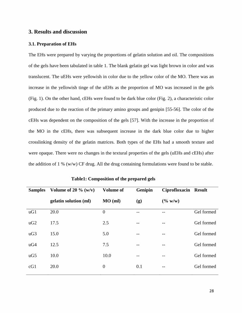
3.1. Preparation of EHs
The EHs were prepared by varying the proportions of gelatin solution and oil. The compositions
of the gels have been tabulated in table 1. The blank gelatin gel was light brown in color and was
translucent. The uEHs were yellowish in color due to the yellow color of the MO. There was an
increase in the yellowish tinge of the uEHs as the proportion of MO was increased in the gels
(Fig. 1). On the other hand, cEHs were found to be dark blue color (Fig. 2), a characteristic color
produced due to the reaction of the primary amino groups and genipin [55-56]. The color of the
cEHs was dependent on the composition of the gels [57]. With the increase in the proportion of
the MO in the cEHs, there was subsequent increase in the dark blue color due to higher
crosslinking density of the gelatin matrices. Both types of the EHs had a smooth texture and
were opaque. There were no changes in the textural properties of the gels (uEHs and cEHs) after
the addition of 1 % (w/w) CF drug. All the drug containing formulations were found to be stable.
Table1: Composition of the prepared gels
Samples Volume of 20 % (w/v)
gelatin solution (ml)
Volume of
MO (ml)
Genipin
(g)
Ciprofloxacin
(% w/w)
Result
uG1 20.0 0 -- -- Gel formed
uG2 17.5 2.5 -- -- Gel formed
uG3 15.0 5.0 -- -- Gel formed
uG4 12.5 7.5 -- -- Gel formed
uG5 10.0 10.0 -- -- Gel formed
cG1 20.0 0 0.1 -- Gel formed

29
cG2 17.5 2.5 0.1 -- Gel formed
cG3 15.0 5.0 0.1 -- Gel formed
cG4 12.5 7.5 0.1 -- Gel formed
cG5 10.0 10.0 0.1 -- Gel formed
uG1D 20.0 0 -- 1.0 Gel formed
uG2D 17.5 2.5 -- 1.0 Gel formed
uG3D 15.0 5.0 -- 1.0 Gel formed
uG4D 12.5 7.5 -- 1.0 Gel formed
uG5D 10.0 10.0 -- 1.0 Gel formed
cG1D 20.0 0 0.1 1.0 Gel formed
cG2D 17.5 2.5 0.1 1.0 Gel formed
cG3D 15.0 5.0 0.1 1.0 Gel formed
cG4D 12.5 7.5 0.1 1.0 Gel formed
cG5D 10.0 10.0 0.1 1.0 Gel formed

30
Fig. 1: uEHs of different compositions (a) uG1, (b) uG2, (c) uG3, (d) uG4 and (e) uG5

31
Fig. 2: cEHs of different compositions (a) cG1, (b) cG2, (c) cG3, (d) cG4 and (e) cG5
3.2. Microscopic evaluation of the emulsions
The micrographs of the molten uEHs have been shown in Fig. 3. The micrographs suggested the
presence of dispersed circular MO droplets within the GS continuum phase. There was an
increase in the size of the dispersed phase droplets with the increase in the MO proportion. The
droplet size distribution of the internal phase in the gels followed Gaussian distribution (Fig. 4a).
This type of distribution is achieved when physical methods are used for the preparation of the
emulsions [58-59]. The sizes of the droplets were in the range of 10-30 µm. 50 % of the droplet‟s

32
population was having the size of 15 microns (approx) (Fig. 4b). The presence of narrow size
distribution gives an indication of a probable stable emulsion [60].
Fig. 3: Light micrographs of MO-in-gelatin sol emulsion. (a) uG1, (b) uG2, (c) uG3, (d) uG4
and (e) uG5 gels.

33
Fig. 4: Droplets size distribution of uEHs, in terms of their (a) % frequency and (b)
cumulative % frequency
.
3.3 Thermal studies
The melting point (Tm) of the uEHs was determined by drop ball method as reported earlier
(table 2) [46]. The Tm of the gels was found to be decreasing as the proportion of MO was
increased. Higher Tm of uG1 may be due to the formation of strong physical polymeric network
when gelatin molecules were dissolved in water. Incorporation of MO within the gelled structure
resulted in the reduction of the intensity of the intermolecular hydrogen bonding which, in turn,
caused reduction in the Tm.
Table 2: The melting point values of uEHs by drop ball method
Samples Tm (°C)
uG1 32.10 ± 2.66
uG2 31.20 ± 1.92

34
uG3 30.90 ± 2.44
uG4 30.50 ± 3.32
uG5 30.00 ± 2.26
3.4. Swelling studies
The swelling behaviour of the crosslinked gels have been shown in Fig. 5. The swelling
behaviour was dependent on the MO content in the gels. The amount and rate of swelling of the
gels were lower in gels with higher proportions of MO. This may be attributed to the lower
amount of gelatin, which is responsible for the absorption of water. The structural integrities of
the gels were intact during the swelling study.
Fig. 5: Swelling behavior of cEHs
3.5. pH measurement
The study of the pH of the pharmaceutical formulation is an important parameter. Various
pharmacopoeia have set standards for the pharmaceutical formulations [61]. This is due to the
reason that the formulations are meant to be in contact with the cells and tissues. Higher or lower
pH values may cause irritation of chemical burn. The results of the study have been tabulated in

35
table 3. The pH of the EHs was found to be in the range of 5.5 to 7.0. This suggested that the
developed gels may be used for transdermal and topical formulations.
Table 3: The pH of hydrogels
Samples pH
uGH1 5.60 ± 0.75
uGH2 5.62 ± 0.81
uGH3 6.00 ± 0.44
uGH4 7.27 ± 0.82
uGH5 5.80 ± 0.44
cGH1 6.54 ± 0.23
cGH2 6.81 ± 0.45
cGH3 6.43 ± 0.62
cGH4 6.75 ± 0.31
cGH5 6.62 ± 0.13
3.6. Hemocompatibility studies
The % hemolysis of the goat blood in the presence of the leachants of the EHs was found to be
below 5% (table 4) suggesting that the gels may be regarded as biocompatibile [62]. Henceforth,
EHs may be tried as drug delivery vehicles.

36
Table 4: % hemolysis of the EHs
Samples % Hemolysis
uG1 2.60 ± 0.55
uG2 2.19 ± 0.82
uG3 1.10 ± 0.62
uG4 0.68 ± 0.47
uG5 0.41 ± 0.79
cG1 3.82 ± 0.32
cG2 2.86 ± 0.65
cG3 1.50 ± 0.49
cG4 1.36 ± 0.19
cG5 0.68 ± 0.67
3.7. In vitro drug release studies
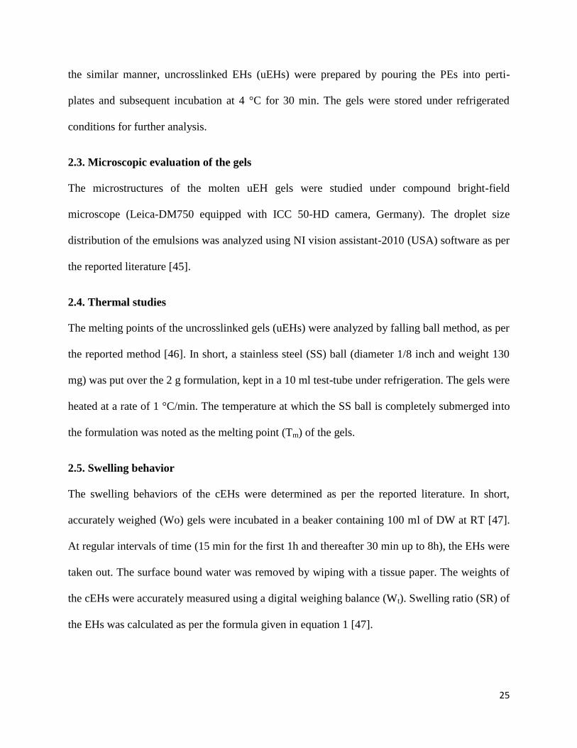
Fig. 6 shows the drug release profile of CF from uEHs and cEHs. The rate of drug release was
found to be dependent on the physicochemical properties of the gels (e.g crosslinking density, %
swelling and impedance). The higher amount of drug release may be associated with gels with
higher % swelling and lower impedance. Higher % swelling leads to higher partition of the drug
from gel matrix into the external aqueous phase. This resulted in higher CPDR value from the
cG1 than the cG3 and cG5. This may be due to the partition coefficient effect of the drug, which
states that the solute distributes itself amongst the two immiscible liquids in a definite
concentration ratio [63-65]. Crosslinking of the gels decreased the net amount of drug release.
37
uEHs have higher cumulative % drug release (CPDR) than the cEHs. The lower CPDR from
cEHs might be due to the hindrance of free movement of water within the cEHs.
Fig. 6: In vitro CPDR profile of CF from (a) uEHs and (b) cEHs
4. Conclusion
The study reports the successful development of a novel gelatin-based EHs using genipin as
crosslinking agent. MO was used as representative oil. The effect of proportion of internal oil
phase on the properties of the gels was studied by various physical techniques. The amount of
drug released was dependent on the oil proportion of the gels. The study suggested that the rate
of release of the drugs may be altered by altering the proportion of the internal oil phase. The
gels were found to be biocompatible and may be used as matrices for the controlled drug
delivery.

38
Development and characterization of gelatin-starch inclusion
physical hydrogels
CHAPTER 2

39
ABSTRACT
The current study deals about the development and characterization of gelatin-starch based
phase-separated hydrogels. The hydrogels were prepared using corn starch, soluble starch and
boiled starch. The hydrogels were characterized by pH and thermal studies, Metronidazole (MZ),
a model antimicrobial drug, was incorporated within the hydrogels. The hydrogels were highly
hemocompatible in nature. All the samples were found to have pH in the range of 5.00-6.00. The
Based on the preliminary results, it was concluded that the developed gels have a good potential
to be used as carriers for bioactive agents.

40
1. Introduction
Hydrogels are three-dimensional cross-linked networks of hydrophilic polymers [27, 66]. Due to
this, hydrogels are able to imbibe and hold water within its structure, which in turn, results in the
migration of water into the core of the polymeric construct. If drug is incorporated into the
polymer construct, the absorption of water results in the dissolution of the drug molecules
thereby creating a concentration gradient. This results in the diffusion of the drug molecules out
of the polymer matrix. The rate of diffusion of the drug out of the hydrogel is dependent on the
physical and chemical properties of the hydrogels. Depending on the nature of the hydrogels and
how it alters the drug release profile, the hydrogels may be used either as matrices for controlled
release or quick release systems [3, 40, 67]. Apart from the pharmaceutical industries, hydrogels
have also been used extensively in food [68] and biomedical [69-71] industries. The wide spread
usage of hydrogels is mainly due to their inherent biocompatible nature [72]. They may be made
biodegradable, designed to support cellular activities [73] and protect cells [74]. The natural
polymers used for the design/construction of hydrogels can be broadly categorized either as
proteins (e.g. collagen [75] and gelatin [76]) or as polysaccharides (e.g. starch [77], alginate and
agarose [78]).
The crosslinking of the polymers to form hydrogels may either be due to physical interactions or
chemical bond formation. Physical interactions include ionic crosslinking, hydrogen bonding and
molecular entanglement, i.e. there is no formation of covalent bonds. Chemical crosslinking
includes formation of covalent bonds during the formation of hydrogels [79]. The mechanical
and thermal properties of the chemically crosslinked hydrogels are far better than the physically
crosslinked hydrogels [79]. The primary disadvantage of the chemically crosslinked hydrogels is
the presence of uncrosslinked starting materials which might cause undesirable side effects [79].
41
Due to this reason, the physically crosslinked hydrogels have been extensively studied in food
industries.
Gelatin is a protein based biopolymer and is obtained by the hydrolysis of collagen from animal
sources [80]. It is inherently biocompatible and biodegradable in nature [81]. It has been used
extensively not only to develop food and pharmaceutical products but also various other products
of biomedical importance [2, 40]. Starch, on the other hand, is a polysaccharide biopolymer [82].
Starch is one of the biopolymers of choice in food, pharmaceutical and biomedical industries.
This is due to its abundance in nature, safe for human consumption and biodegradability [83].
Gelatin has been reported to form phase-separated hydrogels with polysaccharides like starch
and maltodextrin [84-85]. This paper has been designed to study the effect of composition of the
gelatin-starch phase-separated systems on the physical properties of the physical hydrogels.
Apart from this, effects of different grades of starches (e.g. corn starch (CS), soluble starch (SS)
and boiled starch (BS)) on the properties of the hydrogels were studied in-depth.
2. Materials and methods
2.1. Materials
Gelatin, CS, sodium azide, nutrient agar and dialysis tubing (MW cutoff: 60 kDa) were
purchased from Himedia, Mumbai, India. SS was purchased from Merck Specialties Private
Limited, Mumbai, India. Hydrochloric acid (HCl) and sodium citrate were purchased from Loba
Chemie, Mumbai, India. Ethanol was purchased from Changshu Yangyuan Chemical, China.
Metronidazole (MZ) was a kind gift from Aarti drugs, Mumbai, India. Fresh goat blood was
obtained from the local butcher shop. All the experiments were carried out using double distilled
water (DW).
42
2.2. Preparation of the hydrogels
Preparation of the suspensions and solutions: A clear homogeneous 20 % (w/w) solution of
gelatin was prepared by dissolving 20 g of gelatin in 60 g of DW, kept on stirring at 50 °C. After
the dissolution of the gelatin in the DW, the final volume was made to 100 g with DW
maintained at 50 °C (GS).
Three kinds of starches were used, viz. CS, SS and BS, for the development of the hydrogels. 2
% (w/w) CS or SS dispersion was prepared by dispersing 2 g of CS or SS in 98 g of DW. BS
dispersion was prepared by heating 2 % (w/w) of CS dispersion in DW at 80 °C for 30 min. The
final weight of the suspension was made to 100 g using warm DW and subsequently cooled to
room-temperature (RT, 25 °C). The above solutions and suspensions were used for the
preparation of the hydrogels.
Preparation of the hydrogels: Physical hydrogels were prepared by varying the proportions of
gelatin solution (GS) and starch dispersions. The compositions of the hydrogels have been
tabulated in table 1. The hydrogels were prepared by mixing GS and starch dispersions and
subsequently homogenizing the dispersion at 500 rpm for 30 min at 40 °C. The homogenized
mixture was then poured into culture bottles and then stored under refrigerated conditions for
further analysis. Hydrogel made with GS alone served as a control. The MZ-loaded hydrogels
were prepared in a similar manner such that the final concentration of the drug in the hydrogel
was 1% (w/v). MZ was dissolved in the GS-starch dispersion mixture during homogenization.

43
Table 1: Composition of Physical hydrogels
Sample Volume of starch dispersion
(ml)
MZ
(%, w/v)
GS CS SS BS
GH 20.0 0 -- -- --
CS1 16.0 4.0 -- -- --
CS2 12.0 8.0 -- -- --
CS3 8.0 12.0 -- -- --
SS1 16.0 -- 4.0 -- --
SS2 12.0 -- 8.0 -- --
SS3 8.0 -- 12.0 -- --
BS1 16.0 -- -- 4.0 --
BS2 12.0 -- -- 8.0 --
BS3 8.0 -- -- 12.0 --
GHM 20.0 0 -- -- 1
CS1M 16.0 4.0 -- -- 1
CS2M 12.0 8.0 -- -- 1
CS3M 8.0 12.0 -- -- 1
SS1M 16.0 -- 4.0 -- 1
SS2M 12.0 -- 8.0 -- 1
SS3M 8.0 -- 12.0 -- 1
BS1M 16.0 -- -- 4.0 1

44
BS2M 12.0 -- -- 8.0 1
BS3M 8.0 -- -- 12.0 1
2.3. Microscopic studies
The microstructure of the hydrogels was studied using inverted phase contrast (Olympus INVI-
TR attached with SONY digital camera EPL-1, USA) and scanning electron microscopy (JEOL,
JSM-6390, JAPAN). The inverted phase contrast microscopy was carried out by converting the
physical hydrogels into thin smears.
2.4. Thermal studies
The melting point of the hydrogels was determined by drop-ball method as mentioned in the
previous literature [46]. In short, 2 g of the hydrogels were melted and poured in 10 ml of test
tubes. The test tubes were incubated for 15 min in a laboratory refrigerator, maintained at 4 ±1
°C. After refrigeration, a stainless steel (SS) ball (diameter 1/8 inch; weight 130 mg) was placed
gently on top of the hydrogels. The hydrogels were heated at a rate of 1 °C/min in a melting
point apparatus. The melting point (Tm) of the hydrogels was noted when the SS ball completely
submerges [86].
2.5. pH Measurement
The pH of the hydrogels was measured using a digital pH meter (EI digital pH meter, model no:
112, India) [87].
2.6. Hemocompatibility test
Hemocompatibility of the hydrogels were determined as per the reported literature [88]. The %
hemolysis of the blood was calculated as per the equation 1.
45
% 100test Negative
positive Negative
OD ODHemolysis
OD OD
(1)
2.7. In vitro drug release
The drug release study was carried out using a 2-compartment modified Franz diffusion cell, as
per the reported literature [89]. In short, accurately weighed 1.5 g of the drug loaded hydrogel
was loaded in the donor compartment. The donor compartment was lowered towards the receptor
compartment so as to ensure that the dialysis membrane (attached with the donor compartment)
was in contact with the receptor fluid (50 ml of DW), kept on stirring at 100 rpm and the
temperature was maintained at 37 ± 1 °C. The receptor fluid was replaced by fresh DW at regular
intervals (15 min during first hour and subsequently after 30 min during the next 7 h). The
samples were then analyzed using UV-visible spectrophotometer (UV-3200, LABINDIA,
Mumbai, India).
3. Results and discussion
3.1. Preparation of physical hydrogels
The hydrogels were prepared by varying the proportions of GS and starch dispersions. The
organoleptic properties of the hydrogels have been tabulated in table 2. GH was transparent and
light brown in color. Incorporation of the starch dispersions within the hydrogels resulted in the
formation of opaque hydrogels. The colors of the starch incorporated hydrogels were white and
the whiteness of the hydrogels was higher in hydrogels with higher proportions of starch (figure
1). The hydrogels had a pleasant odor and were thermo-reversible in nature. They did not show
flow when kept under refrigerated conditions (figure 1). The hydrogels with higher proportions
of starch showed flow when kept at RT (room-temperature, 25 C). With the further increase in
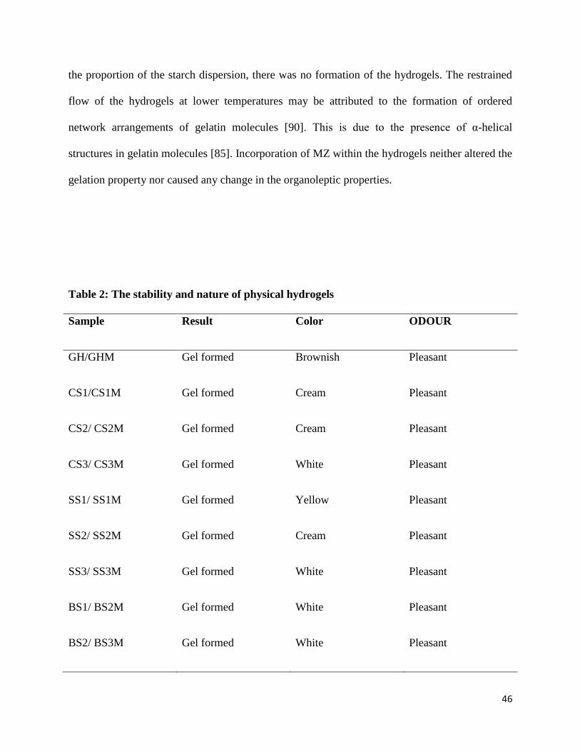
46
the proportion of the starch dispersion, there was no formation of the hydrogels. The restrained
flow of the hydrogels at lower temperatures may be attributed to the formation of ordered
network arrangements of gelatin molecules [90]. This is due to the presence of α-helical
structures in gelatin molecules [85]. Incorporation of MZ within the hydrogels neither altered the
gelation property nor caused any change in the organoleptic properties.
Table 2: The stability and nature of physical hydrogels
Sample Result Color ODOUR
GH/GHM Gel formed Brownish Pleasant
CS1/CS1M Gel formed Cream Pleasant
CS2/ CS2M Gel formed Cream Pleasant
CS3/ CS3M Gel formed White Pleasant
SS1/ SS1M Gel formed Yellow Pleasant
SS2/ SS2M Gel formed Cream Pleasant
SS3/ SS3M Gel formed White Pleasant
BS1/ BS2M Gel formed White Pleasant
BS2/ BS3M Gel formed White Pleasant

47
BS3/ BS3M Gel formed White Pleasant
Figure 1: The stable physical hydrogels (a) GH, (b) CS1, (c) CS2 , (d) CS3, (e) SS1 (f) SS2,
(g) SS3, (h) BS1, (i) BS2 and (j) BS3.

48
3.2. Hydrogel morphology
Figure 2: Phase contrst micrographs of hydrogels (a) GH, (b) CS1, (c) CS2 , (d) CS3, (e)
SS1 (f) SS2, (g) SS3, (h) BS1, (i) BS2 and (j) BS3.
The microstructures of the hydrogels as visualized under inverted phase contrast microscope
have been shown in figure 2. The microstructures of the hydrogels have shown the presence of
phase separated starch inclusions in the form of dark globules in the gelatin continuous matrices .
The increase in the density of starch inclusions was observed with the increase in starch
concentrations. This suggested that the phase separation phenomenon was dependent on the
composition of the hydrogels. An increase in the tendency of the phase separation was observed

49
with the increase in starch concentration. In general, phase separation is a common phenomenon
occuring in protein-polysacharide mixtures. Since both gelatin and starch molecules are not
polyelectrolytes, there are chances of thermodynamic incompatibility which might resulted in the
segregative phase separation [91]. This might have resulted in the increased self-association of
the biopolymers [92]. Phase separation has been found to be dependent on the structure and the
composition of the biopolymers used.
SS is rich in amylodextrin (linear short chained amylose units) and is devoid of amylopectin. The
thermal incompatiblity was found to be lower when linear chained carboxyl containing
polysaccharides were used in the protein-polysaccharide system [91]. Similar results were
obtained when SS was used for the development of the hydrogels, i.e. SS hydrogels showed
small starch droplets/inclusions as compared to the CS and BS hydrogels . The presence of larger
droplets/inclusions in CS and BS hydrogels may be associated with the presence of hyper-
branched polysaccharide units (amylopectin), which lead to the aggregation of polymeric chains
[93].
3.3. Thermal studies
The gel-to-sol transition temperature (Tgs) was determined by drop ball method. GH showed
highest Tgs compared to the other hydrogels. This may be due to the presence of only gelatin
molecules in GH. With the increase in the concentration of the SS, CS and BS, there was a
decrease in the Tgs of all the gels. The CS and SS containing gels have shown similar Tgs. The Tgs
of the BS hydrogels were lower than the CS and the SS hydrogels. This can be attributed to the
reduction in the proportion of the gelatin, the biopolymer responsible for incorporating

50
mechanical strength in the hydrogels. The lower Tgs of the BS hydrogels might be attributed to
the disruption of starch complexes when heated at higher temperatures. This results in the
random orientation of the starch molecules [94].
Table 3: The Tgs values of the physical hydrogels
Sample Tgs (°C)
GH 35.0 ± 1.5
CS1 32.0 ± 3.5
CS2 28.0 ± 1.5
CS3 23.0 ± 1.0
SS1 33.0 ± 2.5
SS2 25.0 ± 3.0
SS3 17.0 ± 2.5
BS1 27.0 ± 3.0
BS2 20.0 ± 2.5
BS3 15.0 ±3.0
3.4. pH Measurement
The pHs of the hydrogels were found to be in the range of 5.4 and 6.0. The results suggested that
the hydrogels may be tried for topical/ transdermal application.
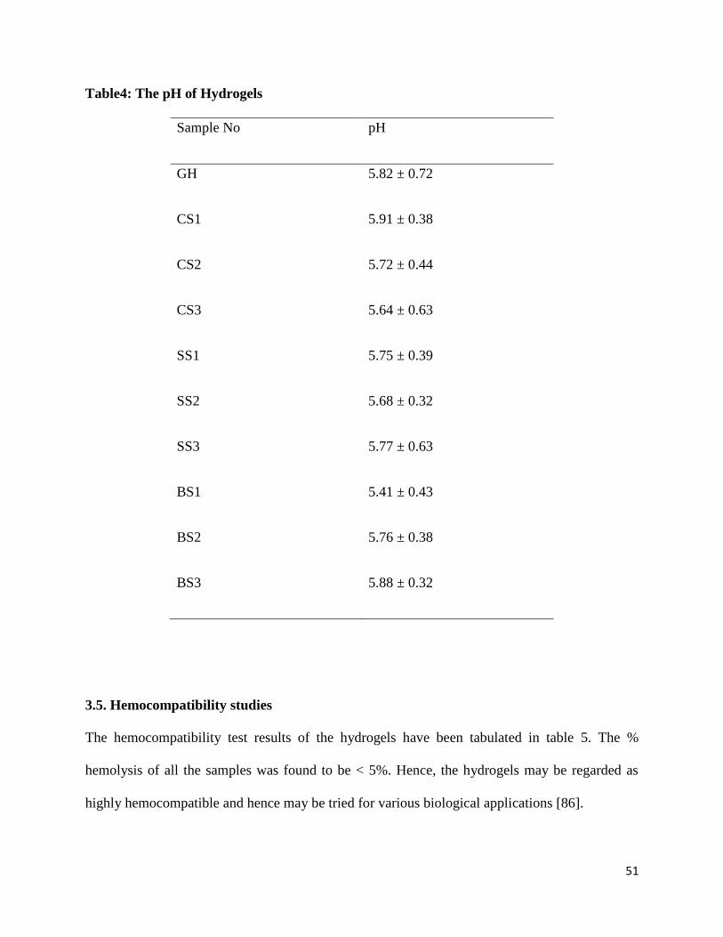
51
Table4: The pH of Hydrogels
Sample No pH
GH 5.82 ± 0.72
CS1 5.91 ± 0.38
CS2 5.72 ± 0.44
CS3 5.64 ± 0.63
SS1 5.75 ± 0.39
SS2 5.68 ± 0.32
SS3 5.77 ± 0.63
BS1 5.41 ± 0.43
BS2 5.76 ± 0.38
BS3 5.88 ± 0.32
3.5. Hemocompatibility studies
The hemocompatibility test results of the hydrogels have been tabulated in table 5. The %
hemolysis of all the samples was found to be < 5%. Hence, the hydrogels may be regarded as
highly hemocompatible and hence may be tried for various biological applications [86].
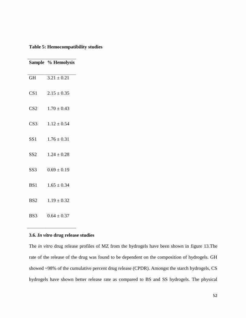
52
Table 5: Hemocompatibility studies
Sample % Hemolysis
GH 3.21 ± 0.21
CS1 2.15 ± 0.35
CS2 1.70 ± 0.43
CS3 1.12 ± 0.54
SS1 1.76 ± 0.31
SS2 1.24 ± 0.28
SS3 0.69 ± 0.19
BS1 1.65 ± 0.34
BS2 1.19 ± 0.32
BS3 0.64 ± 0.37
3.6. In vitro drug release studies
The in vitro drug release profiles of MZ from the hydrogels have been shown in figure 13.The
rate of the release of the drug was found to be dependent on the composition of hydrogels. GH
showed ~98% of the cumulative percent drug release (CPDR). Amongst the starch hydrogels, CS
hydrogels have shown better release rate as compared to BS and SS hydrogels. The physical

53
nature of the hydrogels might have affected the CPDR of the drug from the hydrogels [86, 95].
The CPDR of MZ from the starch hydrogels was in the range of 70 % and 80 %. In general, with
the increase in the starch concentration, there was a reduction in the release rate of MZ. The
release of the drug from the hydrogels followed Higuchian kinetics. This indicated that the
hydrogels acted as a planar matrix for the drug molecules and the release of the drug from the
matrices was diffusion controlled. The Fickian value (n) was calculated from Krossmeyer-
Peppas (KP) model. The „n‟ values were in the range of 0.45 and 0.85 thereby suggesting a non-
Fickian diffusion of the drugs from the hydrogels [96-97].
Figure 13: In vitro drug release profiles of (a) GHM, (b) CSM, (c) SSM and (d) BSM gels.

54
4. Conclusion
Gelatin-starch based physical composite hydrogels were developed by phase-separation
technique. The phase separation of starch inclusions in the rich gelatin phase was confirmed by
microscopic techniques. The mechanical properties of the hydrogels could be varied by altering
the composition of the hydrogels. The drug loaded hydrogels showed good antimicrobial
properties against both E. coli and B. subtilis. The hydrogels were found to be hemocompatible
in nature. Based on the preliminary studies, the developed formulations may be tried as drug and
nutraceutical delivery vehicles in pharmaceutical and food industries.

55
References

56
1. Richter, A., et al., Review on hydrogel-based pH sensors and microsensors. Sensors, 2008. 8(1): p. 561-581.
2. Hoffman, A.S., Hydrogels for biomedical applications. Advanced drug delivery Reviews, 2002. 54(1): p. 3-12.
3. Peppas, N., et al., Hydrogels in pharmaceutical formulations. European journal of pharmaceutics and biopharmaceutics, 2000. 50(1): p. 27-46.
4. Ruel-Gariépy, E. and J.-C. Leroux, In situ-forming hydrogels—review of temperature-sensitive systems. European journal of pharmaceutics and biopharmaceutics, 2004. 58(2): p. 409-426.
5. Boucard, N., et al., The use of physical hydrogels of chitosan for skin regeneration following third-degree burns. Biomaterials, 2007. 28(24): p. 3478-3488.
6. Berger, J., et al., Structure and interactions in covalently and ionically crosslinked chitosan hydrogels for biomedical applications. European journal of pharmaceutics and biopharmaceutics, 2004. 57(1): p. 19-34.
7. Rosiak, J., et al., Radiation crosslinked hydrogels from acrylamide water solutions. Radiation Physics and Chemistry (1977), 1983. 22(3): p. 917-928.
8. Ratner, B.D. and A.S. Hoffman. Synthetic hydrogels for biomedical applications. in Hydrogels for Medical and Related Applications, ACS symposium series. 1976: ACS Publications.
9. Murakami, Y., et al., A novel synthetic tissue‐adhesive hydrogel using a crosslinkable polymeric micelle. Journal of Biomedical Materials Research Part A, 2007. 80(2): p. 421-427.
10. Mauck, R.L., et al., Functional tissue engineering of articular cartilage through dynamic loading of chondrocyte-seeded agarose gels. TRANSACTIONS-AMERICAN SOCIETY OF MECHANICAL ENGINEERS JOURNAL OF BIOMECHANICAL ENGINEERING, 2000. 122(3): p. 252-260.
11. Billiet, T., et al., A review of trends and limitations in hydrogel-rapid prototyping for tissue engineering. Biomaterials, 2012.
12. Fonn, D. and A.S. Bruce, A review of the Holden-Mertz criteria for critical oxygen transmission. Eye & contact lens, 2005. 31(6): p. 247-251.
13. Guzey, D. and D.J. McClements, Formation, stability and properties of multilayer emulsions for application in the food industry. Advances in Colloid and Interface Science, 2006. 128: p. 227-248.
14. Karim, A. and R. Bhat, Gelatin alternatives for the food industry: recent developments, challenges and prospects. Trends in food science & technology, 2008. 19(12): p. 644-656.
15. Tsai, C.C., et al., In vitro evaluation of the genotoxicity of a naturally occurring crosslinking agent (genipin) for biologic tissue fixation. Journal of biomedical materials research, 2000. 52(1): p. 58-65.
16. Li-hau, T.Z.-m.K., Preparation of Alkaline Phosphatase Labeled HBV Probe and Its Application in Serum Detection. Virologica Sinica, 1991. 2: p. 008.
17. Miles, M.J., et al., The roles of amylose and amylopectin in the gelation and retrogradation of starch. Carbohydrate research, 1985. 135(2): p. 271-281.
18. Ochubiojo, E.M. and A. Rodrigues, Starch: From Food to Medicine. 19. Kulygin, O. and M.S. Silverstein, Porous poly (2-hydroxyethyl methacrylate) hydrogels
synthesized within high internal phase emulsions. Soft Matter, 2007. 3(12): p. 1525-1529. 20. Pal, K., A. Banthia, and D. Majumdar, Preparation of novel pH-sensitive hydrogels of
carboxymethyl cellulose acrylates: A comparative study. Materials and manufacturing processes, 2006. 21(8): p. 877-882.
21. Mishra, R., et al., Preparation and characterization of amidated pectin based hydrogels for drug delivery system. Journal of Materials Science: Materials in Medicine, 2008. 19(6): p. 2275-2280.

57
22. Pal, K., A.K. Banthia, and D.K. Majumdar, Polyvinyl alcohol–glycine composite membranes: preparation, characterization, drug release and cytocompatibility studies. Biomedical Materials, 2006. 1(2): p. 49.
23. Thakur, G., et al., Crosslinking of gelatin-based drug carriers by genipin induces changes in drug kinetic profiles in vitro. Journal of Materials Science: Materials in Medicine, 2011. 22(1): p. 115-123.
24. Pal, K., et al., Chitosan based delivery systems on a length scale: nano to macro. 2011. 25. Sagiri, S.S., et al., Encapsulation of vegetable organogels for controlled delivery applications.
Designed Monomers and Polymers, 2012(ahead-of-print): p. 1-11. 26. Roy, S., et al., Polymers in mucoadhesive drug-delivery systems: a brief note. Designed
Monomers and Polymers, 2009. 12(6): p. 483-495. 27. Pal, K., A. Banthia, and D. Majumdar, Polymeric hydrogels: Characterization and biomedical
applications. Designed Monomers and Polymers, 2009. 12(3): p. 197-220. 28. Pal, K., S. Bag, and S. Pal, Development of porous ultra high molecular weight polyethylene
scaffolds for the fabrication of orbital implant. Journal of Porous Materials, 2008. 15(1): p. 53-59.
29. Roy, S., et al., Synthesis of Novel Hydroxypropyl Methyl Cellulose Acrylate—A Novel Superdisintegrating Agent for Pharmaceutical Applications. Materials and manufacturing processes, 2010. 25(12): p. 1477-1481.
30. Pal, K., A.T. Paulson, and D. Rousseau, 14 Biopolymers in Controlled-Release Delivery Systems. Handbook of Biopolymers and Biodegradable Plastics: Properties, Processing and Applications, 2012: p. 329.
31. Pal, K., A.K. Banthia, and D.K. Majumdar, Preparation and characterization of polyvinyl alcohol-gelatin hydrogel membranes for biomedical applications. AAPS PharmSciTech, 2007. 8(1): p. 142-146.
32. Pal, K., A. Banthia, and D. Majumdar, Biomedical evaluation of polyvinyl alcohol–gelatin esterified hydrogel for wound dressing. Journal of Materials Science: Materials in Medicine, 2007. 18(9): p. 1889-1894.
33. Pal, K., A. Banthia, and D. Majumdar, Polyvinyl alcohol—gelatin patches of salicylic acid: preparation, characterization and drug release studies. Journal of biomaterials applications, 2006. 21(1): p. 75-91.
34. Ahmed, A.S.M.N.U., et al., Acceptability of massage with skin barrier-enhancing emollients in young neonates in Bangladesh. Journal of health, population, and nutrition, 2007. 25(2): p. 236.
35. Alam, M.A., et al., Newborn umbilical cord and skin care in Sylhet District, Bangladesh: implications for the promotion of umbilical cord cleansing with topical chlorhexidine. Journal of Perinatology, 2008. 28: p. S61-S68.
36. Sreeramareddy, C., et al., Home delivery and newborn care practices among urban women in western Nepal: a questionnaire survey. BMC pregnancy and childbirth, 2006. 6(1): p. 27.
37. Ahuja, M., et al., Topical Ocular Delivery of NSAIDs. The AAPS Journal, 2008. 10(2): p. 229-241. 38. Yu, M. and M. Vajdy, A novel retinoic acid, catechin hydrate and mustard oil-based emulsion for
enhanced cytokine and antibody responses against multiple strains of HIV-1 following mucosal and systemic vaccinations. Vaccine, 2011. 29(13): p. 2429-2436.
39. Vajdy, M., Immunomodulatory properties of vitamins, flavonoids and plant oils and their potential as vaccine adjuvants and delivery systems. Expert Opinion on Biological Therapy, 2011. 11(11): p. 1501-1513.
40. Mallick, S., et al., Gelatin-Based Emulsion Hydrogels as a Matrix for Controlled Delivery System. Materials and manufacturing processes, 2012. 27(11): p. 1221-1228.

58
41. Thakur, G., et al. Characterization of oil-in-water gelatin emulsion gels: Effect of homogenization time. in Systems in Medicine and Biology (ICSMB), 2010 International Conference on. 2010: IEEE.
42. Taylor, M., K. Ding, and E. Brown, Genipin-Modified Gelatin: Preparation, Characterization, and Application as a Filler for Leather.
43. Liang, H.C., et al., Crosslinking structures of gelatin hydrogels crosslinked with genipin or a water‐soluble carbodiimide. Journal of applied polymer science, 2004. 91(6): p. 4017-4026.
44. Yao, C.H., et al., Preparation of networks of gelatin and genipin as degradable biomaterials. Materials chemistry and physics, 2004. 83(2): p. 204-208.
45. Satapathy, D., et al., Sunflower-oil-based lecithin organogels as matrices for controlled drug delivery. Journal of Applied Polymer Science, 2012: p. n/a-n/a.
46. Terech, P., et al., Rheological properties and structural correlations in molecular organogels. Langmuir, 2000. 16(10): p. 4485-4494.
47. Park, H., et al., Effect of Swelling Ratio of Injectable Hydrogel Composites on Chondrogenic Differentiation of Encapsulated Rabbit Marrow Mesenchymal Stem Cells In Vitro. Biomacromolecules, 2009. 10(3): p. 541-546.
48. Pal, K., A.K. Banthia, and D.K. Majumdar, Preparation of Novel pH-Sensitive Hydrogels of Carboxymethyl Cellulose Acrylates: A Comparative Study. Materials and manufacturing processes, 2006. 21(8): p. 877-882.
49. Pal, K., S. Bag, and S. Pal, Development and Coating of Porous Ultra High Molecular Weight Polyethylene Plates. Trends in Biomaterials and Artificial Organs, 2010. 19(1).
50. Ng, K.W., et al., Evaluation of ultra-thin poly ( -caprolactone) films for tissue-engineered skin. Tissue engineering, 2001. 7(4): p. 441-455.
51. Pal, K., A. Banthia, and D. Majumdar, Development of carboxymethyl cellulose acrylate for various biomedical applications. Biomedical Materials, 2006. 1: p. 85.
52. Sutar, P.B., et al., Development of pH sensitive polyacrylamide grafted pectin hydrogel for controlled drug delivery system. Journal of Materials Science: Materials in Medicine, 2008. 19(6): p. 2247-2253.
53. Mishra, R.K., et al., Preparation and characterization of amidated pectin based hydrogels for drug delivery system. Journal of Materials Science: Materials in Medicine, 2008. 19(6): p. 2275-2280.
54. Nandini, D., et al., Effect of permeation enhancers on the release and permeation kinetics of oxytetracycline hydrochloride organogel formulations. Journal of Young Pharmacists, 2009. 1(4): p. 285.
55. Butler, M.F., Y.-F. Ng, and P.D.A. Pudney, Mechanism and kinetics of the crosslinking reaction between biopolymers containing primary amine groups and genipin. Journal of Polymer Science Part A: Polymer Chemistry, 2003. 41(24): p. 3941-3953.
56. Liang, H.-C., et al., Genipin-crosslinked gelatin microspheres as a drug carrier for intramuscular administration: In vitro and in vivo studies. Journal of Biomedical Materials Research Part A, 2003. 65A(2): p. 271-282.
57. Huang, K.-S., et al., Microfluidic controlling monodisperse microdroplet for 5-fluorouracil loaded genipin-gelatin microcapsules. Journal of Controlled Release, 2009. 137(1): p. 15-19.
58. Reinheimer, K., et al., Fourier Transform Rheology as an innovative morphological characterization technique for the emulsion volume average radius and its distribution. Journal of Colloid and Interface Science, 2012. 380(1): p. 201-212.
59. Silva, K.A., M.H. Rocha-Leão, and M.A.Z. Coelho, Evaluation of aging mechanisms of olive oil–lemon juice emulsion through digital image analysis. Journal of Food Engineering, 2010. 97(3): p. 335-340.

59
60. Thompson, K.L., S.P. Armes, and D.W. York, Preparation of Pickering Emulsions and Colloidosomes with Relatively Narrow Size Distributions by Stirred Cell Membrane Emulsification. Langmuir, 2011. 27(6): p. 2357-2363.
61. Bhatia, V. and R. Barber, The effect of pH variations of ointment bases on the local anesthetic activity of incorporated ethyl aminobenzoate. I. Hydrophilic ointment USP. Journal of the American Pharmaceutical Association, 1955. 44(6): p. 342-343.
62. Pal, K. and S. Pal, Development of porous hydroxyapatite scaffolds. Materials and manufacturing processes, 2006. 21(3): p. 325-328.
63. Chiou, C.T., et al., Partition coefficient and bioaccumulation of selected organic chemicals. Environmental Science & Technology, 1977. 11(5): p. 475-478.
64. Barry, B., Novel mechanisms and devices to enable successful transdermal drug delivery. European journal of pharmaceutical sciences, 2001. 14(2): p. 101-114.
65. Shah, N., et al., Self-emulsifying drug delivery systems (SEDDS) with polyglycolyzed glycerides for improving in vitro dissolution and oral absorption of lipophilic drugs. International journal of pharmaceutics, 1994. 106(1): p. 15-23.
66. Reis, A.V., et al., Synthesis and characterization of a starch‐modified hydrogel as potential carrier for drug delivery system. Journal of Polymer Science Part A: Polymer Chemistry, 2008. 46(7): p. 2567-2574.
67. Atichokudomchai, N. and S. Varavinit, Characterization and utilization of acid-modified cross-linked tapioca starch in pharmaceutical tablets. Carbohydrate polymers, 2003. 53(3): p. 263-270.
68. Abbas, K., S.K. Khalil, and A.S.M. Hussin, Modified starches and their usages in selected food products: a review study. Journal of Agricultural Science, 2010. 2(2): p. P90.
69. Elvira, C., et al., Starch-based biodegradable hydrogels with potential biomedical applications as drug delivery systems. Biomaterials, 2002. 23(9): p. 1955-1966.
70. Sachlos, E. and J. Czernuszka, Making tissue engineering scaffolds work. Review: the application of solid freeform fabrication technology to the production of tissue engineering scaffolds. Eur Cell Mater, 2003. 5(29): p. 39-40.
71. Jagur‐Grodzinski, J., Polymers for tissue engineering, medical devices, and regenerative medicine. Concise general review of recent studies. Polymers for Advanced Technologies, 2006. 17(6): p. 395-418.
72. Sechriest, V.F., et al., GAG‐augmented polysaccharide hydrogel: A novel biocompatible and biodegradable material to support chondrogenesis. Journal of biomedical materials research, 2000. 49(4): p. 534-541.
73. Singh, A., et al., Hydrogels: A review. Int J Pharmaceut Sci Rev Res, 2010. 4(2): p. 97. 74. Peppas, N.A., Hydrogels in medicine and pharmacy. Vol. 3. 1987: CRC press Boca Raton, FL. 75. Tabata, Y., et al., Controlled release of vascular endothelial growth factor by use of collagen
hydrogels. Journal of Biomaterials Science, Polymer Edition, 2000. 11(9): p. 915-930. 76. Tabata, Y. and Y. Ikada, Vascularization effect of basic fibroblast growth factor released from
gelatin hydrogels with different biodegradabilities. Biomaterials, 1999. 20(22): p. 2169-2175. 77. Athawale, V. and V.L. Vidyagauri, Graft copolymerization onto starch. 3: Grafting of acrylamide
using ceric ion initiation and preparation of its hydrogels. Starch‐Stärke, 1998. 50(10): p. 426-431.
78. Coviello, T., et al., Polysaccharide hydrogels for modified release formulations. Journal of Controlled Release, 2007. 119(1): p. 5-24.
79. Vintiloiu, A. and J.-C. Leroux, Organogels and their use in drug delivery — A review. Journal of Controlled Release, 2008. 125(3): p. 179-192.
80. Djagny, K.B., Z. Wang, and S. Xu, Gelatin: a valuable protein for food and pharmaceutical industries: review. Critical reviews in food science and nutrition, 2001. 41(6): p. 481-492.

60
81. Narayani, R. and K.P. Rao, Controlled release of anticancer drug methotrexate from biodegradable gelatin microspheres. Journal of microencapsulation, 1994. 11(1): p. 69-77.
82. STĂNESCU, V.-N., et al., STARCH/CHITOSAN FILM FORMING HYDROGEL. Rev. Roum. Chim, 2011. 56(8): p. 827-832.
83. Röper, H. and H. Koch, The role of starch in biodegradable thermoplastic materials. Starch‐Stärke, 1990. 42(4): p. 123-130.
84. Firoozmand, H. and D. Rousseau, Microstructure and elastic modulus of phase-separated gelatin–starch hydrogels containing dispersed oil droplets. Food Hydrocolloids, 2013. 30(1): p. 333-342.
85. Nickerson, M.T., et al., Some physical and microstructural properties of genipin-crosslinked gelatin–maltodextrin hydrogels. International Journal of Biological Macromolecules, 2006. 38(1): p. 40-44.
86. Sagiri, S.S., et al., Lanolin-based organogels as a matrix for topical drug delivery. Journal of Applied Polymer Science, 2012: p. n/a-n/a.
87. Behera, B., et al., Span-60-based organogels as probable matrices for transdermal/topical delivery systems. Journal of applied polymer science, 2012. 125(2): p. 852-863.
88. Behera, B., et al., Modulating the physical properties of sunflower oil and sorbitan monopalmitate-based organogels. Journal of Applied Polymer Science, 2013. 127(6): p. 4910-4917.
89. Shah, D.K., et al., Development of olive oil based organogels using sorbitan monopalmitate and sorbitan monostearate: A comparative study. Journal of applied polymer science, 2013. 129(2): p. 793-805.
90. Lorén, N., et al., Phase Separation Induced by Conformational Ordering of Gelatin in Gelatin/Maltodextrin Mixtures. Macromolecules, 2000. 34(2): p. 289-297.
91. Doublier, J.L., et al., Protein–polysaccharide interactions. Current Opinion in Colloid & Interface Science, 2000. 5(3–4): p. 202-214.
92. Grinberg, V.Y. and V.B. Tolstoguzov, Thermodynamic incompatibility of proteins and polysaccharides in solutions. Food Hydrocolloids, 1997. 11(2): p. 145-158.
93. Singh, J. and N. Singh, Studies on the morphological and rheological properties of granular cold water soluble corn and potato starches. Food Hydrocolloids, 2003. 17(1): p. 63-72.
94. Sagum, R. and J. Arcot, Effect of domestic processing methods on the starch, non-starch polysaccharides and in vitro starch and protein digestibility of three varieties of rice with varying levels of amylose. Food Chemistry, 2000. 70(1): p. 107-111.
95. Frank, A., S.K. Rath, and S.S. Venkatraman, Controlled release from bioerodible polymers: effect of drug type and polymer composition. Journal of Controlled Release, 2005. 102(2): p. 333-344.
96. Philip, A., M. Srivastava, and K. Pathak, Buccoadhesive gels of glibenclamide: A means for achieving enhanced bioavailability. Drug Delivery, 2009. 16(7): p. 405-415.
97. Sawant, P.D., et al., Drug release from hydroethanolic gels. Effect of drug's lipophilicity (log P), polymer–drug interactions and solvent lipophilicity. International Journal of Pharmaceutics, 2010. 396(1–2): p. 45-52.