
Genetic and technological characterization of lactic acid ...
42
HAL Id: hal-02172525 https://hal.univ-reunion.fr/hal-02172525 Submitted on 22 Oct 2021 HAL is a multi-disciplinary open access archive for the deposit and dissemination of sci- entific research documents, whether they are pub- lished or not. The documents may come from teaching and research institutions in France or abroad, or from public or private research centers. L’archive ouverte pluridisciplinaire HAL, est destinée au dépôt et à la diffusion de documents scientifiques de niveau recherche, publiés ou non, émanant des établissements d’enseignement et de recherche français ou étrangers, des laboratoires publics ou privés. Distributed under a Creative Commons Attribution - NonCommercial| 4.0 International License Genetic and technological characterization of lactic acid bacteria isolated from tropically grown fruits and vegetables Amandine Fessard, Fabienne Remize To cite this version: Amandine Fessard, Fabienne Remize. Genetic and technological characterization of lactic acid bacteria isolated from tropically grown fruits and vegetables. International Journal of Food Microbiology, Elsevier, 2019, 301, pp.61-72. 10.1016/j.ijfoodmicro.2019.05.003. hal-02172525
Transcript of Genetic and technological characterization of lactic acid ...

HAL Id: hal-02172525https://hal.univ-reunion.fr/hal-02172525
Submitted on 22 Oct 2021
HAL is a multi-disciplinary open accessarchive for the deposit and dissemination of sci-entific research documents, whether they are pub-lished or not. The documents may come fromteaching and research institutions in France orabroad, or from public or private research centers.
L’archive ouverte pluridisciplinaire HAL, estdestinée au dépôt et à la diffusion de documentsscientifiques de niveau recherche, publiés ou non,émanant des établissements d’enseignement et derecherche français ou étrangers, des laboratoirespublics ou privés.
Distributed under a Creative Commons Attribution - NonCommercial| 4.0 InternationalLicense
Genetic and technological characterization of lactic acidbacteria isolated from tropically grown fruits and
vegetablesAmandine Fessard, Fabienne Remize
To cite this version:Amandine Fessard, Fabienne Remize. Genetic and technological characterization of lactic acid bacteriaisolated from tropically grown fruits and vegetables. International Journal of Food Microbiology,Elsevier, 2019, 301, pp.61-72. �10.1016/j.ijfoodmicro.2019.05.003�. �hal-02172525�

1
Genetic and technological characterization of lactic acid bacteria isolated 1
from tropically grown fruits and vegetables 2
Amandine Fessarda, Fabienne Remizea* 3
4
a UMR C-95 QualiSud, Université de La Réunion, CIRAD, Université Montpellier, Montpellie SupAgro, 5
Université d’Avignon, ESIROI, 2 rue J. Wetzell, Parc Technologique Universitaire, F-97490 Sainte 6
Clotilde, France 7
8
9
10
[email protected] *Correspondence and reprints 12
13
14
© 2019 published by Elsevier. This manuscript is made available under the CC BY NC user licensehttps://creativecommons.org/licenses/by-nc/4.0/
Version of Record: https://www.sciencedirect.com/science/article/pii/S0168160518307177Manuscript_3bea2363c3af3cdf452b07b413ce9123

2
Abstract 15
Phyllosphere microorganisms are common contaminants of fruit or vegetable containing foods. The 16
aim of this study was to identify and characterize lactic acid bacteria isolated from fruits and 17
vegetables from Reunion Island, regarding possible application in food. Among 77 isolates, a large 18
diversity of species was observed, with isolates belonging to Lactobacillus plantarum (3 isolates), 19
other species of Lactobacillus (3), Lactococcus lactis (13), Leuconostoc pseudomesenteroides (25), 20
Leuconostoc lactis (1), Leuconostoc mesenteroides (7), Leuconostoc citreum (14), Weissella cibaria 21
(4), Weissella confusa (4), other species of Weissella (2) and Fructobacillus tropaeoli (1). Several of 22
these species, although belonging to lactic acid bacteria, are poorly characterized, because of their 23
low occurrence in dairy products. Lactobacillus, Lactococcus, Leuconostoc and Weissella isolates 24
were classified by (GTG)5 fingerprinting in 3, 6, 21 and 10 genetic groups, respectively, suggesting a 25
large intra-species diversity. Several Weissella and Lactobacillus isolates were particularly tolerant to 26
acid and osmotic stress, whereas Lc. pseudomesenteroides 60 was highly tolerant to oxidative stress. 27
Isolates of Weissella 30, 64 and 58, Leuconostoc 60 and 12b, Lactobacillus 75 and Fructobacillus 77 28
present relevant characteristics for their use as starters or as preservative cultures for fruits and 29
vegetables. 30
31
Keywords: diversity; Weissella; Leuconostoc; starter selection; phenotype 32
33

3
1. Introduction 34
For food made from fruits and vegetables, raw material carries numerous microorganisms on its 35
phyllosphere, including both Gram negative and positive bacteria, yeasts and molds, which diversity 36
and number depend on agricultural practices, water quality, environmental conditions, ripening 37
stage and seasons (Leff and Fierer, 2013). Fresh fruits or vegetables may carry foodborne pathogens, 38
resulting into outbreaks (Ramos et al., 2013; Telias et al., 2011). Yeast, molds or bacteria 39
development result in spoilage of minimally processed foods from plant origin (Francis et al., 2012). 40
In recent years, metagenomic studies investigated sources and contamination routes of fresh fruits 41
and vegetables (Alegbeleye et al., 2018; Droby and Wisniewski, 2018; Vepštaitė-Monstavičė et al., 42
2018). It was demonstrated that raw fruits and vegetables were a considerable way of bacterial 43
contamination in canned and ready-to-eat foods (Durand et al., 2015; Guinebretiere et al., 2003; 44
Pothakos et al., 2014). Hopefully raw material contamination not only carries undesirable 45
microorganisms but also bacteria that are useful for food processing or beneficial to food quality. For 46
instance, a dominant lactic acid bacteria (LAB) isolated from tomato surface had inhibitory activities 47
against natural microbial population growth on tomato purée and could be considered as a 48
biological method to control the proliferation of contaminants (Sajur et al., 2007). 49
LAB contribute to food quality thanks to their fermentative activity, their biopreservation or their 50
probiotic properties. They are characterized by a fast growth under moderately acidic and anaerobic 51
conditions and as such are well-adapted for growth in fruit- or vegetable-based foods. Consequently, 52
LAB are frequently detected in traditional spontaneous fermented foods, including fruits- and 53
vegetables-based food (Tamang et al., 2016). During spontaneous fermentation, LAB are dominant 54
and prevent the growth of potential spoilage and pathogenic microorganisms, enhancing the safety 55
and shelf-life of food. These bacteria produce several interesting compounds, such as bacteriocins, 56
vitamins, exopolysaccharides and enzymes, which modify food composition and properties. 57
4
However, LAB represent a minority part of the autochthonous initial microbiota of fruit and 58
vegetable phyllosphere (Leff and Fierer, 2013). Lactobacillus, Leuconostoc, Weissella, Enterococcus 59
and Pediococcus are the LAB genera the most frequently isolated from raw fruits and vegetables (Di 60
Cagno et al., 2013). These genera are also the most frequent in spontaneously fermented fruits and 61
vegetables. The role and the succession stage of LAB involved in food from plant origin fermentation 62
are not clearly understood, especially for Weissella spp. (Fessard and Remize, 2017). Since LAB are 63
naturally present on fruit surface and produce several antimicrobial compounds, their use as 64
biological agent to control and prevent the growth of undesirable microorganisms without change of 65
sensory properties of food is also considered. A better knowledge of LAB from fruits and vegetables 66
constitutes thus an important step for the development of starter or preservative cultures for fruit- 67
and vegetable-based foods. Moreover, the phenotypic diversity of LAB species is well documented 68
for dairy applications but much less for plant origin foods or beverages. Hence, characterization of 69
the autochthonous LAB population from raw fruits and vegetables deserves a deeper investigation. 70
This study describes the identification and the characterization of LAB isolated from fruits and 71
vegetables of Reunion Island. Papaya, tomatoes and pickled cabbage were sampled. Pickled 72
cabbage, called “achards” at Reunion Island, is a mix of vegetables (white cabbage, carrots, green 73
beans, chilli pepper) with vinegar, salt, curcuma and ginger, which arbours high levels of LAB 74
(Fessard et al., 2016). Molecular biology methods such as 16S rRNA, pheS and recA gene sequencing 75
and (GTG)5 fingerprinting were used to identify and classify LAB. LAB from different genetic groups 76
were further characterized phenotypically, with the examination of production of EPS, the influence 77
of temperature on the growth and tolerance to acid, salt, bile salts or hydrogen peroxide in the view 78
of possible use in the food industry. 79

5
2. Materials and methods 80
2.1. Bacterial strains and media 81
LAB were isolated from pickled white cabbage (Brassica oleacera var. capitata), papaya (Carica 82
papaya) and tomatoes (Lycopersicon esculantum) grown in Reunion Island and isolates were stored 83
at -80°C. Isolation was performed as previously described (Fessard et al., 2016). Briefly, from 84
enumeration plates, different colonies were streaked out to obtain pure cultures. All isolates shared 85
the ability to grow on MRS agar with cycloheximide and were catalase negative. Lactobacillus 86
plantarum DSM 2601 and DSM20174, Leuconostoc pseudomesenteroides DSM 20193 and DSM5625, 87
Weissella cibaria DSM 14295 and DSM 15878, Weissella confusa DSM 20196, Weissella koreensis 88
DSM 15830 were used as reference strains for all experiments. Strain 1102001 (W. confusa), isolated 89
from green pea juice from CTCPA was added to the study. 90
2.2. Ability to produce exopolysaccharides (EPS) 91
2.2.1. EPS production from sucrose 92
Homopolysaccharide production test was performed on MRS sucrose agar (40 g.L-1) as previously 93
described (Fessard et al., 2016). Experiments were performed at 25°C, 30°C and 37°C. 94
2.2.2. Sequencing of glycansucrase encoding genes 95
Primers targeting genes encoding enzymes involved in the synthesis of EPS, dextransucrase and 96
levansucrase were used in this study. The primer sets dsrk39-For/dsrk39-Rev and WconDex-97
fw/WconDex-rev were used to detect dextransucrase from W. cibaria and W. confusa strains, 98
respectively (Bounaix et al., 2010b; Malang et al., 2015). The primer sets gtf-fw/gtf-rev and LevV-99
fw/LevV-rev were used to detect glucansucrase and levansucrase respectively, from Leuconostoc 100
strains (Palomba et al., 2012). The PCR reaction was performed in a 50 µL volume with 10 µL of DNA 101
solution. PCR was performed using a Bio-rad S100 Thermal Cycler and were checked by 0.8% agarose 102
gel electrophoresis. PCR products were sequenced by Sanger method with dsrk39-For, WconDex-fw 103

6
and gtf-fw primers as previously described (Fessard et al., 2016). The primer set FTF2-F/FTF2-R was 104
also used to detect fructansucrase from Weissella and Leuconostoc isolates (Bounaix et al., 2009). 105
The obtained sequences were compared with NCBI Nucleotide database using BLASTN program. 106
2.3. Identification of species and typing 107
DNA extraction was performed using Instagen™ protocol (Instagen Matrix, BIORAD, Marnes la 108
Coquette, France). The supernatant containing DNA was stored at -20°C until use. 109
2.3.1. Identification 110
All isolates were identified by 16S rRNA coding region sequencing. In case of uncertainty about the 111
species, pheS gene sequencing was applied for Leuconostoc, Lactococcus, Weissella and 112
Fructobacillus spp. isolates whereas recA gene sequencing was used to identify Lactobacillus spp. 113
isolates, as previously described (Fessard et al., 2017). PCR products were sequenced by Sanger 114
method with FD1m, pheS-21F, PlanF and ParaF primers as previously described (Fessard et al., 115
2016). 116
2.3.2. Genotyping 117
A rep-PCR method based on (GTG)5 primer was used for intra-species genotypic discrimination. PCR 118
was performed according to Versalovic and Schneider (1994) as previously described (Fessard et al., 119
2016). After electrophoresis,, agarose gels were stained with ethidium bromide and the images were 120
acquired with GelDoc (Biorad, Marnes la Coquette, France). The patterns were analyzed with CLIQS 121
1D Pro (Core Laboratory Image Quantification Software, TotalLab, Newcastle upon Tyne, England), 122
which considers both the presence/absence of bands and their relative intensity. The dendrogram 123
was generated using Pearson coefficient correlation and the arithmetic average clustering algorithm 124
(UPGMA). A coefficient of 0.15 was used to delimitate clusters. This coefficient was set up from 125
triplicate electrophoresis of three independent experiments to take into account the experimental 126
variability generated by the analyses (PCR-electrophoresis-image analysis). 127

7
2.4. Tolerance to acid, oxidative and osmotic stress and bile salts 128
Tolerance to stress was performed in sterile 96-well microplates. Bacterial isolates were grown for 129
48 h at 30°C in MRS broth and 20 µL of this bacterial suspension were inoculated into 180 µL of 130
corresponding broth. The control condition was MRS broth (pH 6.5). For acid stress, MRS broth 131
acidified to pH 3.0 or pH 4.5 with 2 mol.L-1 HCl was used. MRS broth containing 5% or 8% NaCl was 132
used for osmotic stress. And for oxidative stress, MRS broth containing 0.025% or 0.05% or 0.075% 133
or 0.1% H2O2 was used. For tolerance to bile salts, MRS broth was supplemented with 0.1%, 0.2% or 134
0.3% of bile salts (Sigma Aldrich). Optical density (OD) at 660 nm was measured (Infinite M200 Pro, 135
Tecan, Lyon, France) just after inoculation (OD0) and after 24h (OD24) or 48h (OD48) of incubation at 136
37°C. For acid, osmotic and oxidative stress, results were expressed as log (OD48/OD0) and indicated 137
as follows: +++ for a log (OD48/OD0) superior to 0.5 considered as a high growth yield; ++ for a log 138
(OD48/OD0) comprised between 0.3 and 0.5 considered as a moderate growth; + for a log (OD48/OD0) 139
comprised between 0.1 and 0.3 considered as a low growth; +/- for a log (OD48/OD0) comprised 140
between 0.08 and 0.1 considered as a very poor growth; - for a log (OD48/OD0) inferior to 0.08 and 141
was considered as no growth. For bile salts condition, results were expressed as a percentage of 142
control (100% corresponding to OD24 in MRS broth without bile salts). Experiments were performed 143
in three independent experiments analysed in triplicate. 144
2.5. Growth parameters 145
Determination of growth parameters was performed in sterile 96-well microplates. Bacterial isolates 146
were grown for 48 h at 30°C. A volume of 20 µL of this bacterial suspension was inoculated into 180 147
µL of MRS broth. Microplates were incubated at 6 different temperatures: 12°C, 18°C, 25°C, 30°C, 148
37°C and 42°C. OD at 660 nm was measured every 2 hours (Infinite M200 Pro, Tecan, Lyon, France). 149
Experiments were performed in three independent experiments analysed in triplicates. The 150
maximum growth rate (µmax) value was deduced from the curve ln (OD 600nm) = f(time) using a 151
primary growth model fitting from Sym’previus software (http://symprevius.eu/en/). Optimum, 152

8
minimal and maximal growth temperature (Topt, Tmin and Tmax) and optimum growth rate (µopt) were 153
deduced from a secondary growth model fitting also from Sym’Previus software (Rosso et al., 1993). 154
The secondary growth model is based on the gamma concept and the model was used to fit the µmax 155
data as a function of temperature. The R² value estimates the goodness of fit of the model: the 156
closest it is to 1, the highest is the fitting of the model to experimental values. Standard deviation 157
was calculated to describe the spread of values towards the model mean. In order to confirm the 158
cardinal temperature values predicted with Sym’Previus software, growth of isolates was also 159
checked after 21 days of incubation at 2°C, 4°C, 6°C, 8°C, 10°C, 12°C, 15°C, 42°C or 45°C as previously 160
described. 161
2.6. Statistical analysis 162
The software XLSTAT (Addinsoft, Paris, France) was used for all statistical analyses. Significant 163
differences versus a control or by pairs were tested with Dunnett’s or Ryan, Einot, Gabriel, Welch q 164
(REGWQ) tests respectively. A confidence interval of 95% was chosen for all statistical tests. 165
166
3. Results 167
3.1. Isolation and identification 168
A total of 77 LAB were isolated: 24 from papayas (6 different fruits), 47 from sliced cabbage (5 169
different samples) and 6 from tomatoes (1 fruit). The highest LAB population was observed for 170
pickled cabbage samples with 8.4 log CFU.g-1. LAB populations of tomato and papaya ranged 171
between 2.9 and 5.1 log CFU.g-1. Isolates were first identified by 16S rRNA gene sequencing but 172
distinction remained uncertain between several species. For further identification, pheS and recA 173
gene sequencing was used respectively for Weissella and Leuconostoc species and for Lactobacillus 174
isolates. 175

9
Sequencing showed the presence of 13 different species (number of isolates): Lb. plantarum (3), Lb. 176
paraplantarum (2), Lb. paralimentarius/kimchii (1), Fructobacillus tropaeoli (1), Lactococcus lactis 177
subsp. lactis (13), Lc. pseudomesenteroides (25), Lc. citreum (14), Lc. mesenteroides (7), Lc. lactis (1), 178
Weissella cibaria (4), W. confusa (4), W. paramesenteroides (1) and W. soli (1) (Table 1). Species with 179
a single isolate, Fb. tropaeoli 77 and W. paramesenteroides 37 on one side, and Lb. 180
paralimentarius/kimchi 71, Lc. lactis 24 and W. soli 58 on the other side, were isolated from papaya 181
and pickled cabbage respectively. 182
For species with multiple isolates, several isolation origins were observed. On the other side, for a 183
given sample, several isolates from the same species could be obtained. Eventually, there was no 184
correlation between a given species and an isolation material. 185
3.2. (GTG)5-fingerprinting 186
All 77 isolates, all reference strains and strain 1102001 were subjected to rep-PCR. (GTG)5 primer 187
generated different patterns which were used for classification into clusters (Table 1 and Fig. 1). 188
(GTG)5 fingerprinting revealed a high diversity of genetic profiles since the 87 isolates or strains were 189
clustered in 48 groups, using a threshold of 0.15. This threshold was set up from triplicate 190
experiments to take into account experimental reproducibility. 191
The eight Lactobacillus spp. were classified into four (GTG)5 groups. The two reference Lb. plantarum 192
strains DSM2601 and DSM20174 were clustered in the same (GTG)5 group. None of our isolates 193
were classified in this group. Isolate 71 identified as Lb. paralimentarius/kimchi showed a specific 194
profile. Lb. paraplantarum isolates (73 and 74) were clustered in the same (GTG)5 group than two Lb. 195
plantarum isolates (17a and 29a). 196
The 13 L. lactis subsp. lactis isolates were allocated to six distinct (GTG)5 group, whereas most of 197
them were isolated from sliced cabbage. Noteworthy, some isolates (6, 14, 19, 40, 41, 53 and 65) 198
from different samples of papaya and sliced cabbage were grouped into the same cluster. 199

10
The 50 Leuconostoc spp. were allocated to 23 (GTG)5 groups. Lc. pseudomesenteroides (27 strains), 200
Lc. mesenteroides (7 strains) and Lc. citreum (15 strains) were spread in 12, 6 and 4 groups, 201
respectively. None of the Lc. pseudomesenteroides isolates were grouped with the reference strains 202
DSM20193 and DSM5625. However, similarity was observed between Lc. pseudomesenteroides 203
isolates 12b, 27b and W. koreensis DSM15830. Two Lc. citreum isolates (33 and 13a) were grouped 204
with the Lc. citreum reference strain DSM20188. Lc. lactis isolate 24 showed a genetic profile distinct 205
from other Leuconostoc spp. 206
A high diversity was observed for the 15 Weissella spp. which were spread into 14 clusters. W. 207
cibaria 64, 21, 30, DSM14295 and DSM15878 patterns presented some similarity but were not 208
classified in the same cluster. (GTG)5 profile of isolate 10b was clearly different from those of other 209
W. cibaria isolates. W. confusa 16 and 17 presented the same profile and showed some similarity 210
with W. confusa 59 and DSM20196 but were not classified in the same cluster. Profiles of W. confusa 211
1102001 and 38 were clearly different. Distinct profiles were observed also for W. soli 58 and W. 212
paramesenteroides 37. 213
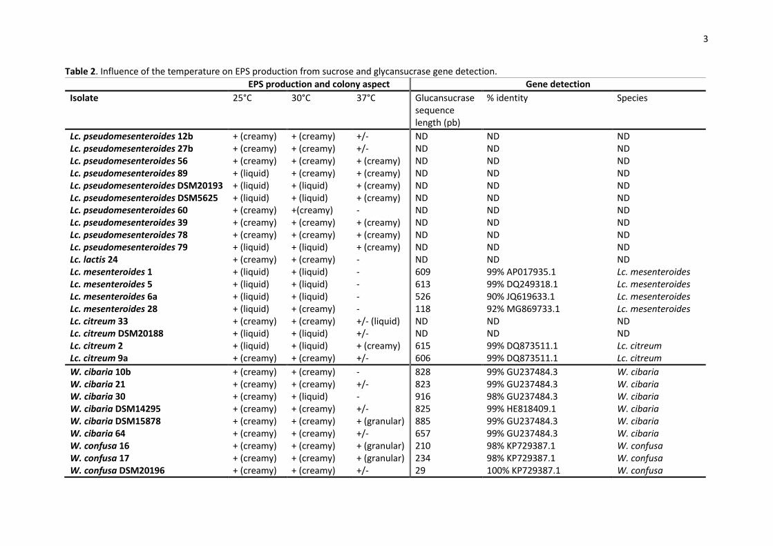
3.3. EPS production and glycansucrase gene detection 214
Neither of the Lactobacillus spp. isolates, nor the L. lactis isolates, nor Fb. tropaeoli 77, W. 215
paramesenteroides 37 and W. soli 58 did produce EPS on sucrose medium (Table 1). On the contrary, 216
all Leuconostoc isolates and all the isolates of other Weissella species produced EPS from sucrose 217
and colony aspect was strain dependent, either liquid or creamy (Tables 1 and 2). 218
For these isolates, EPS production from sucrose was observed at 25°C and 30°C. At 37°C, some 219
isolates (60, 24, 1, 5, 6a, 28, 10b, 30, 38 and 1102001) did not produce EPS, whereas for some other 220
isolates (DSM20193, DSM5625, 79, 33, 2, 16, 17 and 59) a change of the EPS phenotype was noticed 221
(Table 2). The screening for potential glucansucrase genes from Leuconostoc spp. revealed 6 positive 222
strains: Lc. mesenteroides 1, 5, 6a, 28 and Lc. citreum 2, 9a. W. cibaria and W. confusa isolates gave 223
the expected fragment for the amplification of partial dextransucrase gene. Sequencing of the PCR 224

11
products confirmed the similarity to glucansucrase or dextransucrase genes from databases (Table 225
2). Amplification with LevV-fw/LevV-rev and with FTF2-F/FTF2-R primers was not positive suggesting 226
the absence of levansucrase and fructansucrase. 227
3.4. Growth yield in control MRS condition and tolerance to stress 228
Isolates of Weissella, Leuconostoc and Fb. tropaeoli from distinct (GTG)5 group were chosen for 229
further characterization regarding their tolerance to stress, to bile salts and their growth at different 230
temperatures. Results were compared with reference strains and Lactobacillus isolates. 231
Lb. plantarum 75, W. cibaria 64 and 30, W. confusa 1102001 harboured the highest growth yields 232
over 48h compared to other isolates (p < 0.0001), with log OD48/OD0 values of 1.16 ± 0.00, 0.91 ± 233
0.08, 0.86 ± 0.16 and 0.88 ± 0.11, respectively. On the contrary, W. paramesenteroides 37 showed a 234
low growth yield in MRS pH 6.5 37°C compared to other isolates (p < 0.0001) (Table 3). 235
At a low initial pH of 4.5 (Table 3), log OD48/OD0 values were comprised between 0.04 (Lc. 236
pseudomesenteroides 60) and 1.05 (Lb. plantarum 75). At a lower pH of 3, they were comprised 237
between 0.03 (Lc. pseudomesenteroides 56) and 0.22 (W. confusa 1102001). Lb. plantarum 17a and 238
W. cibaria 10b were not affected by exposure to pH 4.5, as no significant differences were detected 239
compared to control condition. Some isolates (73, 75, DSM2601, DSM20193, DSM5625, 39, 78, 240
DSM20188, 21, 30, 64 and 1102001) showed a high growth yield in MRS pH 4.5, but they were 241
clearly affected by this condition compared to control (p < 0.0001). At an initial pH of 3, the growth 242
yield of all isolates was reduced compared to control condition (p < 0.0001) and most of isolates did 243
not succeed to grow in this condition (Table 3). The most acid-tolerant isolates were: Lb. 244
paraplantarum 73; Lb. plantarum 75; Lc. pseudomesenteroides 12b, 27b, DSM5625, DSM20193, 39; 245
Lc. lactis 24; Lc. mesenteroides 28; Lc. citreum 33, 9a; W. cibaria 30, 64, and W. confusa DSM20196, 246
59 and 1102001. 247
12
In moderate saline stress (NaCl 5%), log OD48/OD0 values were comprised between 0.04 (Lc. 248
mesenteroides 5) and 1.16 (Lb. plantarum 75). Lb. plantarum 75, 17a, Lb. paraplantarum 73, W. 249
cibaria DSM15878 and W. soli 58 were the most tolerant to NaCl 5%, and no significant differences 250
were detected compared to control condition (Table 3). Lc. citreum 33, W. confusa 38 and W. 251
paramesenteroides 37 were only moderately affected by NaCl 5% (p < 0.05). Some isolates exhibited 252
high growth in NaCl 5% (1102001, 16, 17, 60, 64, 30 and 59), but compared to control condition, 253
growth was significantly reduced (p < 0.0001). When a stronger saline stress was applied (NaCl 8%), 254
log OD48/OD0 values were comprised between 0.00 (W. cibaria 10b) and 0.97 (Lb. plantarum 75). W. 255
confusa 38 seemed slightly affected by this condition but the difference with control condition was 256
not significant. W. soli 58 was not significantly affected by exposure to NaCl 8%. Isolates 75 and 60 257
revealed a high growth yield in the presence of 8% NaCl, however they were sensitive to this 258
condition (p < 0.001 and p = 0.007, respectively). The growth of all other isolates was severely 259
affected. 260
In a slight oxidative condition, log OD48/OD0 were comprised between 0.00 (5, DSM2601, 2, 79) and 261
1.15 (75). Fb. tropaeoli 77, Lb. plantarum 75, 17a, Lc. pseudomesenteroides DSM20193, DSM5625, 262
60, 39, Lc. citreum 9a, W. cibaria 21, 30, 64, DSM14295, DSM15878 and W. confusa 1102001 were 263
not affected by exposure to 0.025% in H2O2 (Table 3). This concentration partially inhibited the 264
growth of W. confusa 16 and 59 (p < 0.05). The growth of all other isolates was reduced in this 265
condition. With 0.05% H2O2, only four isolates were not affected: Fb. tropaeoli 77, Lc. 266
pseudomesenteroides 60 and W. cibaria 30 and 64. No significant differences were detected 267
between 0.075% and 0.1% H2O2 conditions. This concentration in H2O2 was very efficient to impair 268
growth of almost all LAB isolates. Only Lc. pseudomesenteroides 60 was not affected by 0.1% H2O2. 269
Fb. tropaeoli 77 was also able to grow in this condition but compared to control condition its growth 270
was significantly reduced (p < 0.001). 271
3.5. Tolerance to bile salts 272
13
None of the LAB strains was able to grow in 0.2% or 0.3% of bile salts after 24h (data not shown). 273
Tolerance to bile salts was thus performed with 0.1% over 24h and expressed as a percentage of OD 274
of control (MRS broth without bile salts). Four isolates showed the highest resistance to bile salts: Lc. 275
pseudomesenteroides 12b, W. paramesenteroides 37, Lc. citreum DSM20188 and Lb. plantarum 75, 276
with a percentage of growth comprised between 61% and 83.8% of that observed in control 277
condition (Fig. 3). Tolerance of strain 12b was significantly higher than that of other isolates 278
(p<0.001), except compared to strains 37 and DSM20188. Other Leuconostoc strains showed a 279
relatively good tolerance, with a percentage of growth comprised between 35% and 55.8% of 280
control. Weissella isolates and Lc. pseudomesenteroides 60 were the less tolerant to bile salts, with a 281
percentage of growth comprised between 14.9% and 32.6% of control. The lowest tolerance was 282
observed for Lc. pseudomesenteroides 60 and W. confusa strains 38, 16, DSM20196 and 17. 283
Leuconostoc strains 58, 89, 27b, 2, 39, 56, 79, 33, 78 and Weissella strains 58 and DSM15830 were 284
not able to grow at 37°C in 24h in the presence of 0.1% bile salts: for these isolates, we cannot 285
conclude regarding their tolerance to bile salts. 286
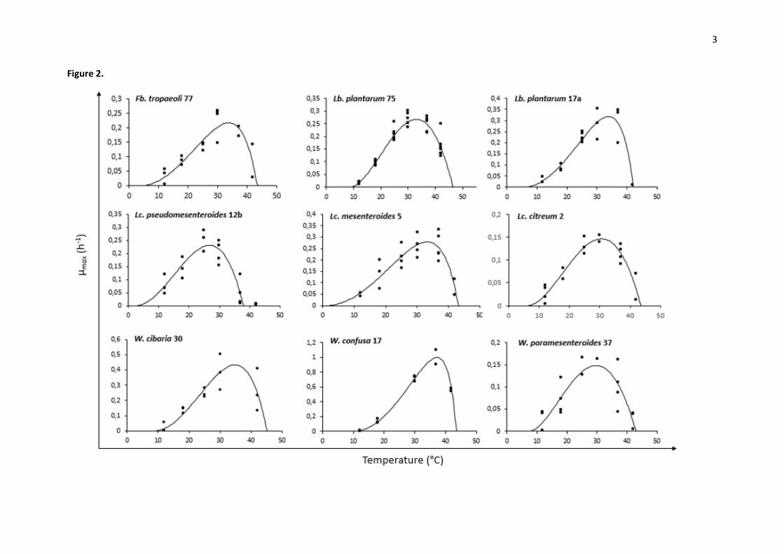
3.6. Temperature effect on growth rate 287
Cardinal temperatures and µopt were determined using Sym’Previus software by plotting µmax as a 288
function of temperature (12°C, 18°C, 25°C, 30°C, 37°C, 42°C) (Fig. 2). Predicted values obtained from 289
Sym’Previus model and possible growth after incubation for 21 days are indicated in Table 4. 290
Optimal growth temperature Topt of LAB isolates ranged between 25.1°C (Lc. citreum 33) and 39.0°C 291
(W. confusa 59). The majority of Leuconostoc spp. harboured Topt comprised between 25.0 and 292
30.0°C. Weissella spp. and Lactobacillus spp. showed Topt over 30°C, above 34°C for Weissella isolates 293
30, 16, 17, 59 and 58. LAB isolates harboured µopt values comprised between 0.107 h-1 (Lc. 294
pseudomesenteroides 27b) and 0.998 h-1 (W. confusa 17) (Table 4). W. confusa isolates (38, 1102001, 295
16, 59, 17) and W. cibaria isolates (64, 30, 10b) showed the highest µopt values. Generally, 296
Leuconostoc isolates shown the lowest µopt values. 297

14
LAB isolates exhibited minimal growth temperatures Tmin comprised between 0.6°C (W. confusa 59) 298
and 16.7°C (W. cibaria DSM15878). Leuconostoc spp. harboured Tmin comprised between 1.0°C (Lc. 299
mesenteroides 5) and 11.9°C (Lc. pseudomesenteroides 79). Weissella spp. harboured Tmin comprised 300
between 0.6°C (W. confusa 59) and 16.7°C (W. cibaria DSM15878). Fructobacillus and Lactobacillus 301
spp. harboured Tmin comprised between 3.7°C (Fb. tropaeoli 77) and 14.9°C (Lb. paraplantarum 73). 302
Generally, Leuconostoc spp. have shown the lowest Tmin values. Some Sym’previus Tmin values were 303
not confirmed by incubation test for 21 days, as for Lc. pseudomesenteroides 39, 89, DSM5625 and 304
DSM20193 or W. confusa 16 and 17. 305
Maximal growth temperature Tmax of LAB isolates ranged between 37.5°C (Lc. pseudomesenteroides 306
78) and 49.3°C (W. confusa 16). The majority of Leuconostoc spp. harboured Tmax comprised 307
between 37.0°C and 43.0°C which was confirmed by incubation for 21 days at 42°C. Incubation for 308
21 days at 45°C confirmed that only two Leuconostoc isolates were able to grow between 42°C and 309
45°C, as Lc. lactis 24 and Lc. pseudomesenteroides 60. Weissella spp. harboured Tmax comprised 310
between 38.1°C (W. soli 58) and 49.3°C (W. confusa 16). Fructobacillus and Lactobacillus spp. 311
harboured Tmax comprised between 40.0°C (Lb. plantarum DSM2601) and 46.5°C (Lb. plantarum 75). 312
In most case, incubation for 21 days resulted in maximal growth temperatures which fell into the 313
confident interval given by the model, except for some strains for which the model mostly proposed 314
higher Tmax. 315
4. Discussion 316
The aim of this paper was to collect and characterize LAB isolates from tropically grown fruits and 317
vegetables for possible application in food industry, especially for fruits and vegetables. 318
In this study, genetic and phenotypic characterization of 77 autochthonous LAB isolated from 319
papaya, tomato and sliced cabbage was performed. Among the species the most frequently 320
detected, Lc. mesenteroides was commonly isolated from fresh fruits and vegetables, as raw prickly 321
pear, sweet cherry and raw peppers (Di Cagno et al., 2016, 2011b, 2009a), Lc. pseudomesenteroides 322

15
and Lc. citreum have been isolated from ripe mulberries, fresh tomato, fresh coffee cherries and 323
banana fruit (Chen et al., 2017, 2010; Leong et al., 2014; Trias et al., 2008). Lc. lactis, hereby isolated 324
from cabbage, is mainly associated with dairy and vegetable-based fermented food, including kimchi 325
(Chen et al., 2012; Cho et al., 2006; Vos et al., 2011). To the best of our knowledge, isolation of Lc. 326
lactis from raw vegetable or ready-to-eat crude vegetable has not been reported. W. cibaria, W. 327
confusa, Lb. plantarum and Lb. paraplantarum were the second-most-commonly detected species. 328
These species are frequently isolated from fresh fruits and vegetables (Chen et al., 2010; Di Cagno et 329
al., 2013, 2011a, 2009b; Emerenini et al., 2013; Trias et al., 2008). Weissella and Lactobacillus 330
frequently occur in spontaneous fermentation of fruits or vegetables (Fessard and Remize, 2017), 331
highlighting their natural adaptation to fruit and vegetable environments. Our study revealed also 332
the presence of isolates of the species W. soli (cabbage), W. paramesenteroides (papaya) and Fb. 333
tropaeoli (papaya). W. paramesenteroides has been isolated from a variety of fermented fruit and 334
vegetable (Chen et al., 2013b, 2013a; Escalante-Minakata et al., 2008; Lan et al., 2009), and a single 335
study reported its isolation from banana fruit (Chen et al., 2017). W. soli has been detected in silage 336
fermentation of vegetable residues (cabbage, Chinese cabbage and lettuce) (Yang et al., 2010). Fb. 337
tropaeoli was first isolated from a flower of Tropaeolum majus in South Africa (Endo et al., 2011) and 338
was further isolated from spontaneous cocoa fermentation together with W. fabalis (Snauwaert et 339
al., 2013). Only recently, Fb. tropaeoli has shown fruit origins (Franquès et al., 2017; Ruiz Rodríguez 340
et al., 2017). Due to its frequent presence in raw milk, L. lactis has been extensively used as starter 341
culture for dairy foods, contributing to the development of texture by producing exopolysaccharides 342
(Casalta and Montel, 2008). In a recent study, two groups have been proposed for L. lactis lactis 343
subspecies, “domesticated” and “environmental”. The latter appears to be the main contributor to 344
genetic and phenotypic diversity within the subspecies (Laroute et al., 2017). The availability of new 345
isolates from our study would be useful to understand the relationships between origin and 346
phenotypic features. Besides, plant material is the main natural habitat of L. lactis and this species 347
has been detected in fresh and frozen corn, corn silks, navy beans, cabbage, lettuce or peas (Vos et 348

16
al., 2011), sprouted seeds and grapefruit juice (Kelly et al., 1996), by-products of pineapple and 349
cherry pulp processing (Garcia et al., 2016), ripe mulberries (Chen et al., 2010) and fresh coffee 350
cherries (Leong et al., 2014). Our results are then consistent with previous detection of these species 351
from fruit and vegetable environments. It also revealed the presence of several species, like Lc. lactis 352
or W. paramesenteroides, rarely isolated from fresh fruit and vegetables but rather from fermented 353
foods. 354
Rep-PCR revealed a high diversity of genetic profiles. The 77 LAB isolates were clustered into 41 355
genetic groups, which is a high number of groups regarding the limited number of species and 356
samples. The highest diversity was observed for Leuconostoc and Weissella isolates. Indeed, the 11 357
Weissella isolates were allocated to 10 genetic groups and were isolated from seven samples from 358
three different raw material. From this observed genetic diversity, we hypothesized a phenotypic 359
diversity which could be used as a stock for bacteria of technological interest. 360
Fermentation and biopreservation of minimally-processed foods from plant origin require selected 361
strains with desirable properties. The use of LAB for this purpose presents many advantages 362
regarding their history of use in foods and their ability to adapt to the specific conditions of these 363
raw materials. The advantages of autochthonous strains rely of the assumption of a higher stability 364
in their natural environment to compete microbial contaminants, and a better adaptative ability to 365
their niche (Beganović et al., 2013; Di Cagno et al., 2009b, 2008; Fessard et al., 2016; Viana de Souza 366
and Silva Dias, 2017). Although there is not a single protocol to select starter or bioprotective or 367
probiotic strains and each application requires a tailored selection, several common traits are 368
considered whatever the context (Bevilacqua et al., 2012; Kostinek et al., 2005; Leroi et al., 2015). 369
They are mainly related to adaptation of strains to technological conditions, such as acidic medium, 370
salt addition or incubation temperature. Moreover, safety of strains is of crucial importance. 371
Regarding probiotic selection, expectations of strain properties are different from those of 372
fermentation starters or biocontrol agents. A step-by-step procedure has been proposed, 373

17
investigating stress tolerance, adhesion ability, antipathogenic activity, safety assessment, host-374
associated functional properties, industrial requirements and omics characterization, before clinical 375
trials (de Melo Pereira et al., 2018). 376
The behaviour of LAB at different temperatures and the determination of growth parameters are 377
thus important to consider for a rational choice of strains for a specific application. Growth of W. 378
cibaria and W. confusa isolates was detected between 6°C and 45°C, in accordance with those found 379
in the literature (Björkroth et al., 2002; Fusco et al., 2015). Leuconostoc isolates had optimum 380
growth between 25.1°C and 33.3°C, and growth up to 37°C was strain dependent, which 381
corresponds to the description of Leuconostoc spp. (Vos et al., 2011). Lc. mesenteroides 1 and 5, Lc. 382
citreum 2 and Lc. pseudomesenteroides 12b have shown particular ability to grow at low 383
temperatures, which it is a technological criteria of importance for their application in fruit and 384
vegetable fermentation (Fessard and Remize, 2017). In our study, µmax values of Leuconostoc isolates 385
were lower than those observed for Weissella or Lactobacillus isolates. They were in the same range 386
than those reported by Ricciardi et al., (2009). and Drosinos et al., (2006). Maximum growth rate is 387
clearly species- and strain-dependent and several factors may affect this value such as temperature, 388
pH, oxygen or presence of toxic compounds. Regarding growth parameters, temperature control 389
appears here to be a potent lever to favour the growth of certain LAB isolates. 390
Low pH and high salt concentration are often used as selective conditions for LAB over food 391
processing steps. Exposure to low pH affected the growth of all LAB strains, but Weissella and 392
Lactobacillus spp. were more tolerant than Leuconostoc spp. Growth of W. cibaria strain in MRS 393
broth adjusted to pH 3 has been reported by Patel et al. (2012). Interestingly, Weissella and 394
Lactobacillus strains were also the most tolerant to salt stress, especially W. soli 58, W. confusa 38 395
and Lb. plantarum 75, which were able to grow in 8% NaCl, in accordance with Lee et al. (2012) and 396
Papamanoli et al. (2003) which reported data for strains from the same species. 397

18
Unexpectedly, Fb. tropaeoli 77, Lc. pseudomesenteroides 60 and W. cibaria 21, 30 and 64 were the 398
most tolerant to hydrogen peroxide, whereas Lb. plantarum strains have already been shown to be 399
highly tolerant to exposure to 0.1% H2O2 for 30 min (Parente et al., 2010). Glutathione (GSH), a non-400
protein thiol compound, has been described in Lactococcus and Lactobacillus spp. and may play a 401
role in the protection towards an oxidative stress (Zhang and Li, 2013). LAB are catalase negative but 402
some strains may possess a manganese-dependent form. 403
Tolerance of LAB to bile salts has been associated with their capacity to metabolize the bile salts (van 404
de Guchte et al., 2002) and constitute an important trait for the selection of cultures which can 405
survive in gut. In our study, LAB isolates were able to grow only in 0.1% of bile salts which is quite 406
low compared to the data reported in the literature. Concentrations of 0.15-0.3% of bile salts have 407
been recommended as a suitable concentration for the selection of probiotic bacteria for human use 408
(Boke et al., 2010). However, experimental time exposure to bile salts is generally limited to 6 hours, 409
which might explain the apparent discrepancy. Generally, Leuconostoc isolates have shown good 410
tolerance to bile salts while W. cibaria and W. confusa isolates were the most sensitive. Lc. 411
pseudomesenteroides 12b, W. paramesenteroides 37, and Lb. plantarum 75 isolates were the most 412
tolerant to bile salts. If some Lb. plantarum strains were shown to be resistant to 2% bile salts 413
(Papamanoli et al., 2003), tolerance to bile salts of Lc. pseudomesenteroides and W. 414
paramesenteroides is not described. 415
Exopolysaccharides are important in the manufacture of dairy products and have gained interest 416
recently for the manufacture of fruit or vegetable puree and smoothies (Di Cagno et al., 2011a; 417
Juvonen et al., 2015). The screening for EPS production performed on sucrose medium revealed that 418
only Leuconostoc spp., W. cibaria and W. confusa isolates produced EPS from sucrose. The literature 419
qualified W. cibaria, W. confusa and Leuconostoc spp. as high producers of EPS (Di Cagno et al., 420
2016; Galle et al., 2010; Maina et al., 2008; Malang et al., 2015; Wolter et al., 2014), and no dextran 421
was produced from sucrose by Fb. tropaeoli strain (Endo et al., 2011), which supports our results. 422

19
However, the production of EPS was also previously reported for Lb. plantarum strains, isolated from 423
sourdough and fish (Di Cagno et al., 2006; Hongpattarakere et al., 2012) and for L. lactis strain 424
isolated from raw milk (Van der Meulen et al., 2007) but our isolates of these species did not 425
produce EPS. The ability of LAB to produce EPS from sucrose is due to the action of one sucrase 426
enzyme, either glucansucrase or fructansucrase (van Hijum et al., 2006). In our study, partial 427
sequencing of glucansucrase genes was positive for W. cibaria, W. confusa, Lc. mesenteroides and Lc. 428
citreum isolates. Several studies already reported glucan production or glucansucrase activity from 429
Lc. mesenteroides and Lc. citreum strains (Bounaix et al., 2010a; Kang et al., 2014; Passerini et al., 430
2015; Song et al., 2016; Zannini et al., 2016) as well as for W. cibaria and W. confusa (Amari et al., 431
2013; Baruah et al., 2017; Bounaix et al., 2010b). Little is known about sucrase enzyme from Lc. 432
pseudomesenteroides spp. and none of our Lc. pseudomesenteroides isolates producing EPS were 433
positive for sucrase enzyme encoding genes. Dextran production constitutes a possible desirable 434
technological trait for our isolates. Our study revealed also that temperatures comprised between 435
25°C and 30°C were optimum for the production of EPS. It has been shown that EPS production was 436
higher at temperature comprised between 15°C and 20°C for W. cibaria (Hu and Gänzle, 2018). W. 437
confusa dextransucrase activity was higher between 20°C and 30°C (Amari et al., 2013). These 438
observations could explain the absence of EPS production at 37°C observed for some isolates. 439
Our study provides a stock of autochthonous LAB species from fruits and vegetables with phenotypic 440
characteristics useful for application in food. Altogether, Weissella strains, especially W. cibaria 64 441
and 30 were particularly tolerant to acidic, osmotic and oxidative conditions and produce EPS. 442
However, until now, this species is not used as commercial starter (Fessard and Remize, 2017). W. 443
soli 58 has shown high tolerance to osmotic conditions but was a relatively poor sugar fermenter in 444
MRS broth. W. paramesenteroides 37 was one of the isolates the most tolerant to bile salts. The 445
group of Leuconostoc isolates harbor a variety of diverse phenotypes. Among those, strain 12b 446
showed a high tolerance to bile salts while strain 60 was particularly tolerant to osmotic and 447
oxidative stress. Lc. mesenteroides 1 and 5 and Lc. citreum 2 were particularly tolerant to low 448

20
temperatures. Lb plantarum 75 was a high sugar fermenter, highly tolerant to low pH, salts and bile 449
salts. For all those strains, no biogenic amine production from lysine, ornithine, tyrosine and 450
histidine, and also the absence of detection of histidine, tyrosine and ornithine decarboxylase genes 451
was observed (data not shown), but the possibility of biogenic amine production via the arginine 452
deiminase pathway has to be checked. 453
W. cibaria 30 and 64, Lc. pseudomesenteroides 12b and 60 and Lb. plantarum 75 are powerful 454
candidates for fruits and vegetables fermentation, whereas W. soli 58, Fb. tropaeoli 77, Lc. 455
mesenteroides 1 and 5 could be investigated as preservative cultures for fruits and vegetables. 456
Despite their frequent detection in foods, genetic and phenotypic features of some LAB species 457
remain poorly documented. LAB species mostly described are L. lactis and Lb. plantarum mainly due 458
to their long and safe history of application, especially for dairy products (Leroy and De Vuyst, 2004). 459
Our study provides a useful description of several autochthonous LAB isolates from other species 460
often encountered in fruits and vegetables, including Lactobacillus, Leuconostoc, Weissella and 461
Fructobacillus spp. The comparison of the core and pan genomes towards available genomes from 462
strains of various origins, of several isolates, which exhibit a potential for their use as starter, would 463
be of particular interest to provide information on niche adaptation and go further on their possible 464
tailored-application. 465
Conflict of interest 466
The authors declare no conflict of interest. 467
Acknowledgements 468
Part of this work was funded by La Reunion regional council and by Federation BioST of the University 469
of La Reunion. We thank Dr Louis Coroller (University of Brest, France) for assistance in growth 470
parameters determination. 471
References 472

21
Alegbeleye, O.O., Singleton, I., Sant’Ana, A.S., 2018. Sources and contamination routes of microbial 473
pathogens to fresh produce during field cultivation: A review. Food Microbiol. 73, 177–208. 474
doi:10.1016/J.FM.2018.01.003 475
Amari, M., Arango, L.F.G., Gabriel, V., Robert, H., Morel, S., Moulis, C., Gabriel, B., Remaud-Siméon, 476
M., Fontagné-Faucher, C., 2013. Characterization of a novel dextransucrase from Weissella confusa 477
isolated from sourdough. Appl. Microbiol. Biotechnol. 97, 5413–5422. doi:10.1007/s00253-012-478
4447-8 479
Baruah, R., Deka, B., Goyal, A., 2017. Purification and characterization of dextransucrase from 480
Weissella cibaria RBA12 and its application in in vitro synthesis of prebiotic oligosaccharides in 481
mango and pineapple juices. LWT - Food Sci. Technol. 84, 449–456. doi:10.1016/J.LWT.2017.06.012 482
Björkroth, K.J., Schillinger, U., Geisen, R., Weiss, N., Hoste, B., Holzapfel, W.H., Korkeala, H.J., 483
Vandamme, P., 2002. Taxonomic study of Weissella confusa and description of Weissella cibaria sp. 484
nov., detected in food and clinical samples. Int. J. Syst. Evol. Microbiol. 52, 141–8. 485
Boke, H., Aslim, B., Alp, G., 2010. The role of resistance to bile salts and acid tolerance of 486
exopolysaccharides (EPSs) produced by yogurt starter bacteria. Arch. Biol. Sci. 62, 323–328. 487
doi:10.2298/ABS1002323B 488
Bounaix, M.-S., Gabriel, V., Robert, H., Morel, S., Remaud-Siméon, M., Gabriel, B., Fontagné-Faucher, 489
C., 2010a. Characterization of glucan-producing Leuconostoc strains isolated from sourdough. Int. J. 490
Food Microbiol. 144, 1–9. doi:10.1016/j.ijfoodmicro.2010.05.026 491
Bounaix, M.-S., Robert, H., Gabriel, V., Morel, S., Remaud-Siméon, M., Gabriel, B., Fontagné-Faucher, 492
C., 2010b. Characterization of dextran-producing Weissella strains isolated from sourdoughs and 493
evidence of constitutive dextransucrase expression. FEMS Microbiol. Lett. 311, 18–26. 494
doi:10.1111/j.1574-6968.2010.02067.x 495
Bounaix, M.S., Gabriel, V., Morel, S., Robert, H., Rabier, P., Remaud-Siméon, M., Gabriel, B., 496
Fontagné-Faucher, C., 2009. Biodiversity of exopolysaccharides produced from sucrose by 497
sourdough lactic acid bacteria. J. Agric. Food Chem. 57, 10889–10897. doi:10.1021/jf902068t 498

22
Casalta, E., Montel, M.C., 2008. Safety assessment of dairy microorganisms: The Lactococcus genus. 499
Int. J. Food Microbiol. 126, 271–273. doi:10.1016/j.ijfoodmicro.2007.08.013 500
Chang, M., Chang, H.C., 2012. Development of a screening method for biogenic amine producing 501
Bacillus spp. Int. J. Food Microbiol. 153, 269–274. doi:10.1016/j.ijfoodmicro.2011.11.008 502
Chen, Y.-S., Wu, H. chung, Wang, C. mei, Lin, C. chun, Chen, Y. ting, Jhong, Y. jyun, Yanagida, F., 503
2013a. Isolation and characterization of lactic acid bacteria from Pobuzihi (fermented 504
cummingcordia), a traditional fermented food in Taiwan. Folia Microbiol. (Praha). 58, 103–109. 505
doi:10.1007/s12223-012-0188-4 506
Chen, Y.-S., Wu, H.C., Pan, S.F., Lin, B.G., Lin, Y.H., Tung, W.C., Li, Y.L., Chiang, C.M., Yanagida, F., 507
2013b. Isolation and characterization of lactic acid bacteria from yan-taozih (pickled peaches) in 508
Taiwan. Ann. Microbiol. 63, 607–614. doi:10.1007/s13213-012-0510-z 509
Chen, Y. sheng, Liao, Y. jou, Lan, Y. shan, Wu, H. chung, Yanagida, F., 2017. Diversity of lactic acid 510
bacteria associated with banana fruits in Taiwan. Curr. Microbiol. 74, 484–490. doi:10.1007/s00284-511
017-1213-2 512
Chen, Y.S., Wu, H., Lo, H., Lin, W., Hsu, W., Lin, C., Lin, P., Yanagida, F., 2012. Isolation and 513
characterisation of lactic acid bacteria from jiang-gua (fermented cucumbers), a traditional 514
fermented food in Taiwan. J. Sci. Food Agric. 92, 2069–2075. doi:10.1002/jsfa.5583 515
Chen, Y.S., Wu, H.C., Yanagida, F., 2010. Isolation and characteristics of lactic acid bacteria isolated 516
from ripe mulberries in Taiwan. Brazilian J. Microbiol. 41, 916–921. doi:10.1590/S1517-517
83822010000400010 518
Cho, J., Lee, D., Yang, C., Jeon, J., Kim, J., Han, H., 2006. Microbial population dynamics of kimchi, a 519
fermented cabbage product. FEMS Microbiol. Lett. 257, 262–267. doi:10.1111/j.1574-520
6968.2006.00186.x 521
Di Cagno, R., Coda, R., De Angelis, M., Gobbetti, M., 2013. Exploitation of vegetables and fruits 522
through lactic acid fermentation. Food Microbiol. 33, 1–10. doi:10.1016/j.fm.2012.09.003 523
Di Cagno, R., De Angelis, M., Limitone, A., Minervini, F., Carnevali, P., Corsetti, A., Gaenzle, M., Ciati, 524

23
R., Gobbetti, M., 2006. Glucan and fructan production by sourdough Weissella cibaria and 525
Lactobacillus plantarum. J. Agric. Food Chem. 54, 9873–9881. doi:10.1021/jf061393+ 526
Di Cagno, R., Filannino, P., Vincentini, O., Lanera, A., Cavoski, I., Gobbetti, M., 2016. Exploitation of 527
Leuconostoc mesenteroides strains to improve shelf life, rheological, sensory and functional features 528
of prickly pear (Opuntia ficus-indica L.) fruit puree. Food Microbiol. 59, 176–189. 529
doi:10.1016/J.FM.2016.06.009 530
Di Cagno, R., Minervini, G., Rizzello, C.G., De Angelis, M., Gobbetti, M., 2011a. Effect of lactic acid 531
fermentation on antioxidant, texture, color and sensory properties of red and green smoothies. Food 532
Microbiol. 28, 1062–71. doi:10.1016/j.fm.2011.02.011 533
Di Cagno, R., Surico, R.F., Minervini, G., De Angelis, M., Rizzello, C.G., Gobbetti, M., 2009a. Use of 534
autochthonous starters to ferment red and yellow peppers (Capsicum annum L.) to be stored at 535
room temperature. Int. J. Food Microbiol. 130, 108–16. doi:10.1016/j.ijfoodmicro.2009.01.019 536
Di Cagno, R., Surico, R.F., Minervini, G., Rizzello, C.G., Lovino, R., Servili, M., Taticchi, A., Urbani, S., 537
Gobbetti, M., Di, R., Fortunata, R., Giuseppe, C., 2011b. Exploitation of sweet cherry (Prunus avium 538
L.) puree added of stem infusion through fermentation by selected autochthonous lactic acid 539
bacteria. Food Microbiol. 28, 900–9. doi:10.1016/j.fm.2010.12.008 540
Di Cagno, R., Surico, R.F., Paradiso, A., De Angelis, M., Salmon, J.-C., Buchin, S., De Gara, L., Gobbetti, 541
M., 2009b. Effect of autochthonous lactic acid bacteria starters on health-promoting and sensory 542
properties of tomato juices. Int. J. Food Microbiol. 128, 473–83. 543
doi:10.1016/j.ijfoodmicro.2008.10.017 544
Droby, S., Wisniewski, M., 2018. The fruit microbiome: A new frontier for postharvest biocontrol and 545
postharvest biology. Postharvest Biol. Technol. 140, 107–112. 546
doi:10.1016/J.POSTHARVBIO.2018.03.004 547
Drosinos, E.H., Mataragas, M., Metaxopoulos, J., 2006. Modeling of growth and bacteriocin 548
production by Leuconostoc mesenteroides E131. Meat Sci. 74, 690–696. 549
doi:10.1016/j.meatsci.2006.05.022 550
24
Durand, L., Planchon, S., Guinebretiere, M.-H., Andre, S., Carlin, F., Remize, F., 2015. Contamination 551
pathways of spore-forming bacteria in a vegetable cannery. Int. J. Food Microbiol. 202, 10–9. 552
doi:10.1016/j.ijfoodmicro.2015.02.019 553
Emerenini, E.C., Afolabi, O.R., Okolie, P.I., Akintokun, A.K., 2013. Isolation and molecular 554
characterization of lactic acid bacteria isolated from fresh fruits and vegetables using nested PCR 555
analysis. Br. Microbiol. Res. J. 3, 368–377. 556
Endo, A., Irisawa, T., Futagawa-Endo, Y., Sonomoto, K., Itoh, K., Takano, K., Okada, S., Dicks, L.M.T., 557
2011. Fructobacillus tropaeoli sp. nov., a fructophilic lactic acid bacterium isolated from a flower. Int. 558
J. Syst. Evol. Microbiol. 61, 898–902. doi:10.1099/ijs.0.023838-0 559
Escalante-Minakata, P., Blaschek, H.P., Barba De La Rosa, A.P., Santos, L., De León-Rodríguez, A., 560
2008. Identification of yeast and bacteria involved in the mezcal fermentation of Agave salmiana. 561
Lett. Appl. Microbiol. 46, 626–630. doi:10.1111/j.1472-765X.2008.02359.x 562
Fessard, A., Bourdon, E., Payet, B., Remize, F., 2016. Identification, stress tolerance, and antioxidant 563
activity of lactic acid bacteria isolated from tropically grown fruits and leaves. Can. J. Microbiol. 62, 564
550–561. doi:10.1139/cjm-2015-0624 565
Fessard, A., Kapoor, A., Patche, J., Assemat, S., Hoarau, M., Bourdon, E., Bahorun, T., Remize, F., 566
2017. Lactic fermentation as an efficient tool to enhance the antioxidant activity of tropical fruit 567
juices and teas. Microorganisms 5, 23. doi:10.3390/microorganisms5020023 568
Fessard, A., Remize, F., 2017. Why are Weissella spp. not used as commercial starter cultures for 569
food fermentation? Fermentation 3, 38. doi:10.3390/fermentation3030038 570
Francis, G.A., Gallone, A., Nychas, G.J., Sofos, J.N., Colelli, G., Amodio, M.L., Spano, G., 2012. Factors 571
affecting quality and safety of fresh-cut produce. Crit. Rev. Food Sci. Nutr. 52, 595–610. 572
doi:10.1080/10408398.2010.503685 573
Franquès, J., Araque, I., Palahí, E., Portillo, M. del C., Reguant, C., Bordons, A., 2017. Presence of 574
Oenococcus oeni and other lactic acid bacteria in grapes and wines from Priorat (Catalonia, Spain). 575
LWT - Food Sci. Technol. 81, 326–334. doi:10.1016/j.lwt.2017.03.054 576

25
Fusco, V., Quero, G.M., Cho, G.-S., Kabisch, J., Meske, D., Neve, H., Bockelmann, W., Franz, C.M.A.P., 577
2015. The genus Weissella: taxonomy, ecology and biotechnological potential. Front. Microbiol. 6, 578
155. doi:10.3389/fmicb.2015.00155 579
Galle, S., Schwab, C., Arendt, E., Gänzle, M., 2010. Exopolysaccharide-forming Weissella strains as 580
starter cultures for sorghum and wheat sourdoughs. J. Agric. Food Chem. 58, 5834–41. 581
doi:10.1021/jf1002683 582
Garcia, E.F., Luciano, W.A., Xavier, D.E., da Costa, W.C.A., de Sousa Oliveira, K., Franco, O.L., de 583
Morais Júnior, M.A., Lucena, B.T.L., Picão, R.C., Magnani, M., Saarela, M., de Souza, E.L., 2016. 584
Identification of lactic acid bacteria in fruit pulp processing byproducts and potential probiotic 585
properties of selected Lactobacillus strains. Front. Microbiol. 7, 1371. doi:10.3389/fmicb.2016.01371 586
Guinebretiere, M.H., Girardin, H., Dargaignaratz, C., Carlin, F., 2003. Contamination flows of Bacillus 587
cereus and spore-forming aerobic bacteria in a cooked, pasteurized and chilled zucchini purée 588
processing line 82, 223–232. 589
Hongpattarakere, T., Cherntong, N., Wichienchot, S., Kolida, S., Rastall, R.A., 2012. In vitro prebiotic 590
evaluation of exopolysaccharides produced by marine isolated lactic acid bacteria. Carbohydr. 591
Polym. 87, 846–852. doi:10.1016/j.carbpol.2011.08.085 592
Hu, Y., Gänzle, M.G., 2018. Effect of temperature on production of oligosaccharides and dextran by 593
Weissella cibaria 10 M. Int. J. Food Microbiol. 280, 27–34. doi:10.1016/j.ijfoodmicro.2018.05.003 594
Juvonen, R., Honkapää, K., Maina, N.H., Shi, Q., Viljanen, K., Maaheimo, H., Virkki, L., Tenkanen, M., 595
Lantto, R., 2015. The impact of fermentation with exopolysaccharide producing lactic acid bacteria 596
on rheological, chemical and sensory properties of pureed carrots (Daucus carota L.). Int. J. Food 597
Microbiol. 207, 109–18. doi:10.1016/j.ijfoodmicro.2015.04.031 598
Kang, H.K., Nguyen, T.T.H., Jeong, H.N., Park, M.E., Kim, D., 2014. Molecular cloning and 599
characterization of a novel glucansucrase from Leuconostoc mesenteroides subsp. mesenteroides 600
LM34. Biotechnol. Bioprocess Eng. 19, 605–612. doi:10.1007/s12257-014-0116-3 601
Kelly, W.J., Asmundson, R. V., Huang, C.M., 1996. Isolation and characterization of bacteriocin-602

26
producing lactic acid bacteria from ready-to-eat food products. Int. J. Food Microbiol. 33, 209–218. 603
doi:10.1016/0168-1605(96)01157-9 604
Lan, W.-T.T., Chen, Y.-S. sheng, Yanagida, F., 2009. Isolation and characterization of lactic acid 605
bacteria from Yan-dong-gua (fermented wax gourd), a traditional fermented food in Taiwan. J. 606
Biosci. Bioeng. 108, 484–7. doi:10.1016/j.jbiosc.2009.06.009 607
Laroute, V., Tormo, H., Couderc, C., Mercier-Bonin, M., Le Bourgeois, P., Cocaign-Bousquet, M., 608
Daveran-Mingot, M.-L., 2017. From genome to phenotype: an integrative approach to evaluate the 609
biodiversity of Lactococcus lactis. Microorganisms 5. doi:10.3390/microorganisms5020027 610
Lee, K.W., Park, J.Y., Jeong, H.R., Heo, H.J., Han, N.S., Kim, J.H., 2012. Probiotic properties of 611
Weissella strains isolated from human faeces. Anaerobe 18, 96–102. 612
doi:10.1016/j.anaerobe.2011.12.015 613
Leff, J.W., Fierer, N., 2013. Bacterial communities associated with the surfaces of fresh fruits and 614
vegetables. PLoS One 8, e59310. doi:10.1371/journal.pone.0059310 615
Leong, K., Chen, Y., Pan, S., Chen, J., Wu, H., Chang, Y., Yanagida, F., 2014. Diversity of lactic acid 616
bacteria associated with fresh coffee cherries in Taiwan. Curr. Microbiol. 68, 440–447. 617
doi:10.1007/s00284-013-0495-2 618
Leroy, F., De Vuyst, L., 2004. Lactic acid bacteria as functional starter cultures for the food 619
fermentation industry. Trends Food Sci. Technol. 15, 67–78. doi:10.1016/j.tifs.2003.09.004 620
Maina, N.H., Tenkanen, M., Maaheimo, H., Juvonen, R., Virkki, L., 2008. NMR spectroscopic analysis 621
of exopolysaccharides produced by Leuconostoc citreum and Weissella confusa. Carbohydr. Res. 343, 622
1446–1455. doi:10.1016/j.carres.2008.04.012 623
Malang, S.K., Maina, N.H., Schwab, C., Tenkanen, M., Lacroix, C., 2015. Characterization of 624
exopolysaccharide and ropy capsular polysaccharide formation by Weissella. Food Microbiol. 46, 625
418–27. doi:10.1016/j.fm.2014.08.022 626
Palomba, S., Cavella, S., Torrieri, E., Piccolo, A., Mazzei, P., Blaiotta, G., Ventorino, V., Pepe, O., 2012. 627
Polyphasic screening, homopolysaccharide composition, and viscoelastic behavior of wheat 628

27
sourdough from a Leuconostoc lactis and Lactobacillus curvatus exopolysaccharide-producing starter 629
culture. Appl. Environ. Microbiol. 78, 2737–2747. doi:10.1128/AEM.07302-11 630
Papamanoli, E., Tzanetakis, N., Litopoulou-Tzanetaki, E., Kotzekidou, P., 2003. Characterization of 631
lactic acid bacteria isolated from a Greek dry-fermented sausage in respect of their technological 632
and probiotic properties. Meat Sci. 65, 859–867. doi:10.1016/S0309-1740(02)00292-9 633
Parente, E., Ciocia, F., Ricciardi, A., Zotta, T., Felis, G.E., Torriani, S., 2010. Diversity of stress 634
tolerance in Lactobacillus plantarum, Lactobacillus pentosus and Lactobacillus paraplantarum: A 635
multivariate screening study. Int. J. Food Microbiol. 144, 270–9. 636
doi:10.1016/j.ijfoodmicro.2010.10.005 637
Passerini, D., Vuillemin, M., Ufarté, L., Morel, S., Loux, V., Fontagné-Faucher, C., Monsan, P., 638
Remaud-Siméon, M., Moulis, C., 2015. Inventory of the GH70 enzymes encoded by Leuconostoc 639
citreum NRRL B-1299 - Identification of three novel α-transglucosylases. FEBS J. 282, 2115–2130. 640
doi:10.1111/febs.13261 641
Patel, A., Prajapati, J.B., Holst, O., Ljungh, A., 2012. Probiotic properties of exopolysaccharide 642
producing lactic acid bacteria isolated from vegetables and traditional Indian fermented foods. Food 643
Biosci. 5, 27–33. doi:10.1016/j.fbio.2013.10.002 644
Pothakos, V., Snauwaert, C., De Vos, P., Huys, G., Devlieghere, F., 2014. Monitoring psychrotrophic 645
lactic acid bacteria contamination in a ready-to-eat vegetable salad production environment. Int. J. 646
Food Microbiol. 185, 7–16. doi:10.1016/j.ijfoodmicro.2014.05.009 647
Ramos, B., Miller, F.A., Brandão, T.R.S., Teixeira, P., Silva, C.L.M., 2013. Fresh fruits and vegetables—648
An overview on applied methodologies to improve its quality and safety. Innov. Food Sci. Emerg. 649
Technol. 20, 1–15. doi:10.1016/j.ifset.2013.07.002 650
Ricciardi, A., Parente, E., Zotta, T., 2009. Modelling the growth of Weissella cibaria as a function of 651
fermentation conditions. J. Appl. Microbiol. 107, 1528–1535. doi:10.1111/j.1365-2672.2009.04335.x 652
Rosso, L., Lobry, J.R., Flandrois, J.P., 1993. An unexpected correlation between cardinal temperatures 653
of microbial growth highlighted by a new model. J. Theor. Biol. 162, 447–63. 654
28
doi:10.1006/jtbi.1993.1099 655
Ruiz Rodríguez, L.G., Aller, K., Bru, E., De Vuyst, L., Hébert, E.M., Mozzi, F., 2017. Enhanced mannitol 656
biosynthesis by the fruit origin strain Fructobacillus tropaeoli CRL 2034. Appl. Microbiol. Biotechnol. 657
101, 6165–6177. doi:10.1007/s00253-017-8395-1 658
Sajur, S.A.A., Saguir, F.M.M., Nadra, M.C.M. de, Manca de Nadra, M.C., 2007. Effect of dominant 659
species of lactic acid bacteria from tomato on natural microflora development in tomato purée. 660
Food Control 18, 594–600. doi:10.1016/J.FOODCONT.2006.02.006 661
Snauwaert, I., Papalexandratou, Z., De Vuyst, L., Vandamme, P., 2013. Characterization of strains of 662
Weissella fabalis sp. nov. and Fructobacillus tropaeoli from spontaneous cocoa bean fermentations. 663
Int. J. Syst. Evol. Microbiol. 63, 1709–1716. doi:10.1099/ijs.0.040311-0 664
Song, L., Miao, M., Jiang, B., Xu, T., Cui, S.W., Zhang, T., 2016. Leuconostoc citreum SK24.002 665
glucansucrase: Biochemical characterisation and de novo synthesis of α-glucan. Int. J. Biol. 666
Macromol. 91, 123–131. doi:10.1016/j.ijbiomac.2016.05.019 667
Tamang, J.P., Watanabe, K., Holzapfel, W.H., 2016. Review: diversity of microorganisms in global 668
fermented foods and beverages. Front. Microbiol. 7, 377. doi:10.3389/fmicb.2016.00377 669
Telias, A., White, J.R., Pahl, D.M., Ottesen, A.R., Walsh, C.S., 2011. Bacterial community diversity and 670
variation in spray water sources and the tomato fruit surface. BMC Microbiol. 11, 81. 671
doi:10.1186/1471-2180-11-81 672
Trias, R., Bañeras, L., Montesinos, E., Badosa, E., 2008. Lactic acid bacteria from fresh fruit and 673
vegetables as biocontrol agents of phytopathogenic bacteria and fungi. Int. Microbiol. 11, 231–236. 674
doi:10.2436/20.1501.01.66 675
Turpin, W., Humblot, C., Guyot, J.-P., 2011. Genetic screening of functional properties of lactic acid 676
bacteria in a fermented pearl millet slurry and in the metagenome of fermented starchy foods. Appl. 677
Environ. Microbiol. 77, 8722–34. doi:10.1128/AEM.05988-11 678
van de Guchte, M., Serror, P., Chervaux, C., Smokvina, T., Ehrlich, S.D., Maguin, E., 2002. Stress 679
responses in lactic acid bacteria. Antonie Van Leeuwenhoek 82, 187–216. 680

29
doi:10.1023/A:1020631532202 681
Van der Meulen, R., Grosu-Tudor, S., Mozzi, F., Vaningelgem, F., Zamfir, M., Font de Valdez, G., De 682
Vuyst, L., 2007. Screening of lactic acid bacteria isolates from dairy and cereal products for 683
exopolysaccharide production and genes involved. Int. J. Food Microbiol. 118, 250–258. 684
doi:10.1016/j.ijfoodmicro.2007.07.014 685
van Hijum, S.A.F.T., Kralj, S., Ozimek, L.K., Dijkhuizen, L., van Geel-Schutten, I.G.H., 2006. Structure-686
function relationships of glucansucrase and fructansucrase enzymes from lactic acid bacteria. 687
Microbiol. Mol. Biol. Rev. 70, 157–76. doi:10.1128/MMBR.70.1.157-176.2006 688
Vepštaitė-Monstavičė, I., Lukša, J., Stanevičienė, R., Strazdaitė-Žielienė, Ž., Yurchenko, V., Serva, S., 689
Servienė, E., 2018. Distribution of apple and blackcurrant microbiota in Lithuania and the Czech 690
Republic. Microbiol. Res. 206, 1–8. doi:10.1016/J.MICRES.2017.09.004 691
Versalovic, J., Schneider, M., De Bruijn, F.J., Lupski, J.R., 1994. Genomic fingerprinting of bacteria 692
using repetitive sequence-based polymerase chain reaction. Methods Mol. Cell. Biol. 5. 693
Vos, P., Garrity, G., Jones, D., Krieg, N., Ludwig, W., 2011. Bergey’s Manual of Systematic 694
Bacteriology: Volume 3: The Firmicutes. 695
Wolter, A., Hager, A.-S.A.-S., Zannini, E., Galle, S., Gänzle, M.G.G., Waters, D.M.M., Arendt, E.K.K., 696
2014. Evaluation of exopolysaccharide producing Weissella cibaria MG1 strain for the production of 697
sourdough from various flours. Food Microbiol. 37, 44–50. doi:10.1016/j.fm.2013.06.009 698
Yang, J., Cao, Y., Cai, Y., Terada, F., 2.573, 2010. Natural populations of lactic acid bacteria isolated 699
from vegetable residues and silage fermentation. J Dairy Sci 93, 3136–3145. doi:10.3168/jds.2009-700
2898 701
Zannini, E., Waters, D.M., Coffey, A., Arendt, E.K., 2016. Production, properties, and industrial food 702
application of lactic acid bacteria-derived exopolysaccharides. Appl. Microbiol. Biotechnol. 100, 703
1121–35. doi:10.1007/s00253-015-7172-2 704
Zhang, Y., Li, Y., 2013. Engineering the antioxidative properties of lactic acid bacteria for improving 705
its robustness. Curr. Opin. Biotechnol. 24, 142–7. doi:10.1016/j.copbio.2012.08.013 706

1
FIGURE LEGENDS 1
Figure 1. Dendrogram obtained from (GTG)5-typing. The dendrogram was generated using Pearson 2
coefficient correlation and the arithmetic average clustering algorithm (UPGMA). The vertical red line 3
indicates the optimization coefficient of 0.15 used to delimitate clusters. A total of 48 genetic group 4
were generated for all isolates and strains. 5
Figure 2. Influence of temperature on the maximum growth rate µmax (h-1) for representative isolates. 6
Curve lines are predicted model calculated by Sym’previus for each isolate. Points are the µmax values 7
calculated by Sym’previus from experimental growth curves. 8
Figure 3. Resistance of LAB isolates to 0.1% of bile salts (mean ± standard deviation). Results are 9
expressed in percentage of OD600nm compared to control without bile salts. Different letters 10
between isolates indicate significant differences with REGWQ test (p<0.05). 11

2
Figure 1.
3
Figure 2.

4
Figure 3.

1
Table 1. Species, origin, EPS production and rep-PCR groups of the isolates. EPS production is indicated as follows: (+) positive phenotype and (-) no production.
Genus (GTG)5 group Species Isolate (origin1,2,3) EPS production
Fructobacillus Fb35 Fb. tropaeoli 77 (P16) -
Lactobacillus Lb1 Lb. paralimentarius/kimchii 71 (C27) -
Lb43 Lb. paraplantarum 73, 74 (C7) -
Lb43 Lb. plantarum 17a (P), 29a (T3) -
Lb44 Lb. plantarum 75 (C7) -
Lb45 Lb. plantarum DSM2601, DSM20174 (pickled cabbage) -
Lactococcus L32 L. lactis 6 (C2), 14, 19 (C4), 40, 41 (P5), 53 (C6), 65 (C7) -
L9 L. lactis 62 (C7) -
L26 L. lactis 20 (C4) -
L2 L. lactis 67 (C7) -
L28 L. lactis 70 (C7) -
ND L. lactis 42 (P5) -
L31 L. lactis 82 (P6) -
Leuconostoc Lc25 Lc. pseudomesenteroides DSM5625 (commercial starter) +
Lc24 Lc. pseudomesenteroides 57 (C6) +
Lc23 Lc. pseudomesenteroides 66 (C6) +
Lc27 Lc. pseudomesenteroides 89 (C7) +
Lc29 Lc. pseudomesenteroides 12 (P2) +
Lc22 Lc. pseudomesenteroides 48, 49, 50, 51, 52, 54, 55, 56 (C6), 61, 63, 68 (C7) +
Lc21 Lc. pseudomesenteroides DSM20193 (sugar cane juice) +
Lc48 Lc. pseudomesenteroides 12b (T), 27b (P) +
Lc34 Lc. pseudomesenteroides 60 (C6) +
Lc6 Lc. pseudomesenteroides 79 (P6) +
Lc7 Lc. pseudomesenteroides 39 (P5) +
Lc8 Lc. pseudomesenteroides 11 (P2), 78, 81, 86, 87 (P6) +
Lc18 Lc. lactis 24 (C5) +
Lc14 Lc. mesenteroides 5a (P), 80 (P6) +
Lc12 Lc. mesenteroides 1 (P1) +
Lc10 Lc. mesenteroides 5 (C2) +

2
Lc13 Lc. mesenteroides 6a (P) +
Lc30 Lc. mesenteroides 28 (C5) +
Lc11 Lc. mesenteroides 47 (C6) +
Lc36 Lc. citreum 8 (P2), 23, 32 (C5) +
Lc37 Lc. citreum 2 (C2), 9 (P2), 29, 31 (C5), 72, 76 (C7), 84, 88
(P6)
+
Lc4 Lc. citreum 9a (T) +
Lc5 Lc. citreum 33 (C5), 13a (T), DSM20188 (ND4) +
Weissella W38 W. cibaria 64 (C7) +
W39 W. cibaria 21 (C4) +
W40 W. cibaria DSM15878 (chili bo) +
W41 W. cibaria DSM14295 (kimchi) +
W42 W. cibaria 30 (C5) +
W33 W. cibaria 10b (T) +
W20 W. confusa 1102001 (green pea juice) +
W15 W. confusa 16, 17 (C4) +
W16 W. confusa DSM20196 (cane sugar) +
W17 W. confusa 59 (C6) +
W3 W. confusa 38 (P5) +
W47 W. koreensis DSM15830 (kimchi) +
W46 W. paramesenteroides 37 (P4) -
W19 W. soli 58 (C6) - 1P: papaya, 2C: cabbage, 3T: tomato, 4ND: not determined
3
Table 2. Influence of the temperature on EPS production from sucrose and glycansucrase gene detection.
EPS production and colony aspect Gene detection
Isolate 25°C 30°C 37°C Glucansucrase
sequence
length (pb)
% identity Species
Lc. pseudomesenteroides 12b
Lc. pseudomesenteroides 27b
Lc. pseudomesenteroides 56
Lc. pseudomesenteroides 89
Lc. pseudomesenteroides DSM20193
Lc. pseudomesenteroides DSM5625
Lc. pseudomesenteroides 60
Lc. pseudomesenteroides 39
Lc. pseudomesenteroides 78
Lc. pseudomesenteroides 79
Lc. lactis 24
Lc. mesenteroides 1
Lc. mesenteroides 5
Lc. mesenteroides 6a
Lc. mesenteroides 28
Lc. citreum 33
Lc. citreum DSM20188
Lc. citreum 2
Lc. citreum 9a
+ (creamy)
+ (creamy)
+ (creamy)
+ (liquid)
+ (liquid)
+ (liquid)
+ (creamy)
+ (creamy)
+ (creamy)
+ (liquid)
+ (creamy)
+ (liquid)
+ (liquid)
+ (liquid)
+ (liquid)
+ (creamy)
+ (liquid)
+ (liquid)
+ (creamy)
+ (creamy)
+ (creamy)
+ (creamy)
+ (creamy)
+ (liquid)
+ (liquid)
+(creamy)
+ (creamy)
+ (creamy)
+ (liquid)
+ (creamy)
+ (liquid)
+ (liquid)
+ (liquid)
+ (creamy)
+ (creamy)
+ (liquid)
+ (liquid)
+ (creamy)
+/-
+/-
+ (creamy)
+ (creamy)
+ (creamy)
+ (creamy)
-
+ (creamy)
+ (creamy)
+ (creamy)
-
-
-
-
-
+/- (liquid)
+/-
+ (creamy)
+/-
ND
ND
ND
ND
ND
ND
ND
ND
ND
ND
ND
609
613
526
118
ND
ND
615
606
ND
ND
ND
ND
ND
ND
ND
ND
ND
ND
ND
99% AP017935.1
99% DQ249318.1
90% JQ619633.1
92% MG869733.1
ND
ND
99% DQ873511.1
99% DQ873511.1
ND
ND
ND
ND
ND
ND
ND
ND
ND
ND
ND
Lc. mesenteroides
Lc. mesenteroides
Lc. mesenteroides
Lc. mesenteroides
ND
ND
Lc. citreum
Lc. citreum
W. cibaria 10b
W. cibaria 21
W. cibaria 30
W. cibaria DSM14295
W. cibaria DSM15878
W. cibaria 64
W. confusa 16
W. confusa 17
W. confusa DSM20196
+ (creamy)
+ (creamy)
+ (creamy)
+ (creamy)
+ (creamy)
+ (creamy)
+ (creamy)
+ (creamy)
+ (creamy)
+ (creamy)
+ (creamy)
+ (liquid)
+ (creamy)
+ (creamy)
+ (creamy)
+ (creamy)
+ (creamy)
+ (creamy)
-
+/-
-
+/-
+ (granular)
+/-
+ (granular)
+ (granular)
+/-
828
823
916
825
885
657
210
234
29
99% GU237484.3
99% GU237484.3
98% GU237484.3
99% HE818409.1
99% GU237484.3
99% GU237484.3
98% KP729387.1
98% KP729387.1
100% KP729387.1
W. cibaria
W. cibaria
W. cibaria
W. cibaria
W. cibaria
W. cibaria
W. confusa
W. confusa
W. confusa

4
W. confusa 59
W. confusa 38
W. confusa 1102001
+ (creamy)
+ (creamy)
+ (creamy)
+ (creamy)
+ (creamy)
+ (creamy)
+ (granular)
-
-
182
39
54
99% KP729387.1
97% KP729387.1
91% KP729387.1
W. confusa
W. confusa
W. confusa
+: observed EPS production on MRS sucrose; +/- : weak EPS production, - : no EPS production; ND: no amplification

5
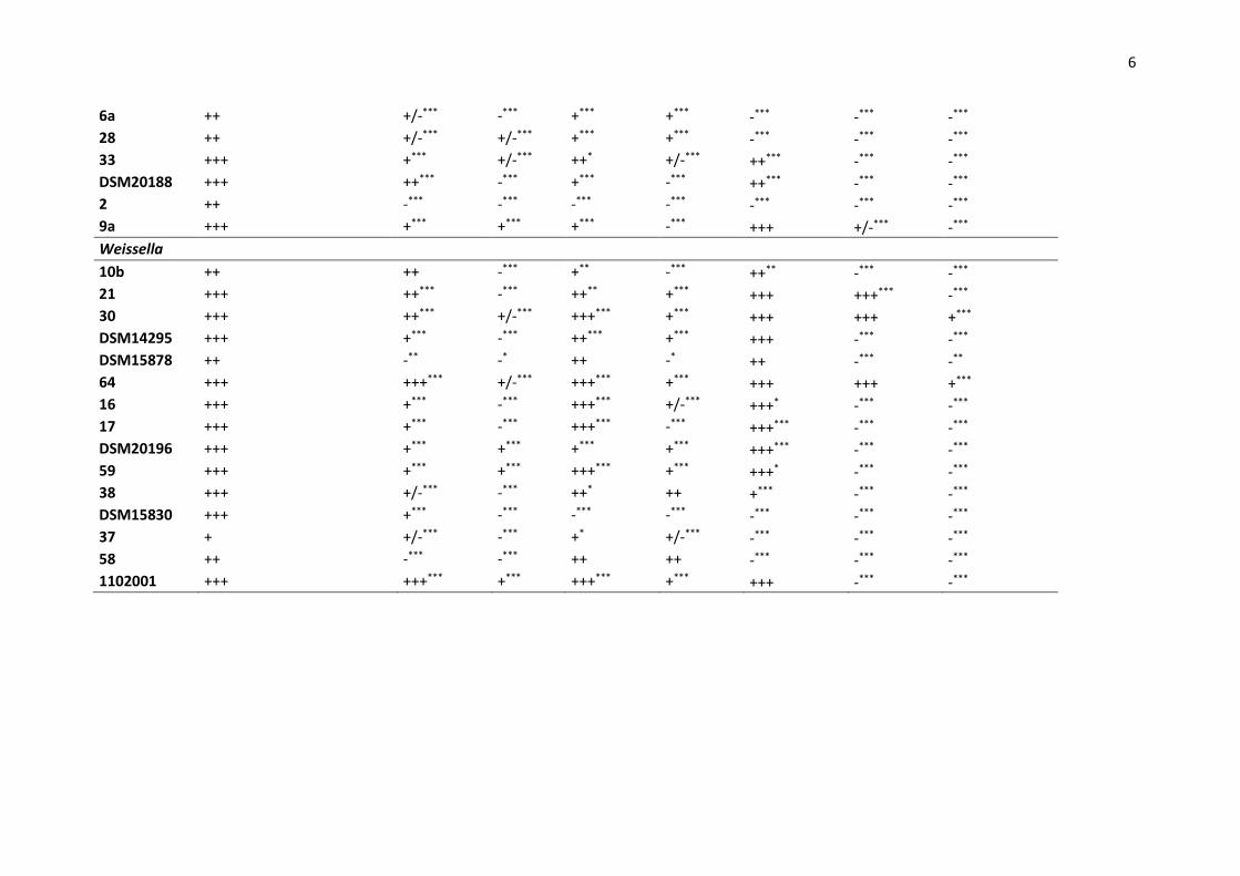
Table 3. Tolerance of isolates to pH, to sodium chloride and to hydrogen peroxide. Results are indicated as follows: (+++) high growth, (++) moderate growth,
(+) poor growth, (+/-) very poor growth and (-) no growth. Significant differences are calculated from Log OD48/OD0 values in acidic or osmotic or oxidative
conditions and indicated as follows: *** p < 0.001, ** p < 0.01, * < 0.05 from Dunnett’s test versus control condition (pH 6.5). No indication of significant
degree means no significant difference (confidence of 95%).
Isolate Control condition pH 4.5 pH 3 NaCl 5% NaCl 8% 0,025% H2O2 0,05% H2O2 0,1% H2O2
Fructobacillus
77 ++ +*** -*** +*** +*** ++ ++ ++***
Lactobacillus
73 +++ ++*** +/-*** +++ -*** -*** -*** -***
75 +++ +++*** +*** +++ +++*** +++ -*** -***
17a +++ +++ -*** +++ +*** +++ -*** -***
DSM2601 +++ ++*** -*** -*** -*** -*** -*** -***
Leuconostoc
12b +++ +*** +*** +*** +/-*** -*** -*** -***
27b +++ +*** +*** +/-*** +/-*** -*** -*** -***
56 +++ +*** -*** +*** -*** -*** -*** -***
89 ++ -*** -*** +*** -*** -*** -*** -***
DSM20193 +++ ++*** +*** ++*** -*** +++ ++*** -***
DSM5625 +++ ++* +*** ++** -*** +++ ++** -***
60 ++ -*** -*** +++** +++** ++ ++ ++
39 +++ ++*** +*** ++*** +/-*** +++ -*** -***
78 +++ +*** -*** +*** -*** +*** -*** -***
79 ++ -*** -*** -*** -*** -*** -*** -***
24 ++ +/-** +/-** +** -** -*** -*** -***
1 ++ +/-*** -*** +/-*** -*** -*** -*** -***
5 +++ +/-*** -*** -*** -*** -*** -*** -***
6
6a ++ +/-*** -*** +*** +*** -*** -*** -***
28 ++ +/-*** +/-*** +*** +*** -*** -*** -***
33 +++ +*** +/-*** ++* +/-*** ++*** -*** -***
DSM20188 +++ ++*** -*** +*** -*** ++*** -*** -***
2 ++ -*** -*** -*** -*** -*** -*** -***
9a +++ +*** +*** +*** -*** +++ +/-*** -***
Weissella
10b ++ ++ -*** +** -*** ++** -*** -***
21 +++ ++*** -*** ++** +*** +++ +++*** -***
30 +++ ++*** +/-*** +++*** +*** +++ +++ +***
DSM14295 +++ +*** -*** ++*** +*** +++ -*** -***
DSM15878 ++ -** -* ++ -* ++ -*** -**
64 +++ +++*** +/-*** +++*** +*** +++ +++ +***
16 +++ +*** -*** +++*** +/-*** +++* -*** -***
17 +++ +*** -*** +++*** -*** +++*** -*** -***
DSM20196 +++ +*** +*** +*** +*** +++*** -*** -***
59 +++ +*** +*** +++*** +*** +++* -*** -***
38 +++ +/-*** -*** ++* ++ +*** -*** -***
DSM15830 +++ +*** -*** -*** -*** -*** -*** -***
37 + +/-*** -*** +* +/-*** -*** -*** -***
58 ++ -*** -*** ++ ++ -*** -*** -***
1102001 +++ +++*** +*** +++*** +*** +++ -*** -***

7
Table 4. Optimum growth rate (µopt) and cardinal growth temperatures (Topt, Tmin and Tmax respectively for optimum, minimal and maximal temperatures).
Results represents the predicted values calculated by Sym’Previus software from three independent experiments. Mean ± standard deviation (SD) is shown.
R² indicates the goodness of fit to the model. Tmin and Tmax observed in 21 days correspond to the minimal and maximal temperature, respectively, at which a
significant growth was observed after 21 days of incubation.
Species Isolate µopt (h-1) ± SD1 Topt (°C) ± SD Tmin (°C) ± SD Tmin observed
in 21 days Tmax (°C) ± SD
Tmax observed
in 21 days R²
Fb. tropaeoli 77 0.217 ± 0.047 33.8 ± 3.7 3.7 ± 6.6 10 43.4 ± 1.9 42 0.768
Lactobacillus 73 0.301 ± 0.038 32.9 ± 1.6 14.9 ± 2.5 6 45.7 ± 3.2 > 45 0.937
75 0.268 ± 0.016 33.0 ± 0.8 8.3 ± 1.1 8 46.5 ± 1.4 42-45 0.954
17a 0.318 ± 0.036 33.9 ± 1.7 5.5 ± 3.5 8 42.1 ± 0.2 42-45 0.92
DSM 2601 0.298 ± 0.156 34.0 ± 2.4 9.8 ± 4.5 8 40.0 ± 12.8 42-45 0.834
Leuconostoc 12b 0.231 ± 0.040 26.7 ± 3.9 2.4 ± 10.5 2-4 37.9 ± 1.0 < 42 0.98
27b 0.107 ± 0.042 28.7 ± 5.1 10.4 ± 5.8 6 42.6 ± 1.4 42 0.723
56 0.263 ± 0.024 28.8 ± 1.1 10.8 ± 0.7 8 42.5 ± 0.2 < 42 0.998
89 0.265 ± 0.016 29.9 ± 1.4 4.4 ± 2.1 8 42.5 ± 0.2 < 42 0.999
DSM 20193 0.127 ± 0.031 30.6 ± 6.6 4.2 ± 2.7 8 42.7 ± 1.2 < 42 0.782
DSM 5625 0.192 ± 0.031 31.0 ± 1.8 11.1 ± 1.6 8 38.3 ± 1.0 < 42 0.894
60 0.186 ± 0.024 33.3 ± 2.4 4.3 ± 6.5 10 50.3 ± 7.8 42-45 0.979
39 0.229 ± 0.052 27.5 ± 2.7 11.2 ± 4.3 6 43.1 ± 1.1 42 0.984
78 0.205 ± 0.107 31.8 ± 6.4 9.0 ± 6.4 6 37.5 ± 1.6 42 0.787
79 0.223 ± 0.047 27.6 ± 2.3 11.9 ± 1.1 8 42.9 ± 0.9 42 0.86
24 0.288 ± 0.118 29.7 ± 5.4 10.5 ± 4.4 8 42.7 ± 1.7 42-45 0.895
1 0.244 ± 0.049 29.6 ± 4.8 3.6 ± 13.5 < 2 45.6 ± 5.1 42 0.52
5 0.279 ± 0.035 33.2 ± 2.5 1.0 ± 7.4 < 2 43.1 ± 1.0 < 42 0.787
6a 0.202 ± 0.007 28.6 ± 0.6 6.6 ± 1.1 6 43.0 ± 0.2 < 42 0.999
28 0.322 ± 0.062 26.1 ± 3.9 8.1 ± 13.2 8 42.2 ± 0.4 < 42 0.967

8
33 0.231 ± 0.012 25.1 ± 1.5 7.1 ± ND 6 43.1 ± 0.4 < 42 0.995
DSM 20188 0.261 ± 0.007 26.9 ± 0.5 3.5 ± 1.4 6 39.2 ± 0.3 > 42 0.999
2 0.146 ± 0.024 30.5 ± 2.9 6.0 ± 4.7 2 43.6 ± 1.5 < 42 0.821
9a 0.191 ± 0.047 31.8 ± 4.3 6.9 ± 5.5 8 42.9 ± 1.2 < 42 0.731
Weissella 10b 0.571 ± 0.144 30.9 ± 3.8 16.2 ± 8.0 8 44.4 ± 2.6 42-45 0.823
21 0.209 ± 0.050 30.5 ± 4.1 8.7 ± 4.9 6 42.9 ± 1.3 42-45 0.794
30 0.433 ± 0.120 34.8 ± 3.7 8.7 ± 4.9 6 45.1 ± 5.0 42-45 0.726
DSM 14295 0.264 ± 0.031 29.8 ± 2.0 6.7 ± 4.8 8 43.1 ± 0.8 42-45 0.876
DSM 15878 0.255 ± 0.052 31.0 ± 2.4 16.7 ± 2.1 6 42.9 ± 1.1 42-45 0.942
64 0.387 ± 0.034 33.8 ± 1.3 6.7 ± 2.3 6 47.0 ± 2.7 42-45 0.981
16 0.418 ± 0.066 32.1 ± 2.8 12.0 ± 0.8 10 49.3 ± 7.3 42-45 0.881
17 0.998 ± 0.080 37.2 ± 0.9 10.1 ± 0.1 8 43.5 ± 0.8 42-45 0.993
DSM 20196 0.199 ± 0.040 33.0 ± 4.5 1.8 ± 20.9 8 42.5 ± 0.9 42-45 0.886
59 0.601 ± 0.185 39.0 ± 4.0 0.6 ± 6.5 6 43.1 ± 4.2 42-45 0.981
38 0.336 ± 0.120 30.5 ± 4.3 8.4 ± 9.2 10 48.2 ± 8.8 42-45 0.599
37 0.148 ± 0.035 29.7 ± 3.6 7.2 ± 6.0 8 43.0 ± 1.3 42-45 0.693
58 0.264 ± 0.570 34.2 ± 10.8 16.2 ± 5.7 6 38.1 ± 13.7 42-45 0.749
1102001 0.359 ± 0.087 31.0 ± 2.5 10.7 ± 2.5 10 47.8 ± 5.7 42-45 0.866
1SD: Standard deviation.


![Lactic Acid - NIHSLactic Acid 乳酸C3H6O3:90.08 (2RS)-2-Hydroxypropanoic acid [50-21-5] Lactic Acid is a mixture of lactic acid and lactic an-hydride. It contains not less than 85.0z](https://static.fdocuments.net/doc/165x107/6107906c3f161736212a9570/lactic-acid-nihs-lactic-acid-ec3h6o39008-2rs-2-hydroxypropanoic-acid.jpg)


