GENERAL ARTHROLOGY - is.muni.cz · Obvious are theoretical knowledges of the general arthrology,...
47
GENERAL ARTHROLOGY RNDr. Michaela Račanská, Ph.D. Lecture 7 – DENTISTRY – Autumn 2013
Transcript of GENERAL ARTHROLOGY - is.muni.cz · Obvious are theoretical knowledges of the general arthrology,...

GENERAL ARTHROLOGYRNDr. Michaela Račanská, Ph.D.
Lecture 7 – DENTISTRY – Autumn 2013

Skeletal junctionsJuncturae seu Systema articulareTwo main types of connections:
1. Synathrosis /fibrous joint, fluent connection/ - union by some kind of theconnective tissue
(fibrous tissue, cartilage, bone)
2. Diarthrosis /synovial joint, connection by touch/ - union by touch (by articularsurfaces and another additional features)

Fibrous joint (synarthrosis)Continuous connections by a layer of connective tissue between bonesnearly immobileThe articulare surface are missing! Differentiation according the type of connective tissue
1) Syndesmosis – articulatio fibrosa, bones are joined by fibrous tissue
2) Synchondrosis – articulatio cartilaginea, bones are joined by cartilage
3) Synostosis – articulatio ossea, bones are joined by bone tissue

Syndesmosis (art. fibrosa)1) connective tissue (ligaments), band of collagen fibrous tissue, (like a rope, ribbon or flat membrane)
Source: http://medical-dictionary.thefreedictionary.com/gomphosis
Source: anatomie Čihák
2) wedging (gomphosis): fixation of tooth to the alveolus
3) sutures between flat skull bones (suturae). The main types of sutures:serrated suture (sutura serrata)squamous suture (sutura squamosa),flat suture (sutura plana)

Synchondrosis (art. cartilaginea)bones are joined by cartilage
Connection using hyaline cartilage (connection of ribs and sternum, between bones of the skull base- in child)
connection using fibrous cartilage (SYMPHYSIS)(intervertebral discs, pubic symphysis (symphysis pubica) between both pelvic bones
Source:http://www.wikiskripta.eu/index.php/Soubor:Gray394.png

Synostosis (art. ossea)bones are joined by bone tissue, for example synostosis sphenooccipitalisConnection of the bones using the bone tissue, the result is growing of two or more bones
Exapmles: sacral bone, coccygeal bone, coxal bone, some skull bones
In adulthood: synostosis of skull sutures - physiological, pathological

DIARTHROSIS (junctura synovialis, articulatio)
Articulation (joint) is movable union of two or more bones by touch
of contact articular surfaces covered by the articular cartilage.

Articular surfaces=facies articulares
(articular fossa=fossa articularis, articular head=caput articulare)
Joint capsule=capsula articularis
(stratum fibrosum and stratum synoviale)
Joint cavity=cavitas articularis
articular fissure filled by synovial fluid (synovia)
Synovial fluid (synovia) – nourishes an articular cartilage,
increases adhesion and decreases friction of contact surfaces
(plicae) or (villi) – are folds of the synovial layer of the articular capsule
and increase inner surface of articular capsule (capsula articularis)
Articular network (rete articulare) – plentiful supplying of joint by arteries, veins and nerves
Special joint apparatus
General features of a joint

Additional features of the jointsa) labrum articulare – fibrocartilaginous ring - broadening of a shallow articular fossa by a stripof cartilage
b) articular discs and meniscs (disci and menisci articulares) – plates of cartilage, which servesas elastic pads, discs divid the articular cavity into two parts, menisci only partly
c) ligaments (ligamenta) are present in the most joints as extracapsular, capsular orintracapsular ligaments
d) articular muscles (musculi articulares) – prevent of a strangulation of articular capsules
e) bursae and synovial pockets (bursae synoviales) – are small cavities close to the joint. Theyare constructed by synovial membrane and synovial fluid. Usually may communicate with thejoint cavity.
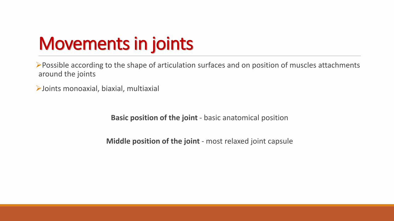
Possible according to the shape of articulation surfaces and on position of muscles attachmentsaround the joints
Joints monoaxial, biaxial, multiaxial
Basic position of the joint - basic anatomical position
Middle position of the joint - most relaxed joint capsule
Movements in joints
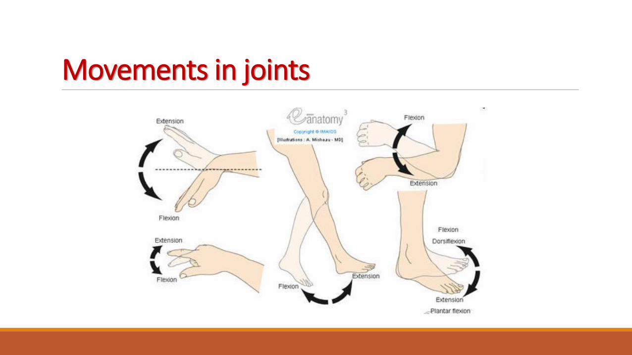
Movements in joints


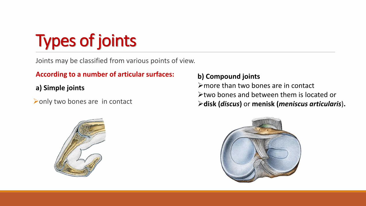
Types of jointsJoints may be classified from various points of view.
According to a number of articular surfaces:
a) Simple joints
only two bones are in contact
b) Compound jointsmore than two bones are in contacttwo bones and between them is located ordisk (discus) or menisk (meniscus articularis).

Classification of joints according to the shape ofarticular surfaces:
spheroidal joint (ball-and-socket joint) (articulatio spheroidea) – head has shape like a sphere or its part),free spheroid joint (arthrodia)spheroid joint with restricted movements (enarthrosis)
ellipsoidal (condyloid) joint (articulatio ellipsoidea)
cylindrical joint:
pivot joint (trochoid) (articulatio trochoidea), wheel joint - the axe of movement is parallel with thelongitudinal axe of bone
hinge joint (articulatio trochlearis); ginglymus - the axe of movement is in the right angle to thelongitudinal axe of bone
saddle joint (sellar) (articulatio sellaris)
plane joint (articulatio plana)
amphiartrosis

AMPHIARTROSIS
ART. PLANA

ARTHRODIA ENARTHROSIS
CYLINDRICAL JOINT:
HINGE JOINT PIVOT JOINT
BALL AND SOCKET

ART. ELLIPSOIDEA (CONDYLOID)
SADDLE JOINT
No rotation!Movements according to the long axis

Classification of joints according to thelevel of moveability and number ofaxis of movements:
Joints with minimal movement:With irregular surfaces – amphiarthrosis
Joints with sliding movements:-Flat joints - articulatio plana
Joints with rotational movements:-Joint surfaces allow rotation along one to three axis One-axis joints (art. cylindroidea and art. trochlearis)Two-axis joints (art. ellipsoidea and art. sellaris)Triaxial joints (art. sphaeroidea)

How to describe jointsObvious are theoretical knowledges of the general arthrology, the knowledges of the special
osteology is obvious.
We are following this outline :
1. Name of the joint,
2. Names of the articular surfaces,
3. Characteristic of the joint capsule
4. Joint auxiliary equipmnet,
5. Type of the joint,
6. Movements in the joint.
An integral part is the description of the joints at the plain x-rays in sagittal and lateral projection
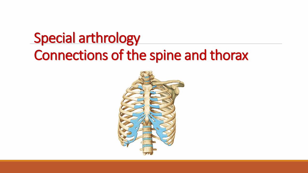
Connections of the spine and thoraxSpecial arthrology

Spine (columna vertebralis)We can observe all types of junctiones on the spineSynartroses and diarthroses as well
Synarthrosis- syndesmosis- ligaments- synchondrosis- disci intervertebrales
- synchondrosis sacrococcygea- synostosis- os sacrum, os coccygis
Diarthrosis- articulationes intervertebrales
Junctions of the spine
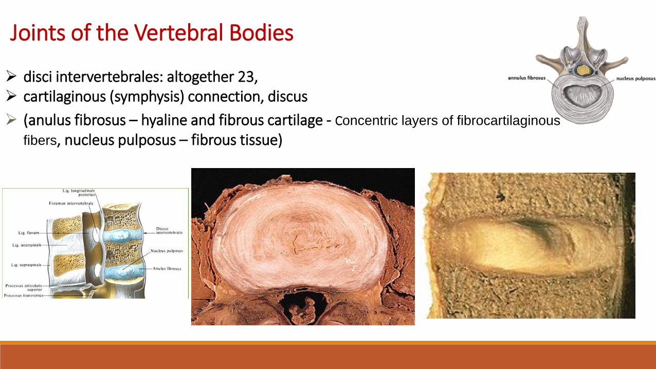
Joints of the Vertebral Bodies
disci intervertebrales: altogether 23, cartilaginous (symphysis) connection, discus
(anulus fibrosus – hyaline and fibrous cartilage - Concentric layers of fibrocartilaginous
fibers, nucleus pulposus – fibrous tissue)

Junctions of vertebral arches- elastic ligaments – ligamenta flava (interarcualia)

Junctions of articular processes of vertebrae•articulationes intervertebrales
between the superior and inferior articular processes of
adjacent vertebrae - zygapophysial/facet joints
sliding movementsangulations of the articular facetsdetermine types of movements- short ligaments:- ligg. intertransversaria- ligg. interspinalia- lig. supraspinale (cervical area) – as sagitally oriented ligamentum nuchae which is goingto the occipital bone

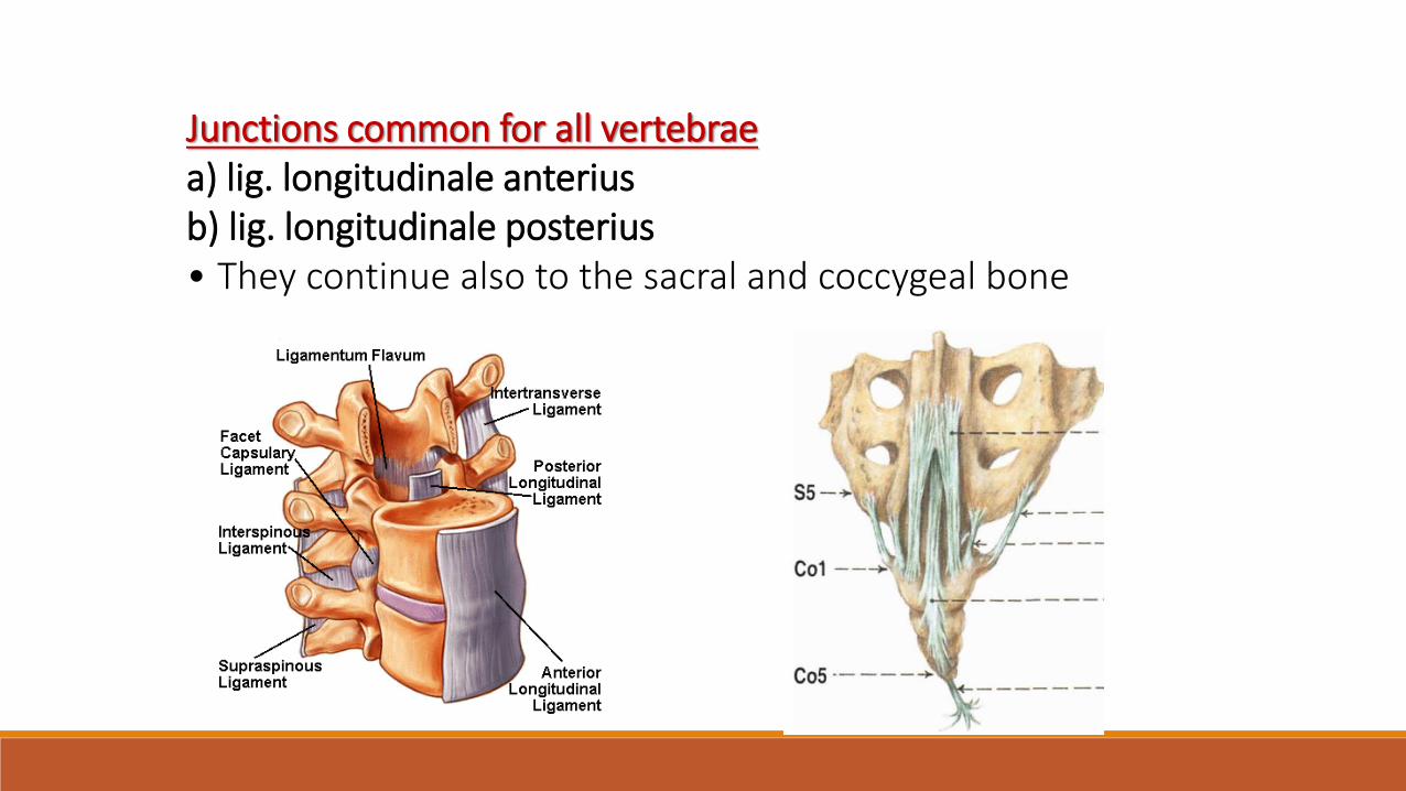
Junctions common for all vertebraea) lig. longitudinale anteriusb) lig. longitudinale posterius• They continue also to the sacral and coccygeal bone

Synostosis• Conection using the bone tissue• Sacral bone: fusion of five sacral vertebrae• Coccygeal bone: fusion of 3 - 5 coccygeal vertebrae

Curvature of vertebral column
1. In the sagittal plane- double S-shaped:lordosis: curvature forwards, cervical C4-5 and lumbar L3-4kyphosis: curvature backwards, thoracic Th6-7 and sacral
2. Curvature in the frontal plane- Skoliosis, mild skoliosis is physiologicaland it is present in all people – in most mild right, in some mild left (if you are right or left-handed)

SHAPE AND MOVEMENTS OF THE SPINE- 35% of body heightMovements• anteflexion, retroflexion, 90° cervical, 23° lumbar, most
stressed and vulnerable is part of the lower cervical vertebrae, Th11-12, L4-S1
• lateroflexion, 30° cervical, 35° lumbar• Rotation and torsion, 60-70° cervical, 25-35° thoracic• Springing movements
Mobility of the vertebral column– depends on the size of intervertebral disc– the mobility is rectricted by: ligaments, articular capsules
and muscles

the cervical vertebrae allow a range of flexion, lateroflexion and rotation coupled with lateroflexion
the thoracic should be particularly mobile in rotation (is limited by the attachment of ribs)
in the lumbar region - anteflexion, retroflexion, lateral flexion

Junctions of the thorax costovertebral joints
art. capitis costae
art. costotransversarium
costochondral joints and interchondral joints
artt. interchondrales (6th-9th)
membrana intercostalis externa, interna
sternocostal joints
artt. sternocostales (2nd-5th)
synchondrosis (1st, 6th, 7th)

Costovertebral Joints
Articulationes capitis costae
AF: head of the rib articulates with the inferior and
superior costal facets of two adjacent thoracic vertebral bodies and the intervening intervertebral disc
AC: firm and it is attached to the margins of AF
special apparatus: lig. capitis costae radiatum, at 2nd – 10th rib: lig. capitis costae intraarticulare
movements: along axis parallel with the neck of the rib
allow elevation and depression of the ribs

Articulationes costotransversariaeAS: foveae costales transversales and art. surface on tuberculum costaeAC: margins of the articular surfacesspecial apparatus: lig. costotransversaria, between collum costae and transversalproccess of the vertebraMovements: along axis which is parallel with collum costae

laterale
laterale

Caput costae + lig.radiatum
Lig.costotransversariumsuperius

Lig.capitis costae radiatum
Lig. costotransv.sup.
Lig. longitudin. ant.

Lig.costotransv.lat.superius
Lig. intertransversariainterspinale (nuchae)

Juncturae sternocostales• Connections between costal cartilages and sternum
1. Synchondrosis sternocostalis: cartilaginous connection with incisuracostalis sterni, regularly at 1st often at 6th and 7th rib
2. Artt. sternocostales:between 2nd to 5th rib and sternumAS: sternal end of costal cartilage, incisura costalissterniAC: to the margins of the articular surfacesSpecial apparatus: ligg. sternocostalia radiata – theyform membrana sterni externa and interna
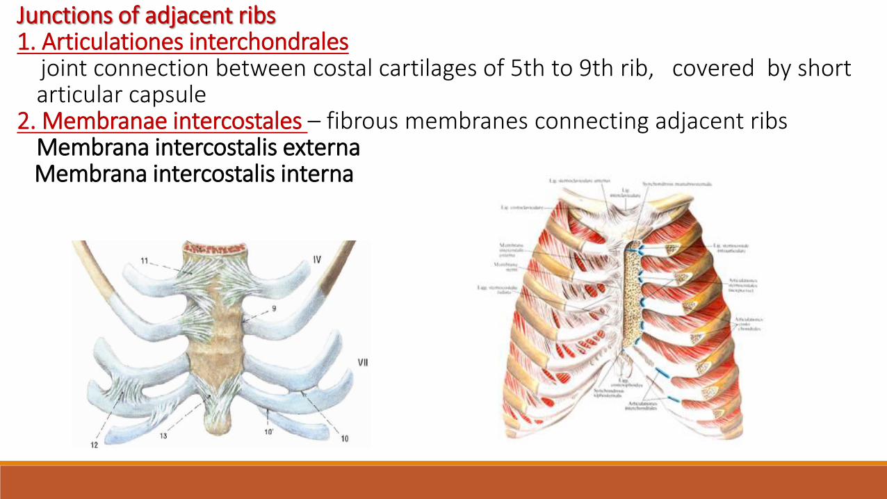
Junctions of adjacent ribs1. Articulationes interchondrales
joint connection between costal cartilages of 5th to 9th rib, covered by shortarticular capsule
2. Membranae intercostales – fibrous membranes connecting adjacent ribsMembrana intercostalis externaMembrana intercostalis interna

Chest cage shape and movements
• Shape of truncated cone• base (apertura thoracis inferior)• apex (apertura thoracis superior)• walls – frontal, dorsal, lateralcavitas thoracisspatia intercostaliaarcus costarumangulus infrasternalis
Movements• in costovertebral connections, axis runs parallel with collum costae• Upward rotation - inspirium
downward rotation- exspirium

Movements of the thoracic wall during inspiration
produce increases in the intrathoracic volume and
diameters of the thorax

Illustrations were copied from:
Atlas der Anatomie des Menschen/Sobotta. Putz,R., und Pabst,R. 20. Auflage. München:
Urban & Schwarzenberg, 1993)
Netter: Interactive Atlas of Human Anatomy. Windows Version 2.0
Čihák R: Anatomie 2 (Splanchnologia). Avicenum, zdravotnické nakladatelství, Praha, 1988.
Drake et al: Gray´s Anatomy for Students. 2010
Archiv of the lecturer