Gallstone disease, Cholangitis, Cholecystectomy, Common...
69
GALLSTONE DISEASE
Transcript of Gallstone disease, Cholangitis, Cholecystectomy, Common...

GALLSTONE DISEASE
TOPICS
Some physiology
Biliary Colic
Acute cholecystitis
Chronic cholecystitis
Cholecystectomy
Cholangitis
Obstructive jaundice
Common bile duct exploration & ERCP

BILE FORMATION
Continuous formation by the liver
Bile flow directly to gallbladder with intact Sphincter of Oddi – bile concentration & storage
Greatest absorptive power per unit area of any structure in the body – permits low pressure state in biliary tree
Bile composition
Water, electrolytes, lipids (cholesterol & phospholipids from liver), bile salts (from cholesterol), proteins, bile pigments
500-1000mL produced per day
Enterohepatic circulation – 80% of conjugated bile acids absorbed in terminal ileum

BILE SECRETION
80% of secreted bile stored in gallbladder in
fasting state
Fasting phase: motilin stimulates
Neurogenic, humoral, chemical stimuli
Vagal stimulation
Secretin from duodenum (released in response to
partially digested fatty acids & proteins)
Gallbladder secretes:
Glycoproteins – protection of mucosal
Hydrogen ions – lowers pH to increase Ca solubility
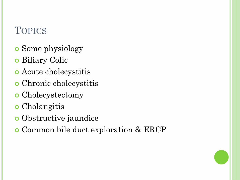
BILE SECRETION CONT...
Neurohormonal control of contraction: Gastric
distension; Vagal stimulation; Peptides
CCK
Released when stimulated by acid, fat & amino acids
into duodenum
from epithelial cells of upper GI tract directly causes:
gallbladder contraction
relaxation of distal CBD & sphincter of Oddi
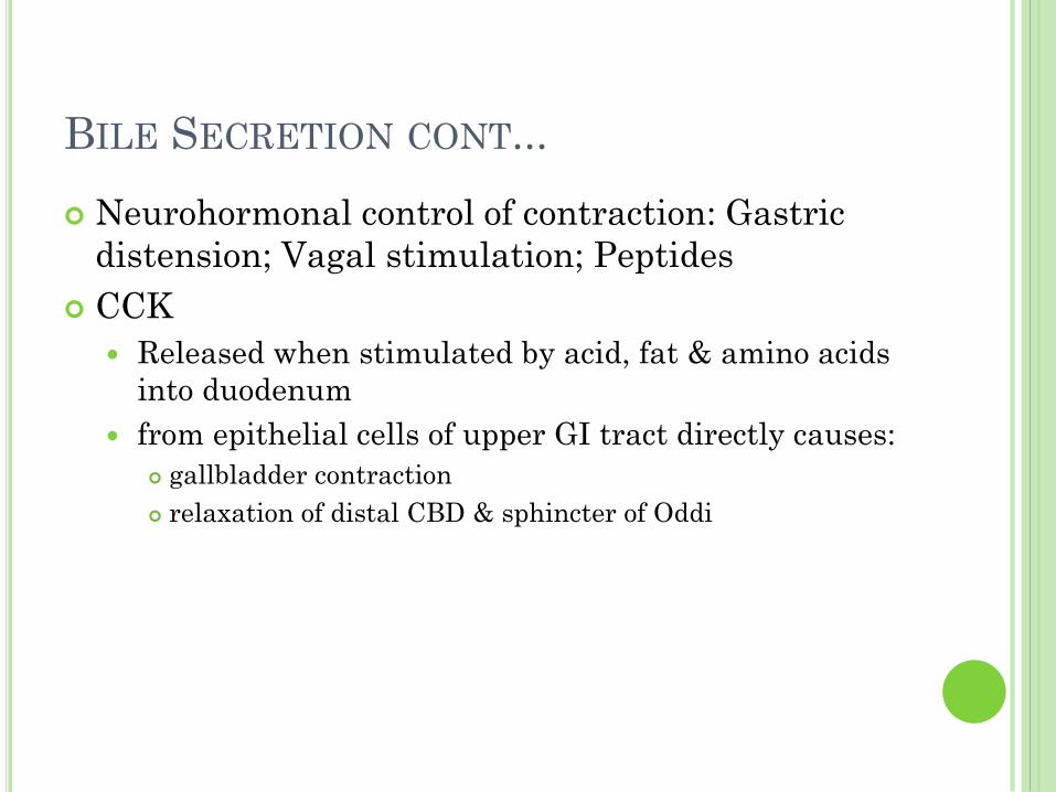
STONE FORMATION
Very simply – solute settling out of solution
Main solutes in bile:
Bile salts, bilirubin, phospholipids, cholesterol
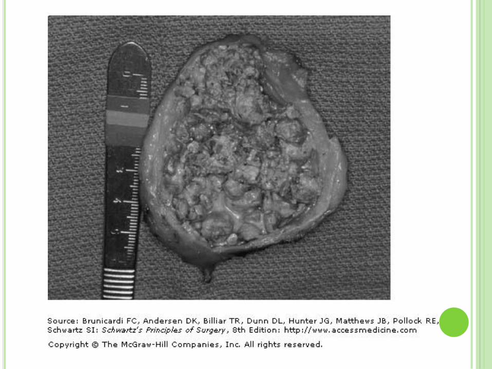
Cholesterol stones (~80%)
Usually ~70% cholesterol by
weight
Pigment stones (~20%)
Black (~15-20%)
Brown (<5%)
CHOLESTEROL STONES
Most are radioluscent (hence decreased utility of
X-rays in diagnosis)
Solubility related to relative concentrations of
cholesterol, bile salts & lecithin (main
phospholipid)
Formed from supersaturation of cholesterol in
bile, mostly in gallbladder
Supersaturation mostly due to cholesterol
hypersecretion (vs low phospholipids or bile salts)

BLACK PIGMENT STONES
Supersaturation of calcium bilirubinate,
carbonate, and phosphate
Small, brittle black stones, mostly in gallbladder
Deconjugation of bilirubin normally at slow rate
Unconjugated bilirubin much less soluble than
conjugated
If large amounts of conjugated bilirubin, have
increased production of deconjugated bilirubin
Hemolytic disorders (hereditary spherocytosis,
sickle cell disease), cirrhosis

BROWN PIGMENT STONES
<1 cm in diameter, brownish-yellow, soft, often mushy
Formed in gallbladder as well as bile ducts – due to stasis Stones mainly composed of precipitated calcium
bilirubinate and bacteria Some bacteria (ex E. coli) secrete beta-glucuronidase –
cleaves bilirubin glucuronide to produce insoluble unconjugated bilirubin
Precipitates with calcium and bacteria – forms brown stones
Asian populations: associated with stasis secondary to parasite infection
Western population: primary bile duct stones in patients with Biliary strictures Other CBD stones that cause stasis and bacterial
contamination
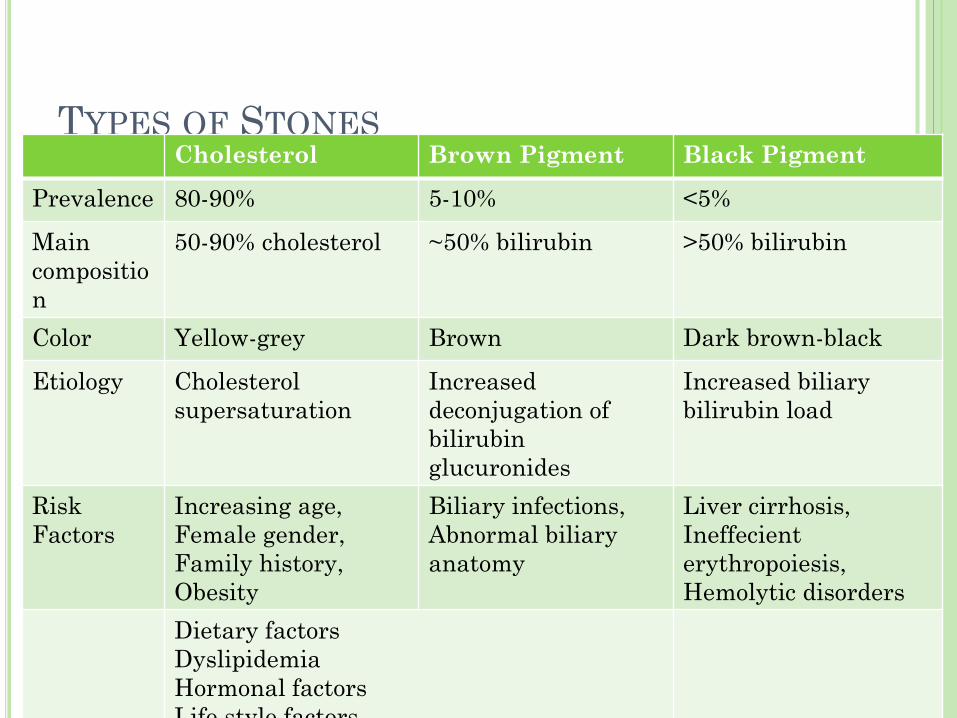
TYPES OF STONES Cholesterol Brown Pigment Black Pigment
Prevalence 80-90% 5-10% <5%
Main
compositio
n
50-90% cholesterol ~50% bilirubin >50% bilirubin
Color Yellow-grey Brown Dark brown-black
Etiology Cholesterol
supersaturation
Increased
deconjugation of
bilirubin
glucuronides
Increased biliary
bilirubin load
Risk
Factors
Increasing age,
Female gender,
Family history,
Obesity
Biliary infections,
Abnormal biliary
anatomy
Liver cirrhosis,
Ineffecient
erythropoiesis,
Hemolytic disorders
Dietary factors
Dyslipidemia
Hormonal factors
Life style factors
Medications
Adapted from J Intern Med 2007
Marschall & Einarsson
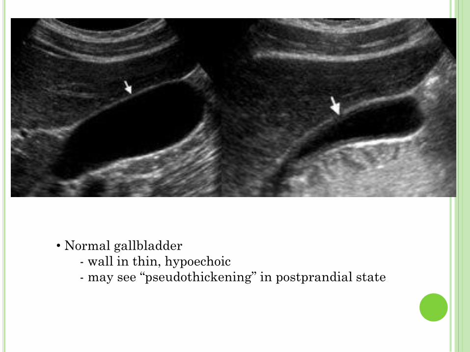
• Normal gallbladder
- wall in thin, hypoechoic
- may see “pseudothickening” in postprandial state
CASE 1
38 female presents to office with recurrent
history of epigastric & RUQ pain, consistently
post-prandial, although not after each meal;
sometimes radiates to the back
Associated with nausea & vomitting
In the past went to walk-in clinic: blood tests
have not shown any abnormalities
Has occasionally taken Abx but is now tired of
the pain and would like definitive treatment
BILIARY COLIC
Definition – pain related to cystic duct
obstruction - very variable: classically RUQ,
aching, post-prandial pain lastly ~ 3 hours
Approximately 3% of people with gallstones will
become symptomatic per year
Once symptomatic, tend to have recurring bouts
Complicated gallstone disease occurs in ~3% of
symptomatic patients per year
I.e. Not common to experience complications from
gallstone disease if not already symptomatic
CLINICAL ETIOLOGY OF BILIARY COLIC
Neurohormonal control of contraction: Gastric
distension; Vagal stimulation; Peptides
CCK – from epithelial cells of upper GI tract –
directly causes gallbladder contraction +
relaxation of sphincter of Oddi
Contraction against obstruction
Gallbladder ischemia from prolonged contraction
Length classically coincides with length of time of
gastric emptying – ie ~3 hours

DIAGNOSIS
History
Classic history vs. not – large range of clinical
presentations
Physical
Usually normal between bouts
Epigastric and/or RUQ pain during bouts
Labs
CBC, ALT, AST, ALP, GGT, Bilirubin
Labs usually normal – no infection; no obstruction
Imaging
U/S, ERCP, EU/S, MRCP, CT, PTC

IMAGING STUDIES
Ultrasound
Shows stones in the gallbladder: sensitivity &
specificity of >90%
(stones acoustically
dense & show
acoustic shadow)

TREATMENT
Symptomatic relief
Analgesia & antiemetics
Definitive treatment
Laparoscopic cholecystectomy
Avoid large meals and fatty foods while waiting for surgery
Diabetic patients – more prone to serious complications
Pregnant patients – can safely undergo surgery if conservative management unsuccessful – open if 3rd trimester
~90% have relief of symptoms post-op – more successful for patients with stones on U/S & typical symptoms

CASE 2
48 male presents in ED with ongoing severe RUQ
pain, radiating to the back
Sharp RUQ pain going on for several hours,
started after dinner out with his wife
Wife documented fever of 38.7 C at home
Arrives at ED – nausea, vomiting x 4, looks
unwell
Labs: WBC is elevated, mild ASL & AST
elevation, normal bilirubin
ACUTE CHOLECYSTITIS
Definition – Acute gallbladder obstruction (longer than that which leads to biliary colic) that leads to gallbladder distension, inflammation & edema
Usually inflammation resolves once obstruction resolves
May however, lead to ischemia and necrosis of gallbladder wall & gallbladder perforation (3-15% of people with acute cholecystitis)
Although obstruction is due is gallstones in ~95% of patients, could be acalculous, or due to tumour obstructing cystic duct
Initial inflammatory process potentially mediated by lysolecithin, bile salts and platelet-activating factor
Bacterial infection in about 50% of cases – difficult to predict who will become secondarily infected (E. coli, Klebsiella, Enterococcus, Enterobacter)
Abscess/empyema forms within gallbladder – may see perforation with cholecystoenteric fistula formation
DIAGNOSIS - HISTORY
Constant, dull epigastric and/or RUQ pain &
tenderness; may radiate to back and between
scapulae
Febrile, anorexia, nausea, vomiting
May see more subtle presentation in diabetic and
elderly – also higher mortality in these
populations
Often history of self-resolving biliary colic with
reports that this pain is more severe than “usual”
colic
DIAGNOSIS – PHYSICAL &
INVESTIGATIONS
General Appears unwell & uncomfortable
Reluctant to move – parietal peritoneum irritation
Vitals coincide with severity of illness: tachycardia, fever
Abdominal exam RUQ tenderness, +ve Murphy’s sign
Guarding
May have palpable gallbladder
Labs CBC (normal to moderate WBC – 12-15,000 cells/mm3) –
higher WBC counts could mean more complicated disease (gangrenous cholecystitis, cholangitis)
ALT, AST – may see mild elevation in transaminases
ALP, GGT, Bilirubin – may be elevated such as with Mirizzi’s syndrome
Imaging U/S, ERCP, EU/S, MRCP, CT, PTC
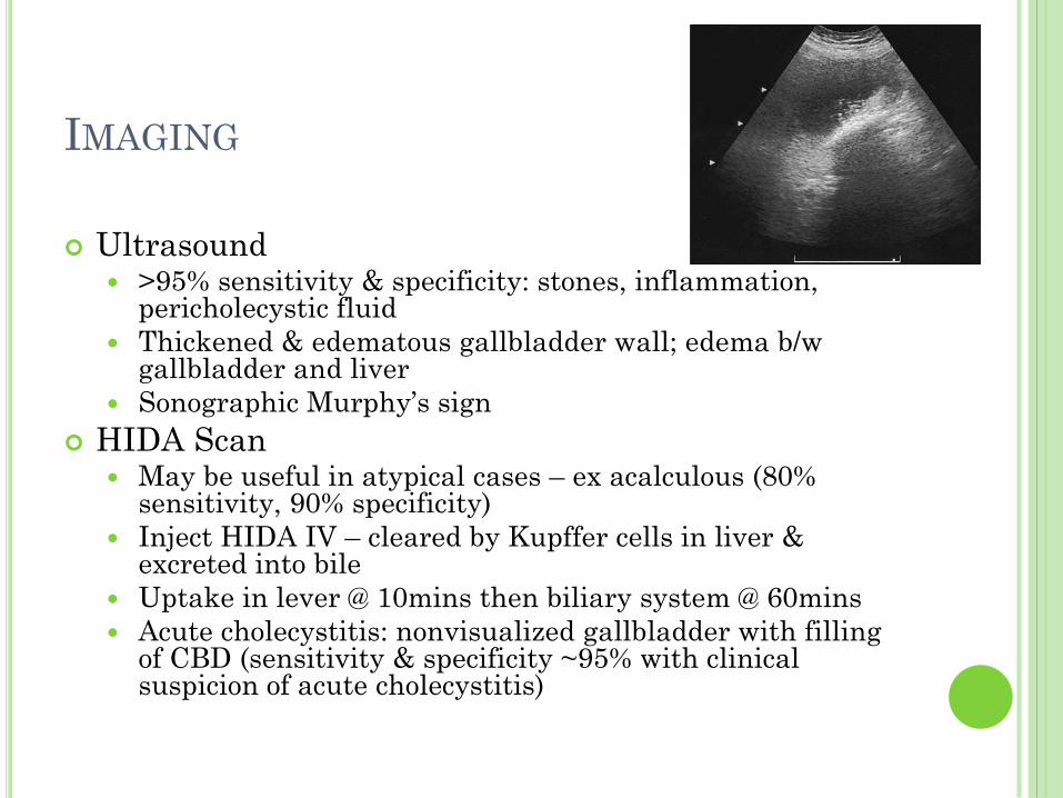
IMAGING
Ultrasound >95% sensitivity & specificity: stones, inflammation,
pericholecystic fluid
Thickened & edematous gallbladder wall; edema b/w gallbladder and liver
Sonographic Murphy’s sign
HIDA Scan May be useful in atypical cases – ex acalculous (80%
sensitivity, 90% specificity)
Inject HIDA IV – cleared by Kupffer cells in liver & excreted into bile
Uptake in lever @ 10mins then biliary system @ 60mins
Acute cholecystitis: nonvisualized gallbladder with filling of CBD (sensitivity & specificity ~95% with clinical suspicion of acute cholecystitis)
TREATMENT
Resuscitation
IV fluids
Analgesia – narcotics
Antibiotics – typically here see Cipro (gram-negatives) + Flagyl (anaerobes)
Cholecystostomy
Goal to drain distended, inflamed gallbladder in those not suited for surgery/unable to tolerate surgery
Future cholecystectomy once recovered and still indicated
Laparoscopic cholecystectomy
within 3 days (preferred over interval surgery after recovery with medical management)
SURGICAL ISSUES
General surgical issues
Tolerance of pneumoperitoneum (cardiorespiratory disease)
Body habitus
Previous surgeries – adhesions, anatomy
Cholecystectomy-specific
Previous history of cholecystitis – may make the surgery
more difficult
Predictors of converting to open: shrunken gallbladder,
thickened gallbladder wall, pericholecystic fluid
Patients at risk for CBD stones – may require pre-op or
intra-op duct imaging
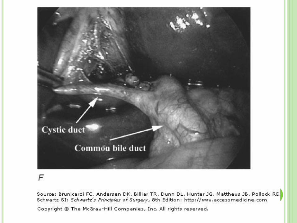
Preoperative planning with imaging: ERCP, EUS, MRCP
SOME HISTORY
First cholecystectomy – 1882 by Dr. Carl
Langenbuch
Laparoscopic cholecystectomy – 1987 by Dr.
Phillipe Mouret
Previous relative contraindications:
Acute cholecystitis, gangrene and empyema of the
gallbladder, biliary-enteric fistulae, obesity,
pregnancy, ventriculoperitoneal shunt, cirrhosis, and
previous upper abdominal procedures
These are now identified as risk factors for
potentially difficult laparoscopic cholecystectomy
BRITISH JOURNAL OF SURGERY 1998
Randomized trial of early versus delayed laparoscopic cholecystectomy for acute cholecystitis
P.B.S. Lai, K.H. Kwong, K.L . Leung, S.P.Y. Kwok, A.C.W. Chan, S.C.S Chung and W.Y. Lau
Previous studies had shown that early open cholecystectomy had shorter hospital stay (with same morbidity & mortality as delayed), but no such trial for laparoscopic technique in treatment of acute cholecystitis
Randomized trail: Early laparoscopic cholecystectomy – withing 24 hours of
randomization
Conservative management (fluids + Abx – amicillin, cefuroxmine, metronidazole) followed by elective laparoscopic cholecystectomy 6-8 weeks later
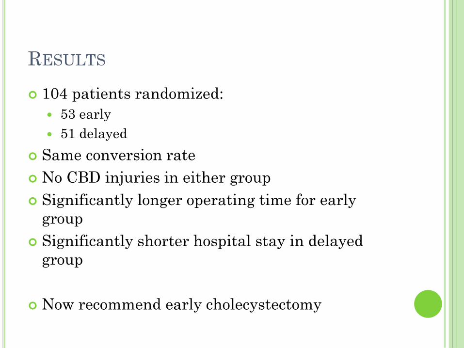
RESULTS
104 patients randomized:
53 early
51 delayed
Same conversion rate
No CBD injuries in either group
Significantly longer operating time for early
group
Significantly shorter hospital stay in delayed
group
Now recommend early cholecystectomy
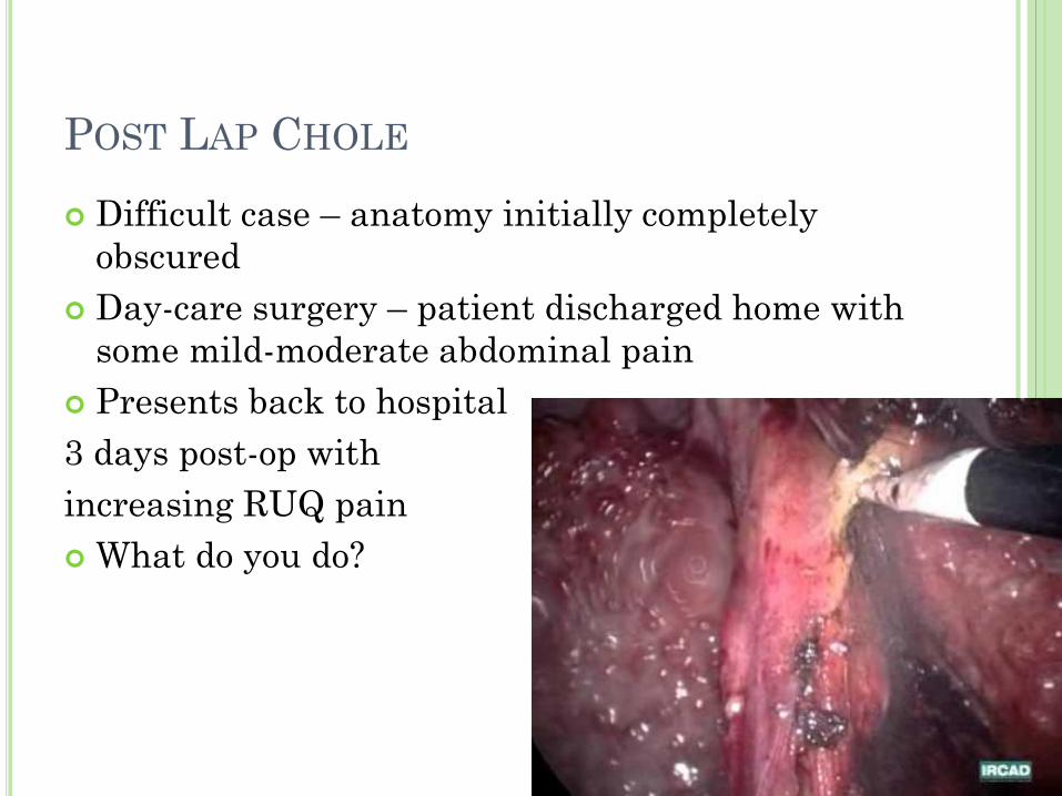
POST LAP CHOLE
Difficult case – anatomy initially completely
obscured
Day-care surgery – patient discharged home with
some mild-moderate abdominal pain
Presents back to hospital
3 days post-op with
increasing RUQ pain
What do you do?
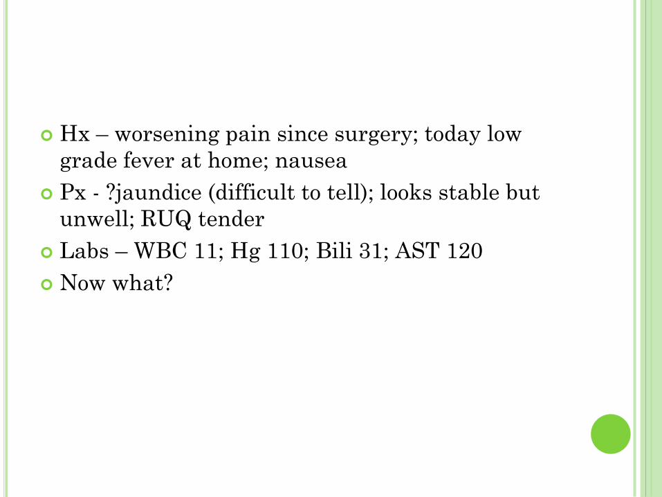
Hx – worsening pain since surgery; today low
grade fever at home; nausea
Px - ?jaundice (difficult to tell); looks stable but
unwell; RUQ tender
Labs – WBC 11; Hg 110; Bili 31; AST 120
Now what?

ULTRASOUND
Biliary collection – how do you approach this?
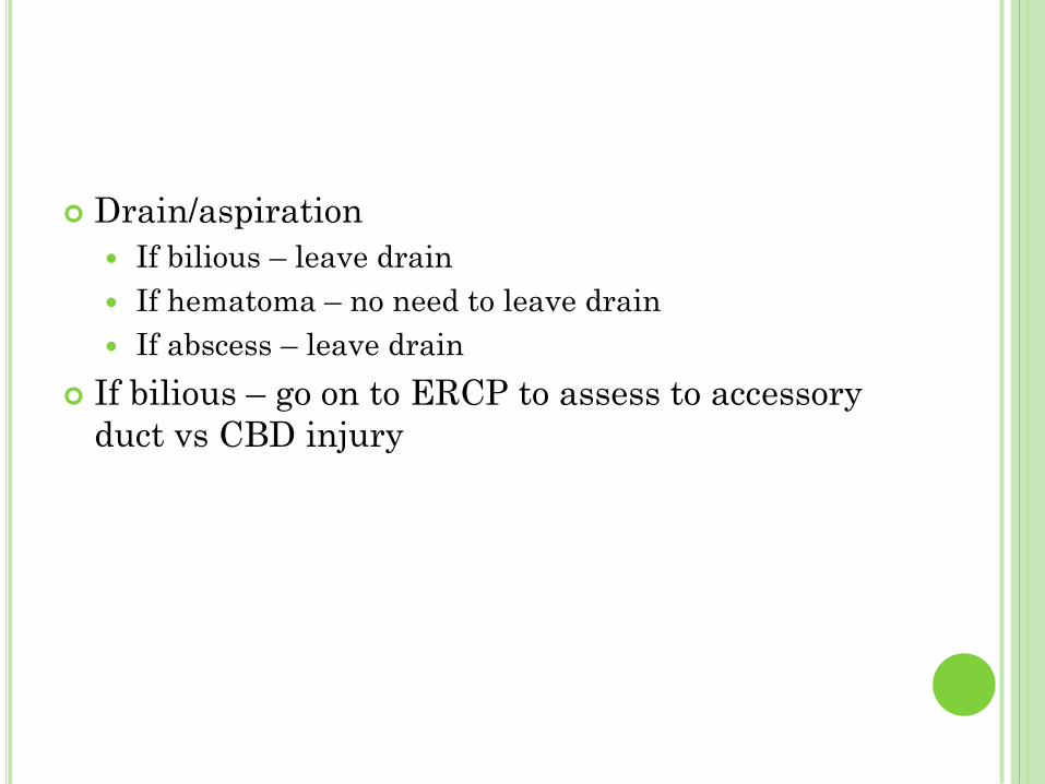
Drain/aspiration
If bilious – leave drain
If hematoma – no need to leave drain
If abscess – leave drain
If bilious – go on to ERCP to assess to accessory
duct vs CBD injury

CASE 3
34 female, teacher
Recurrent attacks of biliary colic
Always subside within 24 hours
Associated with nausea & vomiting
Has been going on for months
Has been to FP several times – normal LFTs, no
episodes of jaundice
Would like definitive treatment
CHRONIC CHOLECYSTITIS
Definition – Ongoing inflammation with
recurrent episodes of biliary colic
About 2/3 of patients present in this way
Various pathological changes to gallbladder –
including scarring & non-funtional gallbladder
Histologically – subepithelial & subserosal
fibrosis + inflammatory cell infiltration
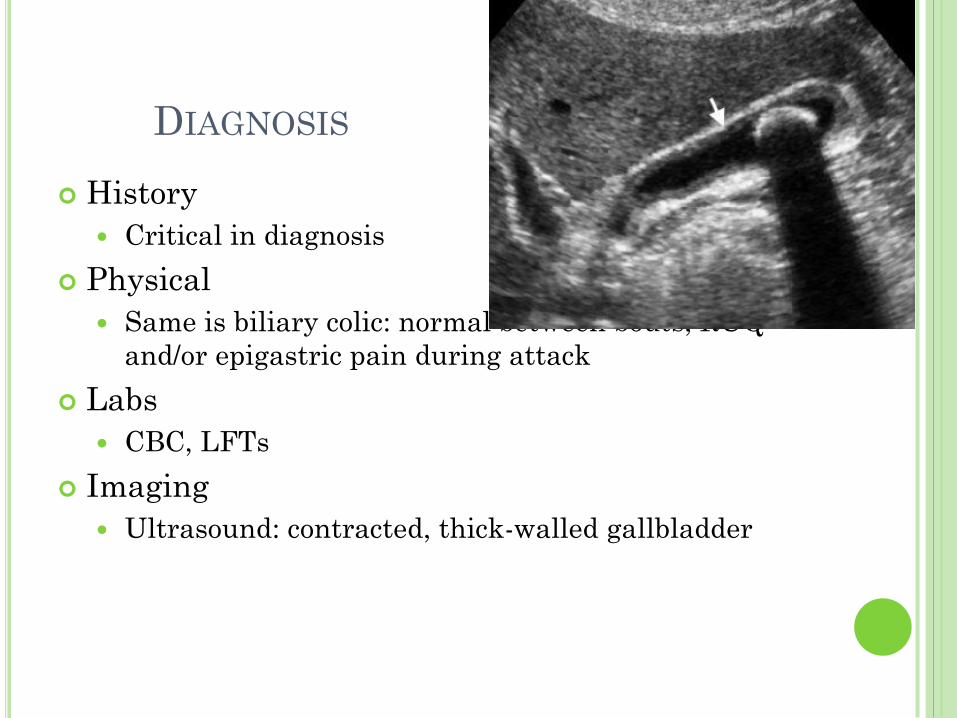
DIAGNOSIS
History
Critical in diagnosis
Physical
Same is biliary colic: normal between bouts; RUQ
and/or epigastric pain during attack
Labs
CBC, LFTs
Imaging
Ultrasound: contracted, thick-walled gallbladder

TREATMENT
Symptomatic management
Analgesia & antiemetics
Elective cholecystectomy
Same recommendations when waiting for surgery as
biliary colic
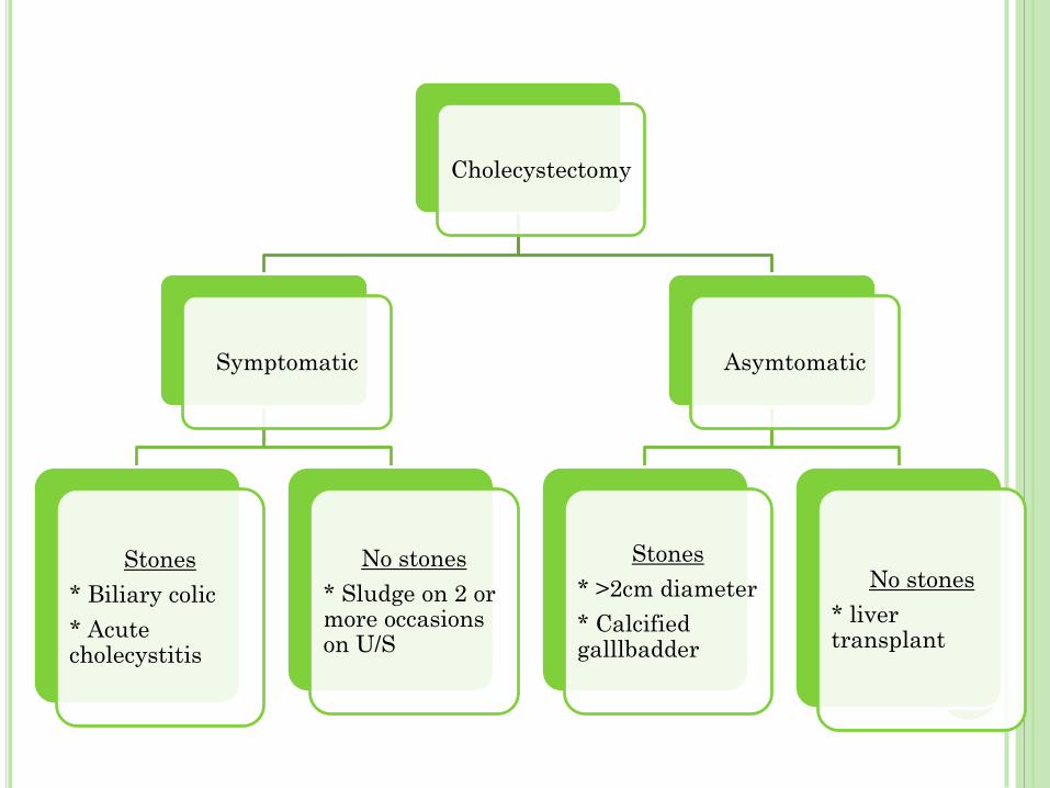
Cholecystectomy
Symptomatic
Stones
* Biliary colic
* Acute cholecystitis
No stones
* Sludge on 2 or more occasions on U/S
Asymtomatic
Stones
* >2cm diameter
* Calcified galllbadder
No stones
* liver transplant
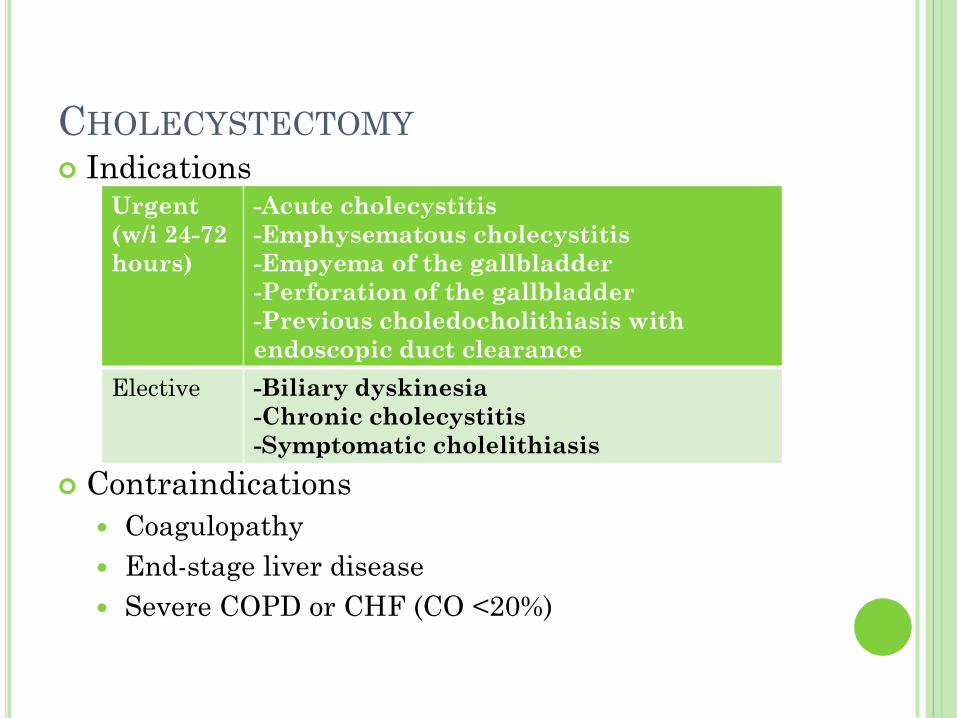
CHOLECYSTECTOMY Indications
Contraindications
Coagulopathy
End-stage liver disease
Severe COPD or CHF (CO <20%)
Urgent
(w/i 24-72
hours)
-Acute cholecystitis
-Emphysematous cholecystitis
-Empyema of the gallbladder
-Perforation of the gallbladder
-Previous choledocholithiasis with
endoscopic duct clearance
Elective -Biliary dyskinesia
-Chronic cholecystitis
-Symptomatic cholelithiasis

CHOLECYSTECTOMY - COMPLICATIONS
General surgical complications
Bleeding
Infection – prophylactic Abx
General anesthetic
Laparoscopic complications
Tolerance of pneumoperitoneum
Cholecystectomy-specific complications
Conversion to open: approximately 5%
Injury to:
Vascular structures
Bowel
Bile duct
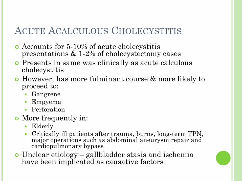
ACUTE ACALCULOUS CHOLECYSTITIS
Accounts for 5-10% of acute cholecystitis presentations & 1-2% of cholecystectomy cases
Presents in same was clinically as acute calculous cholecystitis
However, has more fulminant course & more likely to proceed to: Gangrene
Empyema
Perforation
More frequently in: Elderly
Critically ill patients after trauma, burns, long-term TPN, major operations such as abdominal aneurysm repair and cardiopulmonary bypass
Unclear etiology – gallbladder stasis and ischemia have been implicated as causative factors
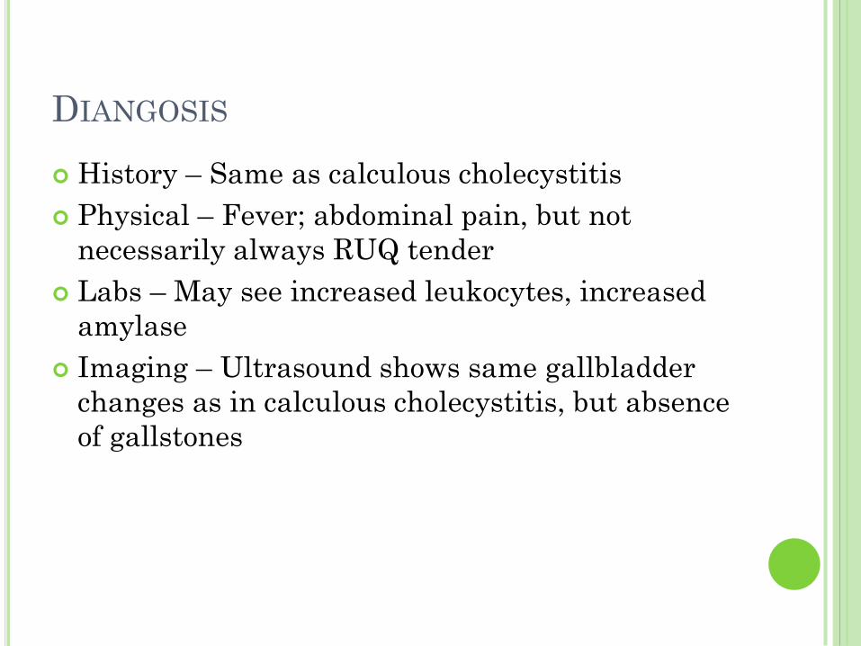
DIANGOSIS
History – Same as calculous cholecystitis
Physical – Fever; abdominal pain, but not
necessarily always RUQ tender
Labs – May see increased leukocytes, increased
amylase
Imaging – Ultrasound shows same gallbladder
changes as in calculous cholecystitis, but absence
of gallstones

TREATMENT
Resuscitation
Fluids
Analgesia
Antibiotics
Surgery
Not usually the first option because usually secondary to underlying severe medical illness
Morbidity rate around 40% because patients often initially critically ill
If unfit for surgery, percutaneous, US or CT-guided cholecystostomy
If diagnosis uncertain, percutaneous cholecystostomy can be both diagnostic and therapeutic - about 90% of patients improve
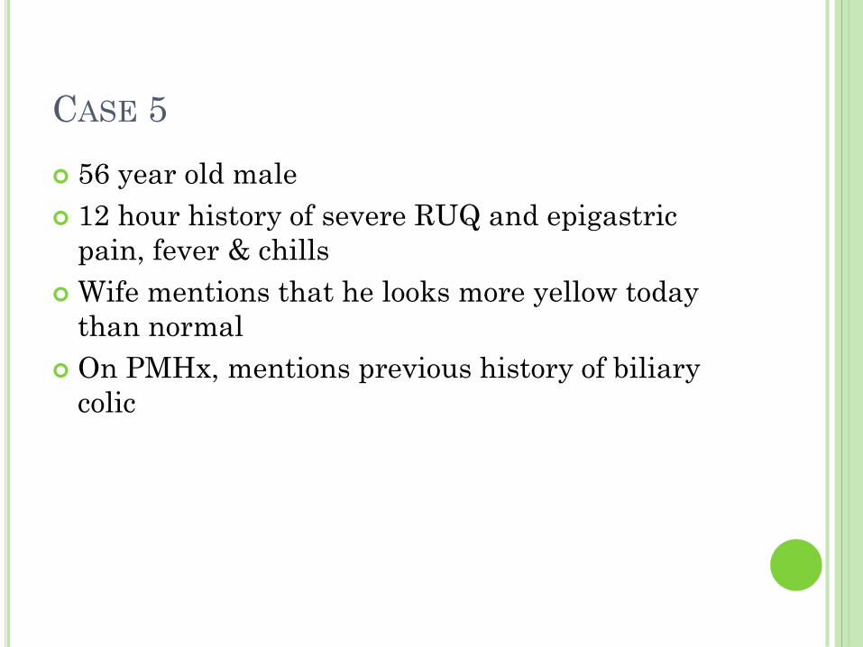
CASE 5
56 year old male
12 hour history of severe RUQ and epigastric
pain, fever & chills
Wife mentions that he looks more yellow today
than normal
On PMHx, mentions previous history of biliary
colic

CHOLANGITIS
Definition – Inflammation of the biliary tree, with ascending bacterial infection associated with partial or complete bile duct obstruction (ie need the obstruction as well as bacterial infection for cholangitis)
One of major complications of gallstone disease (other is gallstone pancreatitis)
Bile normally maintains sterility by constant movement through biliary tree (as well as immunoglobulins)
Most common bacteria: E. coli, Klebsiella pneumoniae, Streptococcus faecalis, and Bacteroides fragilis
Other causes of obstruction:
Benign & malignant strictures
Instrumention
Post-operative anastomotic strictures
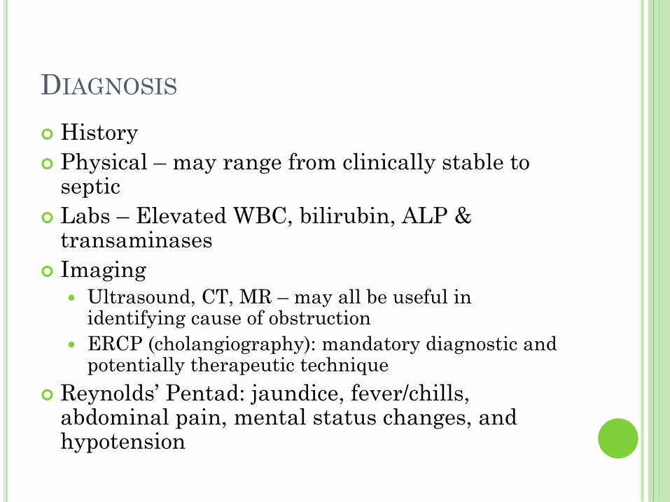
DIAGNOSIS
History
Physical – may range from clinically stable to septic
Labs – Elevated WBC, bilirubin, ALP & transaminases
Imaging
Ultrasound, CT, MR – may all be useful in identifying cause of obstruction
ERCP (cholangiography): mandatory diagnostic and potentially therapeutic technique
Reynolds’ Pentad: jaundice, fever/chills, abdominal pain, mental status changes, and hypotension
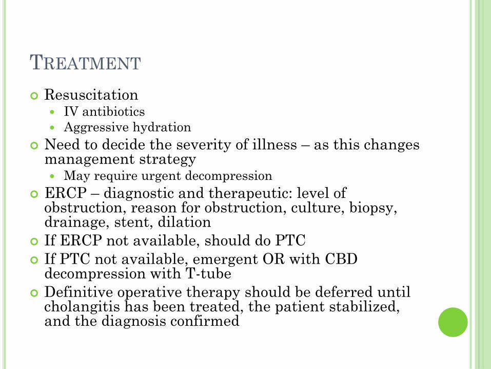
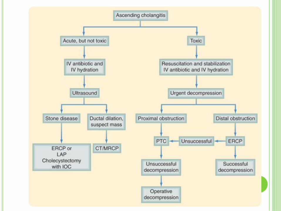
TREATMENT
Resuscitation IV antibiotics
Aggressive hydration
Need to decide the severity of illness – as this changes management strategy May require urgent decompression
ERCP – diagnostic and therapeutic: level of obstruction, reason for obstruction, culture, biopsy, drainage, stent, dilation
If ERCP not available, should do PTC
If PTC not available, emergent OR with CBD decompression with T-tube
Definitive operative therapy should be deferred until cholangitis has been treated, the patient stabilized, and the diagnosis confirmed

Symptomatic Disease
Low Risk (of CBD stones)
No routine cholangiography
Moderate Risk –
*Elevated bilirubin
*Elevated ALP
*Pancreatitis
*Multiple gallstones
MRCP, EUS or intraoperative cholangiogram
High Risk –
*Clinical jaundice
*Cholangitis
*Visible choledocholithiasis
*Dilated CBD on US
Pre-operative ERCP with sphincterotomy if
required

CHOLIDOCHOLITHIASIS
U/S as first imaging modality – however is not
sensitive in this setting
Need to assess/investigate as in initial
presentation of gallstone disease: LFTs, trans-
abdominal U/S
Endoscopic ultrasound and MR – highly effective
in demonstrating CBD stones: need to determine
which is more accessible locally – although other
option is intra-operative cholangiography
Prophylactic Abx should be given to patients with
biliary obstruction or previous evidence of biliary
sepsis
IMAGING
Ultrasound Dilated extrahepatic and intrahepatic ducts
Can be difficult to see stones in the CBD – small stones can get lodged at distal end
CT Scan Used more to delineate the surrounding anatomy, especially if
suspect malignancy (gallbladder, pancreas, extrahepatic biliary system)
PTC (percutaneous transhepatic cholangiography) Most useful for bile duct strictures & tumours (ex. obstructing
cholangiocharcinoma) – anatomy & therapeutic
MRCP Single non-invasive test to define the anatomy
ERCP Diagnostic & therapeutic – sphincterotomy, duct canulation
and stent placement
Endoscopic ultrasound
TREATMENT
Resuscitative treatment
ERCP
Cholecystectomy
If find CBD stones intraoperatively, have choice:
Convert to open
Endoscopic CBD exploration
Laparoscopic sphincterectomy
Intraoperative ERCP
Post-operative ERCP
LANCET 2002
Wait-and-see policy or laparoscopic cholecystectomy after endoscopic sphincterotomy for bile-duct stones: a randomised trial
Djemila Boerma, Erik A J Rauws, Yolande C A Keulemans, Ignace M C Janssen, Clemens JM Bolwerk, Ron Timmer, Egge J Boerma, Huug Obertop, Kees Huibregtse, Dirk J Gouma
Authors noted that after ERCP for choledocholithiasis, only 10% of patients with gallbladder stones develop symptoms – wanted to determine whether it was necessary for all patients post ERCP to have cholecystectomy

LANCET 2002 CONT...
Randomized trial (over 4 years) of 120 patients
with ERCP and stone extraction and proven
stones in gallbladder
Wait and see approach
Laparoscopic cholecystectomy
Primary outcome: symptoms within 2-year
follow-up
Secondary outcomes: complications from
cholecystectomy & QoL

RESULTS
47% of expectant management patients
developed symptoms (vs 2%)
Majority of complications were pain and cholecystitis
Significantly more had to be converted to open
procedure in “wait and see” group – although no
increase in operative complications
37% needed cholecystectomy
Best management is to provide cholecystectomy
at initial presentation post ERCP

INTRAOPERATIVE CHOLANGIOGRAM
Annals of Surgery 1999: Complication of
cholecystectomy: Risks of the laparoscopic
approach and Protective Effect of Intraoperative
Cholangiogram
Study done in a period where CBD injuries were
still double what they were with the open
procedure – whereas now back down to same
rates
Intraoperative cholangiogram recommended in
moderate risk patients to assess for retained
stones
INDICATIONS FOR INTRAOPERATIVE
CHOLANGIOGRAM
Elevated preoperative liver enzymes (AST, ALT, ALP, bilirubin)
Unclear anatomy during laparoscopic dissection
Suspicion of intraoperative injury to biliary tract
Dilated common bile duct on preoperative imaging
Gallstone pancreatitis without endoscopic clearance of common bile duct
Jaundice
Large common bile duct and small stones
Unsuccessful preoperative ERCP for choledocholithiasis
(Sabiston)
LANCET 1998
Randomised trial of laparoscopic exploration of common bile duct versus postoperative endoscopic retrograde cholangiography for common bile duct stones
M Rhodes, L Sussman, L Cohen, M P Lewis
2-year period – 480 patients treated for symptomatic gallstones – 427 had adequate cholangiogram – 80 found to have CBD stones
Randomized to (after intra-op cholangiogram) LECBD:
Transcystic approach and choledochotomy
ERCP as need if LECBD failed
Post-op ERCP: Within 48 hours of surgery
Repeat ERCP within 1 week
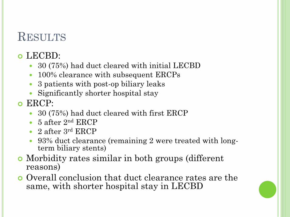
RESULTS
LECBD: 30 (75%) had duct cleared with initial LECBD
100% clearance with subsequent ERCPs
3 patients with post-op biliary leaks
Significantly shorter hospital stay
ERCP: 30 (75%) had duct cleared with first ERCP
5 after 2nd ERCP
2 after 3rd ERCP
93% duct clearance (remaining 2 were treated with long-term biliary stents)
Morbidity rates similar in both groups (different reasons)
Overall conclusion that duct clearance rates are the same, with shorter hospital stay in LECBD
ERCP INDICATIONS – NIH GUIDELINES
ERCP, MRCP and endoscopic US have comparable sensitivity and specificity in the
diagnosis of choledocholithiasis.
If low probability of choledocholithiasis: patients undergoing cholecystectomy do not
need pre-op ERCP
Laparoscopic common bile duct exploration and postoperative ERCP are both safe
and reliable in clearing CBD stones
ERCP + sphincterotomy and stone removal: valuable therapeutic in
choledocholithiasis with jaundice, dilated CBD, acute pancreatitis, or cholangitis.
Biliary cancer: ERCP as palliation of biliary obstruction when surgery is not elected.
ERCP for tissue sampling in pancreatic or biliary cancer (not undergoing surgery)
but not always diagnostic
ERCP is the best means to diagnose ampullary cancers
ERCP has no role in the diagnosis of acute pancreatitis except when biliary
pancreatitis is suspected, where early intervention with ERCP reduces morbidity
and mortality compared with delayed ERCP
ERCP with appropriate therapy is beneficial in selected patients who have either
recurrent pancreatitis or pancreatic pseudocysts.
Patients with type I sphincter of Oddi dysfunction respond to sphincterotomy
Patients with type II SOD should not undergo diagnostic ERCP alone. If SOD
manometer pressures are >40 mmHg, ES is beneficial in some patients.
Avoid unnecessary ERCP – ex. when low likelihood of biliary stone or stricture,
especially in women with recurrent pain, a normal bilirubin, and no other objective
sign of biliary disease
ERCP INDICATIONS – NATIONAL
GUIDELINE CLEARINGHOUSE Jaundice thought to be the result of biliary obstruction
Clinical and biochemical or imaging data suggestive of pancreatic or biliary tract disease
Signs or symptoms suggesting pancreatic malignancy when direct imaging results are equivocal or normal
Pancreatitis of unknown etiology
Preoperative evaluation of chronic pancreatitis or pancreatic pseudocyst
Sphincter of Oddi manometry
Endoscopic sphincterotomy Choledocholithiasis
Papillary stenosis or sphincter of Oddi dysfunction causing disability
Facilitate biliary stent placement or balloon dilatation
Sump syndrome
Choledochocele
Ampullary carcinoma in poor surgical candidates
Access to pancreatic duct
Stent placement across benign or malignant strictures, fistulae, postoperative bile leak, or large common bile duct stones
Balloon dilatation of ductal strictures
Nasobiliary drain placement
Pseudocyst drainage in appropriate cases
Tissue sampling from pancreatic or bile ducts
Pancreatic therapeutics

ERCP INDICATIONS – AMERICAN SOCIETY
OF GASTROINTESTINAL ENDOSCOPY
ERCP is primarily a therapeutic procedure for the management of pancreaticobiliary disorders (C)
Diagnostic ERCP should not be undertaken in the evaluation of pancreaticobiliary pain in the absence of objective findings on other imaging studies (B)
Routine ERCP before laparoscopic cholecystectomy should not be performed (B)
Endoscopic therapy of postoperative biliary leaks and strictures should be undertaken as first-line therapy (B)
ERCP has an important role in patients with recurrent acute pancreatitis and can identify and, in some cases, treat the underlying cause (B)
ERCP is effective in treating symptomatic strictures in chronic pancreatitis (B)
ERCP is effective for the palliation of malignant biliary obstruction (B), for which self-expanding metallic stents have longer patency than plastic stents (A)
ERCP can be used to diagnose and to treat symptomatic pancreatic-duct stones (B)
Pancreatic-duct disruptions or leaks can be effectively treated via the placement of bridging or transpapillary pancreatic stents (B)
SPHINCTER OF ODDI
Regulates flow of bile (and pancreatic juice) into
the duodenum
Prevents the regurgitation of duodenal contents
into the biliary tree
Diverts bile into the gallbladder
Basal resting pressure of about 13 mm Hg above
the duodenal pressure – phasic contractions
Relaxation occurs with a rise in CCK – increases
bile into duodenum

SPHINCTER OF ODDI DYSFUNCTION
Poorly defined syndrome
Pain similar to biliary colic, normal liver function tests, episodes of acute pancreatitis
Unclear pathogenesis; theories: Gallstone migration causing fibrosis of the sphincter
Trauma
Pancreatitis
Congenital anomalies
About 1% of patients undergoing cholecystectomy have sphincter of Oddi dysfunction
Diagnostic clues: Ultrasound: dilated common bile duct (>12 mm diameter) or
increase in common bile duct diameter in response to CCK
ERCP: delayed emptying of contrast medium from CBD
Ampullary manometry: elevated basal sphincter pressure (>40 mm Hg)
Treatment: sphincterotomy

GALLSTONE ILEUS
Passage of a stone through a spontaneous biliary-enteric fistula leading to a mechanical bowel obstruction
Most fistulas between gallbladder and duodenum
F>M
Average age 70yrs
1% of bowel obstructions, up to 25% of bowel obstructions in elderly without history of surgery or hernias
Usually occur after an episode of acute cholecystitis –
Gangrene and perforation of the gallbladder or
Pressure necrosis from an impacted gallstone.
PRESENTATION & DIAGNOSIS
Signs & symptoms of intestinal obstruction:
nausea, vomiting, abdominal pain
½ have history of gallbladder-related symptoms
Tumbling Obstruction: pain may be episodic and
recurrent as the impacted stone temporarily
obstructs the bowel lumen and then dislodges
and moves distally
Abdominal films: evidence of intestinal
obstruction with pneumobilia or a calcified stone
distant from the gallbladder
Most common site of obstruction is the terminal
ileum because of its narrow lumen

RADIOGRAPHIC FINDINGS WITH GALLSTONE
ILEUS
PA barium X-ray: irregular collection of barium in the right upper quadrant (A, arrowheads), representing partial filling of the cystic duct.; jejunum and ileum are markedly dilated, with dilution of the barium in a pattern consistent with small bowel obstruction: abrupt termination of the barium column at the site of an oval intraluminal filling defect (A, arrow). A view of the barium column shows luminal obstruction by a smooth intraluminal mass (B, arrows) with faint calcification of the peripheral rim. (From Kaiser AM etal: Gallstone ileus. N Engl J Med 335:942, 1996.)
MANAGEMENT
Removal of gallstone through a proximal enterotomy
– the stone is “milked” proximally, and then removed
from a healthy portion of bowel
May be necessary to resect any portion of bowel with
ischemia - prevent postimpaction wall necrosis and
leak
Recurrent obstruction in 10% of patients – need to
evaluate for other gallstones
Takedown of the biliary-enteric fistula and
cholecystectomy – risk of recurrent cholecystitis and
cholangitis
If patient unstable or significant inflammation, can
address fistula at a second laparotomy