chronic calculous cholecystitis, gallstone · PDF fileChronic calculous cholecystitis....
13
Chronic calculous cholecystitis. Gallstone disease. Dr. Turumin JL, MD, PhD, DMSci. Web-site: http://www.drturumin.com E-mail: [email protected] Gallstone Disease Chronic calculous cholecystitis Gallstone disease is a disease of hepato-biliary system, caused by cholesterol and/or bilirubin metabolic disorder, and characterized by formation of stones in the gallbladder and/or the biliary tract (1-14). Gallstones are categorized as cholesterol, mixed, black pigment, or brown pigment stones (1-25). Cholesterol gallstones are the main type of gallstones and contain cholesterol as the major chemical constituent. Mixed cholesterol gallstones are composed of more than 50% cholesterol (1- 25). Cholesterol and mixed gallstones are formed from biliary sludge, which stays for a long time in the gallbladder lumen. Biliary sludge consists of calcium bilirubinate granules, cholesterol monohy- drate crystals, and biliary polymerized glycoprotein mucin (1-25). The dynamics of the transforma- tion of biliary sludge into cholesterol stones has been shown as follows: diffused biliary sludge surface biliary sludge precipitating biliary sludge a cholesterol gallstone without acoustic shadow a cholesterol gallstone with acoustic shadow (18). The time of formation of cholesterol stones depends on the intensity of the precipitation processes of cholesterol monohydrate crystals in biliary sludge, and equals 3 to 36 months (1-25). Transformation proportion varies from 5 to 50% depending on the cause (1-25). Black pigment stones are composed of either pure calcium bilirubinate or polymer-like com- plexes consisting of calcium, cooper, and large amounts of mucin glycoproteins. Brown pigment stones are composed of calcium salts of unconjugated bilirubin, with varying amounts of cholesterol and protein. These stones are usually associated with infection (1-25). The natural history of gallstones is typically defined in two separate groups of patients: those with symptomatic gallstones and those who are asymptomatic. The vast majorities of gallstones are asymptomatic and remain asymptomatic (1-58). As a rule, gallstone disease is asymptomatic, which is called “silent” stones. The rate of development of biliary pain is approximately 2% per year for 5 years and then decreases over time. The incidence of complications in patients with asymp- tomatic stones is low, and prophylactic removal of the gallbladder for this condition is not neces- sary (1-58). Patients who had an episode of uncomplicated biliary pain in the year, 38% per year had recurrent biliary pain (1-58). An incidence of recurrent biliary pain as high as 50% per year in those with symptomatic gallstones. 30% of patients with one episode of biliary pain will not have a recurrent episode (1-58). The estimated risk of developing biliary complications is estimated to be 1% to 2% per year and is thought to remain relatively constant over time (1-58). If biliary pain occurs in the right upper abdomen and the gallbladder wall inflames, gallstone disease transforms into chronic calculous cholecystitis. Chronic calculous cholecystitis is an inflammatory disease which affects the gallbladder wall and causes motoric-tonic dysfunctions of the biliary system, accompanied by presence of gall- stones in the gallbladder lumen, and reveals as biliary pain (1-25). The motoric dysfunction of the gallbladder can be caused by increased basal common bile duct resistance, muscle hypertrophy, and chronic inflammation in the gallbladder wall. Biliary colic is the most common presenting symp- tom of cholelithiasis. Approximately 75% of patients with symptomatic gallstone disease seek medical attention because of episodic abdominal pain (1-25). The syndrome of biliary colic is caused by intermittent obstruction of cystic duct by gallstones (1-25). Cholecystectomy should be offered to patients only after significant biliary symptoms develop (59-71). Diagnostic criteria of the chronic calculous cholecystitis 1. Recurrent episodes of biliary pain in the right upper abdomen, sometimes in epigastrium, of- ten with irradiation to the right scapular region. Biliary pains may be in the right hypochon- drium, frequently or occasionally, of different intensity and duration, related to intake of fatty meals (1).
-
Upload
hoangduong -
Category
Documents
-
view
229 -
download
2
Transcript of chronic calculous cholecystitis, gallstone · PDF fileChronic calculous cholecystitis....

Chronic calculous cholecystitis. Gallstone disease. Dr. Turumin JL, MD, PhD, DMSci.
Web-site: http://www.drturumin.com E-mail: [email protected]
Gallstone Disease Chronic calculous cholecystitis
Gallstone disease is a disease of hepato-biliary system, caused by cholesterol and/or bilirubin metabolic disorder, and characterized by formation of stones in the gallbladder and/or the biliary tract (1-14).
Gallstones are categorized as cholesterol, mixed, black pigment, or brown pigment stones
(1-25). Cholesterol gallstones are the main type of gallstones and contain cholesterol as the major chemical constituent. Mixed cholesterol gallstones are composed of more than 50% cholesterol (1-25). Cholesterol and mixed gallstones are formed from biliary sludge, which stays for a long time in the gallbladder lumen. Biliary sludge consists of calcium bilirubinate granules, cholesterol monohy-drate crystals, and biliary polymerized glycoprotein mucin (1-25). The dynamics of the transforma-tion of biliary sludge into cholesterol stones has been shown as follows: diffused biliary sludge surface biliary sludge precipitating biliary sludge a cholesterol gallstone without acoustic shadow a cholesterol gallstone with acoustic shadow (18). The time of formation of cholesterol stones depends on the intensity of the precipitation processes of cholesterol monohydrate crystals in biliary sludge, and equals 3 to 36 months (1-25). Transformation proportion varies from 5 to 50% depending on the cause (1-25).
Black pigment stones are composed of either pure calcium bilirubinate or polymer-like com-
plexes consisting of calcium, cooper, and large amounts of mucin glycoproteins. Brown pigment stones are composed of calcium salts of unconjugated bilirubin, with varying
amounts of cholesterol and protein. These stones are usually associated with infection (1-25). The natural history of gallstones is typically defined in two separate groups of patients: those
with symptomatic gallstones and those who are asymptomatic. The vast majorities of gallstones are asymptomatic and remain asymptomatic (1-58). As a rule, gallstone disease is asymptomatic, which is called “silent” stones. The rate of development of biliary pain is approximately 2% per year for 5 years and then decreases over time. The incidence of complications in patients with asymp-tomatic stones is low, and prophylactic removal of the gallbladder for this condition is not neces-sary (1-58). Patients who had an episode of uncomplicated biliary pain in the year, 38% per year had recurrent biliary pain (1-58). An incidence of recurrent biliary pain as high as 50% per year in those with symptomatic gallstones. 30% of patients with one episode of biliary pain will not have a recurrent episode (1-58). The estimated risk of developing biliary complications is estimated to be 1% to 2% per year and is thought to remain relatively constant over time (1-58).
If biliary pain occurs in the right upper abdomen and the gallbladder wall inflames, gallstone
disease transforms into chronic calculous cholecystitis. Chronic calculous cholecystitis is an inflammatory disease which affects the gallbladder wall
and causes motoric-tonic dysfunctions of the biliary system, accompanied by presence of gall-stones in the gallbladder lumen, and reveals as biliary pain (1-25). The motoric dysfunction of the gallbladder can be caused by increased basal common bile duct resistance, muscle hypertrophy, and chronic inflammation in the gallbladder wall. Biliary colic is the most common presenting symp-tom of cholelithiasis. Approximately 75% of patients with symptomatic gallstone disease seek medical attention because of episodic abdominal pain (1-25). The syndrome of biliary colic is caused by intermittent obstruction of cystic duct by gallstones (1-25).
Cholecystectomy should be offered to patients only after significant biliary symptoms develop
(59-71).
Diagnostic criteria of the chronic calculous cholecystitis 1. Recurrent episodes of biliary pain in the right upper abdomen, sometimes in epigastrium, of-
ten with irradiation to the right scapular region. Biliary pains may be in the right hypochon-drium, frequently or occasionally, of different intensity and duration, related to intake of fatty meals (1).
Chronic calculous cholecystitis. Gallstone disease. Dr. Turumin JL, MD, PhD, DMSci.
Web-site: http://www.drturumin.com E-mail: [email protected]
2
In addition, a biliary pain may occur with one or more of the following symptoms: a. regular or periodical feeling of bitter taste b. nausea, sometimes vomiting c. regular or periodical abdominal bloating and borborygmus d. unstable stool with constipation or diarrhea prevailing
2. Impaired gallbladder emptying. 3. According to ultrasound examination, thickening of the gallbladder wall up to 3-4 mm and
presence of gallstones in the gallbladder lumen.
Causes of the gallbladder evacuation dysfunction, biliary pain and chronic inflammation in the gallbladder wall
1. Pathology of the smooth muscle cells and epithelial cells in the gallbladder wall (high degree of COX-2 expression in the smooth muscle cells and epithelial cells of the gallbladder wall).
2. Hypersecretion of glycoprotein biliary mucin into gallbladder lumen and increase of concen-tration of glycoprotein biliary mucin in the gallbladder bile over the point of polymerization (>2.0 mg/ml) (high degree of COX-2 expression in the epithelial cells of the gallbladder mu-cosa).
3. Contractile dyscoordination of the gallbladder and cystic duct (high degree of COX-2 expres-sion in the smooth muscle cells of the gallbladder and cystic duct).
4. Increased basal cystic duct resistance (high degree of COX-2 expression in the smooth muscle cells of the cystic duct).
5. Increased basal common bile duct resistance (high degree of COX-2 expression in the smooth muscle cells of the sphincter of Oddi).
Mechanism of development of pathologic disorders
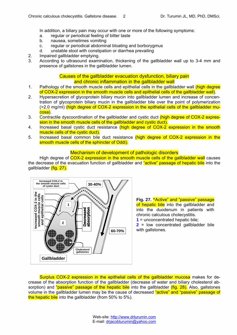
High degree of COX-2 expression in the smooth muscle cells of the gallbladder wall causes the decrease of the evacuation function of gallbladder and “active” passage of hepatic bile into the gallbladder (fig. 27).
Surplus COX-2 expression in the epithelial cells of the gallbladder mucosa makes for de-
crease of the absorption function of the gallbladder (decrease of water and biliary cholesterol ab-sorption) and “passive” passage of the hepatic bile into the gallbladder (fig. 28). Also, gallstones volume in the gallbladder lumen may be the cause of decreased “active” and “passive” passage of the hepatic bile into the gallbladder (from 50% to 5%).
Fig. 27. “Active” and “passive” passage of hepatic bile into the gallbladder and into the duodenum in patients with chronic calculous cholecystitis. 1 = unconcentrated hepatic bile; 2 = low concentrated gallbladder bile with gallstones.
Gallbladder
2
1
30-40%
60-70%
Cholesterolgallstones
Increased COX-2 in the smooth muscle cells
of cystic duct
Incr
ease
d C
OX-
2 in
the
smoo
th m
uscl
e ce
lls
and
epith
elia
l cel
ls
Dec
reas
edab
sorp
tion
Gallbladder
2
1
30-40%
60-70%
Cholesterolgallstones
Increased COX-2 in the smooth muscle cells
of cystic duct
Incr
ease
d C
OX-
2 in
the
smoo
th m
uscl
e ce
lls
and
epith
elia
l cel
ls
Dec
reas
edab
sorp
tion
Gallbladder
2
1
30-40%
60-70%
Cholesterolgallstones
Increased COX-2 in the smooth muscle cells
of cystic duct
Incr
ease
d C
OX-
2 in
the
smoo
th m
uscl
e ce
lls
and
epith
elia
l cel
ls
Dec
reas
edab
sorp
tion
Dec
reas
edab
sorp
tion
Chronic calculous cholecystitis. Gallstone disease. Dr. Turumin JL, MD, PhD, DMSci.
Web-site: http://www.drturumin.com E-mail: [email protected]
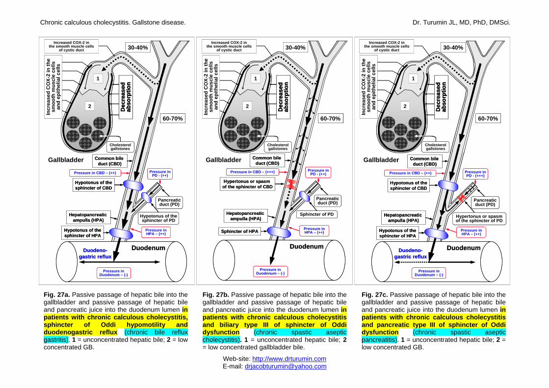
Fig. 27a. Passive passage of hepatic bile into the gallbladder and passive passage of hepatic bile and pancreatic juice into the duodenum lumen in patients with chronic calculous cholecystitis, sphincter of Oddi hypomotility and duodenogastric reflux (chronic bile reflux gastritis). 1 = unconcentrated hepatic bile; 2 = low concentrated GB.
Fig. 27b. Passive passage of hepatic bile into the gallbladder and passive passage of hepatic bile and pancreatic juice into the duodenum lumen in patients with chronic calculous cholecystitis and biliary type III of sphincter of Oddi dysfunction (chronic spastic aseptic cholecystitis). 1 = unconcentrated hepatic bile; 2 = low concentrated gallbladder bile.
Fig. 27c. Passive passage of hepatic bile into the gallbladder and passive passage of hepatic bile and pancreatic juice into the duodenum lumen in patients with chronic calculous cholecystitis and pancreatic type III of sphincter of Oddi dysfunction (chronic spastic aseptic pancreatitis). 1 = unconcentrated hepatic bile; 2 = low concentrated GB.
2
1
30-40%
60-70%
Cholesterolgallstones
Increased COX-2 in the smooth muscle cells
of cystic ductIn
crea
sed
CO
X-2
in th
e sm
ooth
mus
cle
cells
an
dep
ithel
ial c
ells
Dec
reas
edab
sorp
tion
Gallbladder Common bileduct (CBD)
Pancreaticduct (PD)
Hepatopancreaticampulla (HPA)
Pressure inPD - (++)
Pressure in Duodenum – (-)
Hypotonus of thesphincter of CBD
Pressure in CBD – (++)
Hypotonus of thesphincter of HPA
Hypotonus of the sphincter of PD
Pressure inHPA – (++)
Duodeno-gastric reflux
Duodenum
2
1
30-40%
60-70%
Cholesterolgallstones
Increased COX-2 in the smooth muscle cells
of cystic ductIn
crea
sed
CO
X-2
in th
e sm
ooth
mus
cle
cells
an
dep
ithel
ial c
ells
Dec
reas
edab
sorp
tion
Dec
reas
edab
sorp
tion
Gallbladder Common bileduct (CBD)
Pancreaticduct (PD)
Hepatopancreaticampulla (HPA)
Pressure inPD - (++)
Pressure in Duodenum – (-)
Hypotonus of thesphincter of CBD
Pressure in CBD – (++)
Hypotonus of thesphincter of HPA
Hypotonus of the sphincter of PD
Pressure inHPA – (++)
Duodeno-gastric reflux
Duodenum
2
1
30-40%
60-70%
Cholesterolgallstones
Increased COX-2 in the smooth muscle cells
of cystic duct
Incr
ease
d C
OX-
2 in
the
smoo
th m
uscl
e ce
lls
and
epith
elia
l cel
ls
Dec
reas
edab
sorp
tion
Gallbladder Common bileduct (CBD)
Hepatopancreaticampulla (HPA)
Sphincter of HPA
Pancreaticduct (PD)
Sphincter of PD
Pressure inHPA – (++)
Pressure in CBD – (+++)
Hypertonus or spasm of the sphincter of CBD
Pressure inPD - (++)
Pressure in Duodenum – (-)
Duodenum
2
1
30-40%
60-70%
Cholesterolgallstones
Increased COX-2 in the smooth muscle cells
of cystic duct
Incr
ease
d C
OX-
2 in
the
smoo
th m
uscl
e ce
lls
and
epith
elia
l cel
ls
Dec
reas
edab
sorp
tion
Dec
reas
edab
sorp
tion
Gallbladder Common bileduct (CBD)
Hepatopancreaticampulla (HPA)
Sphincter of HPA
Pancreaticduct (PD)
Sphincter of PD
Pressure inHPA – (++)
Pressure in CBD – (+++)
Hypertonus or spasm of the sphincter of CBD
Pressure inPD - (++)
Pressure in Duodenum – (-)
Duodenum
2
1
30-40%
60-70%
Cholesterolgallstones
Increased COX-2 in the smooth muscle cells
of cystic duct
Incr
ease
d C
OX-
2 in
the
smoo
th m
uscl
e ce
lls
and
epith
elia
l cel
ls
Dec
reas
edab
sorp
tion
Gallbladder
Pressure in Duodenum – (-)
Duodeno-gastric reflux
Duodenum
Common bileduct (CBD)
Pressure in CBD – (++)
Hypotonus of thesphincter of HPA
Hypotonus of thesphincter of CBD
Hepatopancreaticampulla (HPA)
Hypertonus or spasm of the sphincter of PD
Pancreaticduct (PD)
Pressure inPD - (+++)
Pressure inHPA – (++)
2
1
30-40%
60-70%
Cholesterolgallstones
Increased COX-2 in the smooth muscle cells
of cystic duct
Incr
ease
d C
OX-
2 in
the
smoo
th m
uscl
e ce
lls
and
epith
elia
l cel
ls
Dec
reas
edab
sorp
tion
Dec
reas
edab
sorp
tion
Gallbladder
Pressure in Duodenum – (-)
Duodeno-gastric reflux
Duodenum
Common bileduct (CBD)
Pressure in CBD – (++)
Hypotonus of thesphincter of HPA
Hypotonus of thesphincter of CBD
Hepatopancreaticampulla (HPA)
Hypertonus or spasm of the sphincter of PD
Pancreaticduct (PD)
Pressure inPD - (+++)
Pressure inHPA – (++)
Chronic calculous cholecystitis. Gallstone disease. Dr. Turumin JL, MD, PhD, DMSci.
Web-site: http://www.drturumin.com E-mail: [email protected]
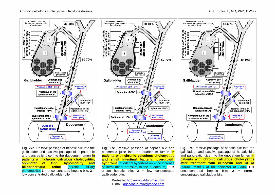
Fig. 27d. Passive passage of hepatic bile into the
gallbladder and passive passage of hepatic bile and pancreatic juice into the duodenum lumen in patients with chronic calculous cholecystitis, sphincter of Oddi hypomotility and biliopancreatic reflux (chronic biliary pancreatitis). 1 = unconcentrated hepatic bile; 2 = low concentrated gallbladder bile.
Fig. 27e. Passive passage of hepatic bile and pancreatic juice into the duodenum lumen in patients with chronic calculous cholecystitis and small intestinal bacterial overgrowth syndrome (duodenal hypertension – the increase of intraluminal pressure in the duodenum). 1 = uncon hepatic bile; 2 = low concentrated gallbladder bile.
Fig. 27f. Passive passage of hepatic bile into the gallbladder and passive passage of hepatic bile and pancreatic juice into the duodenum lumen in patients with chronic calculous cholecystitis after treatment with celecoxib and UDCA (normal motility of the sphincter of Oddi). 1 = unconcentrated hepatic bile; 2 = normal concentrated gallbladder bile.
2
1
30-40%
60-70%
Cholesterolgallstones
Increased COX-2 in the smooth muscle cells
of cystic ductIn
crea
sed
CO
X-2
in th
e sm
ooth
mus
cle
cells
an
dep
ithel
ial c
ells
Dec
reas
edab
sorp
tion
Gallbladder
Pressure in Duodenum – (-)
Duodeno-gastric reflux
Duodenum
Pressure in CBD – (+++)
Common bileduct (CBD)
Hypotonus of thesphincter of HPA
Hypotonus of thesphincter of CBD
Hepatopancreaticampulla (HPA)
Hypotonus of the sphincter of PD
Pressure inHPA – (+++)
Pancreaticduct (PD)
Pressure inPD - (+)
2
1
30-40%
60-70%
Cholesterolgallstones
Increased COX-2 in the smooth muscle cells
of cystic ductIn
crea
sed
CO
X-2
in th
e sm
ooth
mus
cle
cells
an
dep
ithel
ial c
ells
Dec
reas
edab
sorp
tion
Dec
reas
edab
sorp
tion
Gallbladder
Pressure in Duodenum – (-)
Duodeno-gastric reflux
Duodenum
Pressure in CBD – (+++)
Common bileduct (CBD)
Hypotonus of thesphincter of HPA
Hypotonus of thesphincter of CBD
Hepatopancreaticampulla (HPA)
Hypotonus of the sphincter of PD
Pressure inHPA – (+++)
Pancreaticduct (PD)
Pressure inPD - (+)
Gallbladder
2
1
30-40%
60-70%
Increased COX-2 in the smooth muscle cells
of cystic duct
Dec
reas
edab
sorp
tion
Common bileduct (CBD)
Hepatopancreaticampulla (HPA)
Sphincter of HPA
Pancreaticduct (PD)
Sphincter of PD
Pressure inHPA – (+++)
Pressure inPD - (++)
Duodenum
Duodenalhypertension
Pressure in Duodenum – (+++)
Sphincter of CBD
Pressure in CBD – (++)
Incr
ease
d C
OX-
2 in
the
smoo
th m
uscl
e ce
lls
and
epith
elia
l cel
ls
Cholesterolgallstones
Gallbladder
2
1
30-40%
60-70%
Increased COX-2 in the smooth muscle cells
of cystic duct
Dec
reas
edab
sorp
tion
Dec
reas
edab
sorp
tion
Common bileduct (CBD)
Hepatopancreaticampulla (HPA)
Sphincter of HPA
Pancreaticduct (PD)
Sphincter of PD
Pressure inHPA – (+++)
Pressure inPD - (++)
Duodenum
Duodenalhypertension
Pressure in Duodenum – (+++)
Sphincter of CBD
Pressure in CBD – (++)
Incr
ease
d C
OX-
2 in
the
smoo
th m
uscl
e ce
lls
and
epith
elia
l cel
ls
Cholesterolgallstones
2
1
40-60%
40-60%
Cholesterolgallstones
Increased COX-2 in the smooth muscle cells
of cystic duct
Incr
ease
d C
OX-
2 in
the
smoo
th m
uscl
e ce
lls
and
epith
elia
l cel
ls
Dec
reas
edab
sorp
tion
Gallbladder Common bileduct (CBD)
Pancreaticduct (PD)
Hepatopancreaticampulla (HPA)
Pressure inPD - (+)
Pressure in Duodenum – (-)
Pressure in CBD – (+)
Pressure inHPA – (+)
Duodenum
Normal tonus of thesphincter of CBD
Normal tonus of the sphincter of PD
Normal tonus of thesphincter of HPA
2
1
40-60%
40-60%
Cholesterolgallstones
Increased COX-2 in the smooth muscle cells
of cystic duct
Incr
ease
d C
OX-
2 in
the
smoo
th m
uscl
e ce
lls
and
epith
elia
l cel
ls
Dec
reas
edab
sorp
tion
Dec
reas
edab
sorp
tion
Gallbladder Common bileduct (CBD)
Pancreaticduct (PD)
Hepatopancreaticampulla (HPA)
Pressure inPD - (+)
Pressure in Duodenum – (-)
Pressure in CBD – (+)
Pressure inHPA – (+)
Duodenum
Normal tonus of thesphincter of CBD
Normal tonus of the sphincter of PD
Normal tonus of thesphincter of HPA
Chronic calculous cholecystitis. Gallstone disease. Dr. Turumin JL, MD, PhD, DMSci.
Web-site: http://www.drturumin.com E-mail: [email protected]
3
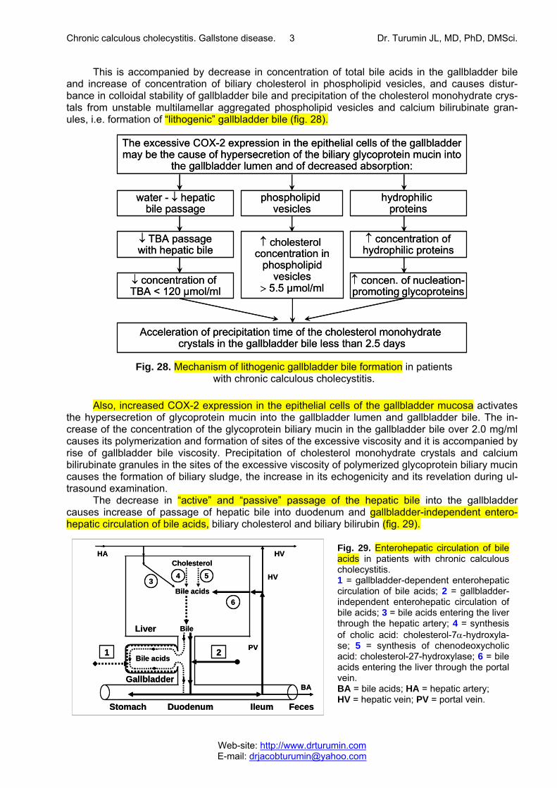
This is accompanied by decrease in concentration of total bile acids in the gallbladder bile and increase of concentration of biliary cholesterol in phospholipid vesicles, and causes distur-bance in colloidal stability of gallbladder bile and precipitation of the cholesterol monohydrate crys-tals from unstable multilamellar aggregated phospholipid vesicles and calcium bilirubinate gran-ules, i.e. formation of “lithogenic” gallbladder bile (fig. 28).
Also, increased COX-2 expression in the epithelial cells of the gallbladder mucosa activates
the hypersecretion of glycoprotein mucin into the gallbladder lumen and gallbladder bile. The in-crease of the concentration of the glycoprotein biliary mucin in the gallbladder bile over 2.0 mg/ml causes its polymerization and formation of sites of the excessive viscosity and it is accompanied by rise of gallbladder bile viscosity. Precipitation of cholesterol monohydrate crystals and calcium bilirubinate granules in the sites of the excessive viscosity of polymerized glycoprotein biliary mucin causes the formation of biliary sludge, the increase in its echogenicity and its revelation during ul-trasound examination.
The decrease in “active” and “passive” passage of the hepatic bile into the gallbladder causes increase of passage of hepatic bile into duodenum and gallbladder-independent entero-hepatic circulation of bile acids, biliary cholesterol and biliary bilirubin (fig. 29).
Fig. 28. Mechanism of lithogenic gallbladder bile formation in patients with chronic calculous cholecystitis.
The excessive COX-2 expression in the epithelial cells of the gallbladdermay be the cause of hypersecretion of the biliary glycoprotein mucin into
the gallbladder lumen and of decreased absorption:
↓ concentration ofTBA < 120 µmol/ml
↑ concen. of nucleation-promoting glycoproteins
↓ TBA passagewith hepatic bile
↑ cholesterolconcentration in
phospholipidvesicles
> 5.5 µmol/ml
↑ concentration ofhydrophilic proteins
water - ↓ hepaticbile passage
phospholipidvesicles
hydrophilicproteins
Acceleration of precipitation time of the cholesterol monohydrate crystals in the gallbladder bile less than 2.5 days
Fig. 29. Enterohepatic circulation of bile acids in patients with chronic calculous cholecystitis. 1 = gallbladder-dependent enterohepatic circulation of bile acids; 2 = gallbladder-independent enterohepatic circulation of bile acids; 3 = bile acids entering the liver through the hepatic artery; 4 = synthesis of cholic acid: cholesterol-7α-hydroxyla-se; 5 = synthesis of chenodeoxycholic acid: cholesterol-27-hydroxylase; 6 = bile acids entering the liver through the portal vein. BA = bile acids; HA = hepatic artery; HV = hepatic vein; PV = portal vein.
34 5
6
PV
HA HV
HV
1 2
BA
Stomach Duodenum Ileum Feces
Gallbladder
Bile acids
BileLiver
Cholesterol
Bile acids3
4 5
6
PV
HA HV
HV
1 2
BA
Stomach Duodenum Ileum Feces
Gallbladder
Bile acids
BileLiver
Cholesterol
Bile acids3
4 5
6
PV
HA HV
HV
1 2
BA
Stomach Duodenum Ileum Feces
Gallbladder
Bile acids
BileLiver
Cholesterol
Bile acids
Chronic calculous cholecystitis. Gallstone disease. Dr. Turumin JL, MD, PhD, DMSci.
Web-site: http://www.drturumin.com E-mail: [email protected]
4
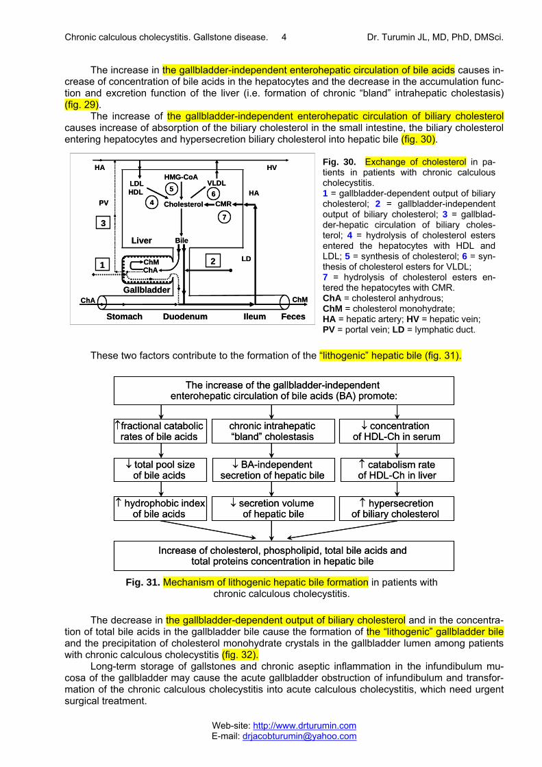
The increase in the gallbladder-independent enterohepatic circulation of bile acids causes in-crease of concentration of bile acids in the hepatocytes and the decrease in the accumulation func-tion and excretion function of the liver (i.e. formation of chronic “bland” intrahepatic cholestasis) (fig. 29).
The increase of the gallbladder-independent enterohepatic circulation of biliary cholesterol causes increase of absorption of the biliary cholesterol in the small intestine, the biliary cholesterol entering hepatocytes and hypersecretion biliary cholesterol into hepatic bile (fig. 30).
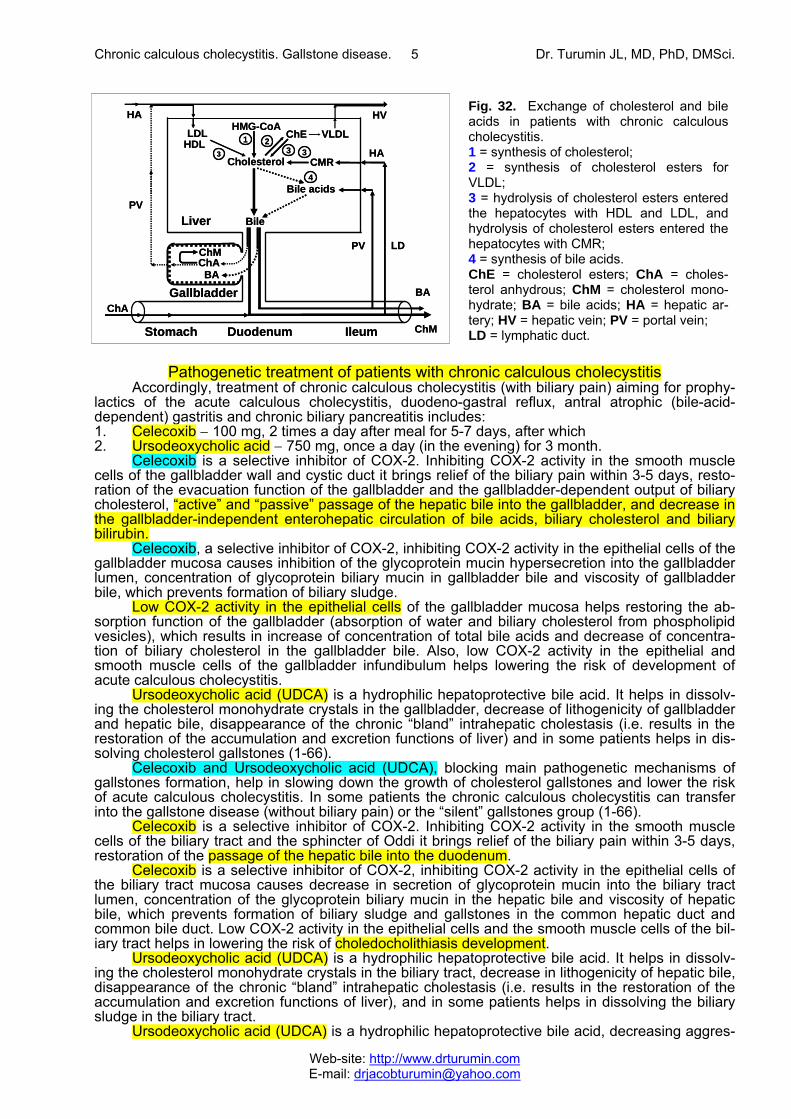
These two factors contribute to the formation of the “lithogenic” hepatic bile (fig. 31).
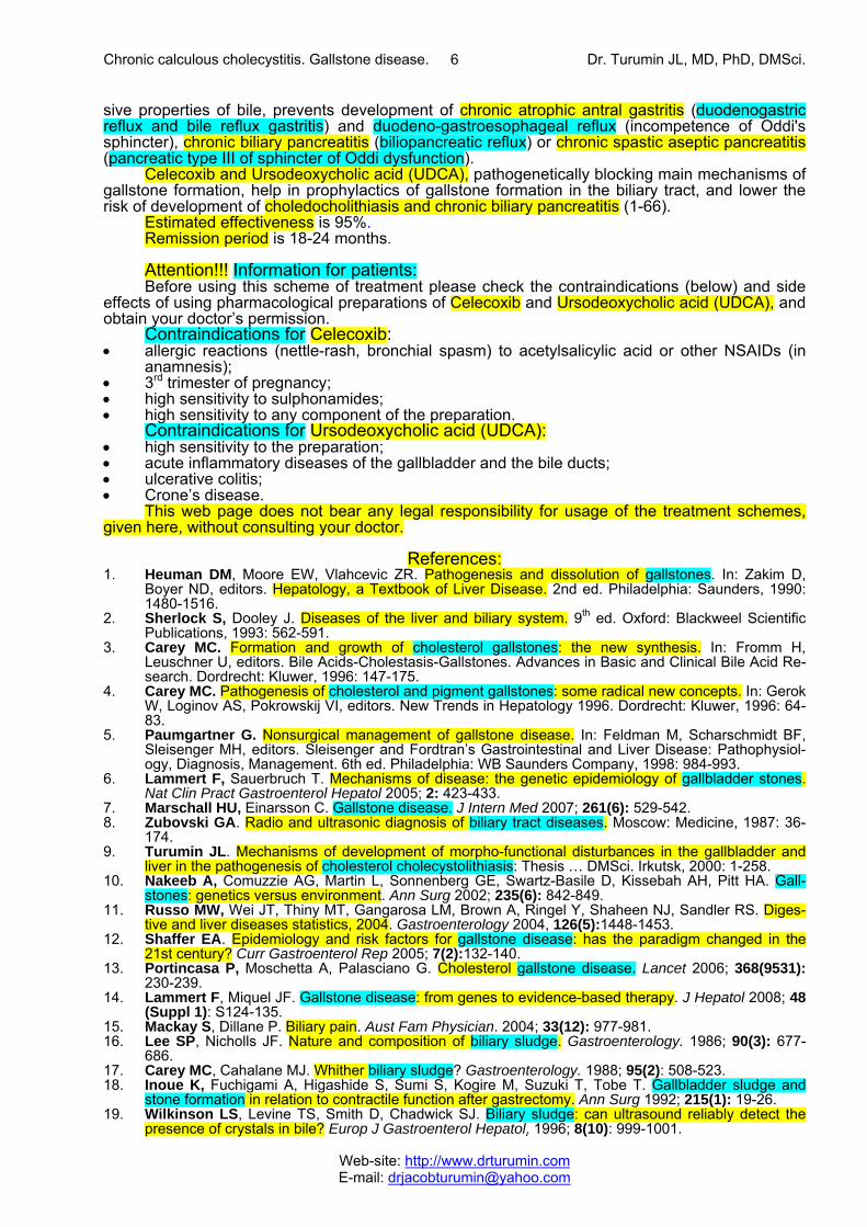
The decrease in the gallbladder-dependent output of biliary cholesterol and in the concentra-
tion of total bile acids in the gallbladder bile cause the formation of the “lithogenic” gallbladder bile and the precipitation of cholesterol monohydrate crystals in the gallbladder lumen among patients with chronic calculous cholecystitis (fig. 32).
Long-term storage of gallstones and chronic aseptic inflammation in the infundibulum mu-cosa of the gallbladder may cause the acute gallbladder obstruction of infundibulum and transfor-mation of the chronic calculous cholecystitis into acute calculous cholecystitis, which need urgent surgical treatment.
Fig. 30. Exchange of cholesterol in pa-tients in patients with chronic calculous cholecystitis. 1 = gallbladder-dependent output of biliary cholesterol; 2 = gallbladder-independent output of biliary cholesterol; 3 = gallblad-der-hepatic circulation of biliary choles-terol; 4 = hydrolysis of cholesterol esters entered the hepatocytes with HDL and LDL; 5 = synthesis of cholesterol; 6 = syn-thesis of cholesterol esters for VLDL; 7 = hydrolysis of cholesterol esters en-tered the hepatocytes with CMR. ChA = cholesterol anhydrous; ChM = cholesterol monohydrate; HA = hepatic artery; HV = hepatic vein; PV = portal vein; LD = lymphatic duct.
4
56
7
Liver Bile
LD
HA HV
HA
ChM
2
ChA
LDLHDL
VLDL
PV
3
ChMChA
Cholesterol
Gallbladder
Stomach Duodenum Ileum Feces
HMG-CoA
CMR
1
4
56
7
Liver Bile
LD
HA HV
HA
ChM
2
ChA
LDLHDL
VLDL
PV
3
ChMChA
Cholesterol
Gallbladder
Stomach Duodenum Ileum Feces
HMG-CoA
CMR
1
4
56
7
Liver Bile
LD
HA HV
HA
ChM
2
ChA
LDLHDL
VLDL
PV
3
ChMChA
Cholesterol
Gallbladder
Stomach Duodenum Ileum Feces
HMG-CoA
CMR
1
Fig. 31. Mechanism of lithogenic hepatic bile formation in patients with chronic calculous cholecystitis.
↑ hydrophobic indexof bile acids
↓ secretion volume of hepatic bile
↑ hypersecretionof biliary cholesterol
↓ total pool sizeof bile acids
↓ BA-independentsecretion of hepatic bile
↑ catabolism rateof HDL-Ch in liver
↑fractional catabolic rates of bile acids
chronic intrahepatic “bland” cholestasis
↓ concentrationof HDL-Ch in serum
Increase of cholesterol, phospholipid, total bile acids andtotal proteins concentration in hepatic bile
The increase of the gallbladder-independent enterohepatic circulation of bile acids (BA) promote:
Chronic calculous cholecystitis. Gallstone disease. Dr. Turumin JL, MD, PhD, DMSci.
Web-site: http://www.drturumin.com E-mail: [email protected]
5
Pathogenetic treatment of patients with chronic calculous cholecystitis
Accordingly, treatment of chronic calculous cholecystitis (with biliary pain) aiming for prophy-lactics of the acute calculous cholecystitis, duodeno-gastral reflux, antral atrophic (bile-acid-dependent) gastritis and chronic biliary pancreatitis includes: 1. Celecoxib − 100 mg, 2 times a day after meal for 5-7 days, after which 2. Ursodeoxycholic acid − 750 mg, once a day (in the evening) for 3 month.
Celecoxib is a selective inhibitor of COX-2. Inhibiting COX-2 activity in the smooth muscle cells of the gallbladder wall and cystic duct it brings relief of the biliary pain within 3-5 days, resto-ration of the evacuation function of the gallbladder and the gallbladder-dependent output of biliary cholesterol, “active” and “passive” passage of the hepatic bile into the gallbladder, and decrease in the gallbladder-independent enterohepatic circulation of bile acids, biliary cholesterol and biliary bilirubin.
Celecoxib, a selective inhibitor of COX-2, inhibiting COX-2 activity in the epithelial cells of the gallbladder mucosa causes inhibition of the glycoprotein mucin hypersecretion into the gallbladder lumen, concentration of glycoprotein biliary mucin in gallbladder bile and viscosity of gallbladder bile, which prevents formation of biliary sludge.
Low COX-2 activity in the epithelial cells of the gallbladder mucosa helps restoring the ab-sorption function of the gallbladder (absorption of water and biliary cholesterol from phospholipid vesicles), which results in increase of concentration of total bile acids and decrease of concentra-tion of biliary cholesterol in the gallbladder bile. Also, low COX-2 activity in the epithelial and smooth muscle cells of the gallbladder infundibulum helps lowering the risk of development of acute calculous cholecystitis.
Ursodeoxycholic acid (UDCA) is a hydrophilic hepatoprotective bile acid. It helps in dissolv-ing the cholesterol monohydrate crystals in the gallbladder, decrease of lithogenicity of gallbladder and hepatic bile, disappearance of the chronic “bland” intrahepatic cholestasis (i.e. results in the restoration of the accumulation and excretion functions of liver) and in some patients helps in dis-solving cholesterol gallstones (1-66).
Celecoxib and Ursodeoxycholic acid (UDCA), blocking main pathogenetic mechanisms of gallstones formation, help in slowing down the growth of cholesterol gallstones and lower the risk of acute calculous cholecystitis. In some patients the chronic calculous cholecystitis can transfer into the gallstone disease (without biliary pain) or the “silent” gallstones group (1-66).
Celecoxib is a selective inhibitor of COX-2. Inhibiting COX-2 activity in the smooth muscle cells of the biliary tract and the sphincter of Oddi it brings relief of the biliary pain within 3-5 days, restoration of the passage of the hepatic bile into the duodenum.
Celecoxib is a selective inhibitor of COX-2, inhibiting COX-2 activity in the epithelial cells of the biliary tract mucosa causes decrease in secretion of glycoprotein mucin into the biliary tract lumen, concentration of the glycoprotein biliary mucin in the hepatic bile and viscosity of hepatic bile, which prevents formation of biliary sludge and gallstones in the common hepatic duct and common bile duct. Low COX-2 activity in the epithelial cells and the smooth muscle cells of the bil-iary tract helps in lowering the risk of choledocholithiasis development.
Ursodeoxycholic acid (UDCA) is a hydrophilic hepatoprotective bile acid. It helps in dissolv-ing the cholesterol monohydrate crystals in the biliary tract, decrease in lithogenicity of hepatic bile, disappearance of the chronic “bland” intrahepatic cholestasis (i.e. results in the restoration of the accumulation and excretion functions of liver), and in some patients helps in dissolving the biliary sludge in the biliary tract.
Ursodeoxycholic acid (UDCA) is a hydrophilic hepatoprotective bile acid, decreasing aggres-
Fig. 32. Exchange of cholesterol and bile acids in patients with chronic calculous cholecystitis. 1 = synthesis of cholesterol; 2 = synthesis of cholesterol esters for VLDL; 3 = hydrolysis of cholesterol esters entered the hepatocytes with HDL and LDL, and hydrolysis of cholesterol esters entered the hepatocytes with CMR; 4 = synthesis of bile acids. ChE = cholesterol esters; ChA = choles-terol anhydrous; ChM = cholesterol mono-hydrate; BA = bile acids; HA = hepatic ar-tery; HV = hepatic vein; PV = portal vein; LD = lymphatic duct.
ChA
LDL
CMR
Bile
ChE VLDL
ChM
BABA
ChM
PV
PV
HA HV
HA
LDChA
HDL
Liver
Stomach Duodenum Ileum
Gallbladder
Cholesterol
Bile acids
HMG-CoA1
32
3 3
4
ChA
LDL
CMR
Bile
ChE VLDL
ChM
BABA
ChM
PV
PV
HA HV
HA
LDChA
HDL
Liver
Stomach Duodenum Ileum
Gallbladder
Cholesterol
Bile acids
HMG-CoA1
32
3 3
4
ChA
LDL
CMR
Bile
ChE VLDL
ChM
BABA
ChM
PV
PV
HA HV
HA
LDChA
HDL
Liver
Stomach Duodenum Ileum
Gallbladder
Cholesterol
Bile acids
HMG-CoA1
32
3 3
4
Chronic calculous cholecystitis. Gallstone disease. Dr. Turumin JL, MD, PhD, DMSci.
Web-site: http://www.drturumin.com E-mail: [email protected]
6
sive properties of bile, prevents development of chronic atrophic antral gastritis (duodenogastric reflux and bile reflux gastritis) and duodeno-gastroesophageal reflux (incompetence of Oddi's sphincter), chronic biliary pancreatitis (biliopancreatic reflux) or chronic spastic aseptic pancreatitis (pancreatic type III of sphincter of Oddi dysfunction).
Celecoxib and Ursodeoxycholic acid (UDCA), pathogenetically blocking main mechanisms of gallstone formation, help in prophylactics of gallstone formation in the biliary tract, and lower the risk of development of choledocholithiasis and chronic biliary pancreatitis (1-66).
Estimated effectiveness is 95%. Remission period is 18-24 months. Attention!!! Information for patients: Before using this scheme of treatment please check the contraindications (below) and side
effects of using pharmacological preparations of Celecoxib and Ursodeoxycholic acid (UDCA), and obtain your doctor’s permission.
Contraindications for Celecoxib: • allergic reactions (nettle-rash, bronchial spasm) to acetylsalicylic acid or other NSAIDs (in
anamnesis); • 3rd trimester of pregnancy; • high sensitivity to sulphonamides; • high sensitivity to any component of the preparation.
Contraindications for Ursodeoxycholic acid (UDCA): • high sensitivity to the preparation; • acute inflammatory diseases of the gallbladder and the bile ducts; • ulcerative colitis; • Crone’s disease.
This web page does not bear any legal responsibility for usage of the treatment schemes, given here, without consulting your doctor.
References:
1. Heuman DM, Moore EW, Vlahcevic ZR. Pathogenesis and dissolution of gallstones. In: Zakim D, Boyer ND, editors. Hepatology, a Textbook of Liver Disease. 2nd ed. Philadelphia: Saunders, 1990: 1480-1516.
2. Sherlock S, Dooley J. Diseases of the liver and biliary system. 9th ed. Oxford: Blackweel Scientific Publications, 1993: 562-591.
3. Carey MC. Formation and growth of cholesterol gallstones: the new synthesis. In: Fromm H, Leuschner U, editors. Bile Acids-Cholestasis-Gallstones. Advances in Basic and Clinical Bile Acid Re-search. Dordrecht: Kluwer, 1996: 147-175.
4. Carey MC. Pathogenesis of cholesterol and pigment gallstones: some radical new concepts. In: Gerok W, Loginov AS, Pokrowskij VI, editors. New Trends in Hepatology 1996. Dordrecht: Kluwer, 1996: 64-83.
5. Paumgartner G. Nonsurgical management of gallstone disease. In: Feldman M, Scharschmidt BF, Sleisenger MH, editors. Sleisenger and Fordtran’s Gastrointestinal and Liver Disease: Pathophysiol-ogy, Diagnosis, Management. 6th ed. Philadelphia: WB Saunders Company, 1998: 984-993.
6. Lammert F, Sauerbruch T. Mechanisms of disease: the genetic epidemiology of gallbladder stones. Nat Clin Pract Gastroenterol Hepatol 2005; 2: 423-433.
7. Marschall HU, Einarsson C. Gallstone disease. J Intern Med 2007; 261(6): 529-542. 8. Zubovski GA. Radio and ultrasonic diagnosis of biliary tract diseases. Moscow: Medicine, 1987: 36-
174. 9. Turumin JL. Mechanisms of development of morpho-functional disturbances in the gallbladder and
liver in the pathogenesis of cholesterol cholecystolithiasis: Thesis … DMSci. Irkutsk, 2000: 1-258. 10. Nakeeb A, Comuzzie AG, Martin L, Sonnenberg GE, Swartz-Basile D, Kissebah AH, Pitt HA. Gall-
stones: genetics versus environment. Ann Surg 2002; 235(6): 842-849. 11. Russo MW, Wei JT, Thiny MT, Gangarosa LM, Brown A, Ringel Y, Shaheen NJ, Sandler RS. Diges-
tive and liver diseases statistics, 2004. Gastroenterology 2004, 126(5):1448-1453. 12. Shaffer EA. Epidemiology and risk factors for gallstone disease: has the paradigm changed in the
21st century? Curr Gastroenterol Rep 2005; 7(2):132-140. 13. Portincasa P, Moschetta A, Palasciano G. Cholesterol gallstone disease. Lancet 2006; 368(9531):
230-239. 14. Lammert F, Miquel JF. Gallstone disease: from genes to evidence-based therapy. J Hepatol 2008; 48
(Suppl 1): S124-135. 15. Mackay S, Dillane P. Biliary pain. Aust Fam Physician. 2004; 33(12): 977-981. 16. Lee SP, Nicholls JF. Nature and composition of biliary sludge. Gastroenterology. 1986; 90(3): 677-
686. 17. Carey MC, Cahalane MJ. Whither biliary sludge? Gastroenterology. 1988; 95(2): 508-523. 18. Inoue K, Fuchigami A, Higashide S, Sumi S, Kogire M, Suzuki T, Tobe T. Gallbladder sludge and
stone formation in relation to contractile function after gastrectomy. Ann Surg 1992; 215(1): 19-26. 19. Wilkinson LS, Levine TS, Smith D, Chadwick SJ. Biliary sludge: can ultrasound reliably detect the
presence of crystals in bile? Europ J Gastroenterol Hepatol. 1996; 8(10): 999-1001.
Chronic calculous cholecystitis. Gallstone disease. Dr. Turumin JL, MD, PhD, DMSci.
Web-site: http://www.drturumin.com E-mail: [email protected]
7
20. Inoue K, Fuchigami A, Higashide S, Sumi S, Kogire M, Suzuki T, Tobe T. Gallbladder sludge and stone formation in relation to contractile function after gastrectomy. Ann Surg. 1992; 215(1): 19-26.
21. Ko CW, Beresford SA, Schulte SJ, Matsumoto AM, Lee SP. Incidence, natural history, and risk factors for biliary sludge and stones during pregnancy. Hepatology 2005; 41(2): 359-365.
22. Das JB, Cosentino CM, Levy MF, Ansari GG, Raffensperger JG. Early hepatobiliary dysfunction dur-ing total parenteral nutrition: an experimental study. J Pediatr Surg 1993; 28(1): 14-18.
23. Spier BJ, Pfau PR, Lorenze KR, Knechtle SJ, Said A. Risk factors and outcomes in post-liver trans-plantation bile duct stones and casts: A case-control study. Liver Transpl 2008; 14(10): 1461-1465.
24. Inoue T, Mashima Y. The pathophysiological characteristics of bile from patients with gallstones: the role of prostaglandins and mucin in gallstone formation. Jap J Surg 1990; 20(1): 10-18.
25. Sunami Y, Tazuma S, Kajiyama G. Gallbladder dysfunction enhances physical density but not bio-chemical metastability of biliary vesicles. Dig Dis Sci 2000; 45(12): 2382-2391.
26. Jüngst D, Del Pozo R, Christoph S, Miquel JF, Eder MI, Lange V, Frimberger E, Von Ritter C, Paumgartner G. Quantification of biliary sludge in patients with cholesterol, mixed and pigment stones. Gastroenterology. 1994; 106(4): Abstr. 912.
27. Jüngst D, Del Pozo R, Christoph S, Miquel JF, Eder MI, Lange V, Frimberger E, Von Ritter C, Paumgartner G. Sedimentation of biliary sludge. Effect on composition of gallbladder bile from patients with cholesterol, mixed and pigment stones. Scand J Gastroenterol. 1996; 31(3): 273-278.
28. Jüngst D, Del Pozo R, Dolu MH, Schneeweiss SG, Frimberger E. Rapid formation of cholesterol crys-tals in gallbladder bile is associated with stone recurrence after laparoscopic cholecystotomy. Hepa-tology. 1997; 25(3): 509-513.
29. Jüngst D, Niemeyer A, Müller I, Zündt B, Meyer G, Wilhelmi M, Del Pozo R. Mucin and phospholipids determine viscosity of gallbladder bile in patients with gallstones. World J Gastroenterol. 2001; 7(2): 203-207.
30. Gründel D, Jüngst C, Straub G, Althaus R, Schneider B, Spelsberg FW, Hüttl TP, Del Pozo R, Jüngst D, Neubrand M. Relation of gallbladder motility to viscosity and composition of gallbladder bile in pa-tients with cholesterol gallstones. Digestion. 2009; 79(4): 229-234.
31. Ilchenko AA, Delyukina O.V. Clinical role of biliary sludge. Consilium Medicum: Gastroenterology. 2005; 2.
32. Pazzi P, Petroni ML, Prandini N, Adam JA, Gullini S, Northfield TC, Jazrawi RP. Postprandial refilling and turnover: specific gallbladder motor function defects in patients with gallstone recurrence. Eur J Gastroenterol Hepatol 2000; 12(7): 787-794.
33. Jazrawi RP, Pazzi P, Petroni ML, Prandini N, Paul C, Adam JA, Gullini S, Northfield TC. Postprandial gallbladder motor function: refilling and turnover of bile in health and in cholelithiasis. Gastroenterology 1995; 109(2): 582-591.
34. Ginanni Corradini S, Ripani C, Della Guardia P, Giovannelli L, Elisei W, Cantafora A, Codacci Pisanelli M, Tebala GD, Nuzzo G, Corsi A, Attili AF, Capocaccia L, Ziparo V. The human gallbladder increases cholesterol solubility in bile by differential lipid absorption: a study using a new in vitro model of isolated intra-arterially perfused gallbladder. Hepatology 1998; 28(2): 314-322.
35. Ginanni Corradini S, Yamashita G, Nuutinen H, Chernosky A, Williams C, Hays L, Shiffman ML, Walsh RM, Svanvik J, Della Guardia P, Capocaccia L, Holzbach RT. Human gallbladder mucosal function: effects on intraluminal fluid and lipid composition in health and disease. Dig Dis Sci 1998; 43(2): 335-343.
36. Corradini SG, Elisei W, Giovannelli L, Ripani C, Della Guardia P, Corsi A, Cantafora A, Capocaccia L, Ziparo V, Stipa V, Chirletti P, Caronna R, Lomanto D, Attili AF. Impaired human gallbladder lipid ab-sorption in cholesterol gallstone disease and its effect on cholesterol solubility in bile. Gastroenterol-ogy 2000; 118(5): 912-920.
37. Corradini SG, Liguori F. Recent studies on the pathogenesis of cholelithiasis: the role of the gallblad-der epithelium. Recenti Prog Med 2001; 92(7-8): 471-476.
38. Shoda J, Ueda T, Ikegami T, Matsuzaki Y, Satoh S, Kano M, Matsuura K, Tanaka N. Increased biliary group II phospholipase A2 and altered gallbladder bile in patients with multiple cholesterol stones. Gastroenterology 1997; 112(6): 2036-2047.
39. Shoda J, Kano M, Asano T, Irimura T, Ueda T, Iwasaki R, Furukawa M, Kamiya J, Nimura Y, Todoroki T, Matsuzaki Y, Tanaka N. Secretory low-molecular-weight phospholipases A2 and their specific re-ceptor in bile ducts of patients with intrahepatic calculi: factors of chronic proliferative cholangitis. Hepatology 1999; 29(4): 1026-1036.
40. Shoda J, Ueda T, Kawamoto T, Todoroki T, Asano T, Sugimoto Y, Ichikawa A, Maruyama T, Nimura Y, Tanaka N. Prostaglandin E receptors in bile ducts of hepatolithiasis patients and the pathobiological significance for cholangitis. Clin Gastroenterol Hepatol 2003; 1(4): 285-296.
41. Myers SI, Inman LR, Kalley-Taylor B, Riva A, Bartula L. Increased intragallbladder pressure stimu-lates gallbladder eicosanoid release. Prostaglandins 1994; 48(1): 53-66.
42. LaMorte WW, Booker ML, Scott TE, Williams LF Jr. Increases in gallbladder prostaglandin synthesis before the formation of cholesterol gallstones. Surgery 1985; 98(3): 445-451.
43. Sasaki H, Tazuma S, Kajiyama G. Effects of 16,16-dimethyl prostaglandin E2 on biliary mucous gly-coprotein and gallstone formation in guinea pigs. Scand J Gastroenterol 1993; 28(6): 495-499.
44. von Ritter C, Niemeyer A, Lange V, Möhrle W, Richter WO, von Meyer L, Brandl H, del Pozo R, Jüngst D. Indomethacin decreases viscosity of gallbladder bile in patients with cholesterol gallstone disease. Clin Invest 1993; 71(11): 928-932.
45. Grossmann EM, Longo WE, Mazuski JE, Panesar N, Kaminski DL. Role of cytosolic phospholipase A2 in cytokine-stimulated prostaglandin release by human gallbladder cells. J Gastrointest Surg 2000; 4(2): 193-200.
Chronic calculous cholecystitis. Gallstone disease. Dr. Turumin JL, MD, PhD, DMSci.
Web-site: http://www.drturumin.com E-mail: [email protected]
8
46. Longo WE, Panesar N, Mazuski JE, Kaminski D. Synthetic pathways of gallbladder mucosal prostanoids: the role of cyclooxygenase-1 and 2. Prostaglandins Leukot Essent Fatty Acids 1999; 60(2): 77-85.
47. Ghosh M, Kawamoto T, Koike N, Fukao K, Yoshida S, Kashiwagi H, Kapoor VK, Agarwal S, Krishnani N, Uchida K, Miwa M, Todoroki T. Cyclooxygenase expression in the gallbladder. Int J Mol Med 2000; 6(5): 527-532.
48. Chen XW, Cai JT. The impact of selective cycloxygenase-2 inhibitor celexibo on the formation of cho-lesterol gallstone. Zhonghua Nei Ke Za Zhi 2003; 42(11): 797-799.
49. Xiao ZL, Rho AK, Biancani P, Behar J. Effects of bile acids on the muscle functions of guinea pig gall-bladder. Am J Physiol Gastrointest Liver Physiol 2002; 283(1): G87-G94.
50. Kano M, Shoda J, Satoh S, Kobayashi M, Matsuzaki Y, Abei M, Tanaka N. Increased expression of gallbladder cholecystokinin: a receptor in prairie dogs fed a high-cholesterol diet and its dissociation with decreased contractility in response to cholecystokinin. J Lab Clin Med 2002; 139(5): 285-294.
51. Nishioka T, Tazuma S, Yamashita G, Kajiyama G. Partial replacement of bile salts causes marked changes of cholesterol crystallization in supersaturated model bile systems. Biochem J 1999; 340 (Pt 2): 445-451.
52. Jüngst C, Sreejayan N, Eder MI, von Stillfried N, Zündt B, Spelsberg FW, Kullak-Ublick GA, Jüngst D, von Ritter C. Lipid peroxidation and mucin secretagogue activity in bile of gallstone patients. Eur J Clin Invest 2007; 37(9): 731-736.
53. Rege RV, Prystowsky JB. Inflammation and a thickened mucus layer in mice with cholesterol gall-stones. J Surg Res 1998; 74(1): 81-85.
54. Myers SI, Bartula LL, Colvin MP, Parkman HP, Braverman AA, Ruggieri MR. Bile duct ligation induced acute inflammation up regulates cyclooxygenase-2 content and PGE2 release in guinea pig gallblad-der smooth muscle cell cultures. Prostaglandins Leukot Essent Fatty Acids 2005; 72(5): 327-333.
55. Kamisawa T, Okamoto A. Biliopancreatic and pancreatobiliary refluxes in cases with and without pan-creaticobiliary maljunction: diagnosis and clinical implications. Digestion 2006; 73(4): 228-236.
56. Beltrán MA, Contreras MA, Cruces KS. Pancreaticobiliary reflux in patients with and without chole-lithiasis: is it a normal phenomenon? World J Surg 2010; 34(12): 2915-2921.
57. Liang TB, Liu Y, Bai XL, Yu J, Chen W. Sphincter of Oddi laxity: An important factor in hepatolithiasis. World J Gastroenterol 2010; 16(8): 1014-1018.
58. Zhang ZH, Wu SD, Wang B, Su Y, Jin JZ, Kong J, Wang HL. Sphincter of Oddi hypomotility and its relationship with duodenal-biliary reflux, plasma motilin and serum gastrin. World J Gastroenterol 2008; 14(25): 4077-4081.
59. Soper NJ, Stockmann PT, Dunnegan DL, Ashley SW. Laparoscopic cholecystectomy. The new “gold standard”? Arch Surg 1992; 127(8): 917-921.
60. Begos DG, Modlin IM. Laparoscopic cholecystectomy: from gimmick to gold standard. J Clin Gastro-enterol 1994; 19(4): 325-330.
61. Moss G. Laparoscopic cholecystectomy and the gold standard. J Laparoendosc Surg 1995; 5(1): 63-64.
62. Ido K, Kimura K. Endoscopic treatment of digestive system diseases. 5. Laparoscopic cholecystec-tomy has become the gold standard of cholecystectomy. Nihon Naika Gakkai Zasshi 1996; 85(9): 1450-1453.
63. Sain AH. Laparoscopic cholecystectomy is the current "gold standard" for the treatment of gallstone disease. Ann Surg 1996; 224(5): 689-690.
64. Bingener-Casey J, Richards ML, Strodel WE, Schwesinger WH, Sirinek KR. Reasons for conversion from laparoscopic to open cholecystectomy: a 10-year review. J Gastrointest Surg 2002; 6(6): 800-805.
65. Bingener J, Richards ML, Schwesinger WH, Strodel WE, Sirinek KR. Laparoscopic cholecystectomy for elderly patients: gold standard for golden years? Arch Surg 2003; 138(5): 531-535.
66. Bueno Lledó J, Planells Roig M, Arnau Bertomeu C, Sanahuja Santafé A, Oviedo Bravo M, García Espinosa R, Martí Obiol R, Espí Salinas A. Outpatient laparoscopic cholecystectomy: a new gold standard for cholecystectomy. Rev Esp Enferm Dig 2006; 98(1): 14-24.
67. Nilsson E, Fored CM, Granath F, Blomqvist P. Cholecystectomy in Sweden 1987-99: a nationwide study of mortality and preoperative admissions. Scand. J. Gastroenterol 2005; 40(12): 1478-1485.
68. Bilhartz LE. Cholesterol gallstone disease: the current status of nonsurgical therapy. Amer J Med Sci 1988; 296(1): 45-56.
69. Fromm H, Malavolti M. Bile acid dissolution therapy of gallbladder stones. Baillieres Clin Gastroen-terol 1992; 6(4): 689-95.
70. Nilsell K. Biliary lipid metabolism in gallstone disease and during gallstone dissolution treatment. Stockholm: Repro-Print AB, 1985: 1-105.
71. Paumgartner G. Nonsurgical management of gallstone disease. In: Feldman M, Scharschmidt BF, Sleisenger MH, editors. Sleisenger and Fordtran’s Gastrointestinal and Liver Disease: Pathophysiol-ogy, Diagnosis, Management. 6th ed. Philadelphia: WB Saunders Company, 1998: 984-993.
References (Celecoxib and UDCA): 1. Chen XW, Cai JT. The impact of selective cycloxygenase-2 inhibitor celexibo on the formation of cho-
lesterol gallstone. Zhonghua Nei Ke Za Zhi. 2003; 42(11): 797-799. 2. Joshi GP. Valdecoxib for the management of chronic and acute pain. Expert Rev Neurother. 2005;
5(1): 11-24. 3. Jayr C. Analgesic effects of cyclooxygenase 2 inhibitors. Bull Cancer. 2004; 91 (Suppl 2): S125-
S131.
Chronic calculous cholecystitis. Gallstone disease. Dr. Turumin JL, MD, PhD, DMSci.
Web-site: http://www.drturumin.com E-mail: [email protected]
9
4. Kumar A, Deed JS, Bhasin B, Kumar A, Thomas S. Comparison of the effect of diclofenac with hyos-cine-N-butylbromide in the symptomatic treatment of acute biliary colic. ANZ J Surg. 2004; 74(7): 573-576.
5. Matheson AJ, Figgitt DP. Rofecoxib: a review of its use in the management of osteoarthritis, acute pain and rheumatoid arthritis. Drugs. 2001; 61(6): 833-865.
6. Akriviadis EA, Hatzigavriel M, Kapnias D, Kirimlidis J, Markantas A, Garyfallos A. Treatment of biliary colic with diclofenac: a randomized, double-blind, placebo-controlled study. Gastroenterology. 1997; 113(1): 225-231.
7. Anez MS, Martínez D, Pacheco JL, González H, Rivera J, Pelaschier E, Uzcátegui L, Romero MD, Molina Z, Roditti de Montilla M. et al. Indomethacin in the treatment of acute cholecystitis and biliary colic. G E N. 1991; 45(1): 32-37.
8. Goldman G, Kahn PJ, Alon R, Wiznitzer T. Biliary colic treatment and acute cholecystitis prevention by prostaglandin inhibitor. Dig Dis Sci. 1989; 34(6): 809-811.
9. Kaminski DL, Deshpande Y, Thomas L, Qualy J, Blank W. Effect of oral ibuprofen on formation of prostaglandins E and F by human gallbladder muscle and mucosa. Dig Dis Sci, 1985; 30(10): 933-940.
10. Ikegami T, Matsuzaki Y, Fukushima S, Shoda J, Olivier JL, Bouscarel B, Tanaka N. Suppressive ef-fect of ursodeoxycholic acid on type IIA phospholipase A2 expression in HepG2 cells. Hepatology. 2005; 41(4): 896-905.
11. Kano M, Shoda J, Irimura T, Ueda T, Iwasaki R, Urasaki T, Kawauchi Y, Asano T, Matsuzaki Y, Ta-naka N. Effects of long-term ursodeoxycholate administration on expression levels of secretory low-molecular-weight phospholipases A2 and mucin genes in gallbladders and biliary composition in pa-tients with multiple cholesterol stones. Hepatology. 1998; 28(2): 302-313.
12. Guarino MP, Carotti S, Morini S, Perrone G, Behar J, Altomare A, Alloni R, Caviglia R, Emerenziani S, Rabitti C, Cicala M. Decreased number of activated macrophages in gallbladder muscle layer of cholesterol gallstone patients following ursodeoxycholic acid. Gut. 2008; 57(12): 1740-1741.
13. Carotti S, Guarino MP, Cicala M, Perrone G, Alloni R, Segreto F, Rabitti C, Morini S. Effect of ursode-oxycholic acid on inflammatory infiltrate in gallbladder muscle of cholesterol gallstone patients. Neuro-gastroenterol Motil. 2010; 22(8): 866-873.
14. Mizuno S, Tazuma S, Kajiyama G. Stabilization of biliary lipid particles by ursodeoxycholic acid. Pro-longed nucleation time in human gallbladder bile. Dig Dis Sci. 1993; 38(4): 684-693.
15. Tazuma S, Sasaki H, Mizuno S, Sagawa H, Hashiba S, Horiuchi I, Kajiyama G. Effect of ursodeoxy-cholic acid administration on nucleation time in human gallbladder bile. Gastroenterology. 1989; 97(1): 173-178.
16. Jüngst C, Sreejayan N, Zündt B, Müller I, Spelsberg FW, Hüttl TP, Kullak-Ublick GA, del Pozo R, Jüngst D, von Ritter C. Ursodeoxycholic acid reduces lipid peroxidation and mucin secretagogue activ-ity in gallbladder bile of patients with cholesterol gallstones. Eur J Clin Invest. 2008; 38(9): 634-639.
17. Fischer S, Müller I, Zündt BZ, Jüngst C, Meyer G, Jüngst D. Ursodeoxycholic acid decreases viscosity and sedimentable fractions of gallbladder bile in patients with cholesterol gallstones. Eur J Gastroen-terol Hepatol. 2004; 16(3): 305-311.
18. Sauter GH, Thiessen K, Parhofer KG, Jüngst C, Fischer S, Jüngst D. Effects of ursodeoxycholic acid on synthesis of cholesterol and bile acids in healthy subjects. Digestion. 2004; 70(2): 79-83.
19. Fahey DA, Carey MC, Donovan JM. Bile acid/phosphatidylcholine interactions in mixed monomolecu-lar layers: differences in condensation effects but not interfacial orientation between hydrophobic and hydrophilic bile acid species. Biochemistry. 1995; 34(34): 10886-10897.
20. Guarino MP, Carotti S, Sarzano M, Alloni R, Vanni M, Grosso M, Sironi G, Maffettone PL, Cicala M. Short-term ursodeoxycholic acid treatment improves gallbladder bile turnover in gallstone patients: a randomized trial. Neurogastroenterol Motil. 2005; 17(5): 680-686.
21. Guarino MP, Cong P, Cicala M, Alloni R, Carotti S, Behar J. Ursodeoxycholic acid improves muscle contractility and inflammation in symptomatic gallbladders with cholesterol gallstones. Gut. 2007; 56(6): 815-820.
22. Mas MR, Comert B, Mas N, Yamanel L, Ozotuk H, Tasci I, Jazrawi RP. Effects of long term hydro-philic bile acid therapy on in vitro contraction of gallbladder muscle strips in patients with cholesterol gallstones. World J Gastroenterol. 2007; 13(32): 4336-4339.
23. Colecchia A, Mazzella G, Sandri L, Azzaroli F, Magliuolo M, Simoni P, Bacchi-Reggiani ML, Roda E, Festi D. Ursodeoxycholic acid improves gastrointestinal motility defects in gallstone patients. World J Gastroenterol. 2006; 12(33): 5336-5343.
24. Xiao ZL, Biancani P, Carey MC, Behar J. Hydrophilic but not hydrophobic bile acids prevent gallblad-der muscle dysfunction in acute cholecystitis. Hepatology. 2003; 37(6): 1442-1450.
25. van de Heijning BJ, van de Meeberg PC, Portincasa P, Doornewaard H, Hoebers FJ, van Erpecum KJ, Vanberge-Henegouwen GP. Effects of ursodeoxycholic acid therapy on in vitro gallbladder con-tractility in patients with cholesterol gallstones. Dig Dis Sci. 1999; 44(1): 190-196.
26. Mendez-Sanchez N, Brink MA, Paigen B, Carey MC. Ursodeoxycholic acid and cholesterol induce enterohepatic cycling of bilirubin in rodents. Gastroenterology. 1998; 115(3): 722-732.
Chronic calculous cholecystitis. Gallstone disease. Dr. Turumin JL, MD, PhD, DMSci.
Web-site: http://www.drturumin.com E-mail: [email protected]
10
27. Beuers U. Drug insight: Mechanisms and sites of action of ursodeoxycholic acid in cholestasis. Nat Clin Pract Gastroenterol Hepatol. 2006; 3(6): 318-328.
28. Pemberton PW, Aboutwerat A, Smith A, Warnes TW. Ursodeoxycholic acid in primary biliary cirrhosis improves glutathione status but fails to reduce lipid peroxidation. Redox Rep. 2006; 11(3): 117-123.
29. Jeong HJ, Kim CG. Pretreatment with ursodeoxycholic acid (UDCA) as a novel pharmacological in-tervention in hepatobiliary scintigraphy. Yonsei Med J. 2005; 46(3): 394-398.
30. Lukivskaya OY, Maskevich AA, Buko VU. Effect of ursodeoxycholic acid on prostaglandin metabo-lism and microsomal membranes in alcoholic fatty liver. Alcohol. 2001; 25(2): 99-105.
31. Bouscarel B, Ceryak S, Robins SJ, Fromm H. Studies on the mechanism of the ursodeoxycholic acid-induced increase in hepatic low-density lipoprotein binding. Lipids. 1995; 30(7): 607-617.
32. Bomzon A, Ljubuncic P. Ursodeoxycholic acid and in vitro vasoactivity of hydrophobic bile acids. Dig Dis Sci. 2001; 46(9): 2017-2024.
33. Ljubuncic P, Said O, Ehrlich Y, Meddings JB, Shaffer EA, Bomzon A. On the in vitro vasoactivity of bile acids. Br J Pharmacol. 2000; 131(3): 387-398.
34. Sinisalo J, Vanhanen H, Pajunen P, Vapaatalo H, Nieminen MS. Ursodeoxycholic acid and endothe-lial-dependent, nitric oxide-independent vasodilatation of forearm resistance arteries in patients with coronary heart disease. Br J Clin Pharmacol. 1999; 47(6): 661-665.
35. Pak JM, Adeagbo AS, Triggle CR, Shaffer EA, Lee SS. Mechanism of bile salt vasoactivity: depend-ence on calcium channels in vascular smooth muscle. Br J Pharmacol. 1994; 112(4): 1209-1215.
36. Ohtake M, Sandoh N, Sakaguchi T, Tsukada K, Hatakeyama K. Enhancement of portal blood flow by ursodeoxycholic acid in partially hepatectomized rats. Surg Today. 1996; 26(2): 142-144.
37. Bomzon A, Ljubuncic P. Bile acids as endogenous vasodilators? Biochem Pharmacol. 1995; 49(5): 581-589.
38. Pak JM, Lee SS. Vasoactive effects of bile salts in cirrhotic rats: in vivo and in vitro studies. Hepatol-ogy. 1993; 18(5): 1175-1181.
39. Benedetti A, Alvaro D, Bassotti C, Gigliozzi A, Ferretti G, La Rosa T, Di Sario A, Baiocchi L, Jezequel AM. Cytotoxicity of bile salts against biliary epithelium: a study in isolated bile ductule fragments and isolated perfused rat liver. Hepatology. 1997; 26(1): 9-21.
40. Itoh S, Kono M, Akimoto T. Psoriasis treated with ursodeoxycholic acid: three case reports. Clin Exp Dermatol. 2007; 32(4): 398-400.
41. Günsar C, Melek M, Karaca I, Sencan A, Mir E, Ortaç R, Canan O. The biochemical and histopa-thological effects of ursodeoxycholic acid and metronidazole on total parenteral nutrition-associated hepatic dysfunction: an experimental study. Hepatogastroenterology. 2002; 49(44): 497-500.
42. Tomida S, Abei M, Yamaguchi T, Matsuzaki Y, Shoda J, Tanaka N, Osuga T. Long-term ursodeoxy-cholic acid therapy is associated with reduced risk of biliary pain and acute cholecystitis in patients with gallbladder stones: a cohort analysis. Hepatology. 1999; 30(1): 6-13.
43. Okoro N, Patel A, Goldstein M, Narahari N, Cai Q. Ursodeoxycholic acid treatment for patients with postcholecystectomy pain and bile microlithiasis. Gastrointest Endosc. 2008; 68(1): 69-74.
44. Guma C, Viola L, Apestegui C, Pinchuk L, Groppa J, Michelini J, Martínez B, Bolaños R, Toselli L. Therapeutic efficacy of ursodeoxycholic acid in persistent gallbladder lithiasis and persistent biliary sludge: preliminary results of a multicenter experience. Acta Gastroenterol Latinoam. 1994; 24(4): 233-237.
45. Ros E, Navarro S, Bru C, Garcia-Pugés A, Valderrama R. Occult microlithiasis in «idiopathic» acute pancreatitis: prevention of relapses by cholecystectomy or ursodeoxycholic acid therapy. Gastroen-terology. 1991; 101(6): 1701-1709.
46. Testoni PA, Caporuscio S, Bagnolo F, Lella F. Idiopathic recurrent pancreatitis: long-term results after ERCP, endoscopic sphincterotomy, or ursodeoxycholic acid treatment. Am J Gastroenterol. 2000; 95(7): 1702-1707.
47. Borda F, Oquiñena S, Borobio E, Vila J, Frauca A, Martínez B. Is pre-operative treatment with urso-deoxycholic acid useful in reducing relapses in acute biliary pancreatitis? An Sist Sanit Navar. 2003; 26(2): 225-229.
48. Okazaki K. Therapy for chronic pancreatitis and the prognosis. Nihon Naika Gakkai Zasshi. 2004; 93(1): 45-50.
49. Saraswat VA, Sharma BC, Agarwal DK, Kumar R, Negi TS, Tandon RK. Biliary microlithiasis in pa-tients with idiopathic acute pancreatitis and unexplained biliary pain: response to therapy. J Gastroen-terol Hepatol. 2004; 19(10): 1206-1211.
50. Venneman NG, van Berge-Henegouwen GP, van Erpecum KJ. Pharmacological manipulation of bil-iary water and lipids: potential consequences for prevention of acute biliary pancreatitis. Curr Drug Targets Immune Endocr Metabol Disord. 2005; 5(2): 193-198.
51. Venneman NG, van Erpecum KJ. Gallstone disease: Primary and secondary prevention. Best Pract Res Clin Gastroenterol. 2006; 20(6): 1063-1073.
52. Tsubakio K, Kiriyama K, Matsushima N, Taniguchi M, Shizusawa T, Katoh T, Manabe N, Yabu M, Kanayama Y, Himeno S. Autoimmune pancreatitis successfully treated with ursodeoxycholic acid. In-tern Med. 2002; 41(12): 1142-1146.
Chronic calculous cholecystitis. Gallstone disease. Dr. Turumin JL, MD, PhD, DMSci.
Web-site: http://www.drturumin.com E-mail: [email protected]
11
53. Okazaki K. Ursodeoxycholic acid as an alternative therapy for autoimmune pancreatitis. Intern Med. 2002; 41(12): 1082-1083.
54. Scarpa PJ, Cappell MS. Treatment with ursodeoxycholic acid of bile reflux gastritis after cholecystec-tomy. J Clin Gastroenterol 1991; 13(5): 601-603.
55. Realini S, Reiner M, Frigerio G. Treatment of dyspeptic disorders, lithiasis and biliary dyskinesia with ursodeoxycholic acid. Analysis of a controlled multicenter study. Schweiz Med Wochenschr. 1980; 110(22): 879-880.
56. Alvisi V, Tralli M, Loponte A, D'Ambrosi A, Pavani F, Ruina M. Ursodeoxycholic acid in the treatment of dyspeptic-painful disorders of biliary origin: report of a controlled multicenter study. Clin Ter. 1982; 100(1): 21-33.
57. Stefaniwsky AB, Tint GS, Speck J, Shefer S, Salen G. Ursodoxycholic acid treatment of bile reflux gastritis. Gastroenterology 1985; 89(5): 1000-1004.
58. Pazzi P, Stabellini G. Effect of ursodeoxycholic acid (UDCA) on biliary dyspepsia in patients without gallstones. Cur Their Res 1985; 37: 685-690.
59. Rosman AS. Efficacy of ursodeoxycholic acid (UDCA) in treating bile reflux gastritis. Gastroenterol-ogy. 1987; 92(1): 269-272.
60. Scalia S, Pazzi P, Stabellini G, Guarneri M. HPLC assay of conjugated bile acids in gastric juice dur-ing ursodeoxycholic acid (Deursil) therapy of bile reflux gastritis. J Pharm Biomed Anal. 1988; 6(6-8): 911-917.
61. Pazzi P, Scalia S, Stabellini G. Bile reflux gastritis in patients without prior gastric surgery: Therapeutic effects of ursodeoxycholic acid. Cur Ther Res 1989; 45: 476-680.
62. Scarpa PJ, Cappell MS, Chen WY, Liao WC. Treatment with ursodeoxycholic acid of bile reflux gastri-tis after cholecystectomy. J Clin Gastroenterol. 1991; 13(5): 601-603.
63. Mathai E, Arora A, Cafferkey M, Keane CT, O'Morain C. The effect of bile acids on the growth and adherence of Helicobacter pylori. Aliment Pharmacol Ther. 1991; 5(6): 653-658.
64. Piepoli AL, Caroppo R, Armentano R, Caruso ML, Guerra V, Maselli MA. Tauroursodeoxycholic acid reduces damaging effects of taurodeoxycholic acid on fundus gastric mucosa. Arch Physiol Biochem. 2002; 110(3): 197-202.
65. Ozkaya M, Erten A, Sahin I, Engin B, Ciftçi A, Cakal E, Caydere M, Demirbaş B, Ustün H. The effect of ursodeoxycholic acid treatment on epidermal growth factor in patients with bile reflux gastritis. Turk J Gastroenterol 2002; 13(4): 198-202.
66. Thao TD, Ryu HC, Yoo SH, Rhee DK. Antibacterial and anti-atrophic effects of a highly soluble, acid stable ursodeoxycholic acid (UDCA) formula in Helicobacter pylori-induced gastritis. Biochem Phar-macol. 2008; 75(11): 2135-2146.