Galactose- 1 -phosphate Uridylyltransferase from ......5610 Biochemistry 1995,34, 5610-5617...
8
5610 Biochemistry 1995,34, 5610-5617 Galactose- 1 -phosphate Uridylyltransferase from Escherichia coli, a Zinc and Iron Metalloenzyme: Frank J. Ruzicka, Joseph E. Wedekind, Jeongmin Kim, Ivan Rayment, and Perry A. Frey* Institute for Enzyme Research, The Graduate School, and Department of Biochemistry, College of Agricultural and Life Sciences, University of Wisconsin-Madison, Madison, Wisconsin 53705 Received October 14, 1994; Revised Manuscript Received January 31, 1995@ ABSTRACT: Galactose- 1 -P uridylyltransferase purified from Escherichia coli cells grown in enriched medium contains approximately 1.2 mol of tightly bound zinc/mol of subunits as well as variable amounts of iron, up to 0.7 mol/mol of subunits, and no detectable Ca, Cd, Cu, Mo, Ni, Co, Mn, As, Pb, or Se. The chelators, 1 , 1 0-phenanthroline, 8-hydroxyquinoline, 8-hydroxyquinoline sulfonate, and 2,2'-bipyridyl remove metal ions from the enzyme and allow the importance of zinc and iron to be evaluated. Dialysis of this enzyme against 2 mM 1 , 10-phenanthroline, 8-hydroxyquinoline sulfonate, and 2,2'-bipyridyl at millimolar concentrations slowly removes both zinc and iron from the enzyme (t112 = 4 days at 24 "C) with concomitant loss of enzymatic activity. In chelation experiments utilizing 1,lO-phenanthroline, residual enzymatic activity was found to be proportional to the zinc content, to the iron content, and to the sum of zinc and iron. UDP-glucose (0.35 mM) protects the enzyme against loss of metal ions and activity in the presence of 1,lO-phenanthroline, whereas glucose-1-P at 70 mM (400 x K,) fails to protect. The enzyme purified from cells grown on a minimal medium containing inorganic salts and glucose supplemented with either ZnS04 or FeS04 shows approximately the same level of enzymatic activity as the enzyme from cells grown on enriched medium. These experiments showed that enzymatic activity is supported by either iron or zinc associated with two sites in the enzyme. Enzyme depleted of metal ions by chelators can be partially reactivated by addition of ZnS04. Uridylyltransferase dialyzed against 5 M urea and EDTA is devoid of metal ions and enzymatic activity but can be reconstituted as an active metalloenzyme containing Zn(II), Fe(II), Co(II), Cd(II), or Mn(I1) by further dialysis against ZnS04, FeS04, CoC12, Cd(OAc)2, or MnC12. The simplest interpretation of available information is that uridylyltransferase contains two metal ion binding sites, at least one of which must be occupied to support enzymatic activity. The metal ions appear to be structural components rather than catalytic components of the enzyme. Galactose- 1 -phosphate uridylyltransferase (hexose-1 -P uri- dylyltransferase, EC 2.7.7.12) catalyzes the interconversion of galactose-1-P and uridine 5'-diphosphate glucose (UDP- glucose)' with glucose-1-P and UDP-galactose: galactose-1-P + UDP-glucose * UDP-galactose + glucose-1-P (1) Galactosemia in humans arises from any of several defects in this enzyme. The defects are inherited as autosomal recessive traits (Kalckar, 1960; Levy & Hammersen, 1978; Reichardt & Woo, 1991). Most mechanistic studies have been carried out on the uridylyltransferase from Escherichia coli, a dimeric enzyme composed of identical subunits. Key aspects of the mech- anism have been c o n f i i e d for the human and yeast enzymes (Frey et al., 1982; Hester & Raushel, 1987). The E. coli enzyme catalyzes eq 1 through a double-displacement mechanism and ping-pong kinetics according to eqs 2 and 3, in which His166 is the nucleophilic catalyst at the active + Supported by Grant GM38480 from the National Institute of General Medical Sciences (P.A.F.) and Grant AR35186 from the National Institute of Arthritis and Musculoskeletal and Skin Diseases (I.R.). @ Abstract published in Advance ACS Abstracts, April 1, 1995. Abbreviations: UDP, uridine S'diphosphate; UDP-glucose, uridine 5'-diphosphate glucose; UDP-galactose, uridine 5'dphosphate galac- tose; EDTA, ethylenediaminetetraacetic acid; IPTG, isopropyl P-D- thiogalactopyranoside; BICINE, N,N-bis(2-hydroxyethyl)glycine; HEPES, N-(2-hydroxyethyl)piperazine-N'-2-ethanesulfonic acid. 0006-2960/95/0434-56 10$09.00/0 E-His'66 + UDP-glucose == E-His'66-UMP + glucose-1-P (2) E-His'66-UMP + galactose-1-P == E-His'66 + UDP-galactose (3) site (Wong & Frey, 1974a,b; Wong et al., 1977; Yang & Frey 1979; Field et al., 1989; Kim et al., 1990). Experiments by site-directed mutagenesis also led to the identification of Hisla as an essential residue, the function of which remains unknown (Kim et al., 1990). The amino acid sequence encompassing the two essential histidine residues is FEN- KGAAMGCSNPHPHGQ; the sequence HPH is conserved in the human, yeast, and Streptomyces enzymes (Reichardt & Berg, 1988). This segment of the protein contains Cys160 and G ~ U ' ~ ~ , both of which are conserved in the consensus sequence for four species. Inasmuch as the side chains of histidine, cysteine, and glutamate are often the ligands for Zn2+ in proteins (Vallee & Auld, 1990a), the presence of these conserved residues raised the question of the possible presence of a metal ion such as Zn2+. We show in this paper that galactose-1-P uridylyltransferase from E. coli is a metalloprotein that contains two divalent metal ions per subunit, zinc and iron, when the cells are grown on enriched medium. The metal ion composition of uridylyltransferase can be varied by growth of cells on minimal media containing variable amounts of divalent metal ions or by chelation and reconstitution. These experiments indicate that the enzyme contains two divalent metal ion binding sites 0 1995 American Chemical Society
Transcript of Galactose- 1 -phosphate Uridylyltransferase from ......5610 Biochemistry 1995,34, 5610-5617...
5610 Biochemistry 1995,34, 5610-5617
Galactose- 1 -phosphate Uridylyltransferase from Escherichia coli, a Zinc and Iron Metalloenzyme:
Frank J. Ruzicka, Joseph E. Wedekind, Jeongmin Kim, Ivan Rayment, and Perry A. Frey* Institute for Enzyme Research, The Graduate School, and Department of Biochemistry, College of Agricultural and Life
Sciences, University of Wisconsin-Madison, Madison, Wisconsin 53705
Received October 14, 1994; Revised Manuscript Received January 31, 1995@
ABSTRACT: Galactose- 1 -P uridylyltransferase purified from Escherichia coli cells grown in enriched medium contains approximately 1.2 mol of tightly bound zinc/mol of subunits as well as variable amounts of iron, up to 0.7 mol/mol of subunits, and no detectable Ca, Cd, Cu, Mo, Ni, Co, Mn, As, Pb, or Se. The chelators, 1 , 1 0-phenanthroline, 8-hydroxyquinoline, 8-hydroxyquinoline sulfonate, and 2,2'-bipyridyl remove metal ions from the enzyme and allow the importance of zinc and iron to be evaluated. Dialysis of this enzyme against 2 mM 1 , 10-phenanthroline, 8-hydroxyquinoline sulfonate, and 2,2'-bipyridyl at millimolar concentrations slowly removes both zinc and iron from the enzyme (t112 = 4 days at 24 "C) with concomitant loss of enzymatic activity. In chelation experiments utilizing 1,lO-phenanthroline, residual enzymatic activity was found to be proportional to the zinc content, to the iron content, and to the sum of zinc and iron. UDP-glucose (0.35 mM) protects the enzyme against loss of metal ions and activity in the presence of 1,lO-phenanthroline, whereas glucose-1-P at 70 mM (400 x K,) fails to protect. The enzyme purified from cells grown on a minimal medium containing inorganic salts and glucose supplemented with either ZnS04 or FeS04 shows approximately the same level of enzymatic activity as the enzyme from cells grown on enriched medium. These experiments showed that enzymatic activity is supported by either iron or zinc associated with two sites in the enzyme. Enzyme depleted of metal ions by chelators can be partially reactivated by addition of ZnS04. Uridylyltransferase dialyzed against 5 M urea and EDTA is devoid of metal ions and enzymatic activity but can be reconstituted as an active metalloenzyme containing Zn(II), Fe(II), Co(II), Cd(II), or Mn(I1) by further dialysis against ZnS04, FeS04, CoC12, Cd(OAc)2, or MnC12. The simplest interpretation of available information is that uridylyltransferase contains two metal ion binding sites, at least one of which must be occupied to support enzymatic activity. The metal ions appear to be structural components rather than catalytic components of the enzyme.
Galactose- 1 -phosphate uridylyltransferase (hexose- 1 -P uri- dylyltransferase, EC 2.7.7.12) catalyzes the interconversion of galactose- 1-P and uridine 5'-diphosphate glucose (UDP- glucose)' with glucose- 1-P and UDP-galactose:
galactose-1-P + UDP-glucose * UDP-galactose + glucose-1-P (1)
Galactosemia in humans arises from any of several defects in this enzyme. The defects are inherited as autosomal recessive traits (Kalckar, 1960; Levy & Hammersen, 1978; Reichardt & Woo, 1991).
Most mechanistic studies have been carried out on the uridylyltransferase from Escherichia coli, a dimeric enzyme composed of identical subunits. Key aspects of the mech- anism have been conf i i ed for the human and yeast enzymes (Frey et al., 1982; Hester & Raushel, 1987). The E. coli enzyme catalyzes eq 1 through a double-displacement mechanism and ping-pong kinetics according to eqs 2 and 3, in which His166 is the nucleophilic catalyst at the active
+ Supported by Grant GM38480 from the National Institute of General Medical Sciences (P.A.F.) and Grant AR35186 from the National Institute of Arthritis and Musculoskeletal and Skin Diseases (I.R.).
@ Abstract published in Advance ACS Abstracts, April 1, 1995. Abbreviations: UDP, uridine S'diphosphate; UDP-glucose, uridine
5'-diphosphate glucose; UDP-galactose, uridine 5'dphosphate galac- tose; EDTA, ethylenediaminetetraacetic acid; IPTG, isopropyl P-D- thiogalactopyranoside; BICINE, N,N-bis(2-hydroxyethyl)glycine; HEPES, N-(2-hydroxyethyl)piperazine-N'-2-ethanesulfonic acid.
0006-2960/95/0434-56 10$09.00/0
E-His'66 + UDP-glucose == E-His'66-UMP + glucose-1-P (2)
E-His'66-UMP + galactose-1-P == E-His'66 + UDP-galactose (3)
site (Wong & Frey, 1974a,b; Wong et al., 1977; Yang & Frey 1979; Field et al., 1989; Kim et al., 1990). Experiments by site-directed mutagenesis also led to the identification of Hisla as an essential residue, the function of which remains unknown (Kim et al., 1990). The amino acid sequence encompassing the two essential histidine residues is FEN- KGAAMGCSNPHPHGQ; the sequence HPH is conserved in the human, yeast, and Streptomyces enzymes (Reichardt & Berg, 1988). This segment of the protein contains Cys160 and G ~ U ' ~ ~ , both of which are conserved in the consensus sequence for four species. Inasmuch as the side chains of histidine, cysteine, and glutamate are often the ligands for Zn2+ in proteins (Vallee & Auld, 1990a), the presence of these conserved residues raised the question of the possible presence of a metal ion such as Zn2+. We show in this paper that galactose-1-P uridylyltransferase from E. coli is a metalloprotein that contains two divalent metal ions per subunit, zinc and iron, when the cells are grown on enriched medium. The metal ion composition of uridylyltransferase can be varied by growth of cells on minimal media containing variable amounts of divalent metal ions or by chelation and reconstitution. These experiments indicate that the enzyme contains two divalent metal ion binding sites
0 1995 American Chemical Society
Metal Ion Content of Galactose- 1 -P Uridylyltransferase
per subunit and that one or both must be occupied to support enzymatic activity.
MATERIALS AND METHODS
Materials. ],IO-Phenanthroline monohydrate was obtained from GFS Chemicals. 8-Hydroxyquinoline, 8-hydroxyquin- oline sulfonate, and 2,2'-bipyridyl were obtained from Aldrich Chemical Co. BICINE and HEPES (Ultrol grade) were obtained from Sigma and Calbiochem, respectively. The following chelators were further purified by recrystallization (2x): 1,lO phenanthroline from 95% aqueous ethanol and 8-hydroxyquinoline from ethanol.
Cell Culture. E. coli BL21 cells transformed with plasmid pTLC5800 (Field et al., 1989) were grown at 37 "C from seed stocks, either in 2 x YT medium (16 g of Bacto tryptone, 10 g of Bacto yeast extract, and 5 g of NaCl per liter of distilled water) or in minimal medium, M9 (3 g of KH2PO4, 6 g of Na2HP04, 0.5 g of NaC1, 1.0 g of NH4C1, 0.25 g of MgS04.7H20, 0.018 g of CaC12.2H20, and 4 g of glucose per liter of distilled water), both containing 100 pg/mL ampicillin. Cells were allowed to grow to a density of approximately 1 OD at 600 nm after which the inducer IPTG was added to a concentration of 1 mM. After an additional 3 h of culture, cells were harvested by centrifugation at 12 OOOg, frozen in liquid nitrogen, and stored at -135 "C.
Purification of Galactose-I -P Uridylyltransferase. Ga- lactose-l-P uridylyltransferase from E. coli was purified by the method of Arabshahi et al. (1986) with the following changes. The enzyme was overexpressed in E. coli BL21 cells transformed with plasmid pTLC5800 (Field et al., 1989). All buffers were scrubbed of metal ions either by dithizone extraction or by treatment with Chelex 100 (Holmquist, 1988) and contained 10 mM P-mercaptoethanol. Desalting steps utilized either Sephadex G25 column chro- matography or two dialyses of 2.5 h duration. In the last chromatographic step, Q-Sepharose Fast Flow was substi- tuted for DEAE-Sephadex A-50 and a linear salt gradient was instituted consisting of 1 L each of 0.05 M NaCl and 0.3 M NaCl in 0.01 M HEPES buffer at pH 7.5. In addition, the purified enzyme was concentrated in an Amicon Ultra- filtration stirred cell prior to drop-freezing in liquid nitrogen. The frozen enzyme was stored at -135 "C. Protein homogeneity was established by amino acid analysis which agreed within f 5% with the expected analysis based on DNA sequence data of the galT gene (Lemaire & Muller- Hill, 1986; Comwell et al., 1987).
Assay of Galactose-I -P Uridylyltransferase. Enzymatic activity was measured by use of the standard coupled assay described by Wong and Frey (1974b). The specific activity of preparations used for these studies ranged from 170 to 190 units/mg of protein.
Metal Analysis. Preliminary metal analyses were con- ducted by inductively coupled plasma emission spectroscopy, the results of which indicated the presence of both zinc and iron, but no other metals, in all samples of the enzyme. Subsequently, samples were analyzed for zinc and iron by graphite furnace atomic absorption spectrometry.
Protein concentrations of analyzed samples were measured by amino acid analysis using the Pic0 Tag system of Waters Associates and were based on the expected amino acid composition of isoleucine, leucine, and phenylalanine (I + L + F = 52) expressed as residues per subunit of enzyme, which is known from the nucleotide
Protein Analysis.
Biochemistry, Vol. 34, No. 16, 1995 5611
sequence of the galT gene in E. coli (Lemaire & Muller- Hill, 1986; Comwell et al., 1987). The extinction coefficient at 280 nm for galactose- 1-P uridylyltransferase monomer was calculated from the A280 measurements on samples of four enzyme preparations, the molar concentrations of which had been established by amino acid analysis. The value of €280
was found to be (7.24 f 0.24) x lo4 M-' cm-'. Metal Chelation. Metal chelation studies were conducted
by dialysis of the enzyme (40-50 p M subunits) at either 4 or 24 "C against 2000 volumes of buffer (0.05 M HEPES at pH 7.5 or 8.0, 0.5 M NaC1, and 10 mM P-mercaptoethanol) containing various chelators. Samples evaluated for metal content were subsequently dialyzed against metal-scrubbed buffer for 2 days at 4 "C (Auld, 1988a). Enzyme treated with salts of either Zn(I1) or Fe(I1) or both were dialyzed against 1000 volumes of buffer containing 100 p M of each metal salt for 2 days at 24 OC and then dialyzed against buffer alone for an additional 2 days at 4 "C. Metal ion reconstitu- tion was conducted inside a Coy anaerobic chamber which contained no detectable dioxygen (< 1 ppm). All dialyses were conducted by use of 1 cm diameter dialysis tubing (Spec trapor-2).
RESULTS
Metal Analyses. Initial analyses of four highly purified but not homogeneous preparations of galactose- 1 -P uridy- lyltransferase by inductively coupled plasma emission spec- troscopy indicated the presence of approximately equivalent amounts of zinc and iron. The average zinc content was 0.72 f 0.24 (SD) mol/mol of dimer, and the average iron content was 0.73 f 0.24 (SD) mol/mol of dimer. The concentration of enzyme in these samples was estimated from measurements of A280 assuming an extinction coefficient of 72 400 M-' cm-'. These initial preparations exhibited a major band upon gel electrophoresis, which migrated at a rate corresponding to galactose- 1 -P uridylyltransferase, and a number of trace impurities. These initial analyses by inductively coupled plasma emission spectroscopy ruled out the presence of Cd, Cu, Mo, Ni, Co, Mn, As, Pb, and Se in galactose- 1 -P uridylyltransferase. The consistent presence of both iron and zinc in substantial and approximately equimolar amounts in the initial analyses suggested that the uridylyltransferase may be a zinc-iron metalloprotein. The metals were unlikely to be associated only with trace impurities because of the amounts found and the fact that there were no major impurities.
To obtain accurate values for the zinc and iron content of galactose- 1 -P uridylyltransferase, we purified six additional samples under carefully controlled conditions by malung use of buffers freed of metal ions. We verified the purities of these preparations by quantitative amino acid analyses and obtained the results shown in Table 1. The amino acid content corresponded closely with the amino acid composi- tion of this protein, as deduced from the amino acid sequence derived by translation of the nucleotide sequence of the gene. The analysis is also similar to that reported by Saito et al. (1967). The purity of four samples was estimated to be '95% based on their amino acid content.
The amounts of zinc and iron associated with the uridy- lyltransferase in six preparations that had been purified by use of buffers freed of divalent metal ions are given in Table 2. The enzyme contains more than 1 mol of zinc and less than 1 mol of iron per mole of subunits, and the combined
Metal Ion Content of Galactose- 1 -P Uridylyltransferase Biochemistry, Vol. 34, No. 16, 1995 5613
Table 3: Effect of Metal Content of Cell Culture Media on the Zinc and Iron Contents of Galactose-1-P Uridvlvltransferase
medium analysis
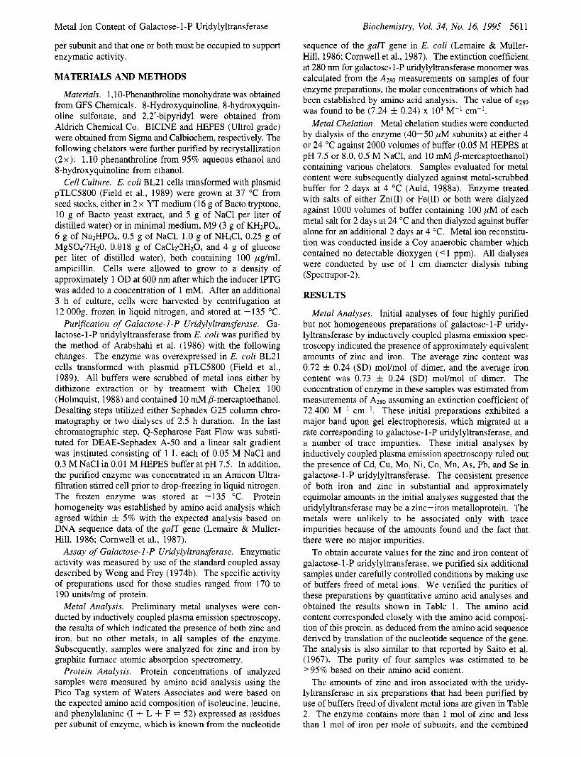
growth medium enzyme Zn (mol/mol)" enzyme Fe (mol/moly Zn i- Fe (moVmol)" specific activityb ZnbM) FebM) (1) enriched medium (2x YT) 1.19 z t 0.13' 0.59 f 0.04 1.78 168 f 2 22 21 (2) M9 + 20 yM ZnS04 1.09 f 0.07 0.23 f 0.01 1.32 174 f 6 21 3 (3) M9 + 100 yM ZnSO4 2.07 f 0.12 0.21 f 0.04 2.28 175 f 7 96 3 (4) M9 + 20 pM FeS04 0.85 f 0.04 1.47 f 0.05 2.32 149 f 3 0.8 22 (5) M9 + 200 pM FeS04 0.12 f 0.01 2.01 f 0.11 2.13 197 f 12 1 196 (6) M9 + 10 yM ZnS04 1.21 f 0.05 0.08 f 0.01 1.29 150 f 3 8 2
Metal content per mole of enzyme subunits. Units per milligram of protein (fSD in quadruplicate assays). MEAN f SD.
h
.I E Q) c) E PI
% ; m c) .I
E 2
Y
h
P c) .I
.I Y 50 -
5 0 1 2 3 4
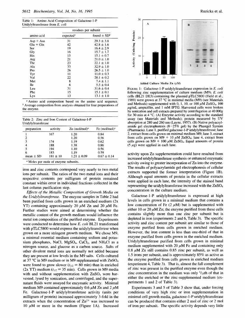
Time (Day) FIGURE 2: Instability of iron-containing uridylyltransferase during dialysis. Samples of iron-uridylyltransferase (40 pM) from experi- ment 5 of Table 3, which had been purified at 4 "C, were dialyzed against 0.05 M HEPES buffer at pH 7.5 containing 0.05 M NaCl and 10 mM P-mercaptoethanol at 24 "C inside the anaerobic chamber in the presence and absence of 0.35 mM UDP-glucose. Aliquots were removed at the indicated times, quenched by diluting into ice-cold buffer, and assayed for enzymatic activity. Symbols: (M) enzyme plus UDP-glucose; (+) enzyme alone. In the latter experiment, the iron content decreased to 0.85 mol/mol in the course of dialysis, and the activity decreased to 30% of its initial value.
on whether the metal ions are zinc or iron, and the amounts of enzyme purified from the cells are comparable. However, the iron-containing enzyme in experiment 5 of Table 3 is less stable toward dialysis at ambient temperature than the zinc-containing enzyme, as shown by the experiment of Figure 2. Both enzymes can be purified at low temperature by the usual procedure and are equally active, but the enzyme containing two irons per subunit and very little zinc quickly loses its activity upon dialysis at 24 OC, whereas the zinc- containing enzyme retains its activity for days when subjected to dialysis. Inactivation of the iron enzyme cannot have resulted from air-oxidation of Fe(I1) because the dialysis was carried out under anaerobic conditions.
Metal Ion Chelation Studies. Chelating agents can often remove divalent metal ions from proteins (Wagner, 1988; Auld, 1988b). The chelation experiments in Figures 3-5 demonstrate a dependence of enzymatic activity on the metal ion content of galactose- 1-P uridylyltransferase. 1,lO- Phenanthroline removes zinc (Figure 3A) and inactivates the enzyme (Figure 3B) in a first-order and chelator concentra- tion-dependent process in the course of 1 week. The half- times for the loss of enzyme activity in the presence of 1,lO- phenanthroline at 24 "C are 4.1 f 1 days at 5 mM and 15 f 1 days at 2 mM 1,lO-phenanthroline, respectively. The half-times for the loss of zinc are 4.0 f 1 days at 5 mM and 14 & 1 days at 2 mM 1,lO-phenanthroline, respectively. The
h
.I c)
E I
I/)
0
$ 1.00 - 3 0.75
1 v
0.50
6 0.25
5
E E3
c) E
200
150
100
50
* I
0 2 4 6 8
Time (Day)
0 2 4 6 8
Time (Day) FIGURE 3: Concentration-dependent loss of Zn and galactose-1-P uridylyltransferase activity by 1,lO-phenanthroline. Galactose- 1 -P uridylyltransferase purified from cells grown on enriched medium contained both iron and zinc. The enzyme (40 pM) was dialyzed against 0, 0.5, 2, or 5 mM 1,lO-phenanthroline (500 volumes) in HEPES buffer (0.05 M, pH 8.0) containing 0.05 M NaCl and 10 mM P-mercaptoethanol24 "C. At selected times, enzyme samples were removed and dialyzed against HEPES buffer for 48 h at 4 "C to remove the chelator. Aliquots were assayed for enzymatic activity and for zinc by graphite furnace atomic absorption spectrophotom- etry. Part A: Zn content versus time. Part B: Enzyme activity versus time. Symbols: (0) no chelator; (A) 0.5 mM 1, lO- phenanthroline; (+) 2 mM 1,lO-phenanthroline; (W) 5 mM 1,lO- phenanthroline.
uridylyl-donor substrate UDP-glucose prevents the loss of zinc and iron and enzymatic activity, as shown in Figure 4A,B, which also shows that the half-times for the extraction of iron and zinc by 1,lO-phenanthroline in the absence of UDP-glucose are the same at 33 "C. Glucose-1-P at a
5614 Biochemistry, Vol. 34, No. 16, 1995 Ruzicka et ai.
- U .- E
I
w M
U
5 s 2 0.25
0 2 4 6
Time (Day)
150 + f. Q B
0
h Y .I
E a
- 0.4
5
s ; v1
1 w
U $ 0.2
s U E
150
100
50
0 0 2 4 6
Time (Day)
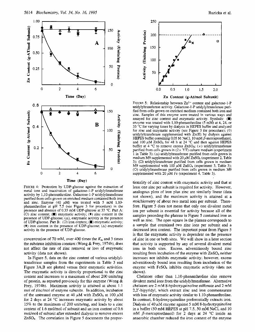
FIGURE 4: Protection by UDP-glucose against the extraction of metal ions and inactivation of galactose- 1-P uridylyltransferase activity by 1,lO-phenanthroline. Galactose- 1 -P uridylyltransferase purified from cells grown on enriched medium contained both iron and zinc. Enzyme (40 pM) was treated with 5 mM 1,lO- phenanthroline at pH 7.5 (see Figure 3 for procedure) in the presence and absence of 0.35 mM UDP-glucose at 33 "C. Part A: (0) zinc content; (W) enzymatic activity; (+) zinc content in the presence of UDP-glucose; (A), enzymatic activity in the presence of UDP-glucose. Part B: (0) iron content; (M) enzymatic activity; (6) iron content in the presence of UDP-glucose; (A) enzymatic activity in the presence of UDP-glucose.
concentration of 70 mM, over 400 times the K , and 3 times the substrate inhibition constant (Wong & Frey, 1974b), does not affect the rate of zinc removal or loss of enzymatic activity (data not shown).
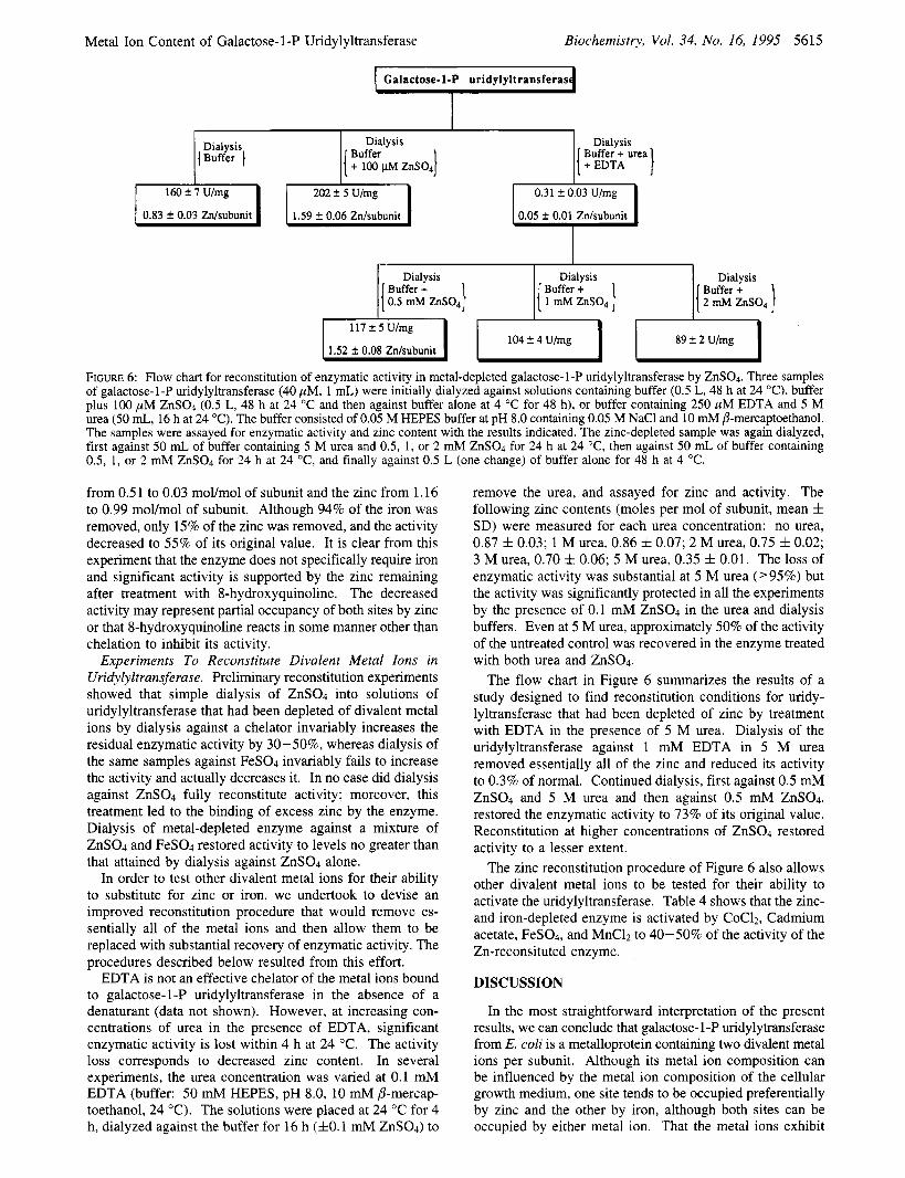
In Figure 5 , data on the zinc content of various uridylyl- transferase samples from the experiments in Table 3 and Figure 3A,B are plotted versus their enzymatic activities. The enzymatic activity is directly proportional to the zinc content and increases to a maximum of about 200 unitdmg of protein, as reported previously for this enzyme (Wong & Frey, 197413). Maximum activity is attained at about 1.1 mol of zinc/mol of enzyme subunits. In addition, incubation of the untreated enzyme at 40 p M with ZnS04 at 100 p M for 2 days at 24 "C increases enzymatic activity by about 15% to the maximum of 200 unitdmg, and leads to a zinc content of 1.6 mol/mol of subunit and an iron content of 0.6 mol/mol of subunit after extended dialysis to remove excess ZnS04. The correlation in Figure 5 documents the propor-
250 b h c *z 200 e U a
% z 150 m U .I
100 z
0.0 0.5 1.0 1.5 2.0
Zn Content (g-At/mol Subunit) FIGURE 5: Relationship between Zn2+ content and galactose-1-P uridylyltransferase activity. Galactose- 1 -P uridylyltransferase puri- fied from cells grown on enriched medium contained both iron and zinc. Samples of this enzyme were treated in various ways and assayed for zinc content and enzymatic activity. Symbols: (W) enzyme was treated with 1,lO-phenanthroline (5 mM) at 4, 24, or 33 "C for varying times by dialysis in HEPES buffer and analyzed for zinc and enzymatic activity (see Figure 3 for procedure); (0) uridylyltransferase supplemented with Zn(I1) by dialysis against HEPES buffer containing 0.05 M NaC1, 10 mM P-mercaptwthanol, and 100 pM ZnS04 for 48 h at 24 "C and then against HEPES buffer at 4 "C to remove excess ZnS04; (x) uridylyltransferase purified from cells grown in (2x YT) culture medium (experiment 1 in Table 3); (A) uridylyltransferase purified from cells grown in medium M9 supplemented with 20 pM ZnSO4 (experiment 2, Table 3); (0) uridylyltransferase purified from cells grown in medium M9 supplemented with 100 pM ZnSOd (experiment 3, Table 3); (0) uridylyltransferase purified from cells grown in medium M9 supplemented with 20 pM Fe (experiment 4, Table 3).
tionality of zinc content with enzymatic activity and that at least one zinc per subunit is required for activity. However, analogous plots of iron plus zinc are similarly linear (data not shown), and the maximum activity is attained at a stoichiometry of about two metal ions per subunit. There- fore, Figure 5 does not mean that only one divalent metal ion per subunit is essential for activity because all of the samples preceding the plateau in Figure 5 contained iron as well as zinc. The open square in the plateau corresponds to a sample that contained two zinc ions per subunit and a decreased iron content. The important point from Figure 5 is that the enzymatic activity is dependent on the presence of zinc in one or both sites. We will show in a later section that activity is supported by any of several divalent metal ions in both sites. Excess, adventitiously bound zinc resulting from incubation of the enzyme with ZnS04 neither increases nor inhibits enzymatic activity; however, excess adventitiously bound iron resulting from incubation of the enzyme with FeS04 inhibits enzymatic activity (data not shown).
Chelators other than 1,lO-phenanthroline also remove divalent metal ions from the uridylyltransferase. Alternative chelators are 2 mM 8-hydroxyquinoline sulfonate and 2 mM 2,2'-bipyridyl, which extract zinc and iron commensurate with loss of enzymatic activity similar to 1,lO-phenanthroline. In contrast, 8-hydroxyquinoline preferentially extracts iron. Dialysis of 40 p M enzyme against 5 mM 8-hydroxyquinoline in a buffer (50 mM HEPES at pH 7.5, 50 mM NaCl, and 10 mM P-mercaptoethanol) for 2 days at 24 "C inside an anaerobic chamber reduced the iron content of the enzyme
Metal Ion Content of Galactose- 1 -P Uridylyltransferase Biochemistry, Vol. 34, No. 16, 1995 5615
Galactose-1-P uridylyltransferas
Dialysis {Buffer 1
Dialysis I ;%k; urea 1 I
0.31 f 0.03 U/mg
Dialysis I( Buffer + 0.5 m M ZnSO,]
Dialysis
2 mM ZnSOp ] 1 Buffer +
1.52 f 0.08 Znhbunit FlodtdUimglI-( I I --
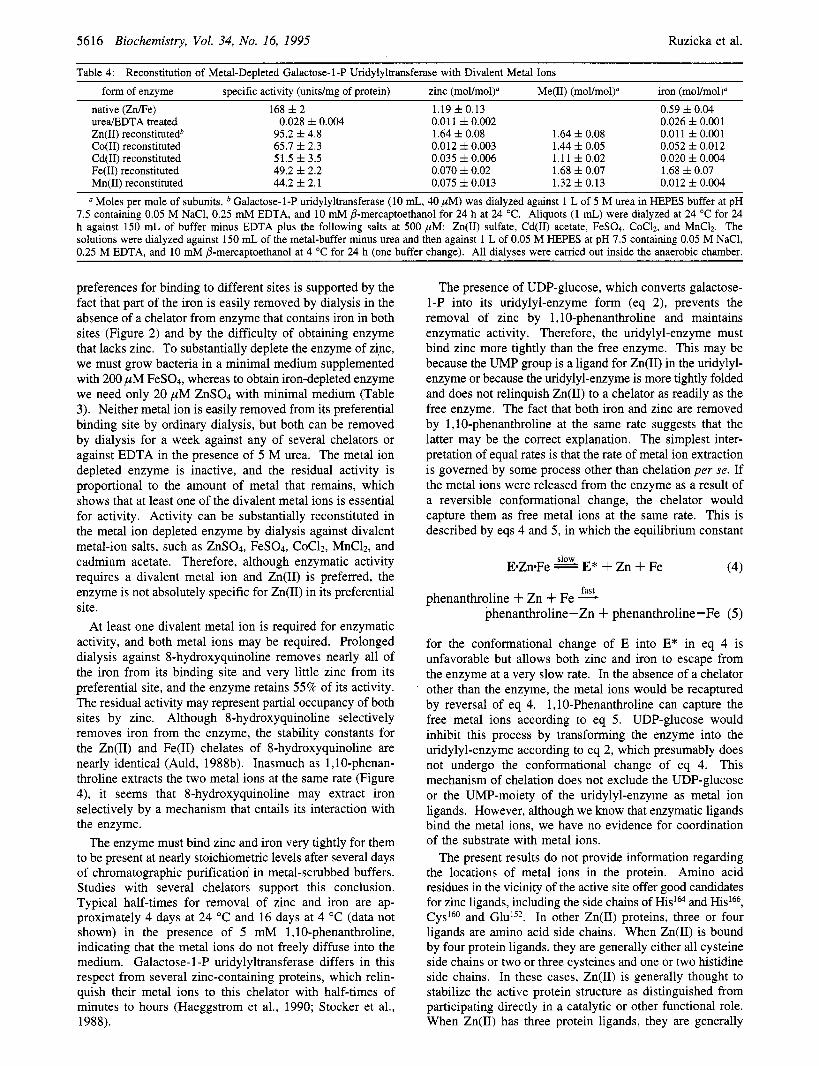
FIGURE 6: Flow chart for reconstitution of enzymatic activity in metal-depleted galactose-l-P uridylyltransferase by ZnS04. Three samples of galactose-l-P uridylyltransferase (40 pM, 1 mL) were initially dialyzed against solutions containing buffer (0.5 L, 48 h at 24 "C), buffer plus 100 pM ZnS04 (0.5 L, 48 h at 24 "C and then against buffer alone at 4 "C for 48 h), or buffer containing 250 pM EDTA and 5 M urea (50 mL, 16 h at 24 "C). The buffer consisted of 0.05 M HEPES buffer at pH 8.0 containing 0.05 M NaCl and 10 mM P-mercaptoethanol. The samples were assayed for enzymatic activity and zinc content with the results indicated. The zinc-depleted sample was again dialyzed, first against 50 mL of buffer containing 5 M urea and 0.5, 1, or 2 mM ZnS04 for 24 h at 24 "C, then against 50 mL of buffer containing 0.5, 1, or 2 mM ZnS04 for 24 h at 24 "C, and finally against 0.5 L (one change) of buffer alone for 48 h at 4 "C.
from 0.51 to 0.03 mol/mol of subunit and the zinc from 1.16 to 0.99 moymol of subunit. Although 94% of the iron was removed, only 15% of the zinc was removed, and the activity decreased to 55% of its original value. It is clear from this experiment that the enzyme does not specifically require iron and significant activity is supported by the zinc remaining after treatment with 8-hydroxyquinoline. The decreased activity may represent partial occupancy of both sites by zinc or that 8-hydroxyquinoline reacts in some manner other than chelation to inhibit its activity.
Experiments To Reconstitute Divalent Metal Ions in Uridylyltransferase. Preliminary reconstitution experiments showed that simple dialysis of ZnS04 into solutions of uridylyltransferase that had been depleted of divalent metal ions by dialysis against a chelator invariably increases the residual enzymatic activity by 30-50%, whereas dialysis of the same samples against FeS04 invariably fails to increase the activity and actually decreases it. In no case did dialysis against ZnS04 fully reconstitute activity; moreover, this treatment led to the binding of excess zinc by the enzyme. Dialysis of metal-depleted enzyme against a mixture of ZnS04 and FeS04 restored activity to levels no greater than that attained by dialysis against ZnS04 alone.
In order to test other divalent metal ions for their ability to substitute for zinc or iron, we undertook to devise an improved reconstitution procedure that would remove es- sentially all of the metal ions and then allow them to be replaced with substantial recovery of enzymatic activity. The procedures described below resulted from this effort.
EDTA is not an effective chelator of the metal ions bound to galactose-l-P uridylyltransferase in the absence of a denaturant (data not shown). However, at increasing con- centrations of urea in the presence of EDTA, significant enzymatic activity is lost within 4 h at 24 "C. The activity loss corresponds to decreased zinc content. In several experiments, the urea concentration was varied at 0.1 mM EDTA (buffer: 50 mM HEPES, pH 8.0, 10 mM P-mercap- toethanol, 24 "C). The solutions were placed at 24 "C for 4 h, dialyzed against the buffer for 16 h ( fO. l mM ZnS04) to
remove the urea, and assayed for zinc and activity. The following zinc contents (moles per mol of subunit, mean f SD) were measured for each urea concentration: no urea, 0.87 f 0.03; 1 M urea, 0.86 f 0.07; 2 M urea, 0.75 f 0.02; 3 M urea, 0.70 f 0.06; 5 M urea, 0.35 f 0.01. The loss of enzymatic activity was substantial at 5 M urea ('95%) but the activity was significantly protected in all the experiments by the presence of 0.1 mM ZnS04 in the urea and dialysis buffers. Even at 5 M urea, approximately 50% of the activity of the untreated control was recovered in the enzyme treated with both urea and ZnS04.
The flow chart in Figure 6 summarizes the results of a study designed to find reconstitution conditions for uridy- lyltransferase that had been depleted of zinc by treatment with EDTA in the presence of 5 M urea. Dialysis of the uridylyltransferase against 1 mM EDTA in 5 M urea removed essentially all of the zinc and reduced its activity to 0.3% of normal. Continued dialysis, first against 0.5 mM ZnS04 and 5 M urea and then against 0.5 mM ZnSO4, restored the enzymatic activity to 73% of its original value. Reconstitution at higher concentrations of ZnS04 restored activity to a lesser extent.
The zinc reconstitution procedure of Figure 6 also allows other divalent metal ions to be tested for their ability to activate the uridylyltransferase. Table 4 shows that the zinc- and iron-depleted enzyme is activated by CoC12, Cadmium acetate, FeS04, and MnClz to 40-50% of the activity of the Zn-reconsituted enzyme.
DISCUSSION
In the most straightforward interpretation of the present results, we can conclude that galactose- l-P uridylytransferase from E. coli is a metalloprotein containing two divalent metal ions per subunit. Although its metal ion composition can be influenced by the metal ion composition of the cellular growth medium, one site tends to be occupied preferentially by zinc and the other by iron, although both sites can be occupied by either metal ion. That the metal ions exhibit
5616 Biochemistry, Vol. 34, No. 16, 1995 Ruzicka et al.
Table 4: Reconstitution of Metal-Depleted Galactose-1-P Uridylyltransferase with Divalent Metal Ions form of enzyme
native (Zn/Fe) ureaEDTA treated Zn(I1) reconstitutedb Co(I1) reconstituted Cd(I1) reconstituted Fe(I1) reconstituted Mn(I1) reconstituted
specific activity (unitdmg of protein) 168 f 2
0.028 f 0.004 95.2 f 4.8 65.7 f 2.3 51.5 f 3.5 49.2 f 2.2 44.2 f 2.1
zinc (moVmo1)" Me(I1) (mol/mol)" iron (mol/mol)" 1.19 f 0.13 0.011 f 0.002 1.64 f 0.08 0.012 f 0.003 0.035 f 0.006 0.070 f 0.02 0.075 f 0.013
0.59 f 0.04 0.026 f 0.001 0.011 f 0.001 0.052 f 0.012 0.020 f 0.004 1.68 f 0.07 0.012 f 0.004
1.64 f 0.08 1.44 f 0.05 1.11 f 0.02 1.68 f 0.07 1.32 f 0.13
Moles per mole of subunits. Galactose-1-P uridylyltransferase (10 mL, 40 pM) was dialyzed against 1 L of 5 M urea in HEPES buffer at pH 7.5 containing 0.05 M NaC1, 0.25 mM EDTA, and 10 mM j3-mercaptoethanol for 24 h at 24 "C. Aliquots (1 mL) were dialyzed at 24 "C for 24 h against 150 mL of buffer minus EDTA plus the following salts at 500 pM: Zn(I1) sulfate, Cd(I1) acetate, FeS04, CoC12, and MnC12. The solutions were dialyzed against 150 mL of the metal-buffer minus urea and then against 1 L of 0.05 M HEPES at pH 7.5 containing 0.05 M NaCl, 0.25 M EDTA, and 10 mM B-mercaptoethanol at 4 "C for 24 h (one buffer change). All dialyses were carried out inside the anaerobic chamber.
preferences for binding to different sites is supported by the fact that part of the iron is easily removed by dialysis in the absence of a chelator from enzyme that contains iron in both sites (Figure 2) and by the difficulty of obtaining enzyme that lacks zinc. To substantially deplete the enzyme of zinc, we must grow bacteria in a minimal medium supplemented with 200 p M FeS04, whereas to obtain iron-depleted enzyme we need only 20 p M ZnS04 with minimal medium (Table 3). Neither metal ion is easily removed from its preferential binding site by ordinary dialysis, but both can be removed by dialysis for a week against any of several chelators or against EDTA in the presence of 5 M urea. The metal ion depleted enzyme is inactive, and the residual activity is proportional to the amount of metal that remains, which shows that at least one of the divalent metal ions is essential for activity. Activity can be substantially reconstituted in the metal ion depleted enzyme by dialysis against divalent metal-ion salts, such as ZnS04, FeS04, CoC12, MnC12, and cadmium acetate. Therefore, although enzymatic activity requires a divalent metal ion and Zn(I1) is preferred, the enzyme is not absolutely specific for Zn(I1) in its preferential site.
At least one divalent metal ion is required for enzymatic activity, and both metal ions may be required. Prolonged dialysis against 8-hydroxyquinoline removes nearly all of the iron from its binding site and very little zinc from its preferential site, and the enzyme retains 55% of its activity. The residual activity may represent partial occupancy of both sites by zinc. Although 8-hydroxyquinoline selectively removes iron from the enzyme, the stability constants for the Zn(I1) and Fe(I1) chelates of 8-hydroxyquinoline are nearly identical (Auld, 1988b). Inasmuch as 1 ,lo-phenan- throline extracts the two metal ions at the same rate (Figure 4), it seems that 8-hydroxyquinoline may extract iron selectively by a mechanism that entails its interaction with the enzyme.
The enzyme must bind zinc and iron very tightly for them to be present at nearly stoichiometric levels after several days of chromatographic purification in metal-scrubbed buffers. Studies with several chelators support this conclusion. Typical half-times for removal of zinc and iron are ap- proximately 4 days at 24 OC and 16 days at 4 "C (data not shown) in the presence of 5 mh4 1,lO-phenanthroline, indicating that the metal ions do not freely diffuse into the medium. Galactose- 1 -P uridylyltransferase differs in this respect from several zinc-containing proteins, which relin- quish their metal ions to this chelator with half-times of minutes to hours (Haeggstrom et al., 1990; Stocker et al., 1988).
The presence of UDP-glucose, which converts galactose- 1-P into its uridylyl-enzyme form (eq 2), prevents the removal of zinc by 1,lO-phenanthroline and maintains enzymatic activity. Therefore, the uridylyl-enzyme must bind zinc more tightly than the free enzyme. This may be because the UMP group is a ligand for Zn(I1) in the uridylyl- enzyme or because the uridylyl-enzyme is more tightly folded and does not relinquish Zn(I1) to a chelator as readily as the free enzyme. The fact that both iron and zinc are removed by 1,lO-phenanthroline at the same rate suggests that the latter may be the correct explanation. The simplest inter- pretation of equal rates is that the rate of metal ion extraction is governed by some process other than chelation per se. If the metal ions were released from the enzyme as a result of a reversible conformational change, the chelator would capture them as free metal ions at the same rate. This is described by eqs 4 and 5, in which the equilibrium constant
slow EZn-Fe E* + Zn + Fe (4)
fast phenanthroline + Zn + Fe -
phenanthroline-Zn + phenanthroline-Fe (5)
for the conformational change of E into E* in eq 4 is unfavorable but allows both zinc and iron to escape from the enzyme at a very slow rate. In the absence of a chelator other than the enzyme, the metal ions would be recaptured by reversal of eq 4. 1,lO-Phenanthroline can capture the free metal ions according to eq 5. UDP-glucose would inhibit this process by transforming the enzyme into the uridylyl-enzyme according to eq 2, which presumably does not undergo the conformational change of eq 4. This mechanism of chelation does not exclude the UDP-glucose or the UMP-moiety of the uridylyl-enzyme as metal ion ligands. However, although we know that enzymatic ligands bind the metal ions, we have no evidence for coordination of the substrate with metal ions.
The present results do not provide information regarding the locations of metal ions in the protein. Amino acid residues in the vicinity of the active site offer good candidates for zinc ligands, including the side chains of Hisla and His166, CysI6O and G ~ U ' ~ ~ . In other Zn(I1) proteins, three or four ligands are amino acid side chains. When Zn(I1) is bound by four protein ligands, they are generally either all cysteine side chains or two or three cysteines and one or two histidine side chains. In these cases, Zn(I1) is generally thought to stabilize the active protein structure as distinguished from participating directly in a catalytic or other functional role. When Zn(I1) has three protein ligands, they are generally
Metal Ion Content of Galactose- I-P Uridylyltransferase
selected from the side chains of histidine, cysteine, and glutamate, with water as the fourth ligand (Vallee & Auld, 1990b). In these cases, the protein is generally an enzyme, and Zn(I1) is thought to participate in catalysis, although it is no doubt also important in maintaining structure in the active site.
Several enzymes have Zn(I1) ligands donated by two histidines that are separated by one or three other amino acids in the sequence (Vallee & Auld, 1990a). For example, in carbonic anhydrase, two histidines in the active site are separated by phenylalanine, and in thermolysins, they are separated by glutamate and two other amino acids. Galactose- 1-P uridylyltransferase from E. coli contains two sequences of HPH, the active site-residues H1&1&166 and the nones- sential histidines in the sequence H9PloH11. The latter sequence is flanked by an aspartate at position eight. Either or both sequences may be considered as possible sources of side chain ligands for metal ions. Moreover, the protein contains six cysteine residues, including Cys5* and C y P , in addition to CYS'~O, that could participate in binding metal ions. With these residues, and a cluster of acidic residues in the sequence E~l,*,AEREDRLQKEYFAE~19*~, there are many possibilities for binding metal ions to this protein.
The finding of Zn in uridylyltransferase places it among the Zn-containing class I1 nucleotidyl transferase enzymes, which include DNA polymerase (Slater et al., 1971; Spring- gate et al., 1973), RNA-dependent DNA polymerase (Cole- man, 1974; Auld et al., 1974), and RNA polymerase (Scrutton et al., 1971). Although stoichiometric amounts of Zn have been found, it is not clear what role it plays in these enzymes. In fact, in the case of DNA polymerase I from E. coli and T7 RNA polymerase, enzymes fully active for DNA polymerization or transcription but devoid of Zn have been purified (Walton et al., 1982; King et al., 1986). DNA polymerase from E. coli is a multifunctional enzyme that catalyzes not only DNA polymerization but also 3'- and 5'- exonuclease activity on distinct substrate binding and catalytic sites. The 3'- exonuclease activity appears to be dependent on divalent cations such as Zn(I1) (Beese & Steitz, 1991). A structural role has been suggested for Zn(I1) in aspartate carbamoyltransferase (Honzatko et al., 1982), another class I1 enzyme.
The present studies do not establish a specific role for metal ions in galactose- 1 -P uridylyltransferase, apart from being required for enzymatic activity. The fact that Co(II), Cd(II), and Mn(I1) support activity 40-50% as well as Zn- (11) or Fe(I1) indicates that the metal ions may be more important in a structural role than as direct participants in catalysis. The X-ray crystallographic studies of galactose- I-P uridylyltransferase in our laboratory are at an advanced stage and will resolve this isssue (Wedekind et al., 1994).
ACKNOWLEDGMENT
We are pleased to acknowledge with thanks the kind assistance given to us by Dr. Bert Vallee, Dr. James Riordan, and Dr. Robert Shapiro during a visit to their laboratory in the critical early stage of this research. We appreciate the access to the use of their facilities in generous measure. The techniques for accurately measuring the metal ion content of uridylyltransferase were acquired under their guidance. We also thank Dr. Vallee for his valuable comments on the manuscript.
Biochemistry, Vol. 34, No. 16, 1995 5617
REFERENCES
Arabshahi, A., Brody, R. S., Smallwood, A., Tsai, T.-C., & Frey,
Auld, D. S. (1988a) Methods Enzymol. 158, 13-14. Auld, D. S. (1988b) Methods Enzymol. 158, 110- 114. Auld, D. S., Kawaguchi, H., Livingston, D. M., & Vallee, B. L.
(1974) Proc. Natl. Acad. Sci. U.S.A. 71, 2091-2095. Beese, L. S., & Steitz, T. A. (1991) EMBO J. 10, 25-33. Coleman, J. E. (1974) Biochem. Biophys. Res. Commun. 60, 641 -
648. Comwell, T. L., Adhya, S. L., Reznikoff, W. S., & Frey, P. A.
(1987) Nucleic Acids Res. 15, 8116. Field, T. L., Reznikoff, W. S., & Frey, P. A. (1989) Biochemistry
28, 2094-2099. Frey, P. A., Wong, L.-J., Sheu, K.-F., & Yang, S.-L. (1982) Methods
Enzymol. 87, 20-36. Haeggstrom, J. Z., Wetterholm, A., Shapiro, R., Vallee, B. L., &
Samuelsson, B. (1990) Biochem. Biophys. Res. Commun. 172, 965-970.
Hester, L. S., & Raushel, F. M. (1987) J. Biol. Chem. 262, 12092-
Holmquist, B. (1988) Methods Enzymol. 158, 6-12. Honzatko, R. B., Crawford, J. L., Monaco, H. L., Ladner, J. E.,
Edwards, B. F. P., Evans, D. R., Warren, S. G., Wiley, D. C., Ladner, R. C., & Lipscomb, W. N. (1982) J. Mol. Biol. 160, 2 19-263.
Kalckar, H. M. (1960) Fed. Proc., Fed. Am. SOC. Exp. Biol. 19, 984-990.
Kim, J., Ruzicka, F., & Frey, P. A. (1990) Biochemistry 29, 10590- 10593.
King, G. C., Martin, C. T., Pham, T. T., & Coleman, J. E. (1986) Biochemistry 25, 36-40.
Layne, E. (1957) Methods Enzymol. 3, 4477454. Lemaire, H. G., & Muller-Hill, B. (1986) Nucleic Acids Res. 14,
Levy, H. L., & Hammersen, G. (1978) J. Pediatr. 92, 871-877. Reichardt, J. K., & Woo, S. L. C. (1991) Proc. Natl. Acad. Sci.
Reichardt, J. K. V., & Berg, P. (1988) Nucleic Acids Res. 16, 9017-
Saito, S . , Ozutsumi, M., & Kurahashi, K. (1967) J . Biol. Chem.
Scrutton, M. C., Wu, C. W., & Goldthwait, D. A. (1971) Proc.
Slater, J. P., Mildvan, A. S . , & Loeb, L. A. (1971) Biochem.
Springgate, C. F., Mildvan, A. S., Abramson, R., Engle, J. L., &
Stocker, W., Wolz, R. L., Zwilling, R., Strydom, D. J., & Auld, D.
P. A. (1986) Biochemistry 25, 5583-5589.
12095.
7705-777 1.
USA 88, 2633-2637.
9026.
242, 2362-2368.
Natl. Acad. Sci. U.S.A. 68, 2497-2501.
Biophys. Res. Commun. 44, 37-43.
Loeb, L. A. (1973) J. Biol. Chem. 248, 5987-5993.
S. (1988) Biochemistry 27, 5026-5032. Vallee, B. L., & Auld, D. S. (1990a) Proc. Natl. Acad. Sci. U.S.A.
87, 220-224. Vallee, B. L., & Auld, D. S . (1990b) Biochemistry 29, 5647-5659. Wagner, F. W. (1988) Methods Enzymol. 158, 21-32. Walton, K. E., FitzGerald, P. C., Hemnann, M. S., & Behnke, W.
D. (1982) Biochem. Biophys. Res. Commun. 108, 1353-1361. Wedekind, J. E., Frey, P. A., & Rayment. I. (1994) Acta Crystal-
logr., Sect. D D50, 329-331. Wong, L.-J., & Frey, P. A. (1974a) J. Biol. Chem. 249, 2322-
2324. Wong, L.-J., & Frey, P. A. (1974b) Biochemistry 13, 3889-3894. Wong, L.-J., Sheu, K.-F., Lee, S.-L., & Frey, P. A. (1977)
Yang, S.-L. L., & Frey, P. A. (1979) Biochemistry 18, 2980-2984. Biochemistry 16, 1010 -1016.
BI942392Q