Misfolding of galactose 1-phosphate uridylyltransferase ...Misfolding of galactose 1-phosphate...
38
Misfolding of galactose 1-phosphate uridylyltransferase can result in type I galactosemia McCorvie, T. J., Gleason, T. J., Fridovich-Keil, J. L., & Timson, D. J. (2013). Misfolding of galactose 1-phosphate uridylyltransferase can result in type I galactosemia. Biochimica et biophysica acta, 1832(8), 1279-1293. https://doi.org/10.1016/j.bbadis.2013.04.004 Published in: Biochimica et biophysica acta Document Version: Peer reviewed version Queen's University Belfast - Research Portal: Link to publication record in Queen's University Belfast Research Portal Publisher rights © 2013, Elsevier B. V. Licensed under the Creative Commons Attribution -NonCommercial-NoDerivs License (https://creativecommons.org/licenses/by-nc-nd/4.0/), which permits distribution and reproduction for non-commercial purposes, provided the author and source are cited. General rights Copyright for the publications made accessible via the Queen's University Belfast Research Portal is retained by the author(s) and / or other copyright owners and it is a condition of accessing these publications that users recognise and abide by the legal requirements associated with these rights. Take down policy The Research Portal is Queen's institutional repository that provides access to Queen's research output. Every effort has been made to ensure that content in the Research Portal does not infringe any person's rights, or applicable UK laws. If you discover content in the Research Portal that you believe breaches copyright or violates any law, please contact [email protected]. Download date:05. Feb. 2020
Transcript of Misfolding of galactose 1-phosphate uridylyltransferase ...Misfolding of galactose 1-phosphate...

Misfolding of galactose 1-phosphate uridylyltransferase can result intype I galactosemia
McCorvie, T. J., Gleason, T. J., Fridovich-Keil, J. L., & Timson, D. J. (2013). Misfolding of galactose 1-phosphateuridylyltransferase can result in type I galactosemia. Biochimica et biophysica acta, 1832(8), 1279-1293.https://doi.org/10.1016/j.bbadis.2013.04.004
Published in:Biochimica et biophysica acta
Document Version:Peer reviewed version
Queen's University Belfast - Research Portal:Link to publication record in Queen's University Belfast Research Portal
Publisher rights© 2013, Elsevier B. V. Licensed under the Creative Commons Attribution -NonCommercial-NoDerivs License(https://creativecommons.org/licenses/by-nc-nd/4.0/), which permits distribution and reproduction for non-commercial purposes, provided theauthor and source are cited.
General rightsCopyright for the publications made accessible via the Queen's University Belfast Research Portal is retained by the author(s) and / or othercopyright owners and it is a condition of accessing these publications that users recognise and abide by the legal requirements associatedwith these rights.
Take down policyThe Research Portal is Queen's institutional repository that provides access to Queen's research output. Every effort has been made toensure that content in the Research Portal does not infringe any person's rights, or applicable UK laws. If you discover content in theResearch Portal that you believe breaches copyright or violates any law, please contact [email protected].
Download date:05. Feb. 2020
Misfolding of galactose 1-phosphate uridylyltransferase canresult in type I galactosemia
Thomas J McCorvie1, Tyler J Gleason2, Judith L Fridovich-Keil2, and David J Timson1,**
1School of Biological Sciences, Queen’s University Belfast, Medical Biology Centre, 97 LisburnRoad, Belfast, BT9 7BL. UK2Department of Human Genetics, Emory University School of Medicine, Atlanta, Georgia, USA
AbstractType I galactosemia is a genetic disorder that is caused by the impairment of galactose-1-phosphate uridylyltransferase (GALT; EC 2.7.7.12). Although a large number of mutations havebeen detected through genetic screening of the human GALT (hGALT) locus, for many it is notknown how they cause their effects. The majority of these mutations are missense, with predictedsubstitutions scattered throughout the enzyme structure and thus causing impairment by othermeans rather than direct alterations to the active site. To clarify the fundamental, molecular basisof hGALT impairment we studied five disease-associated variants p.D28Y, p.L74P, p.F171S,p.F194L and p.R333G using both a yeast model and purified, recombinant proteins. In a yeastexpression system there was a correlation between lysate activity and the ability to rescue growthin the presence of galactose, except for p.R333G. Kinetic analysis of the purified proteinsquantified each variant’s level of enzymatic impairment and demonstrated that this was largelydue to altered substrate binding. Increased surface hydrophobicity, altered thermal stability andchanges in proteolytic sensitivity were also detected. Our results demonstrate that hGALT requiresa level of flexibility to function optimally and that altered folding is the underlying reason ofimpairment in all the variants tested here. This indicates that misfolding is a common, molecularbasis of hGALT deficiency and suggests the potential of pharmacological chaperones andproteostasis regulators as novel therapeutic approaches for type I galactosemia.
KeywordsGALT; yeast model; disease associated mutation; stability; substrate binding; protein misfolding
1. IntroductionType I galactosemia (OMIM #230400) is a genetic disorder that is caused by impairment ofgalactose-1-phosphate uridylyltransferase (GALT; EC 2.7.7.12) [1]. Two other forms ofgalactosemia are also recognized: galactokinase deficiency (type II; OMIM #230200) andUDP-galactose 4′-epimerase deficiency (type III; OMIM #230350) [2;3]. GALT is involvedin the metabolism of galactose and it catalyses the reversible conversion of UDP-glucose
© 2013 Elsevier B.V. All rights reserved.**Author to whom correspondence should be addressed. School of Biological Sciences, Queen’s University Belfast, Medical BiologyCentre, 97 Lisburn Road, Belfast, BT9 7BL. UK., Tel: +44(0)28 9097 5875, Fax: +44(0)28 9097 5877, [email protected].
Publisher's Disclaimer: This is a PDF file of an unedited manuscript that has been accepted for publication. As a service to ourcustomers we are providing this early version of the manuscript. The manuscript will undergo copyediting, typesetting, and review ofthe resulting proof before it is published in its final citable form. Please note that during the production process errors may bediscovered which could affect the content, and all legal disclaimers that apply to the journal pertain.
NIH Public AccessAuthor ManuscriptBiochim Biophys Acta. Author manuscript; available in PMC 2014 August 01.
Published in final edited form as:Biochim Biophys Acta. 2013 August ; 1832(8): 1279–1293. doi:10.1016/j.bbadis.2013.04.004.
NIH
-PA Author Manuscript
NIH
-PA Author Manuscript
NIH
-PA Author Manuscript

and galactose-1-phosphate to UDP-galactose and glucose-1-phosphate via an uridylatedenzyme intermediate [4;5]. Deficiency of human GALT (hGALT) is detected throughnewborn screening in many developed countries minimizing the acute pathology that canotherwise include jaundice, cataracts, vomiting, diarrhea, hepatomegaly, sepsis and neonataldeath [6]. Galactose restriction in the diet can immediately mitigate or prevent these acutemanifestations, but does not appear to prevent longer-term complications that includeovarian failure and disabilities in learning and speech, among other problems [7]. Theunderlying mechanism of these long-term pathologies is not fully known, and the role ofaccumulated galactose-1-phosphate in the process remains controversial [8]. In addition,understanding phenotype-genotype correlations is difficult as compound heterozygosityplays a role in disease [9]. This is because the hGALT protein functions as a dimer (Figure1) [10-12]. However, recently the level of predicted residual GALT activity associated withgenotype of a cohort of school-age children with type I galactosemia was demonstrated toinfluence the level of scholastic achievement of those students in mathematics [13].
To date, 264 variants have been reported from genetic screening of the hGALT gene. Ofthese, 159 are missense mutations and for the majority it is not known how they cause theireffects [14;15]. The most commonly detected severe mutant, Q188R, and selected othershave been studied using a yeast model [16-21] which has provided useful information aboutthe severity of each mutation in vivo. However, detailed functional and structural analyseshave been lacking, as only a small number of variants have been studied in any detail invitro [12;22-27].
It is interesting that although a number of mutations are located in the active site of hGALTand therefore are predicted to affect catalysis directly, the majority are located elsewherethroughout the enzyme’s structure [28;29]. Computational analysis using a homology modelof hGALT has suggested that these mutations alter hydrogen bond networks andhydrophobic interactions. Decreased monomer stability was predicted for over half of thestudied variants, which suggests that they may cause protein misfolding [28;29]. Morerecently it has been shown that disease-associated mutants affect the expression andsolubility of hGALT in an E. coli expression system. Molecular dynamics simulationspredicted that these mutations affect the overall flexibility of the enzyme thus alteringsubstrate affinity [30]. Similarly, previous studies have shown that some mutants can causetemperature sensitivity and decreased levels of expression in yeast [20;21]. Effects on dimerformation have also been detected which further supports the hypothesis that alterations inoverall structure are involved [12;25].
Since misfolding has not been experimentally verified for the majority of hGALT mutants[15] five representative variants, p.D28Y, p.L74P, p.F171S, p.F194L and p.R333G werestudied here with the aim of establishing whether, or not, this is a common feature ofvariants associated with type I galactosemia. These variants have been previously found tobe associated with type I galactosemia (Table S1) and all five variants are classified aspathogenic in the hGALT mutant database [14]. Only p.F171S and p.L74P are located at theactive site (Figure 1) and both have been shown to severely impair enzyme activity (TableS1) [19;20;31]. The remaining three variants are located away from the active site and allfive have been included in a recent molecular modelling study of variant GALT enzymes[29]. Thus the studied set represents a diverse group of mutants, which have previously beenclinically characterised (Table S1) and subject to, at least, some theoretical analysis. Each ofthe five mutants was studied in terms of their effects in vivo using an established yeastmodel and in vitro with the recombinant, purified variant proteins from a bacterialexpression system to determine their stability, substrate binding, ability to dimerise andenzyme kinetics in the forward and reverse directions.
McCorvie et al. Page 2
Biochim Biophys Acta. Author manuscript; available in PMC 2014 August 01.
NIH
-PA Author Manuscript
NIH
-PA Author Manuscript
NIH
-PA Author Manuscript
2. Materials & Methods2.1 Expression of hGALT alleles in yeast
Each hGALT allele was recreated by site-directed mutagenesis of the centromeric yeastvector pMM22.hGALT as described previously [20;21] and confirmed by dideoxysequencing of the entire GALT open reading frame. Creation and analysis of the F171Ssubstitution has been described previously in the context of other studies [19;20]. Theprimers used to generate these alleles are listed in Table S2.
Each plasmid was transformed into each of two haploid strains of Saccharomycescerevisiae: JFy3747 [21], which is deficient in GAL7, the gene encoding endogenous yeastGALT [32], and JFy5555, which is deficient in GAL7 and also deficient in GAL1 andGAL10, which encode the endogenous yeast GALK and GALE enzymes, respectively [32].JFy3747 was used as the host for all growth curve experiments, and JFy5555 was used asthe host for all biochemical studies performed using yeast lysates. All yeast strains weregrown on medium lacking tryptophan to maintain selection for the MM22-based plasmids.
2.2 Enzyme activities from soluble yeast lysatesGALT activity assays were performed using soluble protein lysates from JFy5555expressing each of the desired GALT alleles, essentially as described previously [21] exceptthat progress of the reaction was quantified by monitoring the appearance of UDP-galactose(in nmol UDP-gal/μg protein/min). Because the host yeast were deficient in GALK andGALE as well as endogenous GALT there was essentially no background conversion ofUDP-glc to UDP-gal by GALE in the absence of GALT activity. The average ± SD (n=3) ofGALT enzyme activity for yeast expressing each allele was normalized to the activity levelobserved in yeast expressing wild-type hGALT from the same plasmid backbone.
2.3 Yeast growth studiesColonies of JFy3747 yeast expressing the desired alleles of hGALT were cultured andassessed for growth in the wells of 96 well plates using SGE-trp medium with and without0.01% galactose, as described previously [21]. OD600 readings from any wells that showedevidence of air bubbles or clumping were excluded from analysis. The average ± SD ofOD600 readings from 3 separate wells representing yeast expressing each GALT allele andgalactose condition were plotted.
2.4 Expression and purification of recombinant proteinsThe gene encoding hGALT was amplified by PCR from the IMAGE clone [33] number3922902 and was cloned into the NdeI and EcoRI sites of pET43a using primers, whichincorporated sequence encoding a hexahistidine tag at the 5′ end. The insertion of the geneinto the recombinant expression vector was verified by sequencing and this plasmid wasthen transformed into E. coli Rosetta(DE3) (Merck, Nottingham, UK). Single coloniesresulting from this transformation were picked and grown in 5 ml of LB (supplemented with100 μg.ml−1 ampicillin, 34 μg.ml−1 chloramphenicol, 50 μM ZnCl2), shaking at 30°Covernight. This culture was then diluted into 1 L of LB (supplemented with 100 μg.ml−1
ampicillin and 34 μg.ml−1 chloramphenicol, 50 μM ZnCl2) and grown, shaking at 30 °Cuntil A600nm was between 0.6 and 1.0 (typically 6 h). At this point the culture was inducedwith 1 mM IPTG at 15 °C and grown for a further 20 h. Cells were harvested bycentrifugation at 4,200μg for 20 min and cell pellets were resuspended in buffer R (50 mMHEPES, 5 mM imidazole, pH 7.5, 150 mM NaCl, 10 % (v/v) glycerol, 5 mM DTT). Thesesuspensions were frozen at −80 °C until required.
McCorvie et al. Page 3
Biochim Biophys Acta. Author manuscript; available in PMC 2014 August 01.
NIH
-PA Author Manuscript
NIH
-PA Author Manuscript
NIH
-PA Author Manuscript
The cell suspensions were thawed and the cells broken by sonication on ice (three 30 spulses of 100 W with 30 s gaps in between for cooling). The extract was centrifuged at20,000μg for 20 min to remove insoluble material and the supernatant applied to a 1 mlnickel agarose (Sigma, Poole, UK) column. Once this solution had passed through, thecolumn was washed with 20 ml buffer W (as buffer R, expect with 500 mM NaCl and 20mM imidazole) and the protein eluted with a 2 ml wash of buffer E (buffer W supplementedwith 250 mM imidazole). The eluate was further purified by size exclusion chromatographyon a Sephacryl S-300 (Pharmacia) column (55 ml) at 4 °C with a mobile phase thatconsisted of 50 mM HEPES, pH 7.5, 150 mM NaCl, 10 % (v/v) glycerol, 5 mM DTT. Aflow rate of 1 ml.min−1 was used and 1 ml fractions were collected. Control proteins ofknown molecular mass were used to construct a standard curve and, thus, determine theoligomeric state of hGALT. Protein containing fractions, (judged by absorbance at 280 nm)corresponding to the molecular mass of hGALT dimers (87 kDa), were pooled together.These pooled fractions were then concentrated using Amicon Ultra-4 (Millipore) centrifugalfiltration devices (cut-off of 3 kDa) at 4 °C to a final volume of ≈ 600 μl. The proteinsolution was then divided into 30 μl aliquots and stored frozen at −80 °C.
The Quick Change protocol [34] was used to change the appropriate codons in theexpression vector. Successful mutagenesis was verified by sequencing (MWG-Biotech,Ebersburg, Germany). These mutated plasmids were used to express p.D28Y, p.L74P,p.F171S, p.F194L and p.R333G-hGALT using the same protocol as used with the wild-typeprotein.
Recombinant human UDP-glucose dehydrogenase was expressed and purified as described[35]. The expression and purification of all proteins was monitored by 10 % SDS-PAGE.All protein concentrations were estimated using the Bradford assay [36] with bovine serumalbumin as standard.
2.5 Spectroscopic measurementsIntrinsic fluorescence of each hGALT variant was measured using 5.5 μM protein in 10 mMHEPES, pH 8.8 in a total volume of 180 μl. The binding of 1-anilinonaphthalene-8-sulphonic acid (ANS-1) was used to determine the degree of surface hydrophobicity witheach variant at 5 μM in 10 mM HEPES, pH 8.8 with 100 μM ANS-1 in a total volume was200 μl. Samples with ANS-1 were incubated at room temperature in the dark for 30 minbefore measurement. Fluorescence spectra were measured (in triplicate) at room temperatureusing a Spectra Max Gemini X plate-reader fluorimeter (Molecular Devices. CA, USA) withexcitation at 280 nm, emission 300-500 nm, and a slit width of 10 nm for intrinsicfluorescence. Excitation at 370 nm, emission 420-580 nm, and a slit width of 5 nm wascarried out for ANS-1 binding. Emission spectra were averaged for each variant andcorrected for the emission of buffer only or ANS-1 in buffer only, as appropriate.
2.6 Measurement of the steady state kinetic parameters for galactose-1-phosphateuridylyltransferase
Enzymatic activities of the hGALT variants in the forward reaction were determined using aspectrophotometric coupled assay based on that described previously [19] which couples theproduction of glucose 1-phosphate to its isomerization to glucose 6-phosphate andsubsequent NADP+-dependent oxidation of this compound. The standard reaction wasperformed at 37 °C and contained 10 mM HEPES, pH 8.8, 5 mM DTT, 5 mM glucose 1,6-bisphosphate, 5 mM MgCl2, 0.8 mM NADP+, 0.03 mg glucose-6-phosphate dehydrogenase,and 0.4 mg of phosphoglucomutase. Assays were performed in triplicate in a 96 well plateformat each with a total volume of 150 μl. Kinetic constants were determined for UDP-Glcby varying its concentration from 0.01 to 1.0 mM while the concentration of Gal-1P was
McCorvie et al. Page 4
Biochim Biophys Acta. Author manuscript; available in PMC 2014 August 01.
NIH
-PA Author Manuscript
NIH
-PA Author Manuscript
NIH
-PA Author Manuscript
held at a constant 1.0 mM. Conversely the kinetic constants for Gal-1P were determined byvarying its concentration from 0.01 to 2.0 mM while UDP-Glc was held constant at 0.5 mM.The amount of NADPH produced (detected by absorption at 340 nm) is equivalent to theamount of Glc-1P formed.
Enzymatic activities of the hGALT variants in the reverse reaction were also determinedusing a spectrophotometric coupled assay based on that described previously [37] whichcouples the production of UDP-glucose to the NAD+-dependent oxidation of this compound.The standard reaction was performed at 37 °C and contained 10 mM HEPES, pH 8.8, 5 mMDTT, 5 mM MgCl2, 10 mM NAD+, 1.2 μM human UDP-glucose dehydrogenase. Assayswere performed in triplicate in a 96 well plate format each with a total volume of 150 μl.Kinetic constants were determined for UDP-gal by varying its concentration from 0.01 to1.0 mM while the concentration of Glc-1P was held at a constant 1.0 mM. Conversely thekinetic constants of Glc-1P were determined by varying its concentration from 0.01 to 2.0mM while UDP-gal was held constant at 0.5 mM. The amount NADH produced, measuredat 340 nm, is equivalent to twice the amount of UDP-Glc formed [38].
All reactions were monitored at 340 nm for 40 min at 37 °C using a Multiskan Spectrumspectrophotometer (Thermo Scientific). Controls lacking either one or both substrates wereroutinely included, for both forward and reverse kinetic assays, and always gave theexpected negative results.
The initial rate of product formation was plotted against substrate concentration andanalyzed using non-linear curve fitting of GraphPad Prism (GraphPad Software, CA, USA).The data was fitted to either Michaelis-Menten (1), Michaelis-Menten with substrateinhibition (2) or sigmoidal kinetics (3).
(1)
where is the apparent maximum, limiting rate and is the apparent Michaelisconstant.
(2)
where is the apparent dissociation constant.
(3)
where h is the Hill coefficient and is the concentration of substrate to give a rate equalto half of . [S] is the concentration of the varied substrate for all equations. Thegoodness of fit to these equations was compared using the F test and results are reported forthe best fit to the data
2.7 Chemical cross-linkingBefore the addition of a chemical cross-linker, hGALT variants (5 μM in 10 mM HEPES,pH 8.8) were incubated at 37 °C for 5 min with and without ligands (1 mM). BS3 (Sigma) orglutaraldehyde (Sigma) was then added to final concentrations of 100 μM and 0.25 % (v/v)respectively. Cross-linking was allowed to proceed for 30 min and was then halted by the
McCorvie et al. Page 5
Biochim Biophys Acta. Author manuscript; available in PMC 2014 August 01.
NIH
-PA Author Manuscript
NIH
-PA Author Manuscript
NIH
-PA Author Manuscript

addition of an equal volume of SDS loading buffer (125 mM tris-HCl, pH 6.8, 4% (w/v)SDS, 20% (v/v) glycerol, 1% (w/v) dithiothreitol, 0.002% (w/v) bromophenol blue).Samples were denatured at 95 °C for 5 min before analysis by 10 % SDS-PAGE.
2.8 Limited proteolysishGALT variants (5 μM in 10 mM HEPES, pH 8.8) were incubated at 37 °C for 5 min withand without ligands (1 mM). Thermolysin, trypsin or chymotrypsin (Sigma), as indicated,was then added as final concentrations of 240 nM, 120 nM, and 24 nM respectively.Digestion was carried out for 30 min and was stopped by the addition of an equal volume ofSDS loading buffer. Samples were denatured at 95 °C for 5 min. before analysis by 15 %SDS-PAGE.
2.9 Thermal Inactivation of hGALTThermal inactivation of hGALT variants was judged kinetically using the forward reactionsetup described in section 2.8. Aliquots (100 μl) of each active variant hGALT at 0.5 μMwere incubated in 10 mM HEPES, pH 8.8 for 15 min at temperatures ranging from 30 to 70°C (5 °C increments). These aliquots were chilled in ice immediately after incubation andthe residual activity was determined at 0.5 mM UDP-glucose and 1.0 mM galactose-1-phosphate with 50 nM hGALT. Measurements were carried on three independent assays foreach temperature and the average activity was calculated with standard deviations. Thesewere normalized to the activity of each variant at 30 °C.
2.10 Differential scanning fluorimetry assayDifferential scanning fluorimetry was carried out essentially as previously described [39;40].Protein samples were diluted in 10 mM HEPES, pH 8.8 to a final concentration of 5 μM andany ligands used were added at a final concentration of 1 mM. Sypro orange (Sigma, Poole,UK) was diluted from a 5000μ solution (manufacturer’s concentration definition) into a 50μsolution with 10 mM HEPES, pH 8.8 and was mixed well prior to each use before 1 μl wasadded to each mixture. Reactions were set up in a total volume of 20 μl in 0.2 ml PCR tubesand controls of no protein added were always included.
Reaction mixtures were loaded into a Rotor-Gene Q cycler (Qiagen) and the followingprotocol was used: High resolution melt run (460 nm source, 510 nm detector), 25 °C to 95°C ramp with a 1 °C rise for each step and no gain optimisation. The melting temperatures,(Tm), were calculated using the inbuilt analysis software. The shift in stability correspondingto the change of melting temperature, ΔTm, for each variant and ligand binding werecalculated using equations (4) and (5) respectively.
(4)
(5)
To determine the significance of the differences in Tm the one way ANOVA with Dunnettcomparison test was used.
2.11 In silico analysis of variantsThe homology model of hGALT, PDB 1R3A [41] was used to determine the location ofaltered residues. Additional homology models of p.D28Y, p.L74P, p.F171S, p.F194L andp.R333G hGALT, based on 1R3A, were obtained from the hGALT mutant structuredatabase [28] (http://bioinformatica.isa.cnr.it/GALT/). Structures were viewed using PyMol(http://www.pymol.org/).
McCorvie et al. Page 6
Biochim Biophys Acta. Author manuscript; available in PMC 2014 August 01.
NIH
-PA Author Manuscript
NIH
-PA Author Manuscript
NIH
-PA Author Manuscript

Sequence alignment was carried out using ClustalW2 [42] and all sequences were obtainedfrom the UniProt database (http://www.uniprot.org/). Conserved residues, and thoseinvolved in cofactor and metal binding were identified with ClustalW2.
In determining the effects on stability of hGALT mutants the following programs were used:Dmutant [43], PoPMusic 2.1 [44], Cupsat [45], SDM [46], Eris [47], Concoord/PBSA [48],I-Mutant 2.0 [49], MuPro [50], and Mustab [51]. When appropriate, the structure 1R3A wasused. Both thermal and denaturation options were used of the Cupsat server. Additionallyboth flexible and inflexible backbone options were used of the Eris server. When using theSDM server the mutant structures obtained from the hGALT mutant structure database wereused. The overall consensus of stability change was determined with the percentage ofagreeing predictions. All predictions were determined from the A chain contained in thecoordinate files.
Prediction of intrinsically disordered regions in hGALT was carried out using metaPrDOS[52] and Spine-D [53]; metaPrDOS uses a consensus based approach using multiplepredictors. Both predict the probability of disorder and those residues with a probability of0.5 and higher are deemed disordered. The regions predicted from these two servers weremapped onto the homology model (1R3A) using PyMol.
The FTMap server [54] was used to predict any allosteric sites in hGALT. This serverpredicts potential binding sites of proteins, which can act as the starting pointing ofidentifying ’druggable hotspots’. In addition, this server can predict potential substratebinding and allosteric sites [55]. FTMap uses a fragment-based approach that uses sixteensmall organic molecules to map these potential binding sites. This is based on acrystallographic approach (Multiple Solvent Crystal Structures or MSCS) where structuresare solved in a number of different solvents containing organic solvents [54]. In addition toFTMap, both Q-SiteFinder and Pocket-Finder [56] were used to predict potential bindingpockets. The homology structure 1R3A was submitted to these servers and the resulting siteswere visualised using PyMol.
3. Results3.1 In silico analysis suggests alteration of overall protein charge, surface hydrophobicityand monomer stability due to each amino acid substitution
Previously, a computational approach was used to understand how 107 missense mutationscause their effects on hGALT structure and this suggested changes in residue interactions,surface area and stability [28;29]. Here we extended this work by using a number ofdifferent protein analysis servers to predict how p.D28Y, p.L74P, p.F171S, p.F194L andp.R333G affect hGALT structure and function. This was done to improve the confidence ofour predictions since each of the algorithms has different strengths and weaknesses [57].
Initial analysis using PolyPhen-2 [58] and SIFT [59] suggested that all the mutations were“probably damaging” and “damaging” respectively. In contrast only p.D28Y and p.R333Gwere predicted to have altered overall charges and pI values of the linear protein chain aspredicted using the Protein Calculator version 3.3 (www.scripps.edu/~cdputnam/protcalc.html). However, using the POPS server [60] and the hGALT mutant dimerstructures [28] all were predicted to have increases in surface hydrophobicity (Table S3A).Further analysis using a number of different protein stability prediction servers suggestedthat each mutation results in stability changes of hGALT monomer. Interestingly, the overallconsensus predicted both p.D28Y and p.F194L to be stabilised whereas p.L74P, p.F171Sand p.R333G were predicted to be destabilised (Table S3B). Previous studies using only two
McCorvie et al. Page 7
Biochim Biophys Acta. Author manuscript; available in PMC 2014 August 01.
NIH
-PA Author Manuscript
NIH
-PA Author Manuscript
NIH
-PA Author Manuscript
servers suggested that all are destabilised except p.D28Y where no definite prediction couldbe made [28].
Taken together these analyses suggest that these residues are important in maintaining theoverall structure of hGALT. Furthermore protein sequence alignment revealed L74, F171and R333 were strictly conserved across species, whereas D28 and F194 were not. However,it was revealed that a hydrophilic residue (e.g. aspartate, glutamate or serine) is always atposition 28 and that F194 is conserved in all except yeast, where it is a serine (Figure S1).
3.2 Impact of disease-associated substitutions on hGALT activity measured in solubleyeast lysates
As a first assessment of the functional consequence of each of the patient GALT allelesdescribed here, we expressed each in the context of a haploid strain of Saccharomycescerevisiae (baker’s yeast), JFy5555, which is deficient in the entire endogenous Leloirpathway, and monitored GALT activity measured in vitro in soluble cell lysates. Buddingyeast represents a good, eukaryotic model for inherited metabolic diseases due to theorganism’s short generation time and the ease with which it can be genetically modified[61-64]. However, it cannot recapitulate multicellular or tissue level consequences. Here, weused it to understand how the disease-associated mutations affect GALT activity in a cellularcontext. Two of the patient alleles tested, L74P and F171S, each demonstrated no detectableactivity above background (Table 1). For L74P this was a new finding; for F171S this resulthad been observed previously as part of another study [19]. One allele, R333G,demonstrated detectable, albeit residual (<1%), GALT activity above background, andfinally two alleles, D28Y and F194L, each demonstrated >10% wild-type activity (Table 1).
3.3 Effect of each substitution on the ability of hGALT to rescue galactose stressed yeastAs a test of function in vivo each patient allele was expressed in the haploid yeast strainJFy3747 [21] which is missing endogenous GALT but expresses endogenous galactokinase(Gal1p) and UDP-galactose 4′-epimerase/galactose mutarotase (Gal10p). JFy3747expressing each hGALT allele were inoculated into medium containing 2% glycerol and 2%ethanol in the presence vs. absence of 0.01% galactose. In the absence of galactose all of thecultures grew well (data not shown), but in the presence of 0.01% galactose cleardistinctions were evident. As expected from prior studies [16;20;21] yeast expressing wild-type hGALT grew well, and yeast expressing empty plasmid with no hGALT, completelyfailed to grow (Figure 2). Also as expected, yeast expressing each of the two hGALT alleles(D28Y and F194L) that demonstrated >10% residual GALT activity in vitro demonstratedintermediate growth in the presence of 0.01% galactose (Figure 2). What was surprising,however, was that the allele that demonstrated only marginal residual activity in vitro,R333G, supported growth in the presence of galactose that was more robust than that seenwith either D28Y or F194L. The explanation for this apparent disparity in yeast between invitro and in vivo function for R333G-hGALT remains unclear but underscores thecomplexity of the relationship between mutation, expression, and function in differentcontexts.
3.5 Expression and purification of wild-type and mutant hGALT variantsThe His-tagged wild-type and five mutant proteins were expressed and purified using amodified E. coli expression system along with affinity and size exclusion chromatography.Initial attempts at expression using a previous protocol [26;27] resulted in small amounts ofpoor quality purified protein, which were not amenable to study. Using the E. coli Rosettastrain coupled with decreasing the induction temperature to 15 °C and supplementing thegrowth media with ZnCl2 increased the amount of expressed protein as judged by SDS-PAGE (data not shown). During initial purification attempts precipitation also occurred
McCorvie et al. Page 8
Biochim Biophys Acta. Author manuscript; available in PMC 2014 August 01.
NIH
-PA Author Manuscript
NIH
-PA Author Manuscript
NIH
-PA Author Manuscript
frequently during dialysis, especially when the media or buffers were supplemented withiron (II) ions. However, decreasing the pre-induction temperature from 37 °C to 30 °Cprevented precipitation with ZnCl2-supplemented media, but not those with FeCl2. This islikely to be due to oxidation of Fe2+ to Fe3+. In the E. coli enzyme it has been shown thatZn2+ is essential for maintenance of the structure and that Zn2+ can substitute for Fe2+ at asecond divalent cation binding site [65]. In addition, only Zn2+ has been confirmed to bepresent in hGALT [24]. For these reasons iron supplementation was discontinued.Furthermore size-exclusion chromatography was used instead of dialysis to decrease thepurification time; this allowed the oligomerisation state of each hGALT variant to be judged(Figure 3A, B, C). This protocol resulted in roughly 1.0 mg of highly purified hGALT perlitre of initial bacterial culture (Figure 3D).
All hGALT variants were purified as dimers as judged by size-exclusion chromatography(Figure 3C) and each was expressed and purified successfully using the modified protocol(Figure 3D). Notably both F171S and F194L demonstrated some lower molecular weightcontaminants, which were likely to be degradation products.
3.6 Kinetic analysis of the forward and reverse reactions on the recombinant hGALTvariants reveals perturbed kinetic constants
Kinetic analysis of both the forward (UDP-Glc + Gal-1P → UDP-Gal + Glc-1P) and reversereactions (UDP-Gal + Glc-1P → UDP-Glc + Gal-1P) was carried out on all hGALTvariants. The wild-type protein demonstrated kinetics that fitted to three different kineticmodels and this depended on the variable substrate (Figure 4, Tables 3, 4). In the forwardreaction, varying UDP-Glc resulted in Michaelis-Menten kinetics with substrate inhibition,whereas varying Gal-1P caused the enzyme to demonstrate classical Michaelis-Mentenkinetics. Kinetic analyses of the reverse reaction revealed that varying UDP-Gal resulted insigmoidal kinetics and that varying Glc-1P fitted best to Michaelis-Menten kinetics withsubstrate inhibition. However, the kinetic parameters determined for the wild-type proteinare well within the range of values reported in the literature [23;26;27;37] and substrateinhibition from both UDP-Glc and Glc-1P has been reported previously [19;66].Interestingly hGALT demonstrated positive cooperativity in the reverse reaction with variedUDP-Gal (Hill coefficient of 2.9 ± 0.3) and although there have been no definitive reports ofcooperativity for wild-type hGALT, there have been suggestions of this in purifiedheterodimers of hGALT [12;67]. Submission of the wild-type homology model structure tothree binding site predictors [54;56], resulted in the prediction of an allosteric site at thedimer interface on the opposite side of the enzyme to the active sites (Figure S2).
The variant hGALT proteins demonstrated altered kinetics with two, p.L74P and p.F171S,showing no detectable activity in both the forward and reverse assays in the proteinconcentration range studied (0 to 0.5 μM). This is in agreement with the yeast lysateactivities (Table 1) along with previous reports that these variants are inactive [19;20;31].All other variants showed decreased activity with altered kinetic parameters and decreasedHill coefficients (Tables 3, 4). p.D28Y (Figure S3) had the least altered kcat with onlyslightly lower activity than the wild-type protein. Previously the specificity constants havebeen used as an estimate of the rates of the formation and decay of the uridyl intermediate[23]. Thus, p.D28Y was impaired in terms of uridylylation, for both reactions, as judgedfrom the specificity constants (Tables 3,4). This variant was more prone to substrateinhibition from UDP-Glc with a Ki value nine times lower than that of the wild-type.p.F194L (Figure S4) and p.R333G (Figure S5), however, did not show substrate inhibitionby UDP-Glc and had much lower activity than the wild-type and p.D28Y. For both p.F194Land p.R333G, when UDP-Gal was varied, non-Michaelis-Menten kinetics were observed,with large increases in activity at 700 μM UDP-Gal. This might be due to these proteins’stabilities during storage; however, in the lower substrate range both fitted well to the
McCorvie et al. Page 9
Biochim Biophys Acta. Author manuscript; available in PMC 2014 August 01.
NIH
-PA Author Manuscript
NIH
-PA Author Manuscript
NIH
-PA Author Manuscript

Michaelis-Menten equation. These variants’ specificity constants revealed different levels ofimpairment in uridylylation and deuridylylation for both reactions and each had alteredapparent Km and K0.5 values for all substrates (Tables 3,4).
3.7 Chemical cross-linking reveals that substrate binding alters the dimer interfaceGALT only functions as a dimer and both active sites in the holoenzyme include residuesfrom both polypeptide chains [10;68;69]; it has been shown that specific mutations canaffect dimer formation [12;22;67]. Chemical cross-linking has been a useful tool indetermining whether other mutant proteins, involved in disease, have the potential tooligomerise correctly [70]. Here we employed the cross-linkers BS3 and glutaraldehyde toinvestigate the effects of ligands and point mutations. Both cross-linkers confirmed that allhGALT variants form dimers (Figure 5A; S6A) in agreement with the size-exclusionchromatography experiments (Figure 3C). Trimers were also detected with BS3 and otherhigher molecular weight aggregates were detected when glutaraldehyde was used. Thesemay be artefactual due to the high ratio of cross-linker:protein.
Differences in cross-linking were detected in the presence of each UDP-sugar, sugar-phosphate and appropriate UDP-sugar/sugar-phosphate pair from the forward and reversereactions. A pattern of slightly decreased and slightly increased cross-linking was detectedin the presence of a UDP-sugar and sugar-phosphate respectively (Figure 6B; S6B).Addition of each UDP-sugar/sugar phosphate pair resulted in similar levels of cross-linkingto protein without substrate. Each hGALT variant showed similar responses to substrateswith cross-linkers to the wild-type protein, except for the enzymatically inactive p.L74P andp.F171S. Although these variants could be cross-linked, little change in the crosslinkingpattern was seen in the presence of ligands (Figure 5B; S6B). This pattern was similar forboth cross-linkers suggesting these effects on dimerisation are due to protein-substrateinteractions and not an effect due to the cross-linker itself.
3.8 hGALT mutant proteins show differences in susceptibility to proteases which is alteredin presence of each substrate
Increased susceptibility to proteases can occur as a consequence of structural changes due tomutation [71]. Carrying out limited proteolysis using thermolysin and trypsin revealed thatp.L74P, p.F171S and p.F194L have increased susceptibility to proteolytic degradation.p.R333G showed little change in degradation. In contrast, p.D28Y was more resistant todegradation (Figure 6A; S7A). Digestion with chymotrypsin gave less clear results, butshowed that p.D28Y hGALT is slightly more resistant to degradation whereas p.L74PhGALT is slightly more susceptible (Figure S8A).
Since cross-linking was affected by the presence of each substrate, limited proteolysis wascarried out under similar conditions for all variants. Changes in protease susceptibility in thepresence of each substrate were detected and this followed a similar pattern to that observedin the cross-linking experiments. Increased degradation was detected in the presence of bothsugar-phosphates, whereas a slight decrease in degradation occurred with both UDP-sugars.Each UDP-sugar/sugar-phosphate pair showed similar levels of degradation to proteasetreated protein with sugar-phosphate. This pattern occurred for all hGALT variants exceptfor p.L74P and p.F171S, where the presence of substrate(s) conferred little, or no, change indegradation (Figure 6B; S7B). Additionally, p.F194L demonstrated no increase indegradation in the presence of both UDP-sugar/sugar-phosphate pair. Again the same resultswere obtained for both thermolysin and trypsin, but chymotrypsin resulted in less clearresults for p.F194L and p.R333G (Figure S8B). The common patterns resulting fromdigestion with each protease suggest that these effects on degradation are due to protein-substrate interactions and not an effect on the proteases.
McCorvie et al. Page 10
Biochim Biophys Acta. Author manuscript; available in PMC 2014 August 01.
NIH
-PA Author Manuscript
NIH
-PA Author Manuscript
NIH
-PA Author Manuscript

These findings of altered protease stability are in agreement with the suggestion from insilico analysis that altered protein folding has occurred, but they do not agree with theoverall consensus for each variant. In addition, the decreased effect on stability fromsubstrates for p.L74P and p.F171S demonstrates that these variants have altered substrateinteractions, agreeing with the cross-linking and kinetic experiments.
3.9 hGALT mutants demonstrate altered intrinsic fluorescence and show an increasedsurface hydrophobicity
hGALT contains 24 tryptophan residues per dimer and their excitation resulted in a broademission spectrum for the wild-type protein (Figure S9A). In comparison, all variantsstudied, except p.D28Y, had increased relative emission intensity suggesting changes in themicroenvironment of the tryptophans most likely slight reorientation of one or more of theseresidues towards a more hydrophobic environment or away from polar quenching groups(Figure S9A).
In addition, as in silico analysis suggested changes in surface hydrophobicity, the binding ofthe hydrophobic fluorescent probe ANS-1 to each variant was used to investigate anymisfolding in the ground state. All variants except p.D28Y had an increased ANS-1fluorescence compared to the wild-type protein suggesting p.L74P, p.F171S, p.F194L andp.R333G hGALT all have larger accessible hydrophobic surface areas (Figure 7A; S9B).These fluorescence results suggest all variants tested, except p.D28Y hGALT, have analtered conformation compared to the wild-type.
3.10 hGALT mutants show altered resistance to thermal denaturationThermal stability was first determined through thermal inactivation of enzyme activity.Wild-type hGALT lost activity around 65 °C and p.D28Y was only slightly more resistant tothermal denaturation. p.F194L and p.R333G, however, each lost activity at a much lowertemperatures than the wild-type (50 °C and 55 °C respectively) with p.R333G showing aninactivation profile with increasing activity up to 45 °C (Figure 7B).
Since thermal inactivation was only applicable to those mutants that have activity,differential scanning fluorimetry was carried out on all hGALT variants. This method usesan extrinsic hydrophobic fluorophore (Sypro orange) to detect unfolding in the presence ofincreasing temperature [72]. The melting temperature, Tm, is then calculated as the midpointbetween the maximum and initial minimum fluorescence allowing for comparison ofstability between variants. There is good agreement between DSF and differential scanningcalorimetry [72]. We have previously used this technique to study the stability changes inhuman UDP-galactose 4′-epimerase due to ligand binding and disease-associated mutations[39;40].
Figure 7C shows the unfolding curves of all six hGALT variants and Tm values arepresented in table S4. p.F171S was found to have a relatively high initial fluorescence signalat 30 °C as did p.L74P and p.F194L, to lesser extents. This agrees with the respectiveANS-1 fluorescence results; however, p.R333G showed little difference in initialfluorescence from the wild-type protein and this may be due to differences in the binding ofANS-1 and Sypro orange or the starting temperature of this assay.
Thermal denaturation revealed that each hGALT variant had different Tm values andunfolding profiles with the wild-type having a Tm of 63 °C. p.F194L and p.R333G were lessresistant to denaturation with lower Tm values of 48 °C and 56 °C respectively. In contrast,p.D28Y was slightly more resistant with a Tm of 66 °C. p.L74P and p.F171S were also bothmore resistant to denaturation with Tm values of 69 °C and 70 °C respectively. Thesefindings correlate well with the thermal inactivation results. Taken together these results
McCorvie et al. Page 11
Biochim Biophys Acta. Author manuscript; available in PMC 2014 August 01.
NIH
-PA Author Manuscript
NIH
-PA Author Manuscript
NIH
-PA Author Manuscript

demonstrate that each substitution causes alterations to the thermal stability of the hGALTprotein and that some show misfolding in the ground state.
3.11 Variant hGALT proteins show altered substrate bindingIn addition to determining stabilities of proteins, DSF can also provide information aboutsubstrate binding. Here the presence of a substrate can induce conformational changes whichcan be inferred from the change in Tm (ΔTm) [73]. Tables 5 and S4 present the ΔTm and Tmvalues of each hGALT variant in the presence of various substrates. Figure 7D shows theunfolding curves of WT hGALT in the presence of UDP-Glc and Gal-1P. Wild-type andp.D28Y hGALT showed similar, statistically significant increases in stability in the presenceof all substrates. Here both Glc-1P and Gal-1P resulted in similar ΔTm values ofapproximately 6.5 K whereas UDP-Glc and UDP-Gal resulted in ΔTm values ofapproximately 2.5 K. Both substrate pairs resulted in similar increases of stability of 6.5 K.p.L74P and p.F171S differed, showing no significant changes in stability with each substrateindividually, although p.L74P appeared to be slightly destabilised in the presence of bothsubstrate pairs (ΔTm of −1.5 K). p.F194L also showed increases in stability for allsubstrates, but not to the same extent as the wild-type protein. This variant showeddecreased stability with both sugar phosphates (ΔTm of 1.9 K), UDP-Gal (ΔTm of 0.5 K)and both substrate pairs (ΔTm of 2.2 K). Interestingly, p.R333G showed the most differentbehaviour when compared to the wild-type protein. The presence of UDP-Gal and Gal-1Presulted in no significant changes, whereas UDP-Glc resulted in an increase of 1.6 K andGlc-1P caused a decrease of −2.0 K. Additionally changes in stability in the presence ofeach substrate pair appeared to be the sum of the results of each substrate on its own.
Overall these results indicate that the D28Y substitution does not cause any significantchanges in substrate binding, whereas both L74P and F171S appear to cause a substantiallydecreased ability to bind substrates. p.F194L and p.R333G hGALT still have the ability tobind substrates but not to the same extent as the wild-type protein.
4. Discussion4.1 Structural bioinformatics analyses reveal further details of the effects of eachsubstitution on hGALT structure and function
A previous study used a homology model of hGALT to study the structural effects of 107disease-associated variants of hGALT (http://bioinformatica.isa.cnr.it/GALT/) [29]. Thestructures of the p.D28Y, p.L74P, p.F171S, p.F194L and p.R333G mutant proteins wereincluded in this study and their predicted structures allow for a more indepth analysis of howthese mutations cause their effects.
Asp-28 is located towards the N-terminus and near the dimer interface but is not part of theactive site. This residue forms a salt bridge with Arg-25 (Figure 8A) and its alteration totyrosine is predicted to have no direct effects on substrate interaction. However, Asp-28 alsoforms a salt bridge with His-47 and this residue is in the same loop as Arg-48 and Arg-51that are predicted to form contacts with the sugar and phosphate moieties of the oppositesubunit’s active site [29;69]. Mutating Asp-28 to tyrosine removes these salt bridges (Figure8B). It has been shown that this loop containing equivalent residues in E. coli GALT showsdifferent conformations in the different crystal structures, being partially disordered, and thisis possibly due to interactions between it and the substrate [19;29]. It has been suggestedthat this loop is involved in excluding water from the active site preventing the hydrolysis ofthe uridyl-enzyme and participating in its energetically unfavourable formation [65].
Our studies on the wild-type protein suggest that conformational changes are important insubstrate binding, most likely in these active site loops (Figure 5B, 6B, S6B, S7B, S8B,
McCorvie et al. Page 12
Biochim Biophys Acta. Author manuscript; available in PMC 2014 August 01.
NIH
-PA Author Manuscript
NIH
-PA Author Manuscript
NIH
-PA Author Manuscript

Table 4). These loops in p.D28Y are likely to be less flexible due to the removal of aconnection to the highly flexible N-terminus as determined from predicted regions ofdisorder of WT hGALT (Figure S10). This increases the enzyme’s overall stability (Figure5A, S7A, S8A, 7C), but causes little change in the overall global conformation of the protein(Figure 7A, S9). The decreased flexibility reduces the likelihood of the release of UDP-Glcand Glc-1P thus increasing the apparent inhibition from these substrates (Table 2,3),explaining the kinetic impairment. This is not the first report of an hGALT variant showingincreased inhibition and stability: a patient’s hGALT was more prone to substrate inhibitionby Glc-1P and was more thermally stable than the wild-type protein [74]. The specificmutations involved were not identified but it is likely that they affected these active siteloops. In addition, since p.D28Y appears to affect the other subunit’s active site it can bepredicted that this mutation may be dominant negative and this further supports thehypothesis that there is communication between the active sites.
Leu-74 is located at the dimer interface and active site of hGALT (Figure 8C). Thisresidue’s carbonyl oxygen hydrogen bonds with the side chains of both Cys-130 and Tyr-89,whereas the backbone nitrogen hydrogen bonds to the side chain of Asn-72. The residue ispredicted to be located close to the uracil moiety and it has been suggested that mutating thisresidue to proline removes van der Waal’s contacts between the residue and this part of thesubstrate [10]. Mutating this leucine to proline only appears to remove the hydrogen bondwith Asn-72 on the same polypeptide chain (Figure 8D), as proline’s backbone nitrogencannot hydrogen bond. Leu-74 is flanked by a conserved cysteine and proline [31] and theintroduction of an additional proline would reduce the flexibility of this section of theprotein (Figure S10B) resulting in a more rigid structure. Additionally this variant is alsopredicted to affect the flexibility of Asn-97 (Figure S10B), a residue that makes multiplecontacts with the nucleotide moiety of UDP-Gal (Figure 8D). This interpretation agrees wellwith our in vitro findings that demonstrate that this variant is misfolded (Figure 6, 7A, S7A,S8A, S9), yet thermally stabilised (Figure 7C, Table S4). Therefore this substitution is likelyto cause a conformational change at the active site resulting in improper binding of bothsugar-1-phosphates and UDP-sugars (Figure 5B, 6B, S6B, S7B, S8B, Table 4).
Phe-171 is also located at the dimer interface and active site as judged from the homologymodel of hGALT (Figure 8E). This residue forms a hydrogen bond with Gln-188 of thesame subunit and its phenyl side chain is close to both Asn-172 and Tyr-339 of the otherpolypeptide chain. Asn-172 forms hydrogen bonds to the phosphate moiety of the substrateand Gln-188 is predicted to favour the nucleophilic attack of sugar 1-phosphates on theuridylated enzyme and also stabilises the uridylated intermediate [27;75]. Previous studieshave predicted that this mutation displaces Gln-188 and forms new bonds between it and thehydroxyl of Ser-171 [19]. In contrast, more recent modelling predicted that this hydrogenbond forms between the serine hydroxyl and the carbonyl oxygen of the backbone ofMet-298 [29] (Figure 8F). This repositioning would have severe effects on activity as hasbeen previously determined for this mutant protein [19;20]. In addition, this substitutionreplaces a hydrophobic residue with a polar residue, which may contribute to more generalmisfolding.
It has been suggested that this mutation affects the secondary structure of this region of theprotein, since serine does not favour the formation of β-sheets [19]. Consistent with this,F171S is predicted to increase the disorder of this region (Figure S10B) and is thus likely tocontribute to misfolding of the protein. In this study p.F171S showed similar effects onenzyme function to p.L74P, as expected considering their similar location in the enzyme(Figure 5, 6, S6, S7, S8, S9; Table 4). p.F171S is likely to cause similar conformationalchanges in the active site and result in a protein which is misfolded as suggested from its
McCorvie et al. Page 13
Biochim Biophys Acta. Author manuscript; available in PMC 2014 August 01.
NIH
-PA Author Manuscript
NIH
-PA Author Manuscript
NIH
-PA Author Manuscript
higher surface hydrophobicity (Figure 7A, C). This variant was suggested to be incapable ofsubstrate binding [19] and our results are in agreement with this prediction.
Phe-194 is at the dimer interface and not at the active site (Figure 8G). The backbone of thisphenylalanine forms a hydrogen bond with Ser-192 of the same monomer. Importantly, theside chain of Phe-194 is buried in a largely hydrophobic cleft containing residues from bothpolypeptide chains: Leu-102a, Ala-122a, Ser-192a, Pro-196a, Ile-32b, Leu-43b andTyr-339b (where a and b represent residues from the two subunits). Substitution to leucine atthis position alters these hydrophobic interactions as its side chain points away from thiscleft resulting in increased solvent accessibility (Figure 8H). This is also predicted to cause aslight decrease in the flexibility of the active site residue His-186 (Figure S10B). Thesestructural predictions agree well with our in vitro findings, which showed a higher surfacehydrophobicity (Figure 7A, C) and decreased thermal stability (Figure 7B, C). Therefore thisregion of the protein must be important in stability and this mutation is likely to causeconformational changes as suggested from this variant’s increased intrinsic fluorescence(Figure S9) and decreased resistance to proteolysis (Figure 6, S7, S8). Unlike the active siteloops this section of the protein is highly ordered forming the scaffold for the active site[29]. Misfolding of this section is likely to cause a conformational change at the active site,altering substrate binding (Figure 5B, 6B, S6B, S7B, S8B, Table 4) and decreasing activity(Table 2,3).
Arg-333 is at the dimer interface between the two active sites of the enzyme (Figure 8I).This residue faces Arg-333 of the other monomer and appears not to be involved in anyinteractions. Alteration of this residue to the smaller glycine results in a large void in themiddle of the hGALT dimer (Figure 8J) although not directly affecting any residues in theactive site. However, adjacent to this residue is Lys-334, which is predicted to formhydrogen bonds to the hydroxyl groups of the sugar moiety of each substrate. Interestinglythe E. coli structure demonstrates slight conformational changes depending on the positionof the 4′-hydroxyl group of the substrate’s sugar moiety. This change is predominantly atGlu-317 of the bacterial enzyme and suggests that the corresponding residue in the hGALT,Glu-340, interacts with Lys-334 (Lys-311 in bacterial GALT) only when hGALT is UDP-Gal bound. This lysine also interacts with 3′- and 4′-hydroxyl groups of both UDP-sugars[69]. Arginine side chains are large and Arg-333 possibly reduces the flexibility of thisregion due to the requirement to avoid steric clashes with the equivalent arginine of the othersubunit. Changing either residue to glycine decreases this residue’s size and possibly altersthat subunit’s flexibility at that location. Predictions, however, suggested that this alterationslightly decreases the flexibility of this region (Figure S10B). This alteration of theflexibility, and the void created, may explain this mutant’s decreased thermal stability(Figure 7; Table S4), its increased surface hydrophobicity (Figure 8A) and its alteredintrinsic fluorescence (Figure S9A). Altered flexibility may also cause Lys-334 to change itsinteraction with Glu-340 and the substrates thus interfering with the conformational changewhen the enzyme binds a substrate. This may explain p.R333G’s severely impaired activityin vitro (Table 2,3) and the effects on substrate binding (Figure 5B, 6B, S6B, S7B, S8B,Table 4). The observation of non-Michaelis-Menten kinetics for both F194L and R333G(Table 2,3) may also be due to their increased flexibilities.
4.2 The hGALT variants show characteristics of protein misfoldingAs a large majority of disease-associated mutants of hGALT are not located at the active siteit has been suggested that they are likely to cause their affects by protein misfolding [15;29].In this scenario alteration of the protein sequence reduces the amount of active protein byconformational changes that perturb the active site, thus impairing substrate binding, theformation and decay of the uridylated intermediate and altering the protein’s overallstability. Taken together this study of five hGALT mutants has demonstrated that each
McCorvie et al. Page 14
Biochim Biophys Acta. Author manuscript; available in PMC 2014 August 01.
NIH
-PA Author Manuscript
NIH
-PA Author Manuscript
NIH
-PA Author Manuscript

causes unique changes of these aspects of the protein. All these effects are not mutuallyexclusive and are caused by the removal of important interactions involved in the proteinstructure and in substrate binding. From this it can be concluded that protein misfolding isthe underlying reason for these mutants’ enzymological impairment and adds compellingevidence to this being a common molecular mechanism of hGALT deficiency in patients.
The contribution of uridyl-enzyme intermediate also needs to be considered. hGALT hasbeen shown to be present in vivo as a mixed population of uridylated and deuridylatedenzyme [25]. As such it is likely that the purified hGALT variants in this study are a mixedpopulation of these states. However, there is some uncertainty about the stability of theintermediate as it has been shown kinetically that UMP can dissociate to reform the freeenzyme [27]. Interestingly if the uridylated enzyme is stable there are some hints of theeffects of uridylation in the data presented here. Both p.L74P and p.F171S are inactivevariants and probably do not form the covalent intermediate (similar to the inactive, artificialvariant p.H186G [25]). p.L74P and p.F171S showed a similar level of degradation byproteases (Figure 6A, S7A) and melting temperatures (Table S4) as the wild-type in thepresence of a sugar 1-phosphate. The effects seen in this study on stability from sugar-1-phosphates may be due to deuridylation and the effects of UDP-sugars could be due to dead-end binding [19]. p.L74P and p.F171S may be misfolded and trapped in a deuridylated state.Thus, those variants that are impaired in the formation of the intermediate may be moreprone to protease degradation. Further investigation of the contribution of the covalentintermediate to enzyme stability is required.
4.3 Altered substrate binding: a consequence of protein misfoldingIt has been shown that the ability to rescue galactose stressed null-GALT yeast correlateswith each variant’s activity [20]. This was observed for all variants in this study except forp.R333G (Table S3B; Figure 2). However this is not the first time that a disparity of thisrelationship has been found [20] and it is likely that some variants are more sensitive tocellular environmental factors as also seen with UDP-galactose 4′-epimerase [35]. Activitiesin yeast lysates also correlated well with those of the purified protein and it appears thatenzyme activity is roughly correlated with substrate binding and not overall stability asdetermined by DSF. Inactive variants p.L74P and p.F171S show little change in thermalstability whereas the least impaired variant, p.D28Y, shows similar changes to the wild-typeprotein. p.F194L was the second most impaired active variant and showed clear, but lower,stability changes than the wild-type protein. The least active variant, p.R333G, showed evensmaller changes in stability following substrate binding. This agrees with recent moleculardynamic simulations that predicted that the wild-type protein demonstrates a conformationalchange, becoming more compact when bound to UDP-Gal. Mutants were predicted to beinitially in a more compact structure before UDP-Gal binding and/or did not demonstrate aconformational change after binding suggesting an alteration of substrate binding is thecause of their impairment [30].
4.5 ConclusionsSince a common molecular mechanism of hGALT deficiency appears to be proteinmisfolding it may be possible to develop “pharmacological chaperone” treatments. Here asmall ligand binds at the active site or novel binding pocket causing conformation changesthat increase the stability and activity of the protein. This treatment is already used inphenylketonuria and has been suggested for other diseases [76-78]. Such small moleculesusually bind to cofactor or substrate binding sites and the latter may be appropriate forhGALT. In addition, using the homology model 1R3A and three different binding sitepredictors [54;56] (Figure S2), we have identified an additional binding pocket at the dimerinterface, on the opposite side of the active sites. The possible biological function of this
McCorvie et al. Page 15
Biochim Biophys Acta. Author manuscript; available in PMC 2014 August 01.
NIH
-PA Author Manuscript
NIH
-PA Author Manuscript
NIH
-PA Author Manuscript
predicted binding pocket is not known, but it may be involved in allosteric control of thisenzyme (Figure 4C). The detection of non-Michaelis-Menten kinetics suggests the potentialfor allosteric activation or repression. This pocket could be targeted for pharmacologicalchaperone treatment. In addition the use of proteostasis regulators may also beneficial,decreasing the rate of degradation of unstable mutant proteins [79]. Such therapies forgalactosemia sufferers may alleviate the severity of the disease’s acute or long-termpathology and/or allow for some relaxation of galactose restricted diets. Interestingly thismechanism of a ligand-mediated increase in stability has been used to explain the findingsof increased hGALT activity in HepG2 cells in the presence of high concentrations ofgalactose [80].
In summary we have measured the level of impairment of five hGALT variants in a yeastmodel. Further research on the recombinant purified proteins demonstrated that thesehGALT variants are structurally altered, which affects thermal stability and proteaseresistance. This, in turn, results in their decreased ability to bind substrates and lowerenzymatic activity. These new insights have revealed that the molecular basis of thesevariants’ altered activity is due to protein misfolding and strongly suggests that this is acommon, underlying cause of hGALT deficiency in type I galactosemia. In view of thesefindings we suggest that both pharmacological chaperone and proteostasis regulatortreatments should be investigated.
Supplementary MaterialRefer to Web version on PubMed Central for supplementary material.
AcknowledgmentsTJM thanks the Department of Employment and Learning, Northern Ireland for a PhD studentship. We wish tothank Prof Aaron Maule (Queen’s University, Belfast) for use of a thermal cycler for the thermal scanningfluorimetry assay and Dr. Kostya Panov (Queen’s University, Belfast) for suggesting this assay. We also thank Dr.Ying Liu for her assistance with some of the HPLC analyses. Work conducted in the Fridovich-Keil lab wassupported, in part, by funds from the National Institutes of Health (USA) grant DK059904 (to JLFK). The hGALTexpression clone was constructed by DJT in the laboratory of Prof Richard J Reece (University of Manchester,UK).
Abbreviations
ANS-1 1-anilinonaphthalene-8-sulphonic acid
BS3 suberic acid bis (3-sulfo-N-hydroxysuccinimide ester)
DSF differential scanning fluorimetry
GALT galactose-1-phosphate uridylyltransferase
Gal-1P galactose 1-phosphate
Glc-1P glucose 1-phosphate hGALT: human GALT
NAD+ oxidized nicotinamide adenine dinucleotide
NADH reduced nicotinamide adenine dinucleotide
NADP+ oxidized nicotinamide adenine dinucleotide phosphate
NADPH reduced nicotinamide adenine dinucleotide phosphate
DSF Differential scanning fluorimetry
UDP-Gal uridine diphosphate galactose
McCorvie et al. Page 16
Biochim Biophys Acta. Author manuscript; available in PMC 2014 August 01.
NIH
-PA Author Manuscript
NIH
-PA Author Manuscript
NIH
-PA Author Manuscript

UDP-Glc uridine diphosphate glucose
UDP uridine diphosphate
UMP uridine monophosphate
References1. Isselbacher KJ, Anderson EP, Kurahashi K, Kalckar HM. Congenital galactosemia, a single
enzymatic block in galactose metabolism. Science. 1956; 123:635–636. [PubMed: 13311516]
2. Thoden JB, Timson DJ, Reece RJ, Holden HM. Molecular structure of human galactokinase:implications for type II galactosemia. J.Biol.Chem. 2005; 280:9662–9670. [PubMed: 15590630]
3. Timson DJ. The structural and molecular biology of type III galactosemia. IUBMB Life. 2006;58:83–89. [PubMed: 16611573]
4. Frey PA, Wong LJ, Sheu KF, Yang SL. Galactose-1-phosphate uridylyltransferase: detection,isolation, and characterization of the uridylyl enzyme. Methods Enzymol. 1982; 87:20–36.[PubMed: 6294449]
5. McCorvie TJ, Timson DJ. The structural and molecular biology of type I galactosemia: Enzymologyof galactose 1-phosphate uridylyltransferase. IUBMB Life. 2011; 63:694–700. [PubMed:21793161]
6. Jumbo-Lucioni PP, Garber K, Kiel J, Baric I, Berry GT, Bosch A, Burlina A, Chiesa A, Pico ML,Estrada SC, Henderson H, Leslie N, Longo N, Morris AA, Ramirez-Farias C, Schweitzer-Krantz S,Silao CL, Vela-Amieva M, Waisbren S, Fridovich-Keil JL. Diversity of approaches to classicgalactosemia around the world: a comparison of diagnosis, intervention, and outcomes.J.Inherit.Metab.Dis. 2012; 35:1037–1049. [PubMed: 22450714]
7. Waisbren SE, Potter NL, Gordon CM, Green RC, Greenstein P, Gubbels CS, Rubio-Gozalbo E,Schomer D, Welt C, Anastasoaie V, D’Anna K, Gentile J, Guo CY, Hecht L, Jackson R, JansmaBM, Li Y, Lip V, Miller DT, Murray M, Power L, Quinn N, Rohr F, Shen Y, Skinder-Meredith A,Timmers I, Tunick R, Wessel A, Wu BL, Levy H, Elsas L, Berry GT. The adult galactosemicphenotype. J.Inherit.Metab.Dis. 2012; 35:279–286. [PubMed: 21779791]
8. Berry GT. Is prenatal myo-inositol deficiency a mechanism of CNS injury in galactosemia?J.Inherit.Metab.Dis. 2011; 34:345–355. [PubMed: 21246399]
9. Boutron A, Marabotti A, Facchiano A, Cheillan D, Zater M, Oliveira C, Costa C, Labrune P, BrivetM. French Galactosemia Working Group, Mutation spectrum in the French cohort of galactosemicpatients and structural simulation of 27 novel missense variations. Mol.Genet.Metab. 2012;107:438–447. [PubMed: 22944367]
10. Wedekind JE, Frey PA, Rayment I. Three-dimensional structure of galactose-1-phosphateuridylyltransferase from Escherichia coli at 1.8 A resolution. Biochemistry. 1995; 34:11049–11061. [PubMed: 7669762]
11. Wang BB, Xu YK, Ng WG, Wong LJ. Molecular and biochemical basis of galactosemia.Mol.Genet.Metab. 1998; 63:263–269. [PubMed: 9635294]
12. Christacos NC, Fridovich-Keil JL. Impact of patient mutations on heterodimer formation andfunction in human galactose-1-P uridylyltransferase. Mol.Genet.Metab. 2002; 76:319–326.[PubMed: 12208137]
13. Ryan EL, Lynch ME, Taddeo E, Gleason TJ, Epstein MP, Fridovich-Keil JL. Cryptic residualGALT activity is a potential modifier of scholastic outcome in school age children with classicgalactosemia. J.Inherit.Metab.Dis. 2013 In press.
14. Calderon FR, Phansalkar AR, Crockett DK, Miller M, Mao R. Mutation database for thegalactose-1-phosphate uridyltransferase (GALT) gene. Hum.Mutat. 2007; 28:939–943. [PubMed:17486650]
15. McCorvie TJ, Timson DJ. Structural and molecular biology of type I galactosemia: disease-associated mutations. IUBMB Life. 2011; 63:949–954. [PubMed: 21960482]
16. Fridovich-Keil JL, Jinks-Robertson S. A yeast expression system for human galactose-1-phosphateuridylyltransferase. Proc.Natl.Acad.Sci.U.S.A. 1993; 90:398–402. [PubMed: 8421669]
McCorvie et al. Page 17
Biochim Biophys Acta. Author manuscript; available in PMC 2014 August 01.
NIH
-PA Author Manuscript
NIH
-PA Author Manuscript
NIH
-PA Author Manuscript

17. Fridovich-Keil JL, Quimby BB, Wells L, Mazur LA, Elsevier JP. Characterization of the N314Dallele of human galactose-1-phosphate uridylyltransferase using a yeast expression system.Biochem.Mol.Med. 1995; 56:121–130. [PubMed: 8825075]
18. Fridovich-Keil JL, Langley SD, Mazur LA, Lennon JC, Dembure PP, Elsas JL 2nd. Identificationand functional analysis of three distinct mutations in the human galactose-1-phosphateuridyltransferase gene associated with galactosemia in a single family. Am.J.Hum.Genet. 1995;56:640–646. [PubMed: 7887417]
19. Crews C, Wilkinson KD, Wells L, Perkins C, Fridovich-Keil JL. Functional consequence ofsubstitutions at residue 171 in human galactose-1-phosphate uridylyltransferase. J.Biol.Chem.2000; 275:22847–22853. [PubMed: 10811638]
20. Riehman K, Crews C, Fridovich-Keil JL. Relationship between genotype, activity, and galactosesensitivity in yeast expressing patient alleles of human galactose-1-phosphate uridylyltransferase.J.Biol.Chem. 2001; 276:10634–10640. [PubMed: 11152465]
21. Chhay JS, Openo KK, Eaton JS, Gentile M, Fridovich-Keil JL. A yeast model reveals biochemicalseverity associated with each of three variant alleles of galactose-1P uridylyltransferasesegregating in a single family. J.Inherit.Metab.Dis. 2008; 31:97–107. [PubMed: 18210213]
22. Elsevier JP, Fridovich-Keil JL. The Q188R mutation in human galactose-1-phosphateuridylyltransferase acts as a partial dominant negative. J.Biol.Chem. 1996; 271:32002–32007.[PubMed: 8943248]
23. Quimby BB, Wells L, Wilkinson KD, Fridovich-Keil JL. Functional requirements of the active siteposition 185 in the human enzyme galactose-1-phosphate uridylyltransferase. J.Biol.Chem. 1996;271:26835–26842. [PubMed: 8900165]
24. Wells L, Fridovich-Keil JL. Biochemical characterization of the S135L allele of galactose-1-phosphate uridylyltransferase associated with galactosaemia. J.Inherit.Metab.Dis. 1997; 20:633–642. [PubMed: 9323558]
25. Henderson JM, Wells L, Fridovich-Keil JL. Covalent heterogeneity of the human enzymegalactose-1-phosphate uridylyltransferase. J.Biol.Chem. 2000; 275:30088–30091. [PubMed:10884393]
26. Lai K, Elsas LJ. Structure-function analyses of a common mutation in blacks with transferase-deficiency galactosemia. Mol.Genet.Metab. 2001; 74:264–272. [PubMed: 11592823]
27. Lai K, Willis AC, Elsas LJ. The biochemical role of glutamine 188 in human galactose-1-phosphate uridyltransferase. J.Biol.Chem. 1999; 274:6559–6566. [PubMed: 10037750]
28. d’Acierno A, Facchiano A, Marabotti A. GALT protein database, a bioinformatics resource for themanagement and analysis of structural features of a galactosemia-related protein and its mutants.Genomics Proteomics Bioinformatics. 2009; 7:71–76. [PubMed: 19591794]
29. Facchiano A, Marabotti A. Analysis of galactosemia-linked mutations of GALT enzyme using acomputational biology approach. Protein Eng.Des.Sel. 2010; 23:103–113. [PubMed: 20008339]
30. Tang M, Facchiano A, Rachamadugu R, Calderon F, Mao R, Milanesi L, Marabotti A, Lai K.Correlation assessment among clinical phenotypes, expression analysis and molecular modeling of14 novel variations in the human galactose-1-phosphate uridylyltransferase gene. Hum.Mutat.2012; 33:1107–1115. [PubMed: 22461411]
31. Reichardt JK, Levy HL, Woo SL. Molecular characterization of two galactosemia mutations andone polymorphism: implications for structure-function analysis of human galactose-1-phosphateuridyltransferase. Biochemistry. 1992; 31:5430–5433. [PubMed: 1610789]
32. Johnston M. A model fungal gene regulatory mechanism: the GAL genes of Saccharomycescerevisiae. Microbiol.Rev. 1987; 51:458–476. [PubMed: 2830478]
33. Lennon G, Auffray C, Polymeropoulos M, Soares MB. The I.M.A.G.E. Consortium: an integratedmolecular analysis of genomes and their expression. Genomics. 1996; 33:151–152. [PubMed:8617505]
34. Wang W, Malcolm BA. Two-stage PCR protocol allowing introduction of multiple mutations,deletions and insertions using QuikChange Site-Directed Mutagenesis. BioTechniques. 1999;26:680–682. [PubMed: 10343905]
McCorvie et al. Page 18
Biochim Biophys Acta. Author manuscript; available in PMC 2014 August 01.
NIH
-PA Author Manuscript
NIH
-PA Author Manuscript
NIH
-PA Author Manuscript

35. McCorvie TJ, Wasilenko J, Liu Y, Fridovich-Keil JL, Timson DJ. In vivo and in vitro function ofhuman UDP-galactose 4′-epimerase variants. Biochimie. 2011; 93:1747–1754. [PubMed:21703329]
36. Bradford MM. A rapid and sensitive method for the quantitation of microgram quantities of proteinutilizing the principle of protein-dye binding. Anal.Biochem. 1976; 72:248–254. [PubMed:942051]
37. Markus HB, Wu JW, Boches FS, Tedesco TA, Mellman WJ, Kallen RG. Human erythrocytegalactose-1-phosphate uridylyltransferase. Evidence for a uridylyl-enzyme intermediate by kineticand exchange reaction studies. J.Biol.Chem. 1977; 252:5363–5369. [PubMed: 889611]
38. Ng WG, Donnell GN, Hodgman JE, Bergren WR. Differences in uridine diphosphate galactose-4-epimerase between haemolysates of newborns and of adults. Nature. 1967; 214:283–284.[PubMed: 4291884]
39. McCorvie TJ, Liu Y, Frazer A, Gleason TJ, Fridovich-Keil JL, Timson DJ. Altered cofactorbinding affects stability and activity of human UDP-galactose 4′-epimerase: implications for typeIII galactosemia. Biochim.Biophys.Acta. 2012; 1822:1516–1526. [PubMed: 22613355]
40. Friedman AJ, Durrant JD, Pierce LC, McCorvie TJ, Timson DJ, McCammon JA. The moleculardynamics of Trypanosoma brucei UDP-galactose 4′-epimerase: a drug target for African sleepingsickness. Chem.Biol.Drug Des. 2012; 80:173–181. [PubMed: 22487100]
41. Marabotti A, Facchiano AM. Homology modeling studies on human galactose-1-phosphateuridylyltransferase and on its galactosemia-related mutant Q188R provide an explanation ofmolecular effects of the mutation on homo- and heterodimers. J.Med.Chem. 2005; 48:773–779.[PubMed: 15689161]
42. Larkin MA, Blackshields G, Brown NP, Chenna R, McGettigan PA, McWilliam H, Valentin F,Wallace IM, Wilm A, Lopez R, Thompson JD, Gibson TJ, Higgins DG. Clustal W and Clustal Xversion 2.0. Bioinformatics. 2007; 23:2947–2948. [PubMed: 17846036]
43. Zhou H, Zhang C, Liu S, Zhou Y. Web-based toolkits for topology prediction of transmembranehelical proteins, fold recognition, structure and binding scoring, folding-kinetics analysis andcomparative analysis of domain combinations. Nucleic Acids Res. 2005; 33:W193–W197.[PubMed: 15980453]
44. Dehouck Y, Kwasigroch JM, Gilis D, Rooman M. PoPMuSiC 2.1: a web server for the estimationof protein stability changes upon mutation and sequence optimality. BMC Bioinformatics. 2011;12:151. [PubMed: 21569468]
45. Parthiban V, Gromiha MM, Schomburg D. CUPSAT: prediction of protein stability upon pointmutations. Nucleic Acids Res. 2006; 34:W239–W242. [PubMed: 16845001]
46. Worth CL, Preissner R, Blundell TL. SDM - a server for predicting effects of mutations on proteinstability and malfunction. Nucleic Acids Res. 2011; 39:W215–W222. [PubMed: 21593128]
47. Yin S, Ding F, Dokholyan NV. Eris: an automated estimator of protein stability. Nat.Methods.2007; 4:466–467. [PubMed: 17538626]
48. Benedix A, Becker CM, de Groot BL, Caflisch A, Bockmann RA. Predicting free energy changesusing structural ensembles. Nat.Methods. 2009; 6:3–4. [PubMed: 19116609]
49. Capriotti E, Fariselli P, Casadio R. I-Mutant2.0: predicting stability changes upon mutation fromthe protein sequence or structure. Nucleic Acids Res. 2005; 33:W306–W310. [PubMed:15980478]
50. Cheng J, Randall A, Baldi P. Prediction of protein stability changes for single-site mutations usingsupport vector machines. Proteins. 2006; 62:1125–1132. [PubMed: 16372356]
51. Teng S, Srivastava AK, Wang L. Sequence feature-based prediction of protein stability changesupon amino acid substitutions. BMC Genomics. 2010; 11(Suppl 2):S5. [PubMed: 21047386]
52. Ishida T, Kinoshita K. Prediction of disordered regions in proteins based on the meta approach.Bioinformatics. 2008; 24:1344–1348. [PubMed: 18426805]
53. Zhang T, Faraggi E, Xue B, Dunker AK, Uversky VN, Zhou Y. SPINE-D: accurate prediction ofshort and long disordered regions by a single neural-network based method. J.Biomol.Struct.Dyn.2012; 29:799–813. [PubMed: 22208280]
McCorvie et al. Page 19
Biochim Biophys Acta. Author manuscript; available in PMC 2014 August 01.
NIH
-PA Author Manuscript
NIH
-PA Author Manuscript
NIH
-PA Author Manuscript

54. Brenke R, Kozakov D, Chuang GY, Beglov D, Hall D, Landon MR, Mattos C, Vajda S. Fragment-based identification of druggable ‘hot spots’ of proteins using Fourier domain correlationtechniques. Bioinformatics. 2009; 25:621–627. [PubMed: 19176554]
55. Landon MR, Lieberman RL, Hoang QQ, Ju S, Caaveiro JM, Orwig SD, Kozakov D, Brenke R,Chuang GY, Beglov D, Vajda S, Petsko GA, Ringe D. Detection of ligand binding hot spots onprotein surfaces via fragment-based methods: application to DJ-1 and glucocerebrosidase.J.Comput.Aided Mol.Des. 2009; 23:491–500. [PubMed: 19521672]
56. Laurie AT, Jackson RM. Q-SiteFinder: an energy-based method for the prediction of protein-ligand binding sites. Bioinformatics. 2005; 21:1908–1916. [PubMed: 15701681]
57. Chun S, Fay JC. Identification of deleterious mutations within three human genomes. Genome Res.2009; 19:1553–1561. [PubMed: 19602639]
58. Adzhubei I, Jordan DM, Sunyaev SR. Predicting Functional Effect of Human Missense MutationsUsing PolyPhen-2. Curr.Protoc.Hum.Genet. 2013 Chapter 7. Unit7.20.
59. Kumar P, Henikoff S, Ng PC. Predicting the effects of coding non-synonymous variants on proteinfunction using the SIFT algorithm. Nat.Protoc. 2009; 4:1073–1081. [PubMed: 19561590]
60. Cavallo L, Kleinjung J, Fraternali F. POPS: A fast algorithm for solvent accessible surface areas atatomic and residue level. Nucleic Acids Res. 2003; 31:3364–3366. [PubMed: 12824328]
61. Wells L, Fridovich-Keil JL. The yeast, Saccharomyces cerevisiae, as a model system for the studyof human genetic disease. SAAS Bull.Biochem.Biotechnol. 1996; 9:83–88. [PubMed: 8652137]
62. Botstein D, Fink GR. Yeast: an experimental organism for 21st Century biology. Genetics. 2011;189:695–704. [PubMed: 22084421]
63. Smith MG, Snyder M. Yeast as a model for human disease. Curr.Protoc.Hum.Genet. 2006 Chapter15. Unit 15.6.
64. Coughlan CM, Brodsky JL. Use of yeast as a model system to investigate protein conformationaldiseases. Mol.Biotechnol. 2005; 30:171–180. [PubMed: 15920289]
65. Geeganage S, Frey PA. Significance of metal ions in galactose-1-phosphate uridylyltransferase: anessential structural zinc and a nonessential structural iron. Biochemistry. 1999; 38:13398–13406.[PubMed: 10529216]
66. Bertoli D, Segal S. Developmental aspects and some characteristics of mammalian galactose 1-phosphate uridyltransferase. J.Biol.Chem. 1966; 241:4023–4029. [PubMed: 5920808]
67. Elsevier JP, Wells L, Quimby BB, Fridovich-Keil JL. Heterodimer formation and activity in thehuman enzyme galactose-1-phosphate uridylyltransferase. Proc.Natl.Acad.Sci.U.S.A. 1996;93:7166–7171. [PubMed: 8692963]
68. Wedekind JE, Frey PA, Rayment I. The structure of nucleotidylated histidine-166 of galactose-1-phosphate uridylyltransferase provides insight into phosphoryl group transfer. Biochemistry. 1996;35:11560–11569. [PubMed: 8794735]
69. Thoden JB, Ruzicka FJ, Frey PA, Rayment I, Holden HM. Structural analysis of the H166G site-directed mutant of galactose-1-phosphate uridylyltransferase complexed with either UDP-glucoseor UDP-galactose: detailed description of the nucleotide sugar binding site. Biochemistry. 1997;36:1212–1222. [PubMed: 9063869]
70. Coulter-Mackie MB, Lian Q. Consequences of missense mutations for dimerization and turnoverof alanine:glyoxylate aminotransferase: study of a spectrum of mutations. Mol.Genet.Metab. 2006;89:349–359. [PubMed: 16971151]
71. Fontana A, de Laureto PP, Spolaore B, Frare E, Picotti P, Zambonin M. Probing protein structureby limited proteolysis. Acta Biochim.Pol. 2004; 51:299–321. [PubMed: 15218531]
72. Pantoliano MW, Petrella EC, Kwasnoski JD, Lobanov VS, Myslik J, Graf E, Carver T, Asel E,Springer BA, Lane P, Salemme FR. High-density miniaturized thermal shift assays as a generalstrategy for drug discovery. J.Biomol.Screen. 2001; 6:429–440. [PubMed: 11788061]
73. Ericsson UB, Hallberg BM, Detitta GT, Dekker N, Nordlund P. Thermofluor-based high-throughput stability optimization of proteins for structural studies. Anal.Biochem. 2006; 357:289–298. [PubMed: 16962548]
74. Lang A, Groebe H, Hellkuhl B, von Figura K. A new variant of galactosemia: galactose-1-phosphate uridylytransferase sensitive to product inhibition by glucose 1-phosphate. Pediatr.Res.1980; 14:729–734. [PubMed: 6247691]
McCorvie et al. Page 20
Biochim Biophys Acta. Author manuscript; available in PMC 2014 August 01.
NIH
-PA Author Manuscript
NIH
-PA Author Manuscript
NIH
-PA Author Manuscript

75. Geeganage S, Frey PA. Transient kinetics of formation and reaction of the uridylyl-enzyme formof galactose-1-P uridylyltransferase and its Q168R-variant: insight into the molecular basis ofgalactosemia. Biochemistry. 1998; 37:14500–14507. [PubMed: 9772178]
76. Pey AL, Ying M, Cremades N, Velazquez-Campoy A, Scherer T, Thony B, Sancho J, Martinez A.Identification of pharmacological chaperones as potential therapeutic agents to treatphenylketonuria. J.Clin.Invest. 2008; 118:2858–2867. [PubMed: 18596920]
77. Muntau AC, Gersting SW. Phenylketonuria as a model for protein misfolding diseases and for thedevelopment of next generation orphan drugs for patients with inborn errors of metabolism.J.Inherit.Metab.Dis. 2010; 33:649–658. [PubMed: 20824346]
78. Santos-Sierra S, Kirchmair J, Perna AM, Reiss D, Kemter K, Roschinger W, Glossmann H,Gersting SW, Muntau AC, Wolber G, Lagler FB. Novel pharmacological chaperones that correctphenylketonuria in mice. Hum.Mol.Genet. 2012; 21:1877–1887. [PubMed: 22246293]
79. Mu TW, Ong DS, Wang YJ, Balch WE, Yates JR 3rd, Segatori L, Kelly JW. Chemical andbiological approaches synergize to ameliorate protein-folding diseases. Cell. 2008; 134:769–781.[PubMed: 18775310]
80. Elsas LJ, Webb AL, Langley SD. Characterization of a carbohydrate response element regulatingthe gene for human galactose-1-phosphate uridyltransferase. Mol.Genet.Metab. 2002; 76:287–296.[PubMed: 12208133]
81. Greber-Platzer S, Guldberg P, Scheibenreiter S, Item C, Schuller E, Patel N, Strobl W. Molecularheterogeneity of classical and Duarte galactosemia: mutation analysis by denaturing gradient gelelectrophoresis. Hum.Mutat. 1997; 10:49–57. [PubMed: 9222760]
82. Murphy M, McHugh B, Tighe O, Mayne P, O’Neill C, Naughten E, Croke DT. Genetic basis oftransferase-deficient galactosaemia in Ireland and the population history of the Irish Travellers.Eur.J.Hum.Genet. 1999; 7:549–554. [PubMed: 10439960]
83. Ng WG, Xu YK, Wong LJ, Kaufman FR, Buist NR, Donnell GN. Two adult galactosaemiafemales with normal ovarian function and identical GALT mutations (Q188R/R333G).J.Inherit.Metab.Dis. 2003; 26:75–79. [PubMed: 12872845]
84. Gort L, Boleda MD, Tyfield L, Vilarinho L, Rivera I, Cardoso ML, Santos-Leite M, Giros M,Briones P. Mutational spectrum of classical galactosaemia in Spain and Portugal.J.Inherit.Metab.Dis. 2006; 29:739–742. [PubMed: 17041746]
McCorvie et al. Page 21
Biochim Biophys Acta. Author manuscript; available in PMC 2014 August 01.
NIH
-PA Author Manuscript
NIH
-PA Author Manuscript
NIH
-PA Author Manuscript

• hGALT mutations demonstrate a range of activity changes in vivo and in vitro
• hGALT mutations alter both resistance against proteases and thermal stability
• Severely impairing mutations cause the greatest changes in substrate binding
• Protein misfolding is a fundamental, molecular cause of type I galactosemia
• Thus, pharmacological chaperones may prove to be a viable therapy
McCorvie et al. Page 22
Biochim Biophys Acta. Author manuscript; available in PMC 2014 August 01.
NIH
-PA Author Manuscript
NIH
-PA Author Manuscript
NIH
-PA Author Manuscript

Figure 1. Structure of hGALT and location of mutants analysed in relation to active siteThe homology-modelled structure of the hGALT dimer is presented as a cartoon. Eachmonomer is coloured light orange or light green. The residues altered by the disease-associated mutations are highlighted in dark grey and presented in stick representations. Theactive site residue, His-186 is also represented in a stick model and is highlighted in purple.This figure was visualised in PyMol (www.pymol.org) using PDB entry 1R3A [41]. Readersare directed to the online version of this article for the colour version of this figure.
McCorvie et al. Page 23
Biochim Biophys Acta. Author manuscript; available in PMC 2014 August 01.
NIH
-PA Author Manuscript
NIH
-PA Author Manuscript
NIH
-PA Author Manuscript
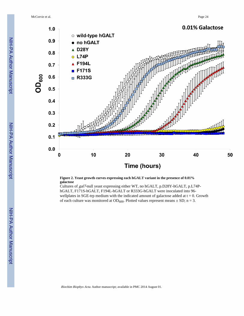
Figure 2. Yeast growth curves expressing each hGALT variant in the presence of 0.01%galactoseCultures of gal7-null yeast expressing either WT, no hGALT, p.D28Y-hGALT, p.L74P-hGALT, F171S-hGALT, F194L-hGALT or R333G-hGALT were inoculated into 96-wellplates in SGE-trp medium with the indicated amount of galactose added at t = 0. Growthof each culture was monitored at OD600. Plotted values represent means ± SD; n = 3.
McCorvie et al. Page 24
Biochim Biophys Acta. Author manuscript; available in PMC 2014 August 01.
NIH
-PA Author Manuscript
NIH
-PA Author Manuscript
NIH
-PA Author Manuscript

Figure 3. Expression and purification of recombinant hGALT from E. coli using size exclusionchromatography(A) WT hGALT purification was resolved by SDS-PAGE (10%) and visualised by stainingwith Coomassie blue. M = molecular mass markers (kDa). (B) Size exclusionchromatography standard curve. The proteins used for standards are as follows A:Ribonuclease (13.7 kDa); B: Chymotrypsinogen A (25 kDa); C: Ovalbumin (43 kDa); D:Serum albumin (67 kDa) E: Alcohol dehydrogenase (149.5 kDa); F: Thyroglobulin (669kDa). (C) Chromatograms of wild-type and mutant hGALT proteins. All the proteins elutedmainly at the expected dimer molecular mass. However p.L74P, p.F171S and p.F194Lshowed increases in the absorbance at approximately one column volume suggesting somedegradation to smaller peptides had occurred. (D) Purified hGALT variants resolved bySDS-PAGE (10 %) and visualised by staining with Coomassie blue. M = molecular massmarkers (kDa). Readers are directed to the online version of this article for the colourversion of this figure.
McCorvie et al. Page 25
Biochim Biophys Acta. Author manuscript; available in PMC 2014 August 01.
NIH
-PA Author Manuscript
NIH
-PA Author Manuscript
NIH
-PA Author Manuscript

Figure 4. The steady state enzyme kinetics for WT hGALT in the forward and reverse reaction(A) The steady state enzyme kinetics for WT hGALT in the forward reaction with variedUDP-Glc and 1 mM Gal-1P. The kinetic data fitted best to the Michaelis-Menten equationwith substrate inhibition with the following constants: ,
, μmol Glc-1P.min−1.mg−1. (B) The steady stateenzyme kinetics in the forward reaction with varied Gal-1P and 500 μM UDP-Glc. Thekinetic data fitted best to the Michaelis-Menten equation with the following constants:
, μmol Glc-1P.min−1.mg−1. (C) The steady stateenzyme kinetics for WT hGALT in the reverse reaction with varied UDP-Gal and 1 mMGlc-1P. The kinetic data fitted best to the Hill equation (sigmoidal kinetics) with the
following constants: , μmol UDP-Glc.min−1.mg−1, h= 2.9. (D) The steady state enzyme kinetics in the reverse reaction with varied Glc-1P and500 μM UDP-Gal. The kinetic data fitted best to the Michaelis-Menten equation withsubstrate inhibition with the following constants: ,
, μmol UDP-Glc.min−1.mg−1. WT hGALT(10 nM) was used for both forward and reverse reactions and controls with no GALT always
McCorvie et al. Page 26
Biochim Biophys Acta. Author manuscript; available in PMC 2014 August 01.
NIH
-PA Author Manuscript
NIH
-PA Author Manuscript
NIH
-PA Author Manuscript

gave no activity. Each point represents the mean of three independent determinations of therate and the error bars represent the standard deviations of these means.
McCorvie et al. Page 27
Biochim Biophys Acta. Author manuscript; available in PMC 2014 August 01.
NIH
-PA Author Manuscript
NIH
-PA Author Manuscript
NIH
-PA Author Manuscript

Figure 5. Chemical cross-linking of hGALT variants with BS3 in comparison to the wild-typehGALT and in the presence of various substrates(A) The hGALT variants showed little difference in their ability to form dimers incomparison to the wild-type. Higher order oligomers were also formed in the presence ofcross-linker. C: control, protein (5 μM) with no cross-linker; +: protein (5 μM) with thecross-linker BS3 (100 μM). (B) Active and inactive hGALT variant show different substratedependent effects on dimerisation as determined by cross-linking. C: control, protein (5 μM)with no cross-linker; +: protein (5 μM) with BS3 (100 μM) in the presence of varioussubstrates (1 mM). Gal-1P: galactose 1-phosphate; UDP-glc: uridine diphosphate glucose;Glc-1P: glucose 1-phosphate; UDP-gal; uridine diphosphate galactose; For: galactose 1-phosphate and uridine diphosphate glucose; Rev: glucose 1-phosphate and uridinediphosphate galactose.
McCorvie et al. Page 28
Biochim Biophys Acta. Author manuscript; available in PMC 2014 August 01.
NIH
-PA Author Manuscript
NIH
-PA Author Manuscript
NIH
-PA Author Manuscript

Figure 6. Limited proteolysis of hGALT variants in comparison to the wild-type hGALT and inthe presence of various substrates(A) The hGALT variants showed differences in susceptibly to degradation by the proteasethermolysin in comparison to the wild-type. C: control, protein (5 μM) with no protease; +:protein (5 μM) with thermolysin (240 nM). (B) WT hGALT showed substrate dependenteffects on the susceptibility to proteolysis where sugar 1-phosphates increased degradationand uridine diphosphate sugars had little effect. Additionally this increased degradation dueto sugar 1-phosphates is the most dominant affect when the protein was in the present ofboth a sugar 1-phosphate and uridine diphosphate sugar. Active and inactive hGALTvariants show different substrate dependent effects on protease sensitivity. C: control,protein (5 μM) with no protease; +: protein (5 μM) with thermolysin (240 mM) in thepresence of various substrates (1 mM). Gal-1P: galactose 1-phosphate; UDP-glc: uridinediphosphate glucose; Glc-1P: glucose 1-phosphate; UDP-gal; uridine diphosphate galactose;For: galactose 1-phosphate and uridine diphosphate glucose; Rev: glucose 1-phosphate anduridine diphosphate galactose.
McCorvie et al. Page 29
Biochim Biophys Acta. Author manuscript; available in PMC 2014 August 01.
NIH
-PA Author Manuscript
NIH
-PA Author Manuscript
NIH
-PA Author Manuscript

Figure 7. Surface hydrophobicity and thermal stability of hGALT variants(A) Relative surface hydrophobicity of each hGALT variant as judged by ANS-1 binding.The emission (at 480 nm) of ANS-1 binding was measured following excitation at 370 nm.The average fluorescence and standard deviation was calculated from three independentassays with each corrected for ANS-1 alone. The significance of change in fluorescence incomparison to the wild-type was determined using one-way ANOVA with a Dunnett’s post-test. (B) Thermal inactivation of wild-type hGALT and active variants (p.D28Y, p.F194Land p.R333G) at 50 nM. Aliquots (100 μl) of each variant hGALT (0.5 μM) were incubatedin HEPES (10 mM), pH 8.8 for 15 min at temperatures ranging from 30 to 70 °C. Residualactivity of the forward reaction was determined with 0.5 mM UDP-Glc and 1.0 mM Gal-1P.Activities were normalized to those at 30 °C. Three independent assays for each temperaturewere carried out and the points show the average activity and the error bars the standarddeviations of these means. (C) Each hGALT variant showed a unique melting curve andsusceptibility to thermal denaturation as judged by DSF. Melting temperatures, (Tm) arepresented in table S2. (D) Melting profiles and melting temperatures of WT hGALT withand without Gal-1P or UDP-Glc. WT hGALT showed a shift in stability when Gal-1P orUDP-Glc was present consistent with binding to the substrate and consequent stabilisationof the substrate. Melting temperatures, (Tm) are presented in table S2. Reaction mixturescontained 5 μM protein, 1 mM ligand and 2.5× Sypro orange (manufacturer’s concentrationdefinition) dissolved in 10 mM HEPES pH 8.8. Controls contained no protein. Assays wereset up in triplicate and the curves were constructed from the mean fluorescence values; the
McCorvie et al. Page 30
Biochim Biophys Acta. Author manuscript; available in PMC 2014 August 01.
NIH
-PA Author Manuscript
NIH
-PA Author Manuscript
NIH
-PA Author Manuscript
error bars represent the standard deviations of these means. Readers are directed to theonline version of this article for the colour version of this figure.
McCorvie et al. Page 31
Biochim Biophys Acta. Author manuscript; available in PMC 2014 August 01.
NIH
-PA Author Manuscript
NIH
-PA Author Manuscript
NIH
-PA Author Manuscript

Figure 8. Amino acids affected by hGALT mutations affect side-chain and substrate interactions(A) Asp-28 is located in the N-terminal loop at the dimer interface and forms a number ofhydrogen bonds, one of which is with His-47. This residue is on the same loop as Arg-48and Arg-51 that form part of subunit B’s active site. (B) p.D28Y results in the removal ofthe interaction between this residue and His-47. (C) Leu-74 is located at the active site andis flanked by Pro-73 and Cys-75. This residue forms hydrogen bonds with Cys-130 andTyr-89 via its backbone oxygen and with Asn-72 via the backbone nitrogen. (D) p.L74Presults in the removal of the hydrogen bond with Asn-72. Leu-74 is predicted to be close tothe uracil moiety of UDP-Glc and possibly interacts hydrophobically. (E) The residuePhe-171 is located at the active site and the dimer interface. It is close to Gln-188 and formsonly backbone interactions with this residue. (F) p.F171S results in an additional hydrogenbond between the side chain hydroxyl and backbone oxygen of Met-298. (G) Phe-194 islocated at the dimer interface and exterior of hGALT. This residue forms hydrogen bondsbetween its backbone oxygen with the hydroxyl of Ser-192. The side chain of Phe-194 isburied in a largely hydrophobic cleft containing residues from both polypeptide chains. (H)p.F194L results in a smaller side chain that points more towards the exterior of the protein.(I) Arg-333 is predicted to be part of a number of loops at the dimer interface between thetwo active sites. This residue is facing the Arg-333 of the other subunit and beside Lys-334that is predicted to interact with the sugar moiety of either substrate [29]. (J) Alteration ofarginine to glycine at residue 333 results in a void at the dimer interface due to the smaller
McCorvie et al. Page 32
Biochim Biophys Acta. Author manuscript; available in PMC 2014 August 01.
NIH
-PA Author Manuscript
NIH
-PA Author Manuscript
NIH
-PA Author Manuscript

size of the side chains. Subunit backbones are ribbon representations with subunit A in palegreen and subunit B in light orange. Selected residues are shown as stick models in whitewith nitrogen, oxygen, phosphate and sulphur atoms in blue, red, orange and yellowrespectively. Residues affected by mutation are in dark grey and hydrogen bonds are shownas black dotted lines. His-186 is shown in purple and the substrate UDP-Glc is shown incyan. Structures are from the hGALT structure [28]. Readers are directed to the onlineversion of this article for the colour version of this figure.
McCorvie et al. Page 33
Biochim Biophys Acta. Author manuscript; available in PMC 2014 August 01.
NIH
-PA Author Manuscript
NIH
-PA Author Manuscript
NIH
-PA Author Manuscript

NIH
-PA Author Manuscript
NIH
-PA Author Manuscript
NIH
-PA Author Manuscript
McCorvie et al. Page 34
Table 1
GALT activity levels observed in null-background yeast expressing each hGALT allele from a centromericplasmid.
hGALT variant nucleotide change % wild-type activity(mean ± SD) n=3
WT N.A. 100 ± 0.00
no GALT N.A. 0.01 ± 0.03
p.D28Y c.82G>T 13.55 ± 5.72
p.L74P c.221T>C 0.01 ± 0.09
p.F171S c.512T>C −0.03 ± 0.01
p.F194L c.580T>C 11.93 ± 2.00
p.R333G c.997C>G 0.60 ± 0.07
Biochim Biophys Acta. Author manuscript; available in PMC 2014 August 01.
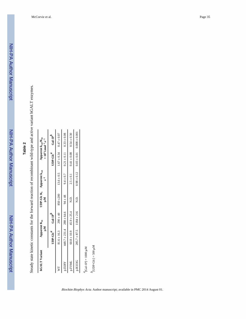
NIH
-PA Author Manuscript
NIH
-PA Author Manuscript
NIH
-PA Author Manuscript
McCorvie et al. Page 35
Tabl
e 2
Stea
dy s
tate
kin
etic
con
stan
ts f
or th
e fo
rwar
d re
actio
n of
rec
ombi
nant
wild
-typ
e an
d ac
tive
vari
ant h
GA
LT
enz
ymes
.
hGA
LT
Var
iant
App
aren
t K
mμ
MU
DP
-Glc
Ki
μM
App
aren
t k c
at
s−1A
ppar
ent
k cat
/Km
× 10
5 l.m
ol−1
.s−1
UD
P-G
lca
Gal
-1P
bU
DP
-Glc
aG
al-1
Pb
WT
81.6
± 1
6.3
290
± 4
085
0 ±
200
13.6
± 0
.51.
67 ±
0.3
40.
47 ±
0.0
7
p.D
28Y
448.
7 ±
231
.428
8 ±
64.
694
± 4
89.
4 ±
0.7
0.21
± 0
.11
0.33
± 0
.08
p.F1
94L
60.8
± 1
0.9
45.9
± 2
5.4
N.D
.2.
5 ±
0.1
0.41
± 0
.08
0.54
± 0
.30
p.R
333G
285.
7 ±
87.
511
84 ±
216
N.D
.0.
98 ±
0.1
20.
03 ±
0.0
10.
008
± 0
.001
a [Gal
-1P]
= 1
000 μ
M
b [UD
P-G
lc]
= 5
00 μ
M
Biochim Biophys Acta. Author manuscript; available in PMC 2014 August 01.
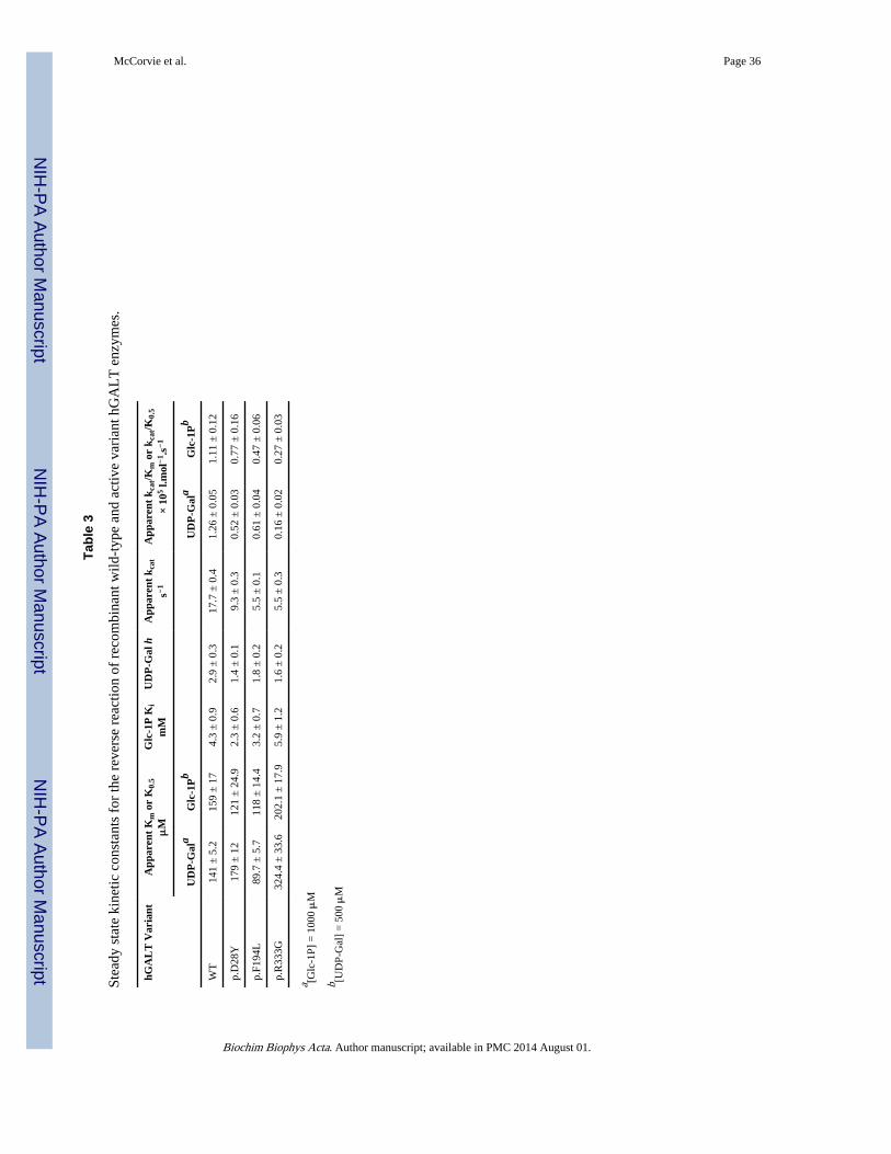
NIH
-PA Author Manuscript
NIH
-PA Author Manuscript
NIH
-PA Author Manuscript
McCorvie et al. Page 36
Tabl
e 3
Stea
dy s
tate
kin
etic
con
stan
ts f
or th
e re
vers
e re
actio
n of
rec
ombi
nant
wild
-typ
e an
d ac
tive
vari
ant h
GA
LT
enz
ymes
.
hGA
LT
Var
iant
App
aren
t K
m o
r K
0.5
μM
Glc
-1P
Ki
mM
UD
P-G
al h
App
aren
t k c
at
s−1A
ppar
ent
k cat
/Km
or
k cat
/K0.
5
× 10
5 l.m
ol−1
.s−1
UD
P-G
ala
Glc
-1P
bU
DP
-Gal
aG
lc-1
Pb
WT
141
± 5
.215
9 ±
17
4.3
± 0
.92.
9 ±
0.3
17.7
± 0
.41.
26 ±
0.0
51.
11 ±
0.1
2
p.D
28Y
179
± 1
212
1 ±
24.
92.
3 ±
0.6
1.4
± 0
.19.
3 ±
0.3
0.52
± 0
.03
0.77
± 0
.16
p.F1
94L
89.7
± 5
.711
8 ±
14.
43.
2 ±
0.7
1.8
± 0
.25.
5 ±
0.1
0.61
± 0
.04
0.47
± 0
.06
p.R
333G
324.
4 ±
33.
620
2.1
± 1
7.9
5.9
± 1
.21.
6 ±
0.2
5.5
± 0
.30.
16 ±
0.0
20.
27 ±
0.0
3
a [Glc
-1P]
= 1
000 μ
M
b [UD
P-G
al]
= 5
00 μ
M
Biochim Biophys Acta. Author manuscript; available in PMC 2014 August 01.

NIH
-PA Author Manuscript
NIH
-PA Author Manuscript
NIH
-PA Author Manuscript
McCorvie et al. Page 37
Tabl
e 4
Cha
nge
of m
eltin
g te
mpe
ratu
res Δ
Tm
of
reco
mbi
nant
wild
-typ
e an
d va
rian
t hG
AL
T e
nzym
es in
the
pres
ence
of
vari
ous
subs
trat
es.
ΔT
m (
K)
hGA
LT
Var
iant
+Gal
-1P
+UD
P-G
lc+G
lc-1
P+U
DP
-Gal
+Gal
-1P
&U
DP
-Glc
+Glc
-1P
&U
DP
-Gal
WT
6.5
± 0
.6**
*2.
5 ±
0.8
***
6.5
± 0
.4**
*2.
5 ±
0.6
***
6.1
± 0
.4**
*6.
6 ±
0.8
***
p.D
28Y
8.8
± 1
.0**
*3.
2 ±
1.4
***
8.0
± 1
.9**
*2.
2 ±
1.5
*8.
0 ±
1.5
***
8.2
± 1
.1**
*
p.L
74P
−0.
7 ±
0.9
−0.
6 ±
1.0
−0.
3 ±
1.1
−1.
4 ±
0.9
−1.
5 ±
0.7
*−
1.5
± 0
.7*
p.F1
71S
−0.
3 ±
1.4
−0.
3 ±
0.9
0.1
± 0
.9−
0.4
± 1
.00.
0 ±
0.9
0.5
± 1
.0
p.F1
94L
1.9
± 0
.5**
*1.
6 ±
0.1
***
1.3
± 0
.5**
*0.
5 ±
0.0
2.1
± 0
.2**
*2.
5 ±
0.0
***
p.R
333G
−1.
1 ±
2.0
1.6
± 0
.8*
−2.
0 ±
0.9
**−
0.2
± 0
.80.
3 ±
0.7
−2.
0 ±
0.8
*
All
ligan
ds w
ere
adde
d to
a f
inal
con
cent
ratio
n of
1 m
M in
10
mM
HE
PES,
pH
8.8
. ΔT
m v
alue
s ar
e re
port
ed a
s th
e m
eans
± S
D o
f th
ree
sepa
rate
exp
erim
ents
.
* P <
0.0
5,
**P
< 0
.001
,
*** P
< 0
.000
1 (O
ne w
ay A
NO
VA
with
Dun
nett
com
pari
son
test
).
Biochim Biophys Acta. Author manuscript; available in PMC 2014 August 01.