
Fungal endophytes of turmeric (Curcuma longa L.) and their ...
17
Vol.:(0123456789) 1 3 World Journal of Microbiology and Biotechnology (2018) 34:49 https://doi.org/10.1007/s11274-018-2431-x ORIGINAL PAPER Fungal endophytes of turmeric (Curcuma longa L.) and their biocontrol potential against pathogens Pythium aphanidermatum and Rhizoctonia solani G. Vinayarani 1 · H. S. Prakash 1 Received: 7 March 2017 / Accepted: 9 March 2018 / Published online: 14 March 2018 © Springer Science+Business Media B.V., part of Springer Nature 2018 Abstract Endophytic fungi have been isolated from the healthy turmeric (Curcuma longa L.) rhizomes from South India. Thirty-one endophytes were identified based on morphological and ITS–rDNA sequence analysis. The isolated endophytes were screened for antagonistic activity against Pythium aphanidermatum (Edson) Fitzp., and Rhizoctonia solani Kuhn., causing rhizome rot and leaf blight diseases in turmeric respectively. Results revealed that only six endophytes showed > 70% suppression of test pathogens in antagonistic dual culture assays. The endophyte T. harzianum TharDOB-31 showed significant in vitro mycelial growth inhibition of P. aphanidermatum (76.0%) and R. solani (76.9%) when tested by dual culture method. The SEM stud- ies of interaction zone showed morphological abnormalities like parasitism, shriveling, breakage and lysis of hyphae of the pathogens by endophyte TharDOB-31. Selected endophytic isolates recorded multiple plant growth promoting traits in in vitro studies. The rhizome bacterization followed by soil application of endophyte TharDOB-31 showed lowest Percent Disease Incidence of rhizome rot and leaf blight, 13.8 and 11.6% respectively. The treatment of TharDOB-31 exhibited significant increase in plant height (85 cm) and fresh rhizome yield/plant (425 g) in comparison with untreated control under greenhouse condition. The confocal microscopy validates the colonization of the TharDOB-31 in turmeric rhizomes. The secondary metabolites in ethyl acetate extract of TharDOB-31 were found to contain higher number of antifungal compounds by high resolution liquid chromatograph mass spectrometer analysis. Thereby, endophyte T. harzianum isolate can be exploited as a potential biocontrol agent for suppressing rhizome rot and leaf blight diseases in turmeric. Keywords Antagonism · Biocontrol · Fungal endophytes · Growth promotion · HR-LCMS · Leaf blight · Rhizome rot · SEM Introduction Turmeric (Curcuma longa L.), a member of the ginger fam- ily (Zingiberaceae) is one of the major rhizomatous spice used worldwide in alternative systems of medicine. The whole plant is aromatic, but the underground rhizomes, fresh or processed are the most valuable commodities. The active compound ‘curcumin’ is believed to have a wide range of biological effects including anti-inflammatory, antioxidant, antitumour, antibacterial and antiviral activities (Gupta et al. 2012a). India is the major producer and exporter of turmeric in the world contributing for 80% of the world production (Thiripurasundari and Selvarani 2014). The fungus P. aphanidermatum and Rhizoctonia solani cause rhizome rot and leaf blight diseases in turmeric plants and reduce commercial value (Park 1934; Roy 1992). It has been noted that chemical fungicides like ridomil, metalaxyl, carbendazim (0.1%) and mancozeb (0.25%) were com- monly used to manage rhizome rot and leaf blight diseases (Rathaiah 1982; Muthukumar et al. 2011). Use of chemical fungicides is of public concern as it causes various human health problems and also pathogens build resistance against fungicides. The increasing awareness of fungicide related hazards has emphasized the need for adopting biological methods as an alternative ecofriendly disease control strategy (Wahab 2009) Biological control measures rely on the use of antago- nistic organisms against target pathogens. Endophytes are * H. S. Prakash [email protected]; [email protected] 1 Department of Studies in Biotechnology, University of Mysore, Manasagangotri, Mysuru 570006, India
Transcript of Fungal endophytes of turmeric (Curcuma longa L.) and their ...

Vol.:(0123456789)1 3
World Journal of Microbiology and Biotechnology (2018) 34:49 https://doi.org/10.1007/s11274-018-2431-x
ORIGINAL PAPER
Fungal endophytes of turmeric (Curcuma longa L.) and their biocontrol potential against pathogens Pythium aphanidermatum and Rhizoctonia solani
G. Vinayarani1 · H. S. Prakash1
Received: 7 March 2017 / Accepted: 9 March 2018 / Published online: 14 March 2018 © Springer Science+Business Media B.V., part of Springer Nature 2018
AbstractEndophytic fungi have been isolated from the healthy turmeric (Curcuma longa L.) rhizomes from South India. Thirty-one endophytes were identified based on morphological and ITS–rDNA sequence analysis. The isolated endophytes were screened for antagonistic activity against Pythium aphanidermatum (Edson) Fitzp., and Rhizoctonia solani Kuhn., causing rhizome rot and leaf blight diseases in turmeric respectively. Results revealed that only six endophytes showed > 70% suppression of test pathogens in antagonistic dual culture assays. The endophyte T. harzianum TharDOB-31 showed significant in vitro mycelial growth inhibition of P. aphanidermatum (76.0%) and R. solani (76.9%) when tested by dual culture method. The SEM stud-ies of interaction zone showed morphological abnormalities like parasitism, shriveling, breakage and lysis of hyphae of the pathogens by endophyte TharDOB-31. Selected endophytic isolates recorded multiple plant growth promoting traits in in vitro studies. The rhizome bacterization followed by soil application of endophyte TharDOB-31 showed lowest Percent Disease Incidence of rhizome rot and leaf blight, 13.8 and 11.6% respectively. The treatment of TharDOB-31 exhibited significant increase in plant height (85 cm) and fresh rhizome yield/plant (425 g) in comparison with untreated control under greenhouse condition. The confocal microscopy validates the colonization of the TharDOB-31 in turmeric rhizomes. The secondary metabolites in ethyl acetate extract of TharDOB-31 were found to contain higher number of antifungal compounds by high resolution liquid chromatograph mass spectrometer analysis. Thereby, endophyte T. harzianum isolate can be exploited as a potential biocontrol agent for suppressing rhizome rot and leaf blight diseases in turmeric.
Keywords Antagonism · Biocontrol · Fungal endophytes · Growth promotion · HR-LCMS · Leaf blight · Rhizome rot · SEM
Introduction
Turmeric (Curcuma longa L.), a member of the ginger fam-ily (Zingiberaceae) is one of the major rhizomatous spice used worldwide in alternative systems of medicine. The whole plant is aromatic, but the underground rhizomes, fresh or processed are the most valuable commodities. The active compound ‘curcumin’ is believed to have a wide range of biological effects including anti-inflammatory, antioxidant, antitumour, antibacterial and antiviral activities (Gupta et al. 2012a). India is the major producer and exporter of turmeric
in the world contributing for 80% of the world production (Thiripurasundari and Selvarani 2014).
The fungus P. aphanidermatum and Rhizoctonia solani cause rhizome rot and leaf blight diseases in turmeric plants and reduce commercial value (Park 1934; Roy 1992). It has been noted that chemical fungicides like ridomil, metalaxyl, carbendazim (0.1%) and mancozeb (0.25%) were com-monly used to manage rhizome rot and leaf blight diseases (Rathaiah 1982; Muthukumar et al. 2011). Use of chemical fungicides is of public concern as it causes various human health problems and also pathogens build resistance against fungicides.
The increasing awareness of fungicide related hazards has emphasized the need for adopting biological methods as an alternative ecofriendly disease control strategy (Wahab 2009) Biological control measures rely on the use of antago-nistic organisms against target pathogens. Endophytes are
* H. S. Prakash [email protected]; [email protected]
1 Department of Studies in Biotechnology, University of Mysore, Manasagangotri, Mysuru 570006, India
World Journal of Microbiology and Biotechnology (2018) 34:49
1 3
49 Page 2 of 17
microorganisms that live within the plant tissue as endo-symbionts, without causing any disease symptoms. Some of them are mutualistic symbionts with beneficial effect on their host such as plant growth promotion, resistance against disease or environmental stress (Colla et al. 2015; Lledo et al. 2016). A large number of endophytic fungi have been isolated from plant species (Nisa et al. 2015). A more precise assessment of diversity and identification of fungi can be achieved using the polymerase chain reaction (PCR) of the internal transcribed spacer (ITS) of the fungal nuclear ribo-somal DNA (Borneman and Hartin 2000; Rodriguez et al. 2009). ITS is recommended as universal barcode to identify fungal species (Schoch et al. 2012).
Endophytic fungi are promising biocontrol agents (BCA’s) as they occupy internal living tissues of plants and due to close proximity to plant pathogens. Endophytic fungi were used as BCA’s like Trichoderma viride, Asper-gillus fumigatus and Penicillium atrovenetum isolated from medicinal plant Artemisia nilagirica against Phytophthora infestans infecting potato (Myrchiang et al. 2014). Similarly, fungal endophytes isolated from healthy Theobroma cacao tissues viz., Colletotrichum gloeosporioides, Clonostachys rosea and Botryosphaeria ribis were screened in vitro for antagonism against major pathogens of cacao. Endophyte C. gloeosporioides and C. rosea significantly decreased pod loss due to black pod rot and incidence of cacao pods with sporulating lesions of M. roreri was reported earlier (Mejia et al. 2008). Further, endophytic C. rosea has been reported to control Botrytis cinerea in roses (Morandi et al. 2000). Endophytes and mycoparasites associated with Theobroma gileri used as BCA’s agents of cocoa diseases (Evans et al. 2003). Endophyte Gliocladium catenulatum isolated from (Theobroma cacao L.) used as biological control of Witches Broom Disease caused by Crinipellis perniciosa (Rubini et al. 2005). Endophytic fungi harbored in Cannabis sativa L. reported as potential BCA’s against two host phytopath-ogens namely Botrytis cinerea and Trichothecium roseum (Kusari et al. 2013). The endophytic fungi were isolated from healthy tissues of vegetable plants viz., cucumber, red pepper, tomato, pumpkin and Chinese cabbage. Among the isolates the endophyte F. oxysporum isolated from roots of red pepper showed potential disease control efficacy against tomato late blight caused by Phytophthora infestans (Kim et al. 2007).
Trichoderma spp. are known to be the most commonly used antagonists against P. aphanidermatum and R. solani (Chet et al. 1981). Apart from their antagonistic activity, Trichoderma strains have also demonstrated growth promot-ing properties and is used as rhizome treatment in turmeric (Ushamalini et al. 2008). Promotion of plant growth is the major contribution of fungal symbiosis. The growth promo-tion of fungal endophytes is attributed to the production of ammonia and phytohormones like indole acetic acid (IAA),
HCN and by production of cell-wall degrading enzymes such as cellulase, pectinase and protease (Fouda et al. 2015).
The production of antibiotic secondary metabolites is often correlated to the biocontrol activity of Trichoderma isolates (Vinale et al. 2006). Trichoderma secondary metab-olites are chemically diverse and their production varies greatly between species and between isolates of the same species (Vinale et al. 2009). The Trichoderma spp. known to produce secondary metabolites with a key role in antago-nistic activities and was used as BCA’s (Vinale et al. 2014).
To the best of our knowledge there is no information available on the potential fungal endophytes on growth pro-motion and biocontrol of rhizome and leaf blight diseases of turmeric. This study was taken up to profile the endo-phytic fungi associated with turmeric and to evaluate their antagonistic activities, biocontrol potential and plant growth promotion both in vitro and in vivo conditions against P. aphanidermatum and R. solani pathogens which cause rhi-zome rot and leaf blight diseases of turmeric respectively.
Materials and methods
Collection of samples
A total of 250 healthy and 50 diseased turmeric samples were collected for endophytes and pathogens (P. aphanider-matum and R. solani) isolation respectively from different agroclimatic regions of Karnataka, Andhra Pradesh, Kerala, Tamilnadu states of India during July–December 2014 (tur-meric plant growth stage was 4–6 months). The samples were collected in polythene bags, labeled and stored in refrigerator at 4 °C and processed within 48 h of collection.
Isolation of endophytic fungi
Healthy turmeric rhizomes were washed in running tap water, then surface sterilized by soaking in 70% ethanol for 1 min, followed by 3.5% sodium hypochloride for 3 min and finally rinsed in sterile distilled water three times at 1 min intervals. After surface sterilization, the epidermis was peeled off and the rhizomes cut into pieces of 5 mm2 using sterile blades. Two hundred segments of each sam-ple were used to isolate fungal endophytes and colonization frequency percentage (CF %) was calculated. Each petrid-ish (90 mm diameter) containing water agar medium sup-plemented with streptomycin sulfate (100 mg/l to suppress bacterial growth) were placed with 10–15 segments (Nalini et al. 2014). To verify the efficacy of surface sterilization of the rhizomes, 100 µl of the last rinse was added on potato dextrose agar (PDA) medium.
The petridishes were incubated at 23 °C with 12 h light and dark cycles up to 3–4 weeks and the segments were
World Journal of Microbiology and Biotechnology (2018) 34:49
1 3
Page 3 of 17 49
examined periodically. The outgrowing fungal colonies were transferred to new petridishes with PDA medium for fur-ther identification. All the endophytic fungal isolates were numbered as DOB# series, maintained in cryovials on PDA layered with 15% glycerol (v/v) at − 80 °C in a deep freezer (Cryoscientific Pvt. Ltd., Chennai, India) at the Department of Studies in Biotechnology, University of Mysore, Mysuru, India. The Colonization frequency of endophytes was calcu-lated according to the method of Fisher and Petrini (1987) as follows,
Isolation and pathogenicity of fungal pathogens
The surface sterilized diseased rhizome pieces (1.5 cm) were blotted dry on sterile filter paper, and placed on P5ARP medium (Pythium selective medium) containing 0.005 g of pimaricin, 0.25 mg of ampicillin, 0.01 g of rifampicin, and 0.10 g of pentachloronitrobenzene per liter in corn meal agar (Jeffers and Martin 1986) for P. aphanidermatum and on PDA medium for R. solani isolation. The plates were incu-bated at room temperature (28–30 °C), the colony morphol-ogy was studied (Van der Plaats-Niterink 1981). Hyphae were successively transferred to the new PDA medium and the cultures were maintained on slants. Pathogenicity of the isolated fungi was established by inoculating to susceptible cultivar ‘Erode local’ (Johnston and Booth 1983; Sriraj et al. 2014).
Morphological identification of endophytes and pathogens
Morphological studies were carried out to identify endo-phytes and pathogens under the stereomicroscope (Leica DM-LS 2 Trinocular Research Microscope). The slides were prepared for light microscopy by staining with lactophenol blue and identified comparing with the description given in standard manuals (Barnett and Hunter 1972; Domsch et al. 1980; Gilma and Joseph 1998).
Molecular characterization of fungal endophytes and pathogens
DNA was extracted using HipurA Fungal DNA Purification kit of Himedia. The quality and quantity of DNA was deter-mined using NanoDrop spectrophotometer (2000 C, Thermo Scientific, Japan). The integrity of the DNA was assayed by 0.7% agarose gel electrophoresis.
The DNA was amplified using universal primers for ITS 1 and ITS 4 region [forward 5′-TCC GTA GGT GAA CCT
Colonization Frequency (CF%) =Number of segments colonized by fungus
Total number of segments× 100
GCG G-3′ and reverse 5′-TCC TCC GCT TAT TGA TAT GC-3′] (White et al. 1990). The PCR reaction was performed in 50 µl final reaction volume containing 5 µl of 10× PCR buffer, 8 µl of 25 mM MgCl2, 2.5 µl of 1.25 mM dNTP, 0.2 µl of each primer (20 µM), 100 ng of DNA and 0.2 µl Taq DNA polymerase (5 U/µl) (Sigma-Aldrich, Bangalore) in a BioRad thermal cycler programmed for initial denaturation at 94 °C for 5 min, followed by 35 cycles of denaturation at 94 °C for 1 min, primer annealing at 56 °C for 1 min, extension at 72 °C for 2 min. At the end of the amplification reaction, a
final extension step was achieved at 72 °C for 10 min. Fifteen microliters of the PCR products from each PCR reaction were electrophoresed on 1% (w/v) agarose gel containing 5 mg/ml of ethidium bromide in a 1×TBE (pH 8.4). To estimate the size of the PCR products, a 100 bp molecular ladder (Sigma-Aldrich, Bangalore) was used. The electrophoresis was carried out using 100 V. The gel was visualized and photographed using Gel Documentation system (Gel Doc 2000, BioRad, California, USA).
Sequencing and phylogenetic analysis
The amplified products were sequenced at Chromous Biotech. Pvt. Ltd, Bangalore. The sequences obtained were further analyzed by BLAST program of National Center for Biotech-nology Information (NCBI, website: http://www.ncbi.nlm.nih.gov). The ITS sequence was compared using nucleotide BLAST with default settings and megablast (highly similar sequences) as the selected program. Species identification was determined from the lowest expect value (E-value) of the BLAST output and the similarity percentage. The analysed sequences were submitted to Genbank (NCBI) and accession numbers were obtained.
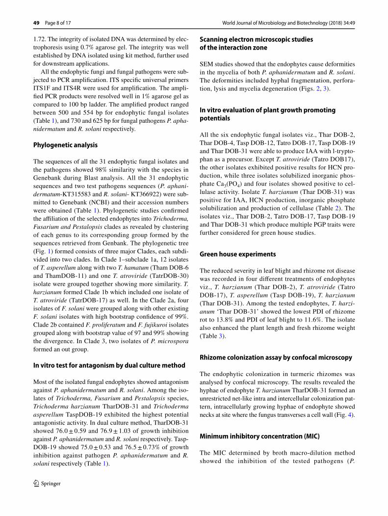
The ITS sequence analysis was done for molecular authenti-cation of the endophytic species. All the nucleotide sequences of isolated endophytes and the sequences of some nearest neighbours of the endophytic fungal isolates retrieved from Genbank (NCBI) were used to construct the phylogenetic tree. All sequences were aligned using CLUSTAL W algorithm (Thompson et al. 1994) and phylogenetic analysis was con-ducted using Mega 6 software. Phylogenetic inference was performed by the Maximum Parsimony (MP) method, with bootstrap values calculated from 1000 replicate runs (Tamura et al. 2013).

World Journal of Microbiology and Biotechnology (2018) 34:49
1 3
49 Page 4 of 17
Assessment of in vitro antagonism by dual culture method
The antagonistic effect of fungal endophytes on P. apha-nidermatum and R. solani pathogens was evaluated using dual culture technique. In this method 5 mm2 size of endo-phyte culture and same size of test pathogen were placed 1 cm away from the periphery of the petriplate containing PDA media in opposite direction. The petriplate inoculated with pathogen alone in the absence of antagonist (endo-phytes) served as control and the experiment was done in triplicates. The inoculated plates were incubated under alternating periods of 12 h darkness and 12 h of daylight at 25 ± 2 °C until the control plates were completely cov-ered by pathogen mycelia (Lahlali et al. 2007). The radial mycelial growth of pathogen towards the antagonistic fun-gus (T) and that on a control plate (C) were measured and the mycelial growth inhibition (I) was calculated using the following formula:
where C is the radial growth measurement of the pathogen in control (main axis), T is the radial growth of the pathogen towards the antagonistic fungus, I is the percent inhibition.
Scanning electron microscope (SEM) studies of the interaction zone
For SEM analysis, the zone of interaction between patho-gen and endophyte from the dual culture was taken and the samples were exposed to 2% osmium tetroxide for 24 h at 20 °C, later it was transferred to copper stubs over double adhesive carbon tape. They were then coated with gold in a POLARON, AU/PD sputter and scanned in SEM, S-3400N model (Japan) at 5.00 kV and the abnormalities in the fungal hyphae were recorded.
In vitro evaluation of plant growth promoting traits of endophytes
In in vitro study of antagonism the six endophytic fun-gal isolates viz., Thar DOB-2, Thar DOB-4, Tasp DOB-12, Tatro DOB-17, Tasp DOB-19 and Thar DOB-31 that showed > 70% inhibition against pathogens (Table 1). These isolates were tested for their plant growth promot-ing (PGP) traits.
Indole‑3‑acetic acid production
For detection of IAA production by endophytic fungal isolates, fungal disc of 1 cm diameter were inoculated to 20 ml of Czapex Dox (CD) broth supplemented with
I = C − T × 100/C
0.5 g−1 of tryptophan and incubated for 7 days at 28 °C, performed in triplicates. Culture suspension (5 ml) was transferred to test tubes and centrifuged at 6000 rpm for 10 min. One milliliters of the supernatant was mixed with 4 ml of Salkowski reagent (Fouda et al. 2015) and the tubes were incubated for 30 min. IAA production was observed as the development of a pink colour. The low IAA production (light pink colour ‘+’); medium IAA pro-duction (Dark pink colour ‘++’); high IAA production (Dense pink colour, ‘+++’) was recorded.
Hydrogen cyanide (HCN) production
HCN production by the fungal agents was tested following the method of Triveni et al. (2013) with slight modifications. The fungal strain was inoculated on PDA slants amended with glycine at 4.4 g/l. Whatman No.1 filter paper strips were dipped in 0.5% picric acid in 2% sodium carbonate solution. The strips were inserted in the top of each test tubes and sealed with parafilm and incubated at 28 °C for 7 days. A change of colour of the filter paper from yellow to light brown, brown or reddish-brown was recorded as weak (+), moderate (++) or strong (+++) reaction respectively.
Screening for cellulolytic activity
Isolated endophytic fungal strains were screened for cellulo-lytic activity by inoculating into petriplates containing agar medium containing carboxymethylcellulose (Pereira and Castro 2014). After 5 days of incubation at 28 °C, the plates were flooded with congo red solution for 15 min (1 mg/l). The plates were destained with 1 M Nacl for 15 min by decanting excess congo red solution. A clear zone was con-sidered as cellulase positive (Gupta et al. 2012b).
Phosphate solubilizing ability
All endophytic fungal isolates were screened for their phos-phate solubilizing activity on Pikovskaya’s medium (Hime-dia) by spot inoculation method and incubated at 28 °C for 7 days. The production of a halo around a growing colony indicated phosphate solubilization (Singh and Reddy 2011).
Efficacy of endophytes on rhizome rot and leaf blight pathogens under green house conditions
Four promising endophytic isolates viz., T. harzianum (Thar DOB-2), T. atroviride (Tatro DOB-17), T. asperellum (Tasp DOB-19), T. harzianum (Thar DOB-31) were selected for green house studies based on in vitro antagonism studies and PGP traits. Two sets of experiments were performed to analyze the efficacy of the endophytes in controlling the rhizome rot and leaf blight diseases of turmeric under green
World Journal of Microbiology and Biotechnology (2018) 34:49
1 3
Page 5 of 17 49
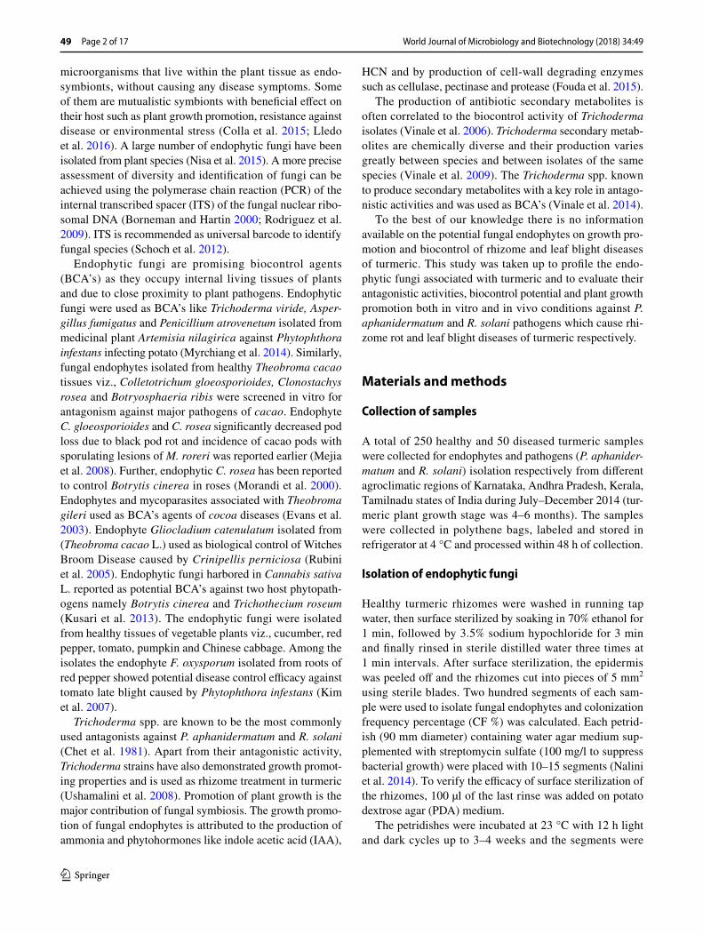
Table 1 Molecular identification of antagonistic fungal endophytes using rRNA gene (ITS1 and ITS4) and their effect on the in vitro mycelial growth of pathogenic fungus P. aphanidermatum and R. solani
Sl. no. Geographic locations
Endophytic fungal isolates
CF% Closest related species
Identity in percent (%)
Amplicon size (BP)
GenBank accession numbers
% Growth inhibition P. aphaniderma-tum
% Growth inhibition of R. solani
1 Dharwad, Kar-nataka
TharDOB-1 10 T. harzianum 99 541 KU365129 63.0 ± 0.93 65.6 ± 0.86
2 Chamarajana-gar, Karnataka
TharDOB-2 14 T. harzianum 99 544 KU365130 73.6 ± 0.74 70.2 ± 0.98
3 Dharwad, Kar-nataka
TharDOB-3 11 T. harzianum 100 545 KU365131 71.0 ± 0.68 69.0 ± 0.43
4 Dharwad, Kar-nataka
TharBOD-4 16 T. harzianum 100 549 KU365132 70.8 ± 0.47 72.2 ± 0.58
5 Dakshina Kannada, Karnataka
PmicDOB-5 10 P. microspora 99 475 KU365133 57.0 ± 0.37 57.4 ± 0.86
6 Chamarajana-gar, Karnataka
ThamDOB-6 16 T. hamatum 99 542 KU365134 69.6 ± 0.86 68.9 ± 0.72
7 Bhavanisagar, Tamilnadu
TharDOB-7 15 T. harzianum 99 531 KU365135 68.3 ± 0.77 69.5 ± 0.76
8 Kollegal, Kar-nataka
TharDOB-8 18 T. harzianum 100 531 KU365136 58.9 ± 0.39 60.2 ± 0.71
9 Dandeli, Karna-taka
FsolDOB-9 9 F. solani 99 519 KU517655 52.0 ± 0.41 53.6 ± 0.34
10 Dakshina Kannada, Karnataka
FsolDOB-10 13 F. solani 99 506 KU517656 55.2 ± 0.86 52.6 ± 0.91
11 Erode, Tamil-nadu
ThamDOB-11 20 T. hamatum 99 542 KU517657 66.8 ± 0.70 63.8 ± 0.84
12 Dharwad, Kar-nataka
TaspDOB-12 12 T. asperellum 100 541 KU662964 72.7 ± 0.93 70.4 ± 0.76
13 Dharwad, Kar-nataka
TaspDOB-13 10.5 T. asperellum 99 542 KU662965 58.0 ± 0.92 60.0 ± 0.45
14 Dharwad, Kar-nataka
TaspDOB-14 14 T. asperellum 99 546 KU662966 73.2 ± 0.56 69.6 ± 0.68
15 Coimbatore, Tamilnadu
TaspDOB-15 22 T. asperellum 99 538 KU662967 62.9 ± 0.72 63.6 ± 0.37
16 Coimbatore, Tamilnadu
TaspDOB-16 26 T. asperellum 99 549 KU662968 59.3 ± 0.64 63.0 ± 0.53
17 Kollegal, Kar-nataka
TatroDOB-17 18.5 T. atroviride 100 547 KU662969 73.8 ± 0.48 72.6 ± 0.90
18 Calicut, Kerala TaspDOB-18 16 T. asperellum 99 541 KU662970 66.0 ± 0.85 68.2 ± 0.4319 Salem, Tamil-
naduTaspDOB-19 20 T. asperellum 100 540 KU662971 75.0 ± 0.53 76.5 ± 0.73
20 Calicut, Kerala TaspDOB-20 14 T. asperellum 99 543 KU662972 60.2 ± 0.72 62.0 ± 0.7121 Kadapa, Andhra
PradeshFproDOB-21 8 F. proliferatum 100 500 KU865563 57.4 ± 0.98 59.0 ± 0.73
22 H.D.Kote, Karnataka
FproDOB-22 18 F. proliferatum 100 424 KU865563 60.1 ± 0.73 61.4 ± 0.63
23 Guntur, Andhra Pradesh
TaspDOB-23 10 T. asperellum 98 450 KU865564 57.8 ± 0.59 60.0 ± 1.00
24 Hassan, Karna-taka
TaspDOB-24 15 T. asperellum 100 546 KU865565 61.8 ± 0.72 62.2 ± 0.96
25 H.D.Kote, Karnataka
TaspDOB-25 20 T. asperellum 99 483 KU865566 62.4 ± 0.80 62.0 ± 0.73
26 Mysore, Karna-taka
FsolDOB-26 24 F. solani 100 439 KU865567 56.0 ± 0.75 58.2 ± 0.92

World Journal of Microbiology and Biotechnology (2018) 34:49
1 3
49 Page 6 of 17
house condition by using turmeric cultivar ‘Erode local’ (susceptible variety). Four replications were maintained for each treatment and each replication consisted of five earthen pots (20 cm diameter) in a completely randomized design (CRD) in a green house, the experiment was repeated twice.
The talc-based formulation of the fungal endophytes was prepared containing population densities of 3 × 10⁶ spores/ml (Shanmugam and Kanoujia 2011). The talc-based for-mulations of endophytic fungal isolates T. harzianum (Thar DOB-2), T. atroviride (Tatro DOB-17), T. asperellum (Tasp DOB-19), T. harzianum (Thar DOB-31) were applied as rhizome treatment and soil application. The rhizomes were surface sterilized with 2% sodium hypochloride for 1 min and soaked in sterile distilled water containing 20 g/l for-mulation. The suspension was drained off after 12 h and the rhizomes were air dried overnight under a sterile air stream. Rhizomes each with three nodes were planted in earthen pots containing sterilized sandy loam soil of 5 kg. For first set of experiment, the pathogen P. aphanidermatum was multiplied on sand-corn meal medium at a ratio of 1:19 (sand–maize inoculum:soil) and were challenge inoculated (Shanmugam et al. 2013). And for second set, the 30 day old BCA treated turmeric plants were challenge inoculated with R. solani by inserting young immature sclerotia (2 sclerotia per sheath) (Sriraj et al. 2014).
Inoculation of pathogens was immediately followed by soil applications (8 g) with talc formulations (individual endophytes) and three times upto 90 days at intervals of 15 days. For R. solani inoculated pots soil application was followed by foliar spray of endophytes (individual endo-phytes) at 108 spores/ml suspended in water. Carbendazim (0.1%) with mancozeb (0.25%) combination was applied as seed treatment and soil drenching served as fungicide con-trol. The rhizomes without treatment and pathogens treated alone served as controls. The intensity of leaf blight disease was recorded after 7 days of inoculation, with 0–9 scale
of the Standard Evaluation System of rice, IIRI (2002) and expressed as Percent Disease Index (Sriraj et al. 2014).
A separate set with four treatments along with untreated control and pathogenic control was maintained for rhizome colonization assay and growth promotion studies. The Per-cent Disease Incidence (PDI) of rhizome rot, plant length and fresh rhizome yield were recorded at the time of harvest (October–February). PDI was calculated using the follow-ing formula:
Rhizome colonization assay by confocal microscopy
BCA (T. harzianum TharDOB-31) treated turmeric rhizomes (as explained earlier) of 60 days old, were removed intact from the soil. The rhizomes were thoroughly washed in run-ning tap water followed by distilled water. The rhizomes were surface sterilized with 2% (W/V) sodium hypochlo-ride solution for 30 s. Experiments were performed twice, and rhizomes from three plants were analyzed for each data. The rhizome material (1 cm) was transferred to trichloro-acetic acid fixation solution [0.15% (wt/vol) trichloroacetic acid in 4:1 (vol/vol) ethanol/chloroform]. Sections from rhizome were hand cut about 1 cm from the surface and approximately 50 µm thick segments were mounted on a microscope slide. Hyphae in rhizome segments were stained by 0.01% acid fuchsin-lactic acid. Subsequently, segments were incubated at room temperature for 10 min in 1× PBS (pH 7.4) containing dye at 10 µg/ml. After incubation the segments were mounted on clean glass slides and examined immediately. Confocal fluorescence images were recorded on Advanced Spectral Confocal Microscope System-LSM 710 (Carl Zeiss, Germany). It was excited with a 568-nm laser line and detected at 557–698 nm, Channels A568 and
PDI =Number of infected plants
Total number of inoculated plants× 100
Table 1 (continued)
Sl. no. Geographic locations
Endophytic fungal isolates
CF% Closest related species
Identity in percent (%)
Amplicon size (BP)
GenBank accession numbers
% Growth inhibition P. aphaniderma-tum
% Growth inhibition of R. solani
27 Wayanad, Kerala
TharDOB-27 16 T. hamatum 100 554 KU865568 66.7 ± 0.81 68.9 ± 0.79
28 Chamarajana-gar, Karnataka
PmicDOB-28 9 P. microspora 100 491 KU865569 56.0 ± 0.88 54.6 ± 0.93
29 Calicut, Kerala FfujDOB-29 6 F. fujikuroi 100 488 KU865570 50.1 ± 1.03 53.0 ± 0.8630 Salem, Tamil-
naduTatrDOB-30 17 T. atroviride 100 496 KU865571 63.1 ± 0.73 61.8 ± 0.67
31 Erode, Tamil-nadu
TharDOB-31 20 T. harzianum 100 550 KU865572 76.0 ± 0.59 76.9 ± 1.03
Values are mean ± SE of three replicates
World Journal of Microbiology and Biotechnology (2018) 34:49
1 3
Page 7 of 17 49
T PMT, laser lines 488 and 543 nm were used (Deshmukh et al. 2006).
Metabolite extraction of endophyte T. harzianum TharDOB‑31
Erlenmeyer flasks (5 l) containing 2 l of potato dextrose broth (PDB) was sterilized and inoculated with two discs (10 mm) of endophyte T. harzianum TharDOB-31 obtained from actively growing margins of PDA cultures. The station-ary cultures were incubated for 31 days at 25 °C devoid of antibiotic, the culture broth was filtered using Whatman No. 1 filter paper and the filtrates was centrifuged at 5000 rpm for 10 min. The obtained supernatant was extracted with ethyl acetate (EtOAc), by liquid–liquid extraction of sec-ondary metabolites using separating funnel. The extract was evaporated to dryness using flash evaporator (Vinale et al. 2006; Buatong et al. 2011) and used for further analysis.
Minimum inhibitory concentration (MIC)
Antifungal properties of crude ethyl acetate extract of endo-phyte T. harzianum TharDOB-31 was tested against the fungal pathogens (P. aphanidermatum and R. solani) using MIC. The MIC of ethyl acetate extract was determined by broth macro dilution method (Pujol et al. 1996). PDB was added with required concentration of DMSO containing endophyte T. harzianum TharDOB-31 ethyl acetate extracts to get a concentration of 20, 40, 60, 80, 100 µg/ml. Conical flask (250 ml) containing 100 ml of PDB was sterilized and inoculated with 5 × 5 mm disc of pathogenic fungal mycelia in each flask and incubated for 7 days at 24 ± 2 °C. The control flasks containing PDB were inoculated only with the fungal mycelial disc and DMSO without extract served as negative control, carbendazim (0.1%) and mancozeb (0.25%) combined 1 mg/ml was considered as positive control. The least concentration at which no visible growth observed was considered as MIC. Three replicates were used per concen-tration treatment and the experiment was repeated twice (Elsherbiny et al. 2016).
HR‑LCMS Analysis
High resolution liquid chromatograph mass spectrometer (HR-LCMS) analysis of ethyl acetate extract of endophyte T. harzianum TharDOB-31 was carried out on a Agilent TOF/Q-TOF M S G6550A System, Model- 1290 Infinity UHPLC System, 1260 infinity Nano HPLC with Chip-cube, 6550 iFunnel Q-TOFs, (mass range: 50–3200 amu, resolution: 40,000 FWHM, high mass accuracy: typically < 1 ppm, ionization method: API; ESI—positive and nega-tive, APCI—positive and negative, direct infusion for mass analysis: MS, MS/MS; binary nano HP-LC system with
mass as detector, UHPLC PDA mass spectrometer). The analysis was carried out at Sophisticated Analytical Instru-ment Facility (SAIF), Indian Institute of Technology, Bom-bay, Powai, Mumbai-400 076, India. The fragmentation pat-terns of mass spectra were compared with those stored in the spectrometer database using National Institute of Standards and Technology Mass Spectral database (NIST-MS).
Statistical analysis
Statistical analyses were performed using MS-Exel version 2007 and SPSS (Version 17). A CRD was used for all the experiments, with three replications for each treatment. Differences between experimental outcomes were assessed using Ducan’s multiple range tests and P ≤ 0.05 was consid-ered statistically significant.
Results
Enumeration of endophytes
From healthy turmeric rhizomes, 31 endophytic fungi were isolated and identified morphologically and molecularly by using ITS–rDNA sequences. The rate of colonization ranged from 6 to 26% in different samples. Trichoderma was the dominant endophyte (Table 1).
Selection of fungal pathogens
Ten isolates of each fungal pathogen were subjected to green house pathogenicity test according to Koch’s postulates. Potent virulent strain of P. aphanidermatum PyDOB-4 and R. solani RhsDOB-3 exhibiting systemic infection within 3 weeks after challenge inoculation were selected for further studies.
Morphological characterization of endophytic fungi and fungal pathogens
The 31 endophytic fungal isolates and fungal pathogens P. aphanidermatum and R. solani were identified based on micro and macroscopic features like colour of the colony, hyphae, spore, sporangia, asexual and sexual structures and oospores. The isolated endophytic fungi were assigned to three genera namely Trichoderma, Fusarium and Pestalopsis (Table 1).
Molecular characterization of endophytic fungi and fungal pathogen
The quality of endophytic and pathogenic genomic DNA was good as evident from the ratio of 260/280, which was
World Journal of Microbiology and Biotechnology (2018) 34:49
1 3
49 Page 8 of 17
1.72. The integrity of isolated DNA was determined by elec-trophoresis using 0.7% agarose gel. The integrity was well established by DNA isolated using kit method, further used for downstream applications.
All the endophytic fungi and fungal pathogens were sub-jected to PCR amplification. ITS specific universal primers ITS1F and ITS4R were used for amplification. The ampli-fied PCR products were resolved well in 1% agarose gel as compared to 100 bp ladder. The amplified product ranged between 500 and 554 bp for endophytic fungal isolates (Table 1), and 730 and 625 bp for fungal pathogens P. apha-nidermatum and R. solani respectively.
Phylogenetic analysis
The sequences of all the 31 endophytic fungal isolates and the pathogens showed 98% similarity with the species in Genebank during Blast analysis. All the 31 endophytic sequences and two test pathogens sequences (P. aphani-dermatum-KT315583 and R. solani- KT366922) were sub-mitted to Genebank (NCBI) and their accession numbers were obtained (Table 1). Phylogenetic studies confirmed the affiliation of the selected endophytes into Trichoderma, Fusarium and Pestalopsis clades as revealed by clustering of each genus to its corresponding group formed by the sequences retrieved from Genbank. The phylogenetic tree (Fig. 1) formed consists of three major Clades, each subdi-vided into two clades. In Clade 1–subclade 1a, 12 isolates of T. asperellum along with two T. hamatum (Tham DOB-6 and ThamDOB-11) and one T. atroviride (TatrDOB-30) isolate were grouped together showing more similarity. T. harzianum formed Clade 1b which included one isolate of T. atroviride (TatrDOB-17) as well. In the Clade 2a, four isolates of F. solani were grouped along with other existing F. solani isolates with high bootstrap confidence of 99%. Clade 2b contained F. proliferatum and F. fujikuroi isolates grouped along with bootstrap value of 97 and 99% showing the divergence. In Clade 3, two isolates of P. microspora formed an out group.
In vitro test for antagonism by dual culture method
Most of the isolated fungal endophytes showed antagonism against P. aphanidermatum and R. solani. Among the iso-lates of Trichoderma, Fusarium and Pestalopsis species, Trichoderma harzianum TharDOB-31 and Trichoderma asperellum TaspDOB-19 exhibited the highest potential antagonistic activity. In dual culture method, TharDOB-31 showed 76.0 ± 0.59 and 76.9 ± 1.03 of growth inhibition against P. aphanidermatum and R. solani respectively. Tasp-DOB-19 showed 75.0 ± 0.53 and 76.5 ± 0.73% of growth inhibition against pathogen P. aphanidermatum and R. solani respectively (Table 1).
Scanning electron microscopic studies of the interaction zone
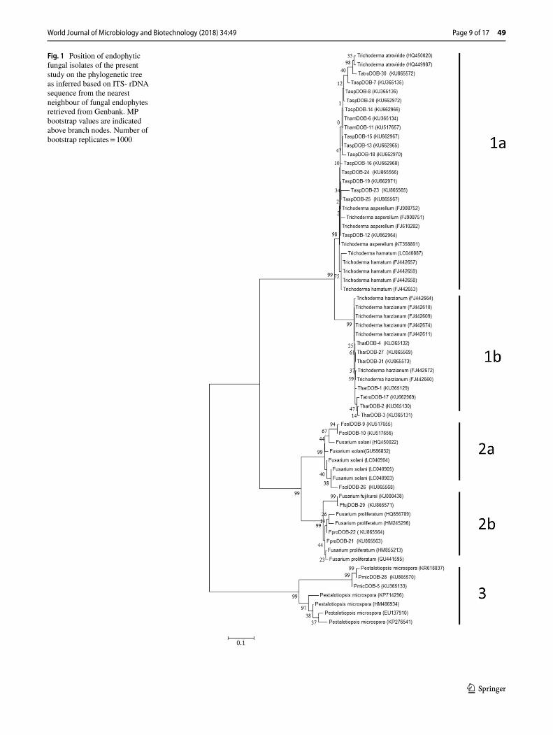
SEM studies showed that the endophytes cause deformities in the mycelia of both P. aphanidermatum and R. solani. The deformities included hyphal fragmentation, perfora-tion, lysis and mycelia degeneration (Figs. 2, 3).
In vitro evaluation of plant growth promoting potentials
All the six endophytic fungal isolates viz., Thar DOB-2, Thar DOB-4, Tasp DOB-12, Tatro DOB-17, Tasp DOB-19 and Thar DOB-31 were able to produce IAA with l-trypto-phan as a precursor. Except T. atroviride (Tatro DOB17), the other isolates exhibited positive results for HCN pro-duction, while three isolates solubilized inorganic phos-phate Ca3(PO4) and four isolates showed positive to cel-lulase activity. Isolate T. harzianum (Thar DOB-31) was positive for IAA, HCN production, inorganic phosphate solubilization and production of cellulase (Table 2). The isolates viz., Thar DOB-2, Tatro DOB-17, Tasp DOB-19 and Thar DOB-31 which produce multiple PGP traits were further considered for green house studies.
Green house experiments
The reduced severity in leaf blight and rhizome rot disease was recorded in four different treatments of endophytes viz., T. harzianum (Thar DOB-2), T. atroviride (Tatro DOB-17), T. asperellum (Tasp DOB-19), T. harzianum (Thar DOB-31). Among the tested endophytes, T. harzi-anum ‘Thar DOB-31’ showed the lowest PDI of rhizome rot to 13.8% and PDI of leaf blight to 11.6%. The isolate also enhanced the plant length and fresh rhizome weight (Table 3).
Rhizome colonization assay by confocal microscopy
The endophytic colonization in turmeric rhizomes was analysed by confocal microscopy. The results revealed the hyphae of endophyte T. harzianum TharDOB-31 formed an unrestricted net-like intra and intercellular colonization pat-tern, intracellularly growing hyphae of endophyte showed necks at site where the fungus transverses a cell wall (Fig. 4).
Minimum inhibitory concentration (MIC)
The MIC determined by broth macro-dilution method showed the inhibition of the tested pathogens (P.
World Journal of Microbiology and Biotechnology (2018) 34:49
1 3
Page 9 of 17 49
Fig. 1 Position of endophytic fungal isolates of the present study on the phylogenetic tree as inferred based on ITS- rDNA sequence from the nearest neighbour of fungal endophytes retrieved from Genbank. MP bootstrap values are indicated above branch nodes. Number of bootstrap replicates = 1000
World Journal of Microbiology and Biotechnology (2018) 34:49
1 3
49 Page 10 of 17
aphanidermatum and R. solani) at 80 µg/ml (MIC) for ethyl acetate extract of T. harzianum TharDOB-31.
HR‑LCMS Analysis
The crude ethyl acetate extract of T. harzianum TharDOB-31 was analyzed using HR-LCMS and the chromatogram of active fractions were given (Fig. 5). From the HR-LCMS analysis TharDOB-31 was found to contain different bioac-tive metabolites, majority of the compounds possess antimi-crobial properties (Table 4).
Discussion
In this study, 31 endophytes were isolated from healthy turmeric rhizomes and the virulent pathogenic strains (P. aphanidermatum PyDOB-4 and R. solani RhsDOB-3) from the diseased rhizome and leaf material. Their identity was confirmed by morphological and ITS–rDNA sequences. The ITS–rDNA sequences were submitted to Genbank (NCBI) (Table 1). The phylogeny showed endophytic isolates grouped with the identical isolates retrieved from Genbank (Fig. 1). A majority of the endophytic isolates belonged to genera namely Trichoderma, Fusarium, and Pestalopsis. The
Trichoderma sp. is one of the major fungi found as an endo-phyte in the healthy turmeric rhizomes (Table 1). All the isolates were screened for antagonism in order to select the isolates that showed the most promising results with regard to growth promotion and biocontrol of rhizome rot and leaf blight diseases in turmeric plants.
The in vitro screening of endophytic isolates for antag-onism against P. aphanidermatum an R. solani reported that endophyte T. harzianum TharDOB-31 followed by T. asperellum TaspDOB-19 exhibited > 70% inhibition (Table 1) of both the pathogens in dual culture assays. Trichoderma spp. showed antagonism against Sclerotium rolfsii, Ceratobasidium cornigerum, Phytophthora para-sitica, P. aphanidermatum, P. myriotylum, and R. solani pathogens, the isolate overgrew the pathogen, covering completely or at least of the surface was reported earlier (Bell et al. 1982). The radial growth of pathogenic fungus was retarded in the dual culture test, and the mycelia of endophytic Trichoderma overgrew the pathogen in petridish (Figs. 2, 3). Similarly, hyphal parasitism of P. aphanider-matum by Trichoderma spp. isolated from soil was reported earlier (Chet et al. 1981).
Endophytes can inhibit pathogen infection and prolif-eration within the host directly via antibiosis, competition and mycoparasitism or indirectly via inducing resistance
Fig. 2 Photographs of dual culture tests and scanning electron micrograph from the inhibition zone between P. aphanidermatum and endophyte T. harzianum TharDOB-31 showing abnormal features: a dual culture test in petri dish; b mycoparasitism; c T. harzianum TharDOB-31 hyphae invading P. aphanidermatum hyphae, and d arrow shows shrivel-ling and lysis of the mycelium along with Trichoderma spores are visible. T = T. harzianum TharDOB-31; P = P. aphanider-matum

World Journal of Microbiology and Biotechnology (2018) 34:49
1 3
Page 11 of 17 49
responses intrinsic to host (Benhamou and Chet 1996; Lahlali and Hijri 2010). The fungal interaction zone between pathogen and BCA (Thar DOB-31) was observed, our SEM results revealed the morphological deformities of mycelia of the pathogens. The antibiosis towards patho-genic fungi could be due to the production antagonistic compounds produced by Trichoderma and are also known to enhance competiveness and thereby control the dis-ease (Yedidia et al. 1999; Howell 2003). Similar observa-tions of hyphal deformities were made in antagonism of Trichoderma spp. against P. aphanidermatum (Chet et al.
1981). Thereby, Trichoderma based biocontrol could be due mainly to the production of hydrolytic enzymes and metabolites (Harman et al. 2004).
The endophyte T. harzianum TharDOB-31 exhibited significant PGP traits like production of cell-wall degrad-ing enzymes, phosphate solubilization, production of IAA and HCN (Table 2). The Trichoderma spp. are reported to have high antibiosis, rhizosphere competency and PGP ability and activates the solubilization of phosphates (Har-man et al. 2004). Cell wall degrading enzymes, such as
Fig. 3 Photographs of dual cul-ture tests and scanning electron micrograph from the inhibition zone between R. solani and endophyte T. harzianum Thar-DOB-31 showing abnormal fea-tures: a dual culture test in petri dish; b and c mycoparasitism; d empty mycelium; e shriveling of mycelium and f arrow shows the breakage of the mycelium of R. solani. T = T. harzianum TharDOB-31; R = R. solani
World Journal of Microbiology and Biotechnology (2018) 34:49
1 3
49 Page 12 of 17
cellulases are important in breakdown of cell walls of oomycete pathogens such as Pythium spp. (Mishra 2010).
The four promising BCA’s viz., T. harzianum (Thar DOB-2), T. atroviride (Tatro DOB-17), T. asperellum (Tasp DOB-19) and T. harzianum (Thar DOB-31) were then tested in the green house for their disease suppression and plant growth promotion abilities compared to untreated and pathogenic controls. Green house results revealed, the endophyte T. harzianum (Thar DOB-31) recorded less PDI (high disease reduction) also enhanced the yield of turmeric when com-pared to untreated control (Table 3). Similar to our reports on turmeric, there are several studies on growth promotion and disease suppression by endophytes in other crops like potato the endophyte T. atroviride and E. nigrum improved yield significantly and decreased the stem disease of potato
plants in greenhouse experiments (Lahlali and Hijri 2010), tomato and pepper the endophyte T. harzianum and T. atro-viride strains promoted the growth and increased the crop productivity as compared to untreated controls (Vinale et al. 2004).
Root colonization by Trichoderma strains frequently enhances root growth and development, crop productivity and resistance to abiotic stress. In the present study endo-phyte T. harzianum Thar DOB-31 showed positive results for colonization of rhizome by confocal microscopic stud-ies (Fig. 4). Our results were supported by earlier reports on some Trichoderma species that colonized root surfaces and caused substantial changes in plant metabolism. Also Trichoderma strains promote plant growth and enhance disease resistance (Harman et al. 2004). The endophyte
Table 2 Characterization of selected endophytic fungal isolates for PGP potentials
IAA production: ‘+’ represent low Indole acetic acid production (light pink colour); ‘++’ represents medium IAA production (dark pink colour); ‘+++’ represents high IAA production (dense pink colour)HCN production: degree of activity (+++>++>+); colour, ranged from yellow (‘+’) to dark brown (‘+++’)Phosphate solubilization: ‘+’ represents clear halo zone < 5 mm on medium; ‘++’ represents halo zone = 5 mm on medium; ‘+++’ represents halo zone > 5 mm on medium; ‘−’ represents no halo zone on mediumCellulase activity: ‘+’ represents positive; ‘++’ represents intermediate; ‘+++’ represents strong activity; ‘−’ represents negative
Sl. no. Isolate no. Species identified IAA production HCN production Phosphate solubiliza-tion
Cel-lulase activity
1 Thar DOB-2 T. harzianum ++ ++ ++ −2 Thar DOB-4 T. harzianum + +++ − +3 Tasp DOB-12 T. asperellum ++ ++ + ++4 Tatro DOB-17 T. atroviride + − ++ +5 Tasp DOB-19 T. asperellum ++ ++ − −6 Thar DOB-31 T. harzianum +++ ++ +++ ++
Table 3 Management of rhizome rot and leaf blight diseases of turmeric caused by P. aphanidermatum and R. solani by fungal endophytes in green house
The values are mean of three replications ± SE. Means in a column followed by same superscript letter are not significantly different according to Duncan’s Multiple range test at P ≤ 0.05
Treatment Rhizome rot Leaf blight
Fresh rhi-zome weight (g)
Plant length (cm) PDI Fresh rhi-zome weight (g)
Plant length (cm) PDI
T. harzianum (Thar DOB-2) 360 ± 2.37d 79.89 ± 1.27c 18.6 ± 0.97c 340 ± 1.27d 76.97 ± 0.83c 15.0 ± 0.37b
T. atroviride (Tatro DOB-17) 410 ± 2.71b 83.17 ± 0.97b 16.4 ± 0.57d 380 ± 1.27b 81.27 ± 0.57b 14.1 ± 0.57c
T. asperellum (Tasp DOB-19) 380 ± 1.97c 80.27 ± 1.21c 15.2 ± 0.37e 365 ± 1.27c 80.50 ± 0.77b 13.4 ± 0.42d
T. harzianum (Thar DOB-31) 425 ± 3.12a 85.71 ± 0.77a 13.8 ± 0.43f 410 ± 1.27a 84.60 ± 1.21a 11.6 ± 0.97e
Carbendazim (0.1%) + mancozeb (0.25%) 300 ± 2.74f 70.88 ± 0.75d 19.4 ± 0.71b 280 ± 1.27e 69.75 ± 0.57d 15.8 ± 0.33b
Untreated control 320 ± 2.57e 57.75 ± 0.57e 0.0 300 ± 1.27e 53.45 ± 0.73e 0.0Pathogenic control 220 ± 2.17g 42.65 ± 0.79f 79.0 ± 0.54a 210 ± 1.27f 40.45 ± 0.43f 75.8 ± 0.37a

World Journal of Microbiology and Biotechnology (2018) 34:49
1 3
Page 13 of 17 49
Trichoderma gamsii exhibited plant growth promotion and antagonism against phytopathogens has been reported (Rinu et al. 2014).
Different formulations using a variety of Trichoderma strains are available commercially for crop production worldwide (Harman 2000) its secondary metabolites
affect plant metabolism and enhance growth (Vinale et al. 2012). The antibiotic fungal metabolites isolated from the biocontrol agent T. harzianum (strains T22 and T39) against phytopathogens Rhizoctonia solani, Pythium ultimum and Gaeumannomyces graminis var. tritici was reported (Vinale et al. 2006). In the present study, crude
Fig. 4 Confocal microscopy observations of BCA (T. har-zianum TharDOB-31) treated 60 day old turmeric rhizome segments for colonization. a The hyphae penetration site is indicated by an arrow in fluorescence image. b Show-ing intracellular mycelium in rhizomes the overlay of T PMT field image and fluorescence image-intracellular hyphae form necks (arrowheads) (scale bars 50 µm)
Fig. 5 HR-LCMS chromatogram of crude ethyl acetate extract of T. harzianum TharDOB-31 showing the bioactive metabolites

World Journal of Microbiology and Biotechnology (2018) 34:49
1 3
49 Page 14 of 17
ethyl acetate extract of endophyte T. harzianum Thar-DOB-31 showed antifungal activity against both the path-ogens and the MIC was observed at 80 µg/m. Also partial characterization of secondary metabolites of endophyte T. harzianum TharDOB-31 by HR-LCMS analysis was reported (Table 4).
The success of Trichoderma strains as BCA’s is due to their high reproductive capacity, ability to survive under unfavorable conditions by utilizing nutrients available, strong aggressiveness against phytopathogenic fungi, and efficiency in promoting plant growth and defence mecha-nism (Benhamou and Chet 1996). Trichoderma as BCA controls the soil-borne pathogens namely ascomycetous, deuteromycetous, basidiomycetous and oomycetous fungi (Omero et al. 1999). Hence, T. harzianum TharDOB-31 can be exploited as potential BCA’s in order to control the rhizome rot and leaf blight diseases in turmeric which helps to reform the chemical fungicide based disease management approaches.
Conclusion
The present study revealed the importance of isolat-ing, screening of endophytes from turmeric rhizome for multiple PGP and biocontrol traits through greenhouse experiments. In this study based on in vitro experiments, endophyte T. harzianum TharDOB-31 exhibited multi-ple PGP traits. Also, the results of greenhouse evidenced endophyte T. harzianum TharDOB-31 suppressed the dis-ease incidence of rhizome rot and leaf blight significantly and markedly enhanced the yield in turmeric compared to untreated control and chemical treatments like car-bendazim–mancozeb. The colonization of T. harzianum TharDOB-31 treated rhizomes was confirmed by confocal microscopic studies. The secondary metabolites of T. har-zianum TharDOB-31 reported bioactive compounds with antimicrobial properties. The study confirms the potential of endophyte T. harzianum TharDOB-31 as biocontrol
Table 4 Major bioactive compounds present in the ethyl acetate extract of Trichoderma harzianum TharDOB-31
Sl. no. Compound name Mass Formula Retention time Biological activity References
1 Diethanolamine 105.0758 C4H11NO2 1.327 Antimicrobial, antifungal, anti-bacterial
Medic-Saric et al. (1980)
2 2-Oxo-3-methylvaleric acid 130.0629 C6H10O3 5.679 Antifungal Guo et al. (2008)3 2-Amino-3-methoxy-benzoic acid 167.0575 C8H9NO3 6.597 Antifungal Park et al. (2001)4 Harmalol 200.094 C12H12N2O 6.942 Antifungal Olmedo et al. (2017)5 Topiramate 339.1009 C12H21NO8S 9.047 Antimicrobial Kruszewska et al. (2012)6 3-Hydroxy-tridecanoic acid 230.186 C13H26O3 9.078 Antifungal Guo et al. (2008)7 Vanillic acid 168.0414 C8H8O4 10.094 Antifungal Kuete et al. (2010)8 3-Methyl-tetradecanedioic acid 272.1966 C15H28O4 10.213 Antimicrobial AbdSharad et al. (2016)9 Dirithromycin 834.5562 C42H78N2O14 11.71 Antimicrobial Roblin and Hammerschlag (1998)10 Punctaporin B 252.1708 C15H24O3 12.28 Antimicrobial Gupta (2016)11 Acarbose 552.2404 C24H40O14 12.292 Antifungal Kruszewska et al. (2008)12 2-Methyl-2E-hexenoic acid 128.0849 C7H12O2 12.431 Antigungal Zhang et al. (2010)13 10-Methyl-1-dodecanol 200.2134 C13H28O 13.283 Antimicrobial Nagoshi et al. (2007)14 9R-hydroxy-10E-octadecenoic acid 298.2504 C18H34O3 14.15 Antimicrobial Abubakar and Majinda (2016)15 Benzenemethanol,
2-(2-aminopropoxy)-3-methyl-196.1108 C11H16O3 14.316 Antifungal Ubaid et al. (2016)
16 Sapindoside A 750.4582 C41H66O12 14.693 Antifungal Khan et al. (2017)17 12-Hydroxy-10-octadecynoic acid 296.2361 C18H32O3 15.64 Antimicrobial Sanabria-Ríos et al. (2014)18 3-Nonaprenyl-4-hydroxybenzoic
acid750.5946 C52H78O3 15.748 Antimicrobial Cho et al. (1998)
19 2-ISOPROPYL-3-Methoxycinnamic acid
220.1106 C13H16O3 17.549 Antimicrobial Naz et al. (2006)
20 Ophiobolin A 400.2635 C25H36O4 17.803 Antimicrobial Li et al. (1995)21 2,3-Dihydroxy stearic acid 316.2606 C18H36O4 19.445 Antimicrobial Choi et al. (2013)22 Calicoferol D 410.3187 C28H42O2 22.47 Antimicrobial Youssef et al. (2011)
World Journal of Microbiology and Biotechnology (2018) 34:49
1 3
Page 15 of 17 49
agent (BCA’s) for sustainable turmeric cultivation. For the best of our knowledge, this is the first report on the endophyte T. harzianum TharDOB-31 as BCA’s against P. aphanidermatum and R. solani pathogens of turmeric. Further studies concerning field applications and stable bioformulations are in progress.
Acknowledgements This work was carried out with the financial assistance from the Department of Science and Technology (DST), Government of India, New Dehli, under the Women Scientist Scheme (DST-WOS A) awarded to Mrs. Vinaya Rani. G (DST sanction No. SR/WOS-A/LS-104/2013) (G) dated 22.04.2014. The authors extend thanks to Dr. K. Ramachandra Kini, Associate Professor, Department of Biotechnology, University of Mysore, Mysore for his help in Phy-logenetic analysis of endophytes and Institute of Excellence (IOE), University of Mysore for providing the instrumentation facility.
Compliance with ethical standards
Conflict of interest The authors declare that they have no conflict of interests regarding the publication of this paper.
References
AbdSharad A, Usup G, Sahrani FK, Ahmad A (2016) Antimicrobial activity and determination of bioactive components from marine Alcaligenes faecalis extract against a sulfate-reducing bacteria. In AIP Conference Proceedings, vol 1784, No 1. AIP Publishing, p 020010
Abubakar MN, Majinda RR (2016) GC-MS analysis and preliminary antimicrobial activity of Albizia adianthifolia (Schumach) and Pterocarpus angolensis (DC). Medicines 3(1):3
Barnett HL, Hunter BB (1972) Illustrated genera of imperfect fungi, 3rd edn. Burgess Publishing Co., Minneapolis
Bell DK, Wells HD, Markham CR (1982) In vitro antagonism of Trichoderma species against six fungal plant pathogens. Phyto-pathology 72(4):379–382
Benhamou N, Chet I (1996) Parasitism of sclerotia of Sclerotium rolf-sii by Trichoderma harzianum: ultrastructural and cytochemical aspects of the interaction. Phytopathology 86(4):405–416
Borneman J, Hartin RJ (2000) PCR primers that amplify fungal rRNA genes from environmental samples. Appl Environ Microbiol 66(10):4356–4360
Buatong J, Phongpaichit S, Rukachaisirikul V, Sakayaroj J (2011) Antimicrobial activity of crude extracts from mangrove fungal endophytes. World J Microbiol Biotechnol 27(12):3005–3008
Chet I, Harman GE, Baker R (1981) Trichoderma hamatum, Its hyphal interactions with Rhizoctonia solani and Pythium sp. Microb Ecol 7:29–38
Cho JY, Moon JH, Seong KY, Park KH (1998) Antimicrobial activ-ity of 4-hydroxybenzoic acid and trans 4-hydroxycinnamic acid isolated and identified from rice hull. Biosci Biotechnol Biochem 62(11):2273–2276
Choi JS, Park NH, Hwang SY, Sohn JH, Kwak I, Cho KK, Choi IS (2013) The antibacterial activity of various saturated and unsatu-rated fatty acids against several oral pathogens. J Environ Biol 34(4):673
Colla G, Rouphael Y, Bonini P, Cardarelli M (2015) Coating seeds with endophytic fungi enhances growth, nutrient uptake, yield and grain quality of winter wheat. Int J Plant Prot 9:171–189
Deshmukh S, Hückelhoven R, Schäfer P, Imani J, Sharma M, Weiss M, Kogel KH (2006) The root endophytic fungus Piri-formospora indica requires host cell death for proliferation during mutualistic symbiosis with barley. Proc Natl Acad Sci 103(49):18450–18457
Domsch KH, Gams W, Anderson TH (1980) Compendium of soil fungi, vol 1. Academic Press, London
Elsherbiny EA, El Khateeb AY, Azzaz NA (2016) Chemical com-position and fungicidal effects of Ocimum basilicum essential oil on Bipolaris and Cochliobolus species. J Agric Sci Technol 18(4):1143–1152
Evans HC, Holmes KA, Thomas SE (2003) Endophytes and myco-parasites associated with an indigenous forest tree, Theobroma gileri, in Ecuador and a preliminary assessment of their potential as biocontrol agents of cocoa diseases. Mycol Prog 2(2):149–160
Fisher PJ, Petrini O (1987) Location of fungal endophytes in tissue of Suaeda fruticosa: a preliminary study. Trans Br Mycol Soc 89:246–249
Fouda AH, Hassan SED, Eid AM, Ewais EED (2015) Biotechnological applications of fungal endophytes associated with medicinal plant Asclepias sinaica (Bioss.). Ann Agric Sci 60(1):95–104
Gilma JC, Joseph C (1998) A manual of soil fungi. Daya Books, New Delhi
Guo L, Wu JZ, Han T, Cao T, Rahman K, Qin LP (2008) Chemical composition, antifungal and antitumor properties of ether extracts of Scapania verrucosa Heeg. and its endophytic fungus Chaeto-mium fusiforme. Molecules 13(9):2114–2125
Gupta A (2016) Exploration of active metabolites from Aegle marmelos and Emblica officinalis for determining its immunosuppressive properties. J Prog Res Biol 3(1):119–126
Gupta SC, Patchva S, Koh W, Aggarwal BB (2012a) Discovery of cur-cumin, a component of golden spice, and its miraculous biological activities. Clin Exp Pharmacol Physiol 39(3):283–299
Gupta P, Samant K, Sahu A (2012b) Isolation of cellulose-degrading bacteria and determination of their cellulolytic potential. Int J Microbiol. https ://doi.org/10.1155/2012/57892 5
Harman GE (2000) Myths and dogmas of biocontrol changes in per-ceptions derived from research on Trichoderma harzinum T-22. Plant Dis 84(4):377–393
Harman GE, Howell CR, Viterbo A, Chet I, Lorito M (2004) Tricho-derma species—opportunistic, avirulent plant symbionts. Nat Rev Microbiol 2(1):43–56
Howell CR (2003) Mechanisms employed by Trichoderma species in the biological control of plant diseases, the history and evolution of current concepts. Plant Dis 87:4–10
Jeffers SN, Martin SB (1986) Comparison of two media selective for Phytophthora and Pythium species. Plant Dis 70:1038–1043
Johnston A, Booth C (1983) Plant pathologist’s pocketbook. Common-wealth Agricultural Bureaux, Slough
Khan H, Khan Z, Amin S, Mabkhot YN, Mubarak MS, Hadda TB, Maione F (2017) Plant bioactive molecules bearing glycosides as lead compounds for the treatment of fungal infection: a review. Biomed Pharmacother 93:498–509
Kim HY, Choi GJ, Lee HB, Lee SW, Lim HK, Jang KS, Kim JC (2007) Some fungal endophytes from vegetable crops and their anti-oomycete activities against tomato late blight. Lett Appl Microbiol 44(3):332–337
Kruszewska JS, Perlinska-Lenart U, Gorka-Niec W, Orlowski J, Zem-bek P, Palamarczyk G (2008) Alterations in protein secretion caused by metabolic engineering of glycosylation pathways in fungi. Acta Biochim Pol 55(3):447–456
Kruszewska H, Zareba T, Tyski S (2012) Examination of antimicrobial activity of selected non-antibiotic medicinal preparations. Act Pol Pharm Drug Res 69:1368–1371
Kuete V, Ngameni B, Mbaveng AT, Ngadjui B, Meye JM, Lall N (2010) Evaluation of flavonoids from Dorstenia barteri for their

World Journal of Microbiology and Biotechnology (2018) 34:49
1 3
49 Page 16 of 17
antimycobacterial, antigonorrheal and anti-reverse transcriptase activities. Acta Trop 116(1):100–104
Kusari P, Kusari S, Spiteller M, Kayser O (2013) Endophytic fungi harbored in Cannabis sativa L: diversity and potential as bio-control agents against host plant-specific phytopathogens. Fun-gal Divers 60(1):137–151
Lahlali R, Hijri M (2010) Screening, identification and evaluation of potential biocontrol fungal endophytes against Rhizoc-tonia solani AG3 on potato plants. FEMS Microbiol Lett 311(2):152–159
Lahlali R, Serrhini MN, Friel D, Jijakli MH (2007) Predictive model-ling of temperature and water activity (solutes) on the in vitro radial growth of Botrytis cinerea Pers. Int J Food Microbiol 114(1):1–9
Li E, Clark AM, Rotella DP, Hufford CD (1995) Microbial metabolites of ophiobolin A and antimicrobial evaluation of ophiobolins. J Nat Prod 58(1):74–81
Lledo S, Rodrigo S, Poblaciones MJ, Santamaria O (2016) Biomass yield, nutritive value and accumulation of minerals in Trifolium subterraneum L. as affected by fungal endophytes. Plant Soil 405(1–2):197–210
Medic-Saric M, Maysinger D, Movrin M, Dvoržak I (1980) Anti-bacterial and antifungal activities of nitroxoline Mannich bases. Chemotherapy 26(4):263–267
Mejia LC, Rojas EI, Maynard Z, Van Bael S, Arnold AE, Hebbar P, Herre EA (2008) Endophytic fungi as biocontrol agents of Theo-broma cacao pathogens. Biol Control 46(1):4–14
Mishra VK (2010) In vitro antagonism of Trichoderma species against Pythium aphanidermatum. J Phytol 2(9):28–35
Morandi MA, Sutton JC, Maffia LA (2000) Effects of host and micro-bial factors on development of Clonostachys rosea and control of Botrytis cinerea in rose. Eur J Plant Pathol 106(5):439–448
Muthukumar A, Eswaran A, Sangeetha G (2011) Induction of sys-temic resistance by mixtures of fungal and endophytic bacterial isolates against Pythium aphanidermatum. Acta Physiol Plant 33(5):1933–1944
Myrchiang P, Dkhar MS, Devi HR (2014) Studies on endophytic fungi associated with medicinally important aromatic plant Artemisia nilagirica (C.B. Clarke) Pamp. and their antagonistic activity against Phytophthora infestans. J Adv Lab Res Biol 4(4):112–119
Nalini MS, Sunayana N, Prakash HS (2014) Endophytic fungal diver-sity in medicinal plants of Western Ghats, India. Int J Biodivers. https ://doi.org/10.1155/2014/49421 3
Naz S, Ahmad S, Rasool SA, Sayeed SA, Siddiqi R (2006) Antibacte-rial activity directed isolation of compounds from Onosma hispi-dum. Microbiol Res 161(1):43–48
Nisa H, Kamili AN, Nawchoo IA, Shafi S, Shameem N, Bandh SA (2015) Fungal endophytes as prolific source of phytochemicals and other bioactive natural products: a review. Microb Pathog 82:50–59
Olmedo GM, Cerioni L, González MM, Cabrerizo FM, Rapisarda VA, Volentini SI (2017) Antifungal activity of β-carbolines on Peni-cillium digitatum and Botrytis cinerea. Food Microbiol 62:9–14
Omero C, Inbar J, Rocha-Ramirez V, Herrera-Estrella A, Chet I, Horwitz BA (1999) G protein activators and cAMP promote mycoparasitic behaviour in Trichoderma harzianum. Mycol Res 103(12):1637–1642
Park M (1934) Report on the work of the Mycology Division. Admn Rep Dir Agric Ceylon, pp 126–133
Park ES, Moon WS, Song MJ, Kim MN, Chung KH, Yoon JS (2001) Antimicrobial activity of phenol and benzoic acid derivatives. Int Biodeterior Biodegradation 47(4):209–214
Pereira SIA, Castro PML (2014) Diversity and characterization of cul-turable bacterial endophytes from Zea mays and their potential as plant growth-promoting agents in metal-degraded soils. Environ Sci Pollut Res 21(24):14110–14123
Pujol I, Guarro J, Llop C, Soler L, Fernández-Ballart J (1996) Com-parison study of broth macrodilution and microdilution antifungal susceptibility tests for the filamentous fungi. Antimicrob Agents Chem 40(9):2106–2110
Rathaiah Y (1982) Rhizome rot of turmeric. Indian Phytopath 35(3):415–417
Rinu K, Sati P, Pandey A (2014) Trichoderma gamsii (NFCCI 2177): a newly isolated endophytic, psychrotolerant, plant growth promoting, and antagonistic fungal strain. J Basic Microbiol 54(5):408–417
Roblin PM, Hammerschlag MR (1998) In vitro activity of a new ketolide antibiotic, HMR 3647, against Chlamydia pneumoniae. Antimicrob Agents Chem 42(6):1515–1516
Rodriguez RJ, White JF Jr, Arnold AE, Redman ARA (2009) Fun-gal endophytes: diversity and functional roles. New phytol 182(2):314–330
Roy AK (1992) Severity of Rhizoctonia solani on the leaves of rice and turmeric. Indian Phytopath 45:344–347
Rubini MR, Silva-Ribeiro RT, Pomella AW, Maki CS, Araújo WL, Dos Santos DR, Azevedo JL (2005) Diversity of endophytic fun-gal community of cacao (Theobroma cacao L.) and biological control of Crinipellis perniciosa, causal agent of Witches’ Broom Disease. Int J Biol Sci 1(1):24
Sanabria-Ríos DJ, Rivera-Torres Y, Maldonado-Domínguez G, Domínguez I, Ríos C, Díaz D, Montano N (2014) Antibacterial activity of 2-alkynoic fatty acids against multidrug-resistant bac-teria. Chem Phys Lipids 178:84–91
Schoch CL, Seifert KA, Huhndorf S, Robert V, Spouge JL, Levesque CA, Miller AN (2012) Nuclear ribosomal internal transcribed spacer (ITS) region as a universal DNA barcode marker for fungi. Proc Natl Acad Sci 109:6241–6246
Shanmugam V, Kanoujia N (2011) Biological management of vascular wilt of tomato caused by Fusarium oxysporum f. sp. lycospersici by plant growth-promoting rhizobacterial mixture. Biol Control 57(2):85–93
Shanmugam V, Gupta S, Dohroo NP (2013) Selection of a compat-ible biocontrol strain mixture based on co-cultivation to control rhizome rot of ginger. Crop Prot 43:119–127
Singh H, Reddy MS (2011) Effect of inoculation with phosphate solu-bilizing fungus on growth and nutrient uptake of wheat and maize plants fertilized with rock phosphate in alkaline soils. Eur J Soil Biol 47(1):30–34
Sriraj PP, Sundravadana S, Alice D (2014) Efficacy of fungicides, botanicals and bioagents against Rhizoctonia solani inciting leaf blight on turmeric (Curcuma longa L.). Afr J Microbiol Res 8(36):3284–3294
Tamura K, Stecher G, Peterson D, Filipski A, Kumar S (2013) MEGA6: molecular evolutionary genetics analysis version 6.0. Mol Biol Evol 30(12):2725–2729
Thiripurasundari K, Selvarani K (2014) Production of turmeric in India: an analysis. Int J Bus Manag 2(9):229
Thompson JD, Higgins DG, Gibson TJ (1994) CLUSTAL W, improv-ing the sensitivity of progressive multiple sequence alignment through sequence weighting, position-specific gap penalties and weight matrix choice. Nucleic Acids Res 22(22):4673–4680
Triveni S, Prasanna R, Shukla L, Saxena AK (2013) Evaluating the biochemical traits of novel Trichoderma-based biofilms for use as plant growth-promoting inoculants. Ann Microbiol 63(3):1147–1156
Ubaid JM, Hussein HM, Hameed IH (2016) Determination of bioactive chemical composition of Callosobruchus maculutus and investiga-tion of its anti-fungal activity. Int J Pharmacogn Phytochem Res 8(8):1385–1397
Ushamalini C, Nakkeeran P, Marimuthu T (2008) Induction of plant defense enzymes in turmeric plants by Trichoderma viride. Arch Phytopathol Plant Prot 41:79–93
World Journal of Microbiology and Biotechnology (2018) 34:49
1 3
Page 17 of 17 49
Van der Plaats-Niterink AJ (1981) Monograph of the genus Pythium. Stud Mycol 21:1–24
Vinale F, Ambrosio GD, Abadi K, Scala F, Marra R, Turrà D, Lorito M (2004) Application of Trichoderma harzianum (T22) and Trichoderma atroviride (P1) as plant growth promoters, and their compatibility with copper oxychloride. J Zhejiang Univ Sci 30(4):425–425
Vinale F, Marra R, Scala F, Ghisalberti EL, Lorito M, Sivasithamparam K (2006) Major secondary metabolites produced by two commer-cial Trichoderma strains active against different phytopathogens. Lett Appl Microbiol 43(2):143–148
Vinale F, Ghisalberti EL, Sivasithamparam K, Marra R, Ritieni A, Ferracane R, Lorito M (2009) Factors affecting the production of Trichoderma harzianum secondary metabolites during the interaction with different plant pathogens. Lett Appl Microbiol 48(6):705–711
Vinale F, Sivasithamparam K, Ghisalberti EL, Ruocco M, Woo S, Lorito M (2012) Trichoderma secondary metabolites that affect plant metabolism. Nat Prod Commun 7:1545–1550
Vinale F, Sivasithamparam K, Ghisalberti EL, Woo SL, Nigro M, Marra R, Manganiello G (2014) Trichoderma secondary metab-olites active on plants and fungal pathogens. Open Mycol J 8(1):127–139
Wahab S (2009) Biotechnological approaches in the management of plant pests, diseases and weeds for sustainable agriculture. J Biopest 2(2):115–134
White TJ, Bruns T, Lee SJWT, Taylor JL (1990) Amplification and direct sequencing of fungal ribosomal RNA genes for phylogenet-ics. PCR Protoc: Guide Methods Appl 18(1):315–322
Yedidia I, Benhamou N, Chet I (1999) Induction of defense responses in cucumber plants (Cucumis sativus L.) by the biocontrol agent Trichoderma harzianum. Appl Environ Microbiol 65:1061–1070
Youssef DA, Miller CW, El-Abbassi AM, Cutchins DC, Cutchins C, Grant WB, Peiris AN (2011) Antimicrobial implications of vita-min D. Dermatoendocrinol 3(4):220–229
Zhang L, Pornpattananangkul D, Hu CM, Huang CM (2010) Develop-ment of nanoparticles for antimicrobial drug delivery. Curr Med Chem 17(6):585–594


![Review Curcumol: From Plant Roots to Cancer RootsPlant tissue culture approach has conventionally recognized as a ... Curcuma longa Common turmeric Rhizome Antifungal [37] Curcuma](https://static.fdocuments.net/doc/165x107/5f7f3f3d7d5b343f5c214108/review-curcumol-from-plant-roots-to-cancer-roots-plant-tissue-culture-approach.jpg)


![Constituents of Representative Curcuma And …...(Curcuma longa L.=C.domestica Valeton) turmeric((1989) [ ] 13 No.10 p7 13 2002 Constituents of Representative Curcuma And Estimation](https://static.fdocuments.net/doc/165x107/5e894bf9686ab57d1b28ac90/constituents-of-representative-curcuma-and-curcuma-longa-lcdomestica-valeton.jpg)


![Assessment Report on Curcuma Longa L. Rhizoma · Curcuma longa rhizome varies from 0.6 to 5% of the dry mass [15]. The dry turmeric rhizomes contain 3-5% curcumin, the curcumin content](https://static.fdocuments.net/doc/165x107/5e0fa7a6b6de3d3894037369/assessment-report-on-curcuma-longa-l-rhizoma-curcuma-longa-rhizome-varies-from.jpg)