Frontal lobe epilepsy - pn.bmj.com · and the diverse, complex semiology that may oc cur with...
14
PRACTICAL NEUROLOGY 260 © 2004 Blackwell Publishing Ltd Lateral-dorsal Medio-ventral Central Medial Premotor Lateral Premotor Ventral Medial-dorsal Classification Prefrontal Lateral-ventral Prefrontal Medial-ventral Lateral Precentral Medial Precentral REVIEW Frontal lobe epilepsy: seizure semiology and presurgical evaluation Dr Aileen McGonigal † and Professor Patrick Chauvel* *Director of Neurophysiology and Neuropsychology and † Clinical Research Fellow in Epileptology, Service de Neurophysiologie Clinique, Hôpital de la Timone and Laboratoire de Neurophysiologie et Neuropsychologie, INSERM EMI 9926, Faculté de Médecine, Marseille, France; Email: [email protected] Practical Neurology, 2004, 4, 260–273 on 6 September 2018 by guest. Protected by copyright. http://pn.bmj.com/ Pract Neurol: first published as 10.1111/j.1474-7766.2004.00244.x on 1 October 2004. Downloaded from
Transcript of Frontal lobe epilepsy - pn.bmj.com · and the diverse, complex semiology that may oc cur with...

PRACTICAL NEUROLOGY260
© 2004 Blackwell Publishing Ltd
Lateral-dorsal
Medio-ventral
CentralMedial PremotorLateral Premotor
VentralMedial-dorsal
Classification
Prefrontal
Lateral-ventral
Prefrontal
Medial-ventral
Lateral
Precentral
Medial
Precentral
REVIEW
Frontal lobe epilepsy:seizure semiology and presurgical evaluation
Dr Aileen McGonigal† and Professor Patrick Chauvel*
*Director of Neurophysiology and Neuropsychology and †Clinical Research Fellow in Epileptology, Service de
Neurophysiologie Clinique, Hôpital de la Timone and Laboratoire de Neurophysiologie et Neuropsychologie, INSERM EMI 9926,
Faculté de Médecine, Marseille, France; Email: [email protected]
Practical Neurology, 2004, 4, 260–273
on 6 Septem
ber 2018 by guest. Protected by copyright.
http://pn.bmj.com
/P
ract Neurol: first published as 10.1111/j.1474-7766.2004.00244.x on 1 O
ctober 2004. Dow
nloaded from

OCTOBER 2004 261
© 2004 Blackwell Publishing Ltd
INTRODUCTIONJohn Hughlings Jackson refl ected that the fron-tal lobe is the brain’s ‘most complex and least organized centre’ (Jackson 1931) and, despite subsequent advances in neuroscience, even by the late 20th century the frontal lobe was still considered to be an ‘uncharted province of the brain’ (Goldman-Rakic 1984). For epileptolo-gists today, frontal lobe epilepsy (FLE) remains the most challenging of all the epilepsies, both in terms of understanding how seizures are or-ganized and how they should be treated. This is very evident in comparison to the now well-defi ned syndrome of mesial temporal lobe epi-lepsy (TLE).
Important advances have been made in re-cent decades, especially in correlations between the clinical and electrical expression of seizures, permitting better understanding of FLE. To-gether with major developments in the fi eld of neuroimaging, these advances are changing the approach to management, particularly in mak-ing curative surgery a real possibility for many more patients than ever before.
Understanding FLE, it can be argued, will also help us understand more about the cer-ebral processes that underlie normal higher brain functions such as the interaction between emotion and decision making (Damasio 1995). Indeed, frontal lobe epilepsy has been described as ‘the next frontier’ (Niedermeyer 1998). From historical observations to futuristic develop-ments: what does all this mean for our routine clinical practice?
AIMS OF THIS REVIEWWe have chosen to focus on the approach to the electroclinical diagnosis and localization of FLE, in other words the combined analysis of the clinical features of seizures (semiology) and electroencephalographic (EEG) data. This approach is particularly important when assess-ing those patients who may be candidates for epilepsy surgery: the 20% or so of all patients with partial epilepsy who are pharmacoresist-ant. In addition we will briefl y discuss recent developments in other aspects of presurgical evaluation.
WHY ARE ELECTROCLINICAL CORRELATIONS SO IMPORTANT IN FRONTAL LOBE EPILEPSY?In epilepsy, as in all neurological practice, the history and physical signs are of paramount im-portance and cannot be replaced by a single test,
no matter how sophisticated. Just as the pres-ence of a right hemiparesis alerts the clinician to look for a lesion in the contralateral motor path-way, so too can the clinical features of a seizure point to the activation (or inhibition) of certain brain regions. At its simplest level, this allows us to relate a sign such as a focal clonic contraction in the hand with seizure activity in the contralat-eral motor cortex. However, in FLE the observed symptoms or signs may be complex, subtle and often occur simultaneously or in rapid succes-sion, frequently refl ecting the activation of dif-ferent structures within a dynamic system, with rapid and unpredictable propagation patterns.
Not only is it challenging to determine from which part of the frontal lobe the seizure arises, but also it is often diffi cult to assess whether a particular attack is indeed a frontal lobe sei-zure at all. Frontal lobe seizures are particularly prone to misdiagnosis as psychogenic non-epi-leptic seizures, due to their sometimes bizarre or atypical appearance, as well as to the fact that surface EEG does not necessarily show interictal or ictal abnormalities (Bautista et al. 1998).
Another possible misdiagnosis of FLE is of a sleep disorder, particularly as a large proportion of frontal seizures arise from sleep. For example, the nocturnal attack disorder originally identi-fi ed as a form of movement disorder – ‘paroxys-mal nocturnal dystonia’ (Lugarasi & Cirignotta 1981) – was subsequently recognized to have an epileptic basis in most cases (Meierkord et al. 1992). The syndrome of autosomal dominant nocturnal frontal lobe epilepsy (ADNFLE) was later described; this is a monogenic disorder with high penetrance, characterized by brief hyperkinetic nocturnal seizures.
Because of these diagnostic diffi culties, cau-tion must be exercised, and an epilepsy specialist rather than a general neurologist or general phy-sician should ideally make the diagnosis.
The localizing value of specifi c semiological features is, in general, less well-understood in FLE, compared with TLE. For this reason, as well as other issues related to the limitations of EEG in FLE, diagnosis and localization are well rec-ognized to be more diffi cult than in other locali-zation-related epilepsies (Manford et al. 1996). Indeed, it is likely that some epilepsy ‘surgical failures’, including cases operated for presumed TLE that do not become seizure-free post-op-eratively, refl ect incorrect presurgical localiza-tion, rather than suboptimal resection (Walsh & Delgado-Escueta 1984). FLE forms the second largest group of potentially operable localiza-
on 6 Septem
ber 2018 by guest. Protected by copyright.
http://pn.bmj.com
/P
ract Neurol: first published as 10.1111/j.1474-7766.2004.00244.x on 1 O
ctober 2004. Dow
nloaded from

PRACTICAL NEUROLOGY262
© 2004 Blackwell Publishing Ltd
area) within the premotor cortex, particularly in the dominant hemisphere (area 44).
• The frontal eye fi elds, which can contribute to ictal versive head and eye movement, lie within the dorsolateral cortex (area 8) in the boundary where the premotor and precentral cortex meet, and may therefore be involved in seizures arising from either of these regions.The organization of the prefrontal cortex,
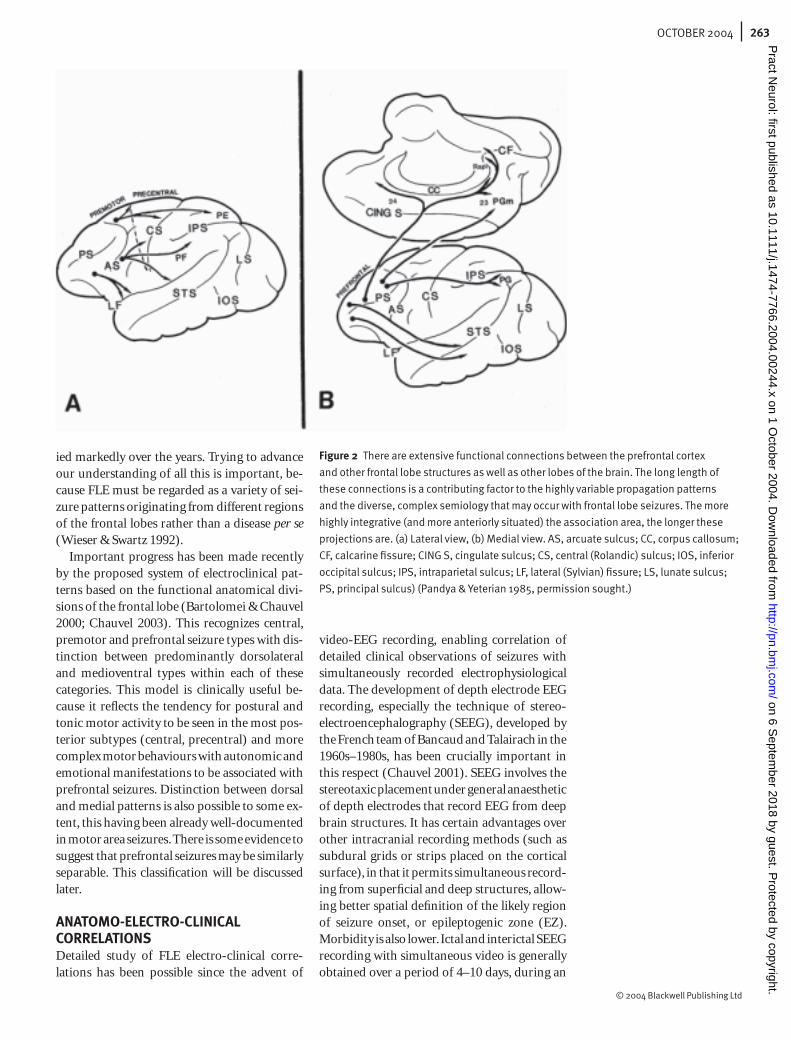
which is predominantly made up of heteromo-dal association areas, is extremely complicated and incompletely understood. It has complicat-ed and long association connections with other brain regions, including limbic and paralimbic areas, which involve a continuum of temporal and frontal lobe structures (particularly the cin-gulate gyrus and the posterior orbital region) (Fig. 2). Incoming sensory information from these areas may be processed, taking account of motivational and emotional states, and used to infl uence decision-making and many aspects of behaviour (Pandya & Yeterian 1985). Patients with prefrontal epilepsy may demonstrate in-terictal behavioural or psychiatric abnormalities, such as lack of spontaneity and poor planning (frontal abulic syndrome), or impulsivity and socially inappropriate behaviour (frontal dis-inhibition syndrome), which may improve fol-lowing surgery (Devinsky et al. 1995).
ATTEMPTS TO CLASSIFY FRONTAL LOBE EPILEPSYAlthough the approach of separating tempo-ral from extra-temporal epilepsy is now estab-lished, and most extra-temporal epilepsies have their origin in the frontal lobe, a widely accepted classifi cation of FLE has not yet been reached. Indeed the nomenclature used by different groups to describe frontal seizure types has var-
tion-related epilepsies after TLE. The reliable electroclinical diagnosis of FLE, as well as the identifi cation of subtypes, is therefore particu-larly important in terms of optimal selection of candidates for epilepsy surgery.
Despite the complex nature of most fron-tal seizures, certain semiological patterns are reproducible and can help to defi ne the likely region(s) involved. The occurrence of localized tonic posturing (face, upper limb, lower limb), emotional behaviour (such as fear) and com-plex motor activity, may direct the clinician to-wards a particular part of the frontal lobe, as will be discussed later. This becomes crucial when a more precise sublobar understanding of locali-zation is required.
FRONTAL LOBE ANATOMY AND RELATION TO SEMIOLOGYThe frontal lobe is the largest lobe in the brain (accounting for about 40% of cerebral cortex) (Fig. 1). This large size contributes to diagnostic diffi culties. There are multiple diverse propaga-tion patterns, and there is the problem of lim-ited EEG sampling, particularly from relatively ‘hidden’ regions such as medial and basal (or-bitofrontal) cortex (Bautista et al. 1998). The functional anatomical divisions of precentral, premotor and prefrontal cortex provide a use-ful model for thinking about semiology and will be briefl y described:• The precentral region consists of primary
motor cortex, Brodmann’s area 4.• The premotor cortex consists principally of
the lateral and medial components of area 6, the latter corresponding to the supplemen-tary motor area (SMA).
• There is some representation of language (in-cluding the region formally known as Broca’s
Figure 1 Cytoarchitectonic
diagram of the frontal lobe
(prefrontal cortex shown in
colour), with Brodmann’s areas
(Petrides & Pandya 1994). (a)
Lateral view, (b) Medial aspect.
Reprinted from The Frontal Lobes,
Computational Modelling and
Neuropsychology: Handbook of
Neuropsychology, Vol 9, Boller F,
Spinnler H, Hendler JA, 1994, with
permission from Elsevier.
10
�� ��
47/12
1045A
46
45B
44
9/46v6
9/46d
9
8B
8Ad
8Av
6
4
8B
9
CC 24
32
25
14
10
on 6 Septem
ber 2018 by guest. Protected by copyright.
http://pn.bmj.com
/P
ract Neurol: first published as 10.1111/j.1474-7766.2004.00244.x on 1 O
ctober 2004. Dow
nloaded from
OCTOBER 2004 263
© 2004 Blackwell Publishing Ltd
ied markedly over the years. Trying to advance our understanding of all this is important, be-cause FLE must be regarded as a variety of sei-zure patterns originating from different regions of the frontal lobes rather than a disease per se (Wieser & Swartz 1992).
Important progress has been made recently by the proposed system of electroclinical pat-terns based on the functional anatomical divi-sions of the frontal lobe (Bartolomei & Chauvel 2000; Chauvel 2003). This recognizes central, premotor and prefrontal seizure types with dis-tinction between predominantly dorsolateral and medioventral types within each of these categories. This model is clinically useful be-cause it refl ects the tendency for postural and tonic motor activity to be seen in the most pos-terior subtypes (central, precentral) and more complex motor behaviours with autonomic and emotional manifestations to be associated with prefrontal seizures. Distinction between dorsal and medial patterns is also possible to some ex-tent, this having been already well-documented in motor area seizures. There is some evidence to suggest that prefrontal seizures may be similarly separable. This classifi cation will be discussed later.
ANATOMO-ELECTRO-CLINICAL CORRELATIONSDetailed study of FLE electro-clinical corre-lations has been possible since the advent of
video-EEG recording, enabling correlation of detailed clinical observations of seizures with simultaneously recorded electrophysiological data. The development of depth electrode EEG recording, especially the technique of stereo-electroencephalography (SEEG), developed by the French team of Bancaud and Talairach in the 1960s–1980s, has been crucially important in this respect (Chauvel 2001). SEEG involves the stereotaxic placement under general anaesthetic of depth electrodes that record EEG from deep brain structures. It has certain advantages over other intracranial recording methods (such as subdural grids or strips placed on the cortical surface), in that it permits simultaneous record-ing from superfi cial and deep structures, allow-ing better spatial defi nition of the likely region of seizure onset, or epileptogenic zone (EZ). Morbidity is also lower. Ictal and interictal SEEG recording with simultaneous video is generally obtained over a period of 4–10 days, during an
Figure 2 There are extensive functional connections between the prefrontal cortex
and other frontal lobe structures as well as other lobes of the brain. The long length of
these connections is a contributing factor to the highly variable propagation patterns
and the diverse, complex semiology that may occur with frontal lobe seizures. The more
highly integrative (and more anteriorly situated) the association area, the longer these
projections are. (a) Lateral view, (b) Medial view. AS, arcuate sulcus; CC, corpus callosum;
CF, calcarine fi ssure; CING S, cingulate sulcus; CS, central (Rolandic) sulcus; IOS, inferior
occipital sulcus; IPS, intraparietal sulcus; LF, lateral (Sylvian) fi ssure; LS, lunate sulcus;
PS, principal sulcus) (Pandya & Yeterian 1985, permission sought.)
on 6 Septem
ber 2018 by guest. Protected by copyright.
http://pn.bmj.com
/P
ract Neurol: first published as 10.1111/j.1474-7766.2004.00244.x on 1 O
ctober 2004. Dow
nloaded from

PRACTICAL NEUROLOGY264
© 2004 Blackwell Publishing Ltd
inpatient stay in a specialist videotelemetry unit. The decision about where to place the electrodes is based on the hypotheses regarding the likely brain structures involved in the EZ. These hy-potheses are formed by the epilepsy team and are based on the ensemble of all the available non-invasive data [standard EEG and video-EEG, MRI, single photon emission computer-ized tomography (SPECT), positron emission tomography (PET), etc.], and including detailed semiological observations. This technique can be very useful in those patients with FLE who are potential surgical candidates, but where standard non-invasive investigations are insuf-fi cient to allow localization of the EZ (for ex-ample normal or nonlocalizing MRI). Potential contra-indications to surgery (e.g. involvement of language areas) can also be studied during the recording. The method is well-established in several European countries for epilepsy pre-surgical evaluation, but remains much less used elsewhere, including in the UK and US. An ex-ample of this electroclinical approach is shown in Fig. 3.
Figure 3 Stereo electroencephalographic (SEEG) intracerebral recording of a dorsolateral
prefrontal seizure. (a) This patient had seizures characterized by semipurposeful
behaviour, proximal tonic posturing and vocalization. The combination of clinical,
electrophysiological and imaging data suggested likely involvement of the dorsolateral
prefrontal region. (b) The electrical onset of the seizure is clearly seen in his ictal SEEG
recording, with a build-up of high amplitude rhythmic spikes (red arrows) followed by
a high frequency rapid discharge (blue arrows). The electrodes involved lie within a
localized part of the dorsolateral prefrontal region. (c) Representation of propagation
patterns as recorded with SEEG, superimposed on a 3D MRI reconstruction. The
patient subsequently underwent localized cortical resection, with no postoperative
neurocognitive defi cit, and remains seizure-free at 1-year follow-up. With thanks to the
staff of the Epilepsy Unit, Hôpital de la Timone, Marseille, France.
a b
c
on 6 Septem
ber 2018 by guest. Protected by copyright.
http://pn.bmj.com
/P
ract Neurol: first published as 10.1111/j.1474-7766.2004.00244.x on 1 O
ctober 2004. Dow
nloaded from

OCTOBER 2004 265
© 2004 Blackwell Publishing Ltd
GENERAL FEATURES OF FRONTAL SEIZURE SEMIOLOGYRecent reviews have revealed the diversity of presentation of frontal seizure semiology and some have sought to defi ne clinical or electro-clinical subtypes (Bancaud & Talairach 1992; Talairach et al. 1992a; Chauvel et al. 1995; Wil-liamson & Engel 1997; So 1998; Swartz et al. 1998; Bartolomei & Chauvel 2000; Jobst et al. 2000; Kotagal et al. 2003). Those (relatively few) series with confi rmation of frontal lobe origin by depth studies and/or subsequent surgical cure by frontal resection have been particularly useful.
Frontal seizure semiology is extremely di-verse, but certain features are agreed to suggest frontal lobe origin. Seizures may be brief with sudden onset and termination; often arise from sleep; may occur in clusters; have a tendency to rapid secondary generalization; and produce minimal postictal confusion (Williamson et al. 1985; Williamson & Engel 1997). Clonic activity and asymmetric tonic posturing are typical of frontal seizures and of all the possible frontal sei-zure symptoms and signs, motor manifestations remain the most frequent and important, ob-served in 90% of patients (Chauvel et al. 1995).
Complex gestural manifestations or patterns of behaviour seem to be particularly characteris-tic of certain FLEs and are increasingly recognized (Williamson et al. 1985; Bancaud & Talairach 1992; Chauvel et al. 1995; Manford et al. 1996; Jobst 2000). Gestural automatisms include fum-bling or exploratory movements with the hand directed toward self or environment, such as tap-ping or grabbing of objects or bedclothes; more complex behaviours might include snapping the fi ngers, crossing and uncrossing the legs, or more dramatic pedalling movements, thrashing or hit-ting. Some of these complex manifestations may seem to be to a greater or lesser degree adapted to the environment, indicating a degree of re-tained awareness and autonomy. Although the term ‘hypermotor seizures’ was proposed by the Cleveland group to describe such seizures char-acterized by motor agitation associated with an emotional quality, this has not been uniformly accepted as a useful description (So 1998).
DIFFICULTIES OF ELECTROCLINICAL CORRELATION IN FLEUnlike TLE, where the sites of epileptic discharge likely to be responsible for certain ictal clinical signs are relatively well-recognized, the neuro-physiological organization of many types of frontal lobe seizure remains poorly understood.
Several recent FLE studies have attempted to demonstrate a consistent sublobar localization for given ictal symptoms or signs but have found limited correlations (Manford et al. 1996; Jobst et al. 2000; Kotagal et al. 2003). This has led some authors to question whether it is possible to rely at all on semiological analysis when attempting to localize frontal seizures (Manford 1996).
However, such conclusions may to some de-gree refl ect the limitations of using statistical cluster analysis to try to correlate an isolated clinical sign with a ‘focus’ of epileptic activity, rather than considering patterns of clinical signs and the concept of an epileptic ‘network’ that involves several sites and which gives rise to ictal phenomena depending on the interplay of a dy-namic system.
The underlying mechanism of the complex ictal behaviour patterns seen in FLE remains an interesting and disputed question. One theory, proposed by Jackson and being revisited today, is that epileptic activity may disrupt the con-trol normally exerted by higher brain centres, thus allowing the ‘release’ or disinhibition of more primitive, stereotyped behaviours. Such release phenomena might be comparable with the forced grasping or ‘utilization behaviour’ fi rst described by Lhermitte (1983) in patients with bilateral frontal lesions, now recognized to form part of the spectrum of abnormal motor responses that occur in the context of imbal-ance between internally generated control of movement and response to environmental cues (Archibald et al. 2001). The stepping refl ex is another example of such release phenomena, and the kicking movements or pelvic thrust-ing during frontal seizures may be interpreted as relating to this. Recent electrophysiological data supporting the role of epileptic networks in frontal lobe ictal phenomena argue in favour of a ‘functional uncoupling’ of this nature (Wend-ling et al. 2003; Gavaret et al. 2004).
This area therefore remains one of the most important for further study if we are to advance in our ability to understand FLE, and success-fully select FLE patients for surgical treatment.
ANATOMIC-FUNCTIONAL CLASSIFICATION OF FRONTAL LOBE SEIZURESWhile the recent anatomical and functional cate-gorization of frontal seizure subtypes proposed by Chauvel and colleagues remains preliminary, it provides a useful way to consider the localizing value of certain patterns of seizure semiology (Fig. 4)
Seizures may
be brief with
sudden onset
and termination;
often arise from
sleep; may occur
in clusters; have
a tendency to
rapid secondary
generalization;
and produce
minimal postictal
confusion
on 6 Septem
ber 2018 by guest. Protected by copyright.
http://pn.bmj.com
/P
ract Neurol: first published as 10.1111/j.1474-7766.2004.00244.x on 1 O
ctober 2004. Dow
nloaded from

PRACTICAL NEUROLOGY266
© 2004 Blackwell Publishing Ltd
Precentral (primary
motor) area (BA 4):
clonic jerks, sometimes
tonic posturing or
cortical myoclonus
Premotor areas
including SMA
(BA 6):
asymmetric tonic
posturing,
sometimes more
complex motor
phenomena
Frontal eye
fields (BA 8):
version of gaze
and/or head
version
Expressive language
areas
(BA 44, 45)
Frontal operculum:
facial contraction,
hypersalivation
Dorsolateral
prefrontal
region:
complex
automatisms,
semi-
purposeful
behaviour,
“forced acting”;also frontal
absences
a
Precentral
(primary motor)
area (leg
representation):
clonic jerks,
sometimes tonic
posturing or
cortical myoclonus
Premotor region: SMA (BA 6)
asymmetric tonic posturing,
sometimes more complex motor
phenomena
Frontal eye
fields (BA 8):
version of
gaze and/or
head version
Ventromesial
prefrontal
region:
hyperkinetic
motor
behaviour, ictal
expression of
emotion (fear)
b
Figure 4 Summary of elements
of frontal seizure semiology in
relation to precentral, premotor
and prefrontal regions. (a)
dorsolateral view (b) medial
view. BA, Brodmann’s area; SMA,
supplementary motor area.
on 6 Septem
ber 2018 by guest. Protected by copyright.
http://pn.bmj.com
/P
ract Neurol: first published as 10.1111/j.1474-7766.2004.00244.x on 1 O
ctober 2004. Dow
nloaded from

OCTOBER 2004 267
© 2004 Blackwell Publishing Ltd
Pre-central seizuresThe best-defi ned frontal seizure type was rec-ognized by Jackson over 100 years ago (in Jack-son 1931). His description of focal seizures with contralateral clonic movements arising from the precentral (primary motor) region remains valid today. The characteristic feature is the slow progression from one body part to another ad-jacent segment – the ‘Jacksonian march’. The body part involved in the seizure indicates the region of motor cortex activity according to the somatotopic representation in the motor cortex, so that it is possible to distinguish dorsal from medial precentral seizures.
In the light of depth electrode data, it is now recognized that seizures arising from the pre-central cortex may also appear rather different to the classical Jacksonian focal clonic seizure. For example, contralateral clonic movements may be accompanied by more complex bilateral tonic posturing, or predominantly distal partial myoclonus (Chauvel et al. 1992).
Another form of epilepsy associated with the Rolandic (central) region, in other words motor (frontal precentral) and also sensory (parietal postcentral) areas, is refl ex epilepsy. (Vignal et al. 1998). This rare phenomenon (about 1% of partial epilepsies) manifests as seizures that are triggered by cutaneous stim-ulation or movement of a specifi c body part. They may manifest as tonic posturing (often asymmetric), clonic jerks or a combination of both; sometimes there is also a sensory compo-nent (e.g. tingling in the arm followed by clonic jerks in the same territory). The aetiology may be related to hyperexcitability of the sensori-motor Rolandic cortex.
In the related but separate entity of startle sei-zures, a sudden or unexpected sensory stimu-lus, usually a noise, can provoke a motor startle response characterized by tonic motor signs, such as bilateral upper and lower limb postur-ing, that is often asymmetric. Such seizures were classically described in the context of infantile hemiplegia due to a cortical lesion involving the motor area, and their cortical origin in the pre-central and premotor region has been demon-strated (Chauvel et al. 1992).
Another specifi c type of seizure more rarely arising from central regions is epilepsia partialis continua, a form of ‘partial somatomotor sta-tus epilepticus’. This can remain focal for hours, days, weeks or even months because of long-loop refl ex mechanisms in the sensorimotor cortex (Biraben & Chauvel 1997). Its cortical origin has
been shown by stereo-electroencephalography (SEEG) and by scalp-EEG using back averaging techniques. The underlying cause in some is an autoimmune process, which is now recognized to be the mechanism for Rasmussen’s encepha-litis; vascular lesions and tumours may also be responsible.
Premotor seizuresThis region includes the supplementary motor area (SMA). Seizures arising in the SMA were originally described on the basis of the results of electrical stimulation of normal cortex. However, the spectrum of semiology for sei-zures involving the SMA is now recognized to be wider than originally suggested (Bancaud & Talairach 1992; Chauvel et al. 1992). Premotor seizures are characterized by postural and tonic signs, which are predominantly proximal, usu-ally bilateral and asymmetrical. The upper limbs are most often involved, producing the classical ‘fencing posture’ or a variety of other tonic pos-tures (Fig. 5). Adversion (turning) of the head and eyes is often associated with this, due to in-volvement of the frontal eye fi elds. The direction of adversion may be ipsilateral or contralateral to the site of epileptic activity, depending on its timing within the seizure, and is therefore not a consistently reliable guide to lateralization of
Figure 5 This patient has seizures arising from the left supplementary motor area in the
premotor frontal region. Note the characteristic asymmetric tonic posturing of upper limbs
during one of her typical seizures. With thanks to Dr JP Vignal, Nancy, France.
on 6 Septem
ber 2018 by guest. Protected by copyright.
http://pn.bmj.com
/P
ract Neurol: first published as 10.1111/j.1474-7766.2004.00244.x on 1 O
ctober 2004. Dow
nloaded from

PRACTICAL NEUROLOGY268
© 2004 Blackwell Publishing Ltd
frontal origin, although whether it was possible to distinguish them electroclinically from tem-poral lobe seizures was disputed for a decade or so. A frontal lobe origin for this pattern of seizure was later confi rmed by depth EEG re-cordings (Williamson et al. 1985). Subsequent workers developed distinctions between those with a dorsal origin from those arising from an-terior cingulate, orbitofrontal, and frontopolar areas (Bancaud & Talairach 1992).
Currently available data suggest that the clear-est differentiation can be made between ventral and dorsal patterns.
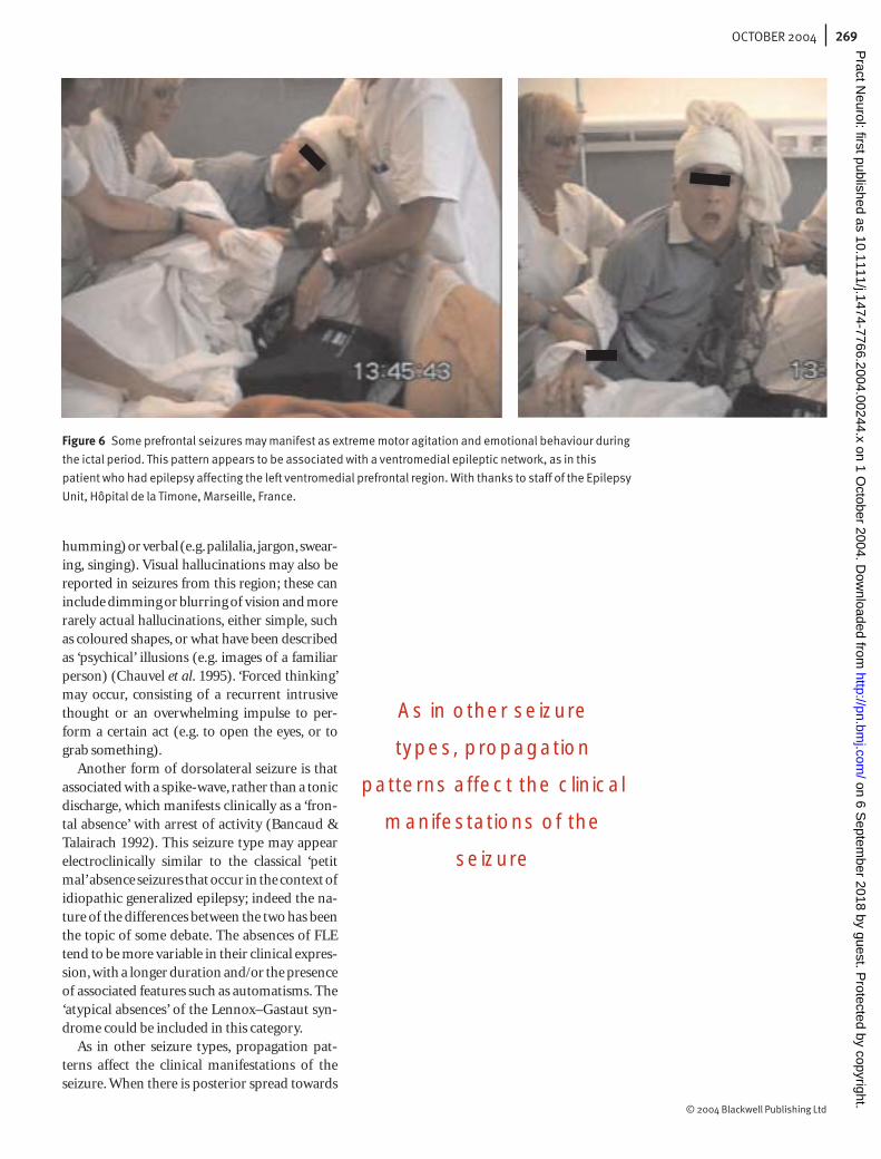
Ventro-medial prefrontal seizuresSeizures arising from ventral or ventro-medial regions appear to correspond to those initially described as ‘complex partial seizures of fron-tal lobe origin’ (Williamson et al. 1985), as de-scribed above. Some begin with what appears to be a dramatic reaction to fear, with a frightened facial expression, screaming and abrupt agita-tion (Fig. 6). This intense behavioural reac-tion to fear is very different from the pattern of temporal lobe seizures that include a subjective sensation of fear. There may be complex, appar-ently purposeful gesticulation, such as kicking or punching, bipedal cycling movements or attempts to escape. Autonomic signs such as mydriasis, tachycardia and facial fl ushing are common, as well as peri-ictal urination. It ap-pears that a consistent role for a ventro-medial epilepsy network can be demonstrated for those seizures involving ictal fear-related behaviour (Biraben et al. 2001).
Dorso-lateral prefrontal seizuresCertain clinical features suggest the involvement of the dorsal prefrontal region, notably tonic de-viation of the eyes preceding head version, and gestural automatisms that may be directed to-wards the same location as the gaze. These move-ments may appear semipurposeful, for example a patient may appear to be reaching towards something in their visual fi eld. There may also be a compulsive element to the behaviour (‘forced acting’). Motor patterns in this seizure type are usually complex, such as semirhythmic tapping of the hands or feet, or grasping motions. They are often associated with asymmetrical tonic or dystonic posturing of upper and/or lower limbs. Vocalization may occur; unlike the pattern seen in medioventral seizures, this often does not appear to have an initial emotional modifi cation. Such vocalizations may be non-verbal (e.g. groaning,
the epilepsy. Speech arrest or vocalization (char-acteristically palilalia) may also occur if there is involvement of the speech area in the frontal operculum. Spread to involve the operculum and the lower central region may also give rise to facial clonic jerks and salivation. More com-plex movements of all four limbs can follow these main signs. Initial subjective sensations or ‘auras’ occur infrequently in this seizure type, but some patients describe sensory symptoms such as ill-defi ned tightness or tingling, which may be generalized or localized (Williamson & Engel 1997). Secondary generalization is infre-quent.
Prefrontal seizuresPrefrontal seizures remain the least well-charac-terized of all frontal seizures. In fact distinguish-ing between prefrontal and premotor origin can be diffi cult, as seizures may involve both areas. Seizure patterns that arise from the prefrontal region refl ect its highly complex organization, so that in comparison with the relatively simple elements of more posterior frontal seizures, pre-frontal semiology is extremely diverse and may be highly idiosyncratic.
The fi rst observations of prefrontal seizures were published in the 1970s (Tharp 1972; Lud-wig et al. 1975). These were described as brief attacks typically occurring in clusters, often at night, and characterized by a frightened ap-pearance, agitation, repetitive semipurposeful behaviour and vocalizations that could be non-verbal (screaming) or verbal (expletives). It was initially speculated that these were of orbito-
The direction of adversion
may be ipsilateral or
contralateral to the
site of epileptic activity,
depending on its timing
within the seizure,
and is therefore not a
consistently reliable guide
to lateralization of the
epilepsy
on 6 Septem
ber 2018 by guest. Protected by copyright.
http://pn.bmj.com
/P
ract Neurol: first published as 10.1111/j.1474-7766.2004.00244.x on 1 O
ctober 2004. Dow
nloaded from
OCTOBER 2004 269
© 2004 Blackwell Publishing Ltd
humming) or verbal (e.g. palilalia, jargon, swear-ing, singing). Visual hallucinations may also be reported in seizures from this region; these can include dimming or blurring of vision and more rarely actual hallucinations, either simple, such as coloured shapes, or what have been described as ‘psychical’ illusions (e.g. images of a familiar person) (Chauvel et al. 1995). ‘Forced thinking’ may occur, consisting of a recurrent intrusive thought or an overwhelming impulse to per-form a certain act (e.g. to open the eyes, or to grab something).
Another form of dorsolateral seizure is that associated with a spike-wave, rather than a tonic discharge, which manifests clinically as a ‘fron-tal absence’ with arrest of activity (Bancaud & Talairach 1992). This seizure type may appear electroclinically similar to the classical ‘petit mal’ absence seizures that occur in the context of idiopathic generalized epilepsy; indeed the na-ture of the differences between the two has been the topic of some debate. The absences of FLE tend to be more variable in their clinical expres-sion, with a longer duration and/or the presence of associated features such as automatisms. The ‘atypical absences’ of the Lennox–Gastaut syn-drome could be included in this category.
As in other seizure types, propagation pat-terns affect the clinical manifestations of the seizure. When there is posterior spread towards
Figure 6 Some prefrontal seizures may manifest as extreme motor agitation and emotional behaviour during
the ictal period. This pattern appears to be associated with a ventromedial epileptic network, as in this
patient who had epilepsy affecting the left ventromedial prefrontal region. With thanks to staff of the Epilepsy
Unit, Hôpital de la Timone, Marseille, France.
As in other seizure
types, propagation
patterns affect the clinical
manifestations of the
seizure
on 6 Septem
ber 2018 by guest. Protected by copyright.
http://pn.bmj.com
/P
ract Neurol: first published as 10.1111/j.1474-7766.2004.00244.x on 1 O
ctober 2004. Dow
nloaded from

PRACTICAL NEUROLOGY270
© 2004 Blackwell Publishing Ltd
single lesion is seen on MRI, it is necessary to un-derstand its relation to the epileptogenic zone, as the two do not necessarily fully overlap (Ta-lairach et al. 1992b). Anatomo-electro-clinical correlation carried out using SEEG as devised by Talairach, which was developed well before the era of MRI, does not necessarily depend on the presence of a visible lesion on neuroimaging. In addition, some series have reported good out-comes in patients with normal imaging, based on presurgical evaluation incorporating semi-ological analysis and intracerebral recording as well as other noninvasive data (Talairach et al. 1992a; Zentner et al. 1996; Swartz et al. 1998; Siegel et al. 2001).
Much current research is therefore being directed towards the detection of focal lesions that are not visible with currently optimal MRI (Knowlton 2004). It seems likely that the small surgical series reporting successful outcomes despite normal imaging refl ect those patients with ‘invisible’ focal lesions, many of which are focal cortical dysplasias, and which the ensem-ble of clinical data including intracerebral EEG have correctly identifi ed. It is clear that develop-ment of less invasive methods that might per-mit the confi dent detection of such lesions is an important area for future work. Such methods include functional imaging, of which positron emission tomography (PET) appears particu-larly promising. Although most work relates to medial temporal epilepsy, a recent study has highlighted the potential importance of [11C]
fl umazenil PET, which appears to have better
a b c
premotor and motor regions, secondary gener-alization is frequent.
This proposed classifi cation remains some-what theoretical and is the subject of ongoing study but progress in the identifi cation of elec-troclinical patterns has the potential to greatly advance the surgical treatment of FLE.
DEVELOPMENTS IN THE PRESURGICAL EVALUATION OF FRONTAL LOBE EPILEPSYWhen considering the overall evolution of sur-gery for epilepsy, the importance of develop-ments in neuroimaging over the past 15 years cannot be overestimated – these have revolu-tionized the optimum selection and treatment of potential epilepsy surgical candidates (Dun-can 1997). Given the diffi culties in the diagno-sis and localization of FLE as described above, imaging plays an extremely important role. Magnetic resonance imaging (MRI), including techniques such as diffusion tensor imaging, has developed to the point where the great major-ity of patients with localization-related epilepsy can be shown to have an underlying cortical le-sion (Fig. 7). Many authors emphasize the cor-relation between the presence of a visible focal lesion and good surgical outcome (Mosewich et al. 2000).
However, there remain around 20% of pa-tients with localization-related epilepsy who have no lesion visible on current optimum MRI with expert review, and others with dual or multifocal pathology. Moreover, even when a
Figure 7 Brain MRI demonstrating the presence of a small dysplasia (arrow) in the left
medial frontal lobe (prefrontal region). (a) Coronal inversion-recovery sequence, (b)
Coronal FLAIR, (c) Axial T2.
on 6 Septem
ber 2018 by guest. Protected by copyright.
http://pn.bmj.com
/P
ract Neurol: first published as 10.1111/j.1474-7766.2004.00244.x on 1 O
ctober 2004. Dow
nloaded from

OCTOBER 2004 271
© 2004 Blackwell Publishing Ltd
1 sec
a
sensitivity and specifi city in detecting metabolic abnormalities than 2-[11F] fl uoro-2-deoxy-d-
glucose (FDG) PET in neocortical epilepsy and
which may be useful in helping to detect extra-
temporal epileptogenic zones even when MRI is
normal (Hammers et al. 2003).
Developments in non-invasive neurophysi-ological techniques include magneto-elec-troencephalography (MEG), which, though limited to a few centres, seems to be particularly useful in neocortical compared with temporal epilepsy and has been validated by intracranial comparison studies (Barkley & Baumgartner 2003). Source localization techniques using high-resolution scalp EEG also appear promis-ing (Gavaret et al. 2004) (Fig. 8).
Figure 8 High resolution EEG can be superimposed on a patient’s MR scan to represent the region of greatest
interictal activity and this can be particularly useful where imaging is non-localizing. This fi gure shows
interictal spikes recorded from a 27-year-old woman with medioventral frontal lobe epilepsy. (a) Interictal
spikes occurred in brief runs with maximal amplitude over electrodes FP1, F7 and FPZ (left fronto-polar
region), (b) Source localization was then performed using a technique called MUSIC (Mosher et al. 1992),
which showed the maximal contribution to be in the anterior part of the left cingulate gyrus. This localization
was later validated during a depth EEG recording that demonstrated both interictal and ictal epileptic activity
arising from this region. Cortectomy was subsequently performed and histopathology showed Taylor’s
dysplasia. The patient was seizure-free at 2 years post-operatively. With thanks to Dr Martine Gavaret,
Hôpital de la Timone, Marseille, France.
Much current research is
being directed towards
the detection of focal
lesions that are not visible
with currently optimal MRI
b
on 6 Septem
ber 2018 by guest. Protected by copyright.
http://pn.bmj.com
/P
ract Neurol: first published as 10.1111/j.1474-7766.2004.00244.x on 1 O
ctober 2004. Dow
nloaded from

PRACTICAL NEUROLOGY272
© 2004 Blackwell Publishing Ltd
The notion of ‘totally non-invasive presurgi-cal epilepsy evaluation’ in the future (Knowl-ton 2004) is also supported by the potential of functional MRI for localization of language and memory function prior to deciding on surgical resection. This may eventually be able to replace tests such as the WADA and mapping by direct cortical stimulation.
ACKNOWLEDGEMENTSThis paper was reviewed by Dr Richard Roberts, Dundee, Scotland.
Aileen McGonigal gratefully acknowleges support from the European Federation of Neurological Sciences and the Glasgow Neuro-science Foundation.
REFERENCESArchibald SJ, Mateer CA & Kerns KA (2001) Utilization
Behaviour. Clinical Manifestations and Neurological Mechanisms. Neuropsychology Review, 11, 177–30.
Bancaud J & Talairach J (1992) Clinical Semiology of Frontal Lobe Seizures. Advances in Neurology, 57, 3–59.
Barkley GL & Baumgartner C (2003) MEG and EEG in epilepsy. Journal of Clinical Neurophysiology, 20, 163–78.
Bartolomei F & Chauvel P (2000) Seizure symptoms and cerebral localization. frontal lobe and rolandic seizures. In: Intractable Focal Epilepsy (eds Oxbury JM, Polkey CE, Duchowny TM), pp55–62. WB Saunders, Philadelphia .
Bautista RE, Spencer DD & Spencer SS (1998) EEG fi nd-ings in frontal lobe epilepsies. Neurology, 50, 1765–71.
Biraben A & Chauvel P (1997) Epilepsia Partialis Con-tinua. In: Epilepsy. A Comprehensive Textbook (eds Engel J & Pedley TA), pp. 2447–53. Lippincott-Raven Publications.
Biraben A, Taussig D, Thomas P et al. (2001) Fear as the main feature of epileptic seizures. Journal of Neurol-ogy, Neurosurgery and Psychiatry, 70, 186–91.
Chauvel P (2001) Contributions of Jean Talairach and Jean Bancaud to Epilepsy Surgery. In: Epilepsy Surgery, 2nd edn (ed. Luders H), pp. 35–41. Lippincott Wil-liams & Wilkins.
Chauvel P (2003) Can we classify frontal lobe seizures?. In. Frontal Seizures and Epilepsies in Children (eds Beaumanoir A, Andermann F, Chauvel P, Mira L & Zifkin B), pp. 59–64. John Libbey Eurotext.
Chauvel P, Kliemann F, Vignal JP et al. (1995) The Clini-cal Signs and Symptoms of Frontal Lobe Seizures: Phenomenology and Classifi cation. In: Epilepsy and the Functional Anatomy of the Frontal Lobe (eds Jas-
CONCLUSIONS• The study of the semiology and electro-
clinical correlation of frontal seizures continues to hold an important place in the understanding of frontal epilepsy and cerebral function.
• This type of detailed study has been made possible largely because of modern video-EEG techniques, especially those using depth recording, and those that have cor-related electroclinical data with postop-erative outcome.
• Recent years have seen major advances in classifying FLE: by establishing the ten-dency for frontal seizure types to be related to functional divisions of the frontal lobe and to be organized along antero-poste-rior and dorso-ventral gradients.
• There can be few treatments for chronic disease with the potential to make as huge an impact on an individual’s quality of life as curative epilepsy surgery. This treat-ment has the potential to transform a young person’s life from one with intrac-table seizures and the inevitably associated risks of increased mortality, social disad-vantage and psychological vulnerability, to a normal existence.
• Although FLE remains in general more dif-fi cult and less common than TLE, thanks to the major developments in electroclini-cal correlation and neuroimaging, it is be-coming possible to envisage surgery as a possibility for many future FLE patients
• A multimodal approach, combining elec-tro-clinical methods with the best avail-able imaging techniques, allows complex FLE cases to be tackled in centres with ap-propriate expertise
• The ultimate aim will be to achieve a reli-able, non-invasive presurgical approach in as many patients as possible.
• One of the biggest challenges remains the surgical treatment of those patients with normal imaging. It seems likely that current developments in electroclinical correlation and functional imaging tech-niques will permit advancement in the near future.
on 6 Septem
ber 2018 by guest. Protected by copyright.
http://pn.bmj.com
/P
ract Neurol: first published as 10.1111/j.1474-7766.2004.00244.x on 1 O
ctober 2004. Dow
nloaded from

OCTOBER 2004 273
© 2004 Blackwell Publishing Ltd
per HH, Riggio S & Glodman-Rakic PS), pp. 115–125. Raven Pres, New York.
Chauvel P, Trottier S, Vignal JP et al. (1992) Somatomo-tor seizures of frontal lobe origin. Advances in Neurol-ogy, 57, 185–232.
Damasio A (1995) Descartes’ Error. Putman Publishing Group.
Devinsky O, Morrell MJ & Vogt B (1995) Contribution of anterior cingulate cortex to behaviour. Brain, 118, 279–306.
Duncan JS (1997) Neuroimaging and Epilepsy. Brain, 120, 339–77.
Gavaret M, McGonigal A, Badier JM & Chauvel P (2004) Physiology of Frontal Lobe Seizures. Pre-ictal, Ictal and Inter-ictal Relationships. In: Advances in Clinical Neurophysiology (supplement to Clinical Neurophysi-ology Vol 57) (eds Hallet M, Schomer DL, Massey JM). Elsevier BV, Amsterdam.
Goldman-Rakic PS (1984) The frontal lobes: uncharted provinces of the brain. Trends in Neurosciences, 7, 425–9.
Hammers A, Koepp MJ, Richardson MP et al. (2003) Grey and white matter fl umazenil binding in neo-cortical epilepsy with normal MRI: a PET study of 44 patients. Brain, 126, 1300–18.
Jackson JH (1931) Selected Writings of John Hughlings Jackson (2 vols), (ed. Taylor J). Hodder and Stoughton, London.
Jobst BC, Siegel AM, Thadani VM et al. (2000) Intrac-table Seizures of Frontal Lobe Origin. Clinical Char-acteristics, Localising Signs and Results of Surgery. Epilepsia, 41, 1139–52.
Knowlton RC (2004) Multimodality imaging in par-tial epilepsies. Current Opinion in Neurology, 17, 165–72.
Kotagal P, Arunkamur G, Hammel J et al. (2003) Com-plex partial seizures of frontal lobe onset: statistical analysis of ictal semiology. Seizure, 12, 268–81.
Lhermitte F (1983) Utilisation behaviour and its relation to lesions of the frontal lobes. Brain, 106, 237–5
Ludwig B, Ajmone-Marsan C & Van Buren J (1975) Cer-ebral seizures of probable orbitofrontal origin. Epi-lepsia, 16, 141–58.
Lugarasi E & Cirignotta F (1981) Hypnogenic paroxys-mal dystonia: epileptic seizures or a new syndrome? Sleep, 4, 129–38.
Manford M, Fish D & Shorvon S (1996) An analysis of clinical seizure patterns and their localizing value in frontal and temporal lobe epilepsies. Brain, 119, 17–40.
Meierkord H, Fish DR, Smith SJM et al. (1992) Is noc-turnal paroxysmal dystonia a form of frontal lobe epi-lepsy? Movement Disorders, 7, 38–42.
Mosewich RK, O’Brien TJ, Cascino GD et al. (2000) Fac-tors Predictive of the Outcome of Frontal Lobe Epi-lepsy Surgery. Epilepsia, 41, 843–9.
Mosher JC, Lewis PS & Leahy RM (1992) Multiple dipole
modelling and localization from spatio-temporal MEG data. IEEE Trans Biomed Eng, 39, 541–57.
Niedermeyer E (1998) Frontal lobe epilepsy: the next frontier. Clinical Electroencephalography, 29, 163–9.
Pandya DN & Yeterian EH (1985) Architecture and Connections of Cortical Association Areas. In: Cer-ebral Cortex, Vol. 4: Association and Auditory Cortices (eds Peters A & Jones EG), p. 40. Plenum Press, New York.
Petrides M & Pandya DN (1994) Comparative architec-tonic analysis of the human and the macqque frontal cortex. In: Handbook of Neuropsychology (eds Boller F & Grafman J), pp. 17–58. Elsevier, Amsterdam.
Siegel A, Jobst BC, Thadani VM et al. (2001) Medically Intractable, Localization-related Epilepsy with Nor-mal MRI. Presurgical Evaluation and Surgical Out-come in 43 Patients. Epilepsia, 42, 883–8.
So N (1998) Mesial Frontal Epilepsy. Epilepsia, 39, S49–S61.
Swartz BE, Delgado-Escueta AV, Walsh GO et al. (1998) Surgical outcomes in pure frontal lobe epilepsy and foci that mimic them. Epilepsy Research, 29, 97–108.
Talairach J, Bancaud J, Bonis A et al. (1992a) Surgical Therapy for Frontal Epilepsies. Advances in Neurol-ogy, 57, 707–732.
Talairach J, Tournoux P, Musolino A, Missir O (1992b) Stereotactic exploration in frontal epilepsy. In: Frontal Lobe Seizures and Epilepsies (eds Chauvel P, Delgado-Escueda AV, Halgren E & Bancaud J), pp. 651–88. Raven Press, New York.
Tharp BR (1972) Orbital frontal seizures: An unique electroencephalographic and clinical syndrome. Epi-lepsia, 13, 627–42.
Vignal JP, Biraben A, Chauvel PY & Reutens DC (1998) Refl ex Partial Seizures of Sensorimotor Cortex (In-cluding Cortical Refl ex Myoclonus and Startle Epi-lepsy). Advances in Neurology, 75, 207–26.
Walsh GO & Delgado-Escueta AV (1984) Type II com-plex partial seizures: poor results of anterior temporal lobectomy. Neurology, 34, 1–13.
Wendling F, Bartolomei F, Bellanger JJ, Bourien J & Chauvel P (2003) Epileptic fast intracerebral EEG activity: evidence for spatial decorrelation at seizure onset. Brain, 126, 1449–59.
Wieser HG, Swartz BE, Delgado-Escueta AV et al. (1992) Differentiating Frontal Lobe seizures from Temporal Lobe Seizures. Advances in Neurology, 57, 267–85.
Williamson P & Engel J (1997) Complex partial seizures. In. Epilepsy: a Comprehensive Textbook (eds Engel J & Pedley T), pp. 557–66. Lippincott-Raven, New York.
Williamson PD, Spencer DD, Spencer SS et al. (1985) Complex partial seizures of frontal lobe origin. An-nals of Neurology, 18, 497–504.
Zentner J, Hufnagel A, Ostertun B et al. (1996) Surgical Treatment of Extratemporal Epilepsy. Clinical, Radi-ologic, and Histopathologic Findings in 60 Patients. Epilepsia, 37, 1072–80.
on 6 Septem
ber 2018 by guest. Protected by copyright.
http://pn.bmj.com
/P
ract Neurol: first published as 10.1111/j.1474-7766.2004.00244.x on 1 O
ctober 2004. Dow
nloaded from