Chevalley De Rivaz, Descrizione delle acque termominerali e delle stufe d'Ischia (1837)

1
Clinical Pathways
Fragility Fractures
Ghassan MaaloufDepartment of Orthopaedics
St. George Hospital, Balamand University Beirut, Lebanon
Karsten DreinhöferDepartment of Orthopaedics
Ulm University Ulm, Germany

2
Content
• Fragility fractures are under-diagnosed and under-treated
• Development of a pathway– Management of fragility patients
– Risk assessment
– Diagnosis
– Treatment • Basic treatment• Pharmacological• Exercise
• Established pathways
3
Alarming facts

4
• 852/2386 women identified as having sustained fractures over the age of 50
• 43 (5%) of those women have been previouslyassessed by DEXA scan to determine absolute fracture risk and make informed management decisions
• 81 (9.5%) women with ≥1 fracture on treatment
Brankin et al.Curr Med Res Opin 2005; 21:475-482
Osteoporosis assessment of non-vertebral fracture patients
Coatbridge Prior Fracture Program

5
385 patients with fragility fractures
“Have you ever heard of osteoporosis?”
NO: 20 % / YES: 80 %
“Do you think that the fracture you have experienced could be due to a fragility of your bones?”
NO: 73 % / YES: 27 %
An Osteoporosis Clinical Pathway for the Medical Management of Patients with Low Trauma Fracture
Chevalley et al. Osteoporos Int 2002; 13:450-455
Awareness and knowledge of osteoporosis in fracture patients

6
n=934 women >60 years old
020406080
100120140
Pat
ient
s(n
)
Fractureidentified by
studentradiologists
Fracturenoted in radiology
report
Fracturenoted inmedical record
Receivedosteoporosis
treatment
132
65
23 25
Gehlbach et al. Osteoporos Int 2000; 11:577-582
Osteoporotic vertebral compression fractures –only 20% receive treatment
7
Multinational Survey of Osteoporotic Fracture Management
Survey of 3422 orthopaedic surgeons from 6 countries
• 90% do not routinely measure bone density following the firstfracture
• 75% are lacking appropriate knowledge about osteoporosis
Dreinhöfer et al. Osteoporos Int 2005; 16:S44-S54

8
Consequence
Development of pathways for orthopaedic surgeons

9
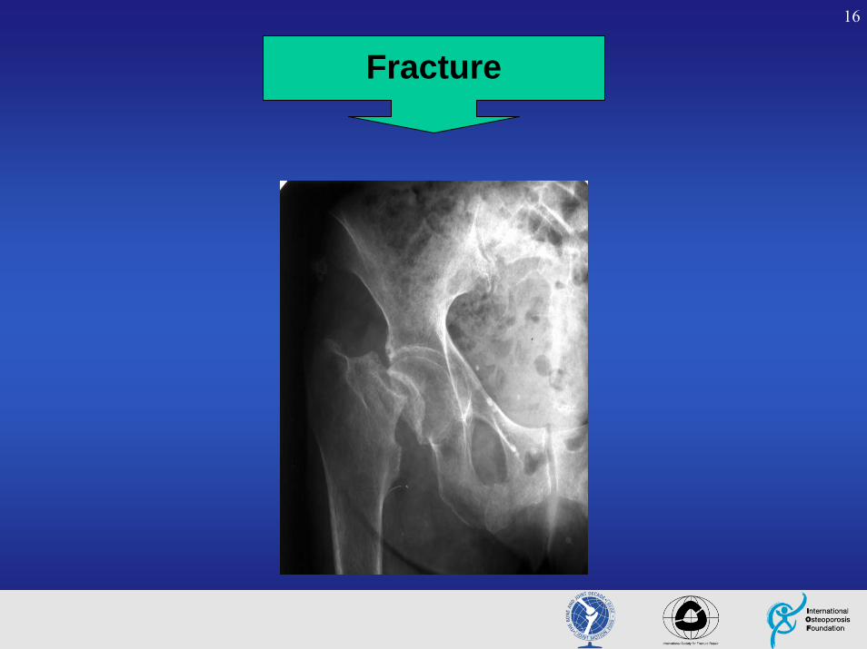
Fracture
Accident pattern / Risk assessment
inadequate trauma with / without risk factors or adequate with risk factors
adequate traumawithout risk factors
No further evaluation
History / Physical examination
Demographic Factors
Lab / X-ray thoracic/lumbar spine in 2 planes
Bone Densitometry
? Secondary Osteoporosisor other bone disease
Woman premenopausalMan < 65 Years
? Secondary Osteoporosisor other bone disease
? Primary Osteoporosis
Woman postmenopausalMan > 65 Years
? Primary Osteoporosis
Vertebral fracture, T ≤ -2,0 orperipheral fracture, T ≤ -2,5 or
steroid-induced osteoporosis, T ≤ -1,5
Further evaluation
Further evaluation
Further evaluation
ConsultationFall evaluation and prevention
Basic treatment (Calcium + Vitamin D)Special pharmaceutical treatment
(Alendronat / Risedronat / Etidronat / Ibandronat)(Raloxifen / Teriparatid / Strontium)
Process according to evidence based DVO-guidelines
ConsultationFall evaluation and prevention
Basic treatment (Calcium + Vitamin D)
Vertebral fracture, T > -2,0 orperipheral fracture, T > -2,5 or
steroid-induced osteoporosis, T > -1,5

10
Fracture

11
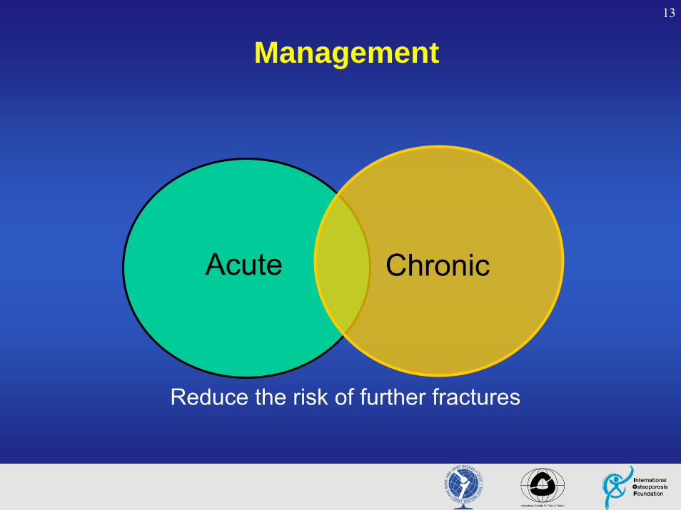
Management
Acute Chronic

12
Acute management
• Surgical repair and/or appropriate orthopaedic management
• Pain relief
• Physiotherapy
13
Management
Acute Chronic
Reduce the risk of further fractures

14
Call for action

15
What to do?
4 steps:
• Identification
• Prevention
• Pharmacological intervention
• Follow-up and rehabilitation
16
Fracture

17
Fracture
Accident pattern

18
Fracture
Accident pattern Risk assessment
Ten-Year Fracture Risk

19
Ri sk fac t ors
50%
40%
30%
20%
10%Ten-year fracture risk
Age
Absolute risk
Kanis et al. Osteoporos Int 2005; 16:581-589
Assessment of fracture risk
20
Key risk factors for fractures(RR > 2)
• Age
• Bone mineral density
• Prior fragility fracture
• Family history of osteoporotic fracture

21
Distribution of bone mineral densityin women of different ages, and the prevalence of osteoporosis (blue)
Kanis et al. J Bone Miner Res 1994; 9:1137-41
Age and osteoporosis
22
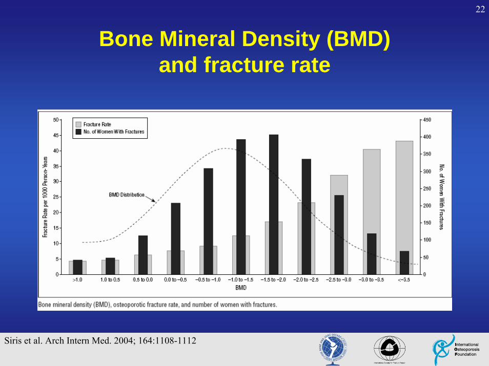
Bone Mineral Density (BMD) and fracture rate
Siris et al. Arch Intern Med. 2004; 164:1108-1112

23
Strong risk factors for fractures(RR > 2)
• Menopause < 45
• Glucocorticoids
• Immobilization
• BMI < 19
• Anorexia Nervosa
• Propensity to fall
• Malabsorption
• Chronic renal failure
• Transplantation
• Hypogonadism
Kanis et al. Osteoporos Int. 2005; 16:581-9

24
Moderate risk factors(1 < RR < 2)
• Rheumatoid arthritis
• Bechterew disease
• Anticonvulsants
• Calcium intake < 500 mg/d
• Diabetes mellitus
• Estrogen deficiency
• Primary hyperparathyroidism
• Hyperthyroidism
• Smoking
• Alcohol excess
Kanis et al. Osteoporos Int. 2005; 16:581-9

25
Accident pattern / Risk assessment
adequate traumawithout risk factors
No further evaluation
Fracture

26
Accident pattern / Risk assessment
inadequate trauma with / without risk factors or adequate trauma with risk factors
adequate traumawithout risk factors
No further evaluation
History / Physical examination
Fracture

27
History / physical examination
• Weight / height
• Menarche / menopause
• Nutrition
• Medication (past and present)
• Level of activity
• Fracture history
• Fall history
• Risk factors for secondary osteoporosis
28
High risk for secondary osteoporosis
• Severe chronic liver or kidney disease
• Steroid medication ( >7.5mg for more than 6 months)
• Malabsorption (e.g. Crohn´s disease)
• Rheumatoid arthritis
• Systemic inflammatory disorders
• Hyperthyroidism
• Primary hyperparathyroidism
• Antiepileptic medication

29
Accident pattern / Risk assessment
inadequate trauma with / without risk factors or adequate with risk factors
adequate traumawithout risk factors
No further evaluation
History / Physical examination? Secondary Osteoporosis
or other bone diseaseFurther evaluation
Fracture

30
Accident pattern / Risk assessment
inadequate trauma with / without risk factors or adequate with risk factors
adequate traumawithout risk factors
No further evaluation
History / Physical examination? Secondary Osteoporosis
or other bone disease? Primary Osteoporosis
Further evaluation
Fracture

31
Accident pattern / Risk assessment
inadequate trauma with / without risk factors or adequate with risk factors
adequate traumawithout risk factors
No further evaluation
History / Physical examination
Demographic Factors
? Secondary Osteoporosisor other bone disease
? Primary Osteoporosis
Further evaluation
Fracture

32
Demographic factors
• Man vs. woman
• Woman premenopausal vs. postmenopausal
• Man older than 65 years vs. younger than 65 years
• EthnicityCaucasian or Asian compared to Black

33
Demographic factors
0.0
0.4
0.8
1.2
1.6
10 20 30 40 50 60 70 80 90 Age (years)
White women
Black women
White men
Black men
Lumbar BMD (g/cm2)

34
Osteoporosis in men
Primary osteoporosis (50%)
• Idiopathic
Secondary osteoporosis (50%)
• Glucocorticoid excess (15%)• Hypogonadism (10%)• Alcoholism (7%)• Hypercalciuria (2%)• Smoking• Gastrointestinal disorders• Immobilization• Others
Bilezikian. J Clin Endocrinol Metab, 1999; 84:3431-3434

35
Men and women have equivalent risks of fracture
for a given level of bone mineral density
Nguyen et al. Am J Epidemiol. 1996; 144:255-263Legrand et al. Osteoporos Int. 1999; 10:265-270
Osteoporosis in men

36
Accident pattern / Risk assessment
inadequate trauma with / without risk factors or adequate with risk factors
adequate traumawithout risk factors
No further evaluation
History / Physical examination
Demographic Factors
? Secondary Osteoporosisor other bone disease
Woman premenopausalMan < 65 Years
? Primary Osteoporosis
Further evaluation
Further evaluation
Fracture

37
Accident pattern / Risk assessment
inadequate trauma with / without risk factors or adequate with risk factors
adequate traumawithout risk factors
No further evaluation
History / Physical examination
Demographic Factors
? Secondary Osteoporosisor other bone disease
Woman premenopausalMan < 65 Years
? Primary Osteoporosis
Woman postmenopausalMan > 65 Years
Further evaluation
Further evaluation
Fracture

38
Accident pattern / Risk assessment
inadequate trauma with / without risk factors or adequate with risk factors
adequate traumawithout risk factors
No further evaluation
History / Physical examination
Demographic Factors
Lab / X-ray thoracic/lumbar spine in 2 planes
? Secondary Osteoporosisor other bone disease
Woman premenopausalMan < 65 Years
? Primary Osteoporosis
Woman postmenopausalMan > 65 Years
Further evaluation
Further evaluation
Fracture

39
Laboratory tests
• SR / CRP • Blood count • Calcium• Phosphate• Alkaline Phosphatase (AP)• GGT• Creatinin• Basal TSH• Protein-Immunoelectrophoresis

40
X-Ray
Thoracic and lumbar spine in 2 planes for patients with:
• Back pain
• Progressive kyphosis
• Loss of height > 4 cm
• - 2.5 < Bone Mineral Density < -1.0

41
Accident pattern / Risk assessment
inadequate trauma with / without risk factors or adequate with risk factors
adequate traumawithout risk factors
No further evaluation
History / Physical examination
Demographic Factors
Lab / X-ray thoracic/lumbar spine in 2 planes
? Secondary Osteoporosisor other bone disease
Woman premenopausalMan < 65 Years
? Secondary Osteoporosisor other bone disease
? Primary Osteoporosis
Woman postmenopausalMan > 65 Years
? Primary Osteoporosis
Further evaluation
Further evaluation
Further evaluation
Fracture

42
Accident pattern / Risk assessment
inadequate trauma with / without risk factors or adequate with risk factors
adequate traumawithout risk factors
No further evaluation
History / Physical examination
Demographic Factors
Lab / X-ray thoracic/lumbar spine in 2 planes
Bone Densitometry
? Secondary Osteoporosisor other bone disease
Woman premenopausalMan < 65 Years
? Secondary Osteoporosisor other bone disease
? Primary Osteoporosis
Woman postmenopausalMan > 65 Years
? Primary Osteoporosis
Further evaluation
Further evaluation
Further evaluation
Process according to evidence based DVO-guidelines
Fracture

43
0
5
10
15
20
25
30
35
-5 -4 -3 -2 -1 0
% v
erte
bral
frac
ture
T–score
–1SD
RR2 x
Watts. Arch Intern Med. 2001; 161:772.
Fracture risk depending on bone mineral density

44
Fracture risk depending on bone mineral density
02468
101214161820
-3 -2 -1 0 1 2
hipcalcaneusradius
Rel
ativ
e ris
k hi
p fra
ctur
e
BMD (SD)T= -2.5
Cummings et al. Lancet 1993; 341:72-75
Age > 65 years:

45
Ten-year probability of hip fracture in Swedish men and women, according to age and T- score assessed at the femoral neck by dual X-rayabsorptiometry
Green dotted line = probability at which intervention is cost-effective
Kanis et al. Osteoporos Int 2001; 12: 989-95

46
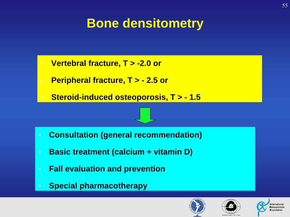
Bone densitometry
• Vertebral fracture, T > -2.0 or
• Peripheral fracture, T > - 2.5 or
• Steroid-induced osteoporosis, T > - 1.5

47
Bone densitometry
• Consultation (general recommendation)
• Basic treatment (calcium + vitamin D)
• Fall evaluation and prevention
• Vertebral fracture, T > -2.0 or
• Peripheral fracture, T > - 2.5 or
• Steroid-induced osteoporosis, T > - 1.5

48
General recommendations
• Regular physical activity and daily outdoor activities (at least 30 minutes)
• Adequate nutrition
• Sufficient basic intake of calcium(1000-1500 mg calcium per day) through adequate nutrition(milk, milk products, green vegetables, calcium-rich mineral water)
• Avoidance of cigarettes, alcohol intake (<30g per day)

49
Basic treatment
• Postmenopausal women whose nutrition does not provide appropriate daily calcium intake of 1500 mg:
– Daily supplement of 1000 mg of calcium
• For institutionalized and/or immobile women over 65 years of age, and for all women over 75:
– Daily supplement of 1200 mg of calcium + 800 IE vitamin D3 (Cholecalciferol)

50
Fall evaluation
• History of circumstances surrounding the fall
• Drugs, acute or chronic medical problems, mobility levels
• Examination of vision, gait and balance, function of the leg joints
• Examination of basic neurological function, including mental status, muscle strength, peripheral nerves of the legs, proprioception, reflexes, and tests of cortical, extrapyramidal,cerebellar function
• Assessment of basic cardiovascular status,including heart rate and rhythm, postural pulse and blood pressure and, ifappropriate, heart rate and blood pressure responses to carotidsinus stimulation
Woolf et al. BMJ 2003; 327:89-95

51
General deterioration associated with ageing• Poor postural control• Defective proprioception• Reduced walking speed• Weakness of legs• Slow reaction time• Various comorbidities
Problems with balance, gait, or mobility• Joint disease• Cerebrovascular disease• Peripheral neuropathy• Parkinson’s disease• Alcohol• Various drugs
Visual impairment• Impaired visual acuity• Cataracts• Glaucoma• Retinal degeneration
Risk factors for falling
Impaired cognition or depression• Alzheimer’s disease• Cerebrovascular disease
“Blackouts”• Hypoglycaemia• Postural hypotension• Cardiac arrhythmia• Transient ischaemic attack, acute onset• Cerebrovascular attack• Epilepsy• Vertebrobasilar insufficiency• Carotid sinus syncope• Neurocardiogenic (vasovagal) syncope
Woolf et al. BMJ 2003; 327:89-95
Intrinsic factors
52
Extrinsic factors
Personal hazards• Inappropriate footwear or clothing
Multiple drug therapy• Sedatives• Hypotensive drugs
Environmental factors
Hazards indoors or at home• Bad lighting• Steep stairs, lack of grab rails• Slippery floors, loose rugs• Pets, grandchildren’s toys• Cords for telephone and electrical
appliances
Hazards outdoors• Uneven pavements, streets, paths• Lack of safety equipment• Snowy and icy conditions• Traffic and public transportation
Risk factors for fallingExtrinsic and environmental factors
Woolf et al. BMJ 2003; 327:89-95

53
Accident pattern / Risk assessment
inadequate trauma with / without risk factors or adequate with risk factors
adequate traumawithout risk factors
No further evaluation
History / Physical examination
Demographic Factors
Lab / X-ray thoracic/lumbar spine in 2 planes
Bone Densitometry
? Secondary Osteoporosisor other bone disease
Woman premenopausalMan < 65 Years
? Secondary Osteoporosisor other bone disease
? Primary Osteoporosis
Woman postmenopausalMan > 65 Years
? Primary Osteoporosis
Further evaluation
Further evaluation
Further evaluation
Process according to evidence based DVO-guidelines
ConsultationFall evaluation and prevention
Basic treatment (Calcium + Vitamin D)
Vertebral fracture, T > -2,0 orperipheral fracture, T > -2,5 or
steroid-induced osteoporosis, T > -1,5
Fracture

54
Bone densitometry
• Vertebral fracture, T > -2.0 or
• Peripheral fracture, T > - 2.5 or
• Steroid-induced osteoporosis, T > - 1.5
55
Bone densitometry
• Vertebral fracture, T > -2.0 or
• Peripheral fracture, T > - 2.5 or
• Steroid-induced osteoporosis, T > - 1.5
• Consultation (general recommendation)
• Basic treatment (calcium + vitamin D)
• Fall evaluation and prevention
• Special pharmacotherapy
56
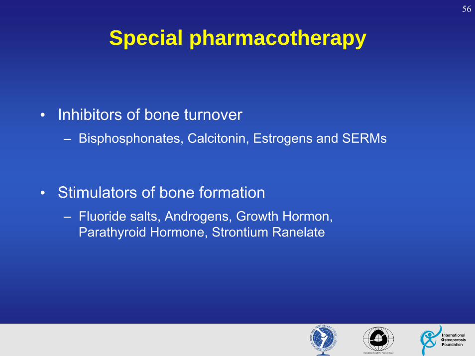
Special pharmacotherapy
• Inhibitors of bone turnover– Bisphosphonates, Calcitonin, Estrogens and SERMs
• Stimulators of bone formation– Fluoride salts, Androgens, Growth Hormon,
Parathyroid Hormone, Strontium Ranelate

57
Bisphosphonates• Alendronate (FOSAMAX®)• Risedronate (ACTONEL®)• Ibandronate (BONVIVA®)• Zoledronate (ACLASTA®)
SERMs• Raloxifene (EVISTA®)
Stimulators of bone formation• rh-PTH (FORTEO®)• Strontium Ranelate (PROTELOS®)
Special pharmacotherapy

58
Accident pattern / Risk assessment
inadequate trauma with / without risk factors or adequate with risk factors
adequate traumawithout risk factors
No further evaluation
History / Physical examination
Demographic Factors
Lab / X-ray thoracic/lumbar spine in 2 planes
Bone Densitometry
? Secondary Osteoporosisor other bone disease
Woman premenopausalMan < 65 Years
? Secondary Osteoporosisor other bone disease
? Primary Osteoporosis
Woman postmenopausalMan > 65 Years
? Primary Osteoporosis
Vertebral fracture, T ≤ -2,0 orperipheral fracture, T ≤ -2,5 or
steroid-induced osteoporosis, T ≤ -1,5
Further evaluation
Further evaluation
Further evaluation
ConsultationFall evaluation and prevention
Basic treatment (Calcium + Vitamin D)Special pharmaceutical treatment
(Alendronat / Risedronat / Etidronat / Ibandronat )(Raloxifen, Teriparatid, Strontium)
Process according to evidence based DVO-guidelines
ConsultationFall evaluation and prevention
Basic treatment (Calcium + Vitamin D)
Vertebral fracture, T > -2,0 orperipheral fracture, T > -2,5 or
steroid-induced osteoporosis, T > -1,5
Fracture

59
Exercise
Strengthening of muscles
Improving muscle function
Group experience
Social function

60
ExerciseStrengthening of muscles Improving muscle function
Falls Bone mass
Fractures
Quality of life

61
ExerciseStrengthening of muscles Improving muscle function
Falls Bone mass
Fractures
Quality of life

62
Tai Qi reduces fall risk
Quin et al. Arch Phys Med Rehabil. 2002; 83:1355-9Wolff et al. J Am Geriatr Soc. 1996; 44:489-97

63
ExerciseStrengthening of muscles Improving muscle function
Falls Bone mass
Fractures
Quality of life

64
Low mechanical signals strengthen long bones
• Low magnitude mechanical signals areanabolic to bone if applied at a high frequency (15–90 Hz)
• Low-magnitude mechanical signals canincrease:– cancellous bone volume fraction– trabecular thickness– trabecular numberand enhance bone stiffness and strength
Rubin et al. Nature 2001; 412:603-604

65
Rubin et al: J Bone Miner Res. 2004; 19:343-351
One-year prospective, randomized, double-blind, and placebo-controlledtrial of 70 postmenopausal women:
Brief periods (<20 minutes) of a low-level (0.2g, 30 Hz) vibration appliedduring quiet standing can effectivelyinhibit bone loss in the spine and femur
Prevention of postmenopausal bone loss bylow-magnitude, high-frequency mechanical stimuli

66
ExerciseStrengthening of muscles Improving muscle function
Falls Bone mass
Fractures
Quality of life

67
Established pathways

68
Glasgow Fracture Discharge Program
• To identify patients at increased risk of osteoporotic fracture
• To offer these patients appropriate information on osteoporosis and its management
• To provide advice to GPs on suitable interventions
Objectives:
Hospital Wards - In-patientFracture Clinic - Out-patient
Osteoporosis Service

69
• Identify prospective patients >50 years who sustain a fracture
• Use DXA to identify those with OP and at highest risk of future fracture
• Provide advice to GPs on appropriate interventions
• Provide lifestyle modification advice to individual patients
• Provide information for patients on fall prevention
• Refer patients to physiotherapy-led exercise classes
• Facilitate investigation and treatment of OP after admission to orthopaedics and introduce this into ICP
• Collect data for audit
A comprehensive service
Glasgow Fracture Discharge Program

70
Results of the Glasgow experience
• Better awareness of fragility fractures
• Improved rate of post-fracture follow-up
• Better management
• Better patient satisfaction

71
Osteoporosis clinical pathway (OCP)Geneva
• Enrolment of patients with low trauma fracture• Collection of data, additional diagnostic examination
• Educational program for patients and their families
• Advice on specific anti-osteoporotic therapy
Chevalley et al. Osteoporos Int. 2002; 13:450-455

72
“ Writing a guideline may be difficult, but determining how best to implement the guideline is even more difficult ”
Gross et al 2001

73
The surgeon’s responsibilities
• Identify the orthopaedic patient with risk factors and fragility fractures
• Inform the patient about the need for an osteoporosis evaluation
• Investigate whether osteoporosis is an underlying cause of the fracture
• Ensure that appropriate intervention is initiated
• Educate the patient and their family
Bouxsein et al. J Am Acad Orthop Surg. 2004; 12:385-95