FRACP talk - heart failure, cardiomoypathy and cardiac ... · Causes of sudden cardiac death ......
13
Heart failure • Refers to the syndrome of fluid retention and breathlessness, caused by cardiac disease • Usually biventricular in children due to ventricular interdependence and child specific pathology C i ld • Causes include: - left to right shunts, - valvular disease - myocardial dysfunction - high output heart failure (AVM’s, anaemia, hormonal disturbances) Heart failure • Cardiac changes include: - Decreased stroke volume and cardiac output - Increased end-diastolic pressure - Ventricular dilatation or hypertrophy - Impaired filling (diastolic dysfunction) - Reduced ejection fraction (systolic dysfunction) • Vascular changes include: - Increased systemic vascular resistance - Decreased arterial pressure - Impaired organ perfusion - Decreased arterial compliance - Increased venous pressure - Increased blood volume Compensatory mechanisms during heart failure • Cardiac: - Frank-Starling mechanism - Ventricular dilatation or hypertrophy - Tachycardia • Autonomic nerves: Increased sympathetic adrenergic activity - Increased sympathetic adrenergic activity - Reduced vagal activity to the heart • Neurohormal activation: - Renin-angiotensin-aldosterone system - Vasopressin (antidiuretic hormone) - Circulating catecholamines - Natriuretic peptides Frank-Starling curves Increasing afterload or decreasing inotropy Decreasing afterload or increasing inotropy Sympathetic activation in chronic heart failure Renin-angiotensin-aldosterone axis in heart failure
-
Upload
nguyentruc -
Category
Documents
-
view
214 -
download
0
Transcript of FRACP talk - heart failure, cardiomoypathy and cardiac ... · Causes of sudden cardiac death ......

Heart failure
• Refers to the syndrome of fluid retention and breathlessness, caused by cardiac disease
• Usually biventricular in children due to ventricular interdependence and child specific pathology
C i l d• Causes include:- left to right shunts, - valvular disease- myocardial dysfunction- high output heart failure (AVM’s, anaemia, hormonal
disturbances)
Heart failure
• Cardiac changes include:- Decreased stroke volume and cardiac output- Increased end-diastolic pressure- Ventricular dilatation or hypertrophy- Impaired filling (diastolic dysfunction)- Reduced ejection fraction (systolic dysfunction)
• Vascular changes include:- Increased systemic vascular resistance- Decreased arterial pressure- Impaired organ perfusion- Decreased arterial compliance- Increased venous pressure- Increased blood volume
Compensatory mechanisms during heart failure
• Cardiac:- Frank-Starling mechanism- Ventricular dilatation or hypertrophy- Tachycardia
• Autonomic nerves:Increased sympathetic adrenergic activity- Increased sympathetic adrenergic activity
- Reduced vagal activity to the heart
• Neurohormal activation:- Renin-angiotensin-aldosterone system- Vasopressin (antidiuretic hormone)- Circulating catecholamines- Natriuretic peptides
Frank-Starling curves
Increasing afterload or decreasing inotropy
Decreasing afterload or increasing inotropy
Sympathetic activation in chronic heart failure Renin-angiotensin-aldosterone axis in heart failure

Norepinephrine concentrations and prognosis in chronic heart failure Effects of natriuretic peptides
Heart failure – a self perpetuating cycle CardiomyopathiesDefinition
• WHO definition (1996): “Diseases of the myocardium associated with cardiac dysfunction”
- Dilated cardiomyopathyy y
- Hypertrophic cardiomyopathy
- Restrictive cardiomyopathy
- Unclassified: Arrhythmogenic RV dysplasia, LV non-compaction
Dilated cardiomyopathy:overview
• Characterised by dilatation and impaired ventricular contraction
• May be genetic, post-viral, drug or toxin induced, metabolic, mitochondrial, connective tissue associated or due to HIV
I i f t l i i f l t t b l d d• In infants, anomalous coronary origin from a pulmonary artery must be excluded
• Late histological findings are non-specific
• Usually presents with heart failure
• Accompanying diastolic dysfunction may include impaired ventricular relaxation and non-compliance
Dilated cardiomyopathy:echocardiogram

Dilated cardiomyopathygenetic mutations
• Up to 25% of dilated CM is caused by genetic mutations
• 1st gene identified was dystrophin (X-linked CM); others include actin, desmin and lamin A/C (dominant and recessive)
• Actin, desmin and dystrophin are cytoskeletal proteins with roles in force transmission, cytoskeletal stability, calcium homeostasis, myocyte differentiation, myofibrillogenesis
• Lamin is a nuclear protein; commonest mutation and is associated with conducting system disease
• Dystrophin, desmin and lamin mutations can be associated with skeletal muscle disease
Dilated cardiomyopathy:viral disease
• Common pathogenic viruses include adenovirus, enterovirus, CMV, influenza
• About 20% of subjects with dilated CM have virus by PCR
• In subjects with myocarditis, 35-40% viral yieldj y y
• Mechanisms of damage are both acute (dystrophin cleavage) and delayed (lymphocytic infiltrate)
• Adenovirus typically causes little lymphocytic infiltrate
Myocarditis:mouse model
Acute myocarditis Subacute myocarditis Chronic myocarditis
Viral M t iViral infection
Myocyte necrosis
Macrophage activation
Infiltrating mononuclear cells
Viraemia Viral clearing Viral absence4 days 14 days
CytokinesNatural killer cells
Nitric oxide
Cytotoxic T lymphocytesB lymphocytes
Neutralising antibodies
Fibrosis
Dilatation
Death
Myocarditis –histologic variation
Diffuse mononuclear
Focal mononuclear infiltratemononuclear
infiltrateinfiltrate
Myocardial oedema –no infiltrate
Myocardial fibrosisand hypertrophy
Mitochondrial function
• Mitochondria are the power plants of cells
• They convert fat, sugar and proteins to ATP
• Other roles include gluconeogenesis, amino acid and steroid synthesis, ROS and apoptosis
IIIIIIIVV ATP
Mitochondrial diseasestypical organ involvement
Brain: seizures, dementia, infarcts, leukoencephalopathy
Eye: optic atrophy, pigmentary degeneration, cataracts
Ear: deafness
M l k l t l thMuscle: skeletal myopathy
Heart: cardiomyopathy (HCM, DCM), conduction defects
Kidney: tubular dysfunction
Liver: hepatic dysfunction, bile stasis
Bone marrow: pancytopaenia, specific cell line failure
Blood, urine, CSF: increased lactate

Mitochondrial diseasesRespiratory chain Complex 1 deficiency
cardiomyopathy
180
200
220
240
200
220
240
200
220
240
% %%Muscle Liver Heart
0
20
40
60
80
100
120
140
160
180
I II II+III III IV CS0
20
40
60
80
100
120
140
160
180
I II II+III III IV CS0
20
40
60
80
100
120
140
160
180
I II II+III III IV CS
Hypertrophic cardiomyopathy
• Primary cardiac disorder with a heterogeneous expression and diverse clinical course
• Characterised by left ventricular hypertrophy in the absence of dilatation, or conditions capable of producing LVHconditions capable of producing LVH
• Non-obstructive in around 75% of cases
• Prevalence in the general population is around 0.2%
Hypertrophic cardiomyopathy:echocardiogram
Hypertrophic cardiomyopathymorphological characteristics
• Distribution of hypertrophy is usually asymmetric
• Any pattern possible but anterior ventricular septum predominantly involved
• Spontaneous LV remodeling with increase in wall thickness during adolescence, and a decrease in wall thickness with aging
Hypertrophic cardiomyopathygenetic defects
• Mendelian trait with autosomal dominant inheritance
• Mutations involve genes that encode for sarcomeric proteins
• 10 different proteins implicated and >200 described mutations ( ll li h i )(allelic heterogeneity)
• Around 50% of cases represent spontaneous mutations
• Hypertrophy may be secondary to altered sensitivity to calcium and impaired contractility

Hypertrophic cardiomyopathycontractile protein mutations
HCM - age related penetranceNimura et al; NEJM 1998
100
Cardiac beta-myosin heavy chainCardiac troponin TCardiac myosin-binding protein C
0102030405060708090
10-19 20-29 30-39 40-49 50-59 >60
Hypertrophic cardiomyopathyclinical considerations
• In adults, some mutations are associated with development of hypertrophy beyond middle life
• Disease penetrance may be incomplete below 60 years of ageDisease penetrance may be incomplete below 60 years of age
• With some mutations there is variable disease expression within a kindred
• Electrocardiographic abnormalities may precede development of overt hypertrophy
Paediatric HCMaetiological considerations
• Contractile protein abnormality
• Syndromes: Noonan, Beckwith-Wiedemann, LEOPARD, Friedreich’s ataxia
• Metabolic: Carnitine deficiency, Fatty acid oxidation defects, Glycogen storage disease, MPS, Mannosidosis, Fucosidosis, lipodystrophy
• Mitochondrial myopathies
• Neonatal hyperinsulinaemia
Paediatric HCMmorphological considerations
• Congenital heart disease and inappropriate hypertrophy
• Subpulmonary RV outflow obstruction
• Pulmonary valve stenosis (Noonan syndrome)
• Atrial septal defect or stretched PFO
• Subaortic membrane
• Anomalous mitral cord insertion into the IVS
• Anomalous papillary muscle insertion directly into the anterior mitral leaflet
Causes of sudden cardiac deathin young people
Maron BJ et al. Circulation. 1996;94:850-56.
Congenital coronary
anomalies (19%)
Mildly increased cardiac mass (10%)
Ruptured aorta 5%
Myocarditis 3%
Hypertrophiccardiomyopathy (36%)
Aortic stenosis 4%
5%Tunneled LAD 5%
ARVC 3%MVP 2%
CAD 2%Other 6%

Mortality in HCMMaron et al; Circulation 2000
12
14
16StrokeHeart failureSudden death
talit
y
0
2
4
6
8
10
% M
ort
Age at Death or Most Recent Evaluation (years)
5-15 16-25 26-35 36-45 46-55 56-65 66-75 >75
Hypertrophic cardiomyopathysubstrate for SCD
• Disorganised cellular architecture
• Abnormal intramural coronary arteries with thickened walls and narrow lumens
R l t fib i dj t• Replacement fibrosis adjacent adjacent to intramural vessels
Maron BJ; Lancet 1997
Adult hypertrophic cardiomyopathyrisk factors for sudden death
Implantable defibrillator
M di l th (?)
Cardiac arrest/sustained VTFamily history of sudden deathRecurrent syncopeMultiple-repetitive NSVT
Highest
Intermediate
Lowest
Medical therapy (?)Exercise hypotensionMassive LVHMalignant genotype?
Relation of wall thickness to sudden deathSpirito P et al, NEJM 2000
1214161820
11
18.2
dden
Dea
thso
n –
yr)
02468
1012
Maximal Left-Ventricular-Wall Thickness (mm)
02.6
7.4
11.0
<15 16 - 19 20 - 24 25 - 29 > 30
Inci
denc
e of
Sud
(per
100
0 pe
rs
Restrictive cardiomyopathy
• Basic defect unknown
• Diastolic dysfunction with normal wall thickness and systolic function
• Primary: endomyocardial fibrosis, Loeffler’s, and primary RCM
• Infiltrative: Irradiation, sarcoid, amyloid
• Metabolic: Glyocogen storage disease Fabry’s disease• Metabolic: Glyocogen storage disease, Fabry s disease, haemachromatosis
• Mixed HCM and RCM may be due to Troponin I mutation
• Relentless downhill course
Restrictive cardiomyopathy:echocardiogram

Arrhythmogenic right ventricular dysplasia
• Progressive fibro-fatty replacement of right ventricular myocardium with relative septal sparing
• May be autosomal dominant with incomplete penetrance or• May be autosomal dominant with incomplete penetrance or autosomal recessive
• Presentation with arrhythmias and sudden death is common, particularly in adolescents and young adults
Paediatric cardiomyopathyinvestigations
• ECG, CXR, cardiac ultrasound
• Serum carnitine, pyruvate, lactate, urine metabolic screen
• Viral PCR and culture of available tissues/fluids
• Metabolic consults; consider liver and skeletal muscle biopsy
• Screen first degree relatives
• Genotype and skeletal muscle biopsy if no improvement
Alternatives to heart transplantationmedical therapy
• ACE inhibitors, beta-blockers and aldosterone antagonists improve outcomes in adults with left ventricular dysfunction
• Carvedilol and bisoprolol have been shown to reduce mortality, decrease cardiovascular hospitalisation, improve LV function and
lit f lifquality of life
• With current therapy, 5 year survival for patients who are NYHA III at presentation is comparable to that of transplantation
• Prospective, randomised studies are lacking in paediatric patients but retrospective and limited prospective data suggests a similar benefit in children with cardiomyopathy
• The impact of beta-blocker therapy on ventricular function in children with congenital heart disease remains uncertain
Alternatives to heart transplantationcardiac resynchronisation
• LBBB with ventricular dyssynchrony is mechanically disadvantageous
• Cardiac resynchronisation therapy improved symptoms, exercise tolerance and quality of life in several randomised trials
• The traditional criteria for resynchronisation include:
- optimal medical therapyp py
- depressed LV ejection fraction
- wide QRS duration complex (duration >120ms) with left bundle branch block morphology
• Not all patients respond and mechanical dyssynchrony is not necessarily related to electrical dyssynchrony
• More recently echocardiographic criteria for ventricular dyssynchrony have been proposed, including M-mode, difference in ventricular pre-ejection intervals, analysis of regional wall motion analysis and tissue Doppler
Alternatives to heart transplantationcardiac resynchronisation
• Data on efficacy of resynchronisation therapy in children and in subjects with CHD is limited
• We have placed biventricularWe have placed biventricular pacemakers in 16 children with CM (7) and congenital heart disease (9), none of whom have so far required transplantation
• In those with dilated CM, the mean baseline LVEF was 36%, compared to 59% at latest follow-up
30
35
Lancet 357 2001
5 yr mortality %5 yr mortality %
Wall thickness alone is not a god predictor of sudden death
0
5
10
15
20
25
3RF 2RF 1RF 0RF<15mm
≥ 30mm
15-19 mm20-24 mm
25-29 mm
LV wall thickness
Decisions about the use of ICD’s need to be made within the context of other known risk factors
Alternatives to heart transplantationICD therapy
• Some subjects with CM and CHD are mainly at risk of sudden death
• Decisions about ICD’s are usually made on the basis of the underlying disease, family history, symptoms, documentation of arrhythmias and the results of an EP studyarrhythmias and the results of an EP study
• ICD therapy rather than cardiac transplantation should be considered for these patients
• We have placed 26 ICD’s in children with CM (12), CHD (3) and primary arrhythmias (11), one of whom has subsequently been transplanted
Paediatric cardiac transplantationindications
• Severe heart disease (CM, CHD, anthracycline toxicity) with depressed LV function, symptoms and anticipated poor 12 month survival, despite optimal medical therapy, p p py
• Patients with palliated cardiac malformations who have a poor quality of life
babi
lity
babi
lity
.7
.8
.91
DCM
HCM
Freedom from death/transplant
Surv
ival
pro
bSu
rviv
al p
rob
YearsYears0 1 2 3 4 5 6 7 8 9 10 11 12
0.1.2.3.4.5.6
DCM
RCM
LVNC
Paediatric cardiac transplantationcontraindications
• Active neoplasm
• Inadequate pulmonary arteries
• Degenerative CNS, neuromuscular or metabolic disease
• Severe elevation of pulmonary vascular resistance without acute reactivity
Recipient assessment
• Try and make a firm diagnosis: genotyping, mitochondrial work-up and storage of DNA
• Quantify ventricular function and PVRI
• Consider any other available therapies
• Assess arrhythmic potential• Assess arrhythmic potential
• Neuro-psychometric assessment
• Let family meet team members (surgeons, ICU physician, transplant coordinator, social worker, psychologist) and other transplant families
• Allow several detailed conversations before canvassing a decision
• Discuss issues life support and extended ICU therapy in advance
• Periodically reassess the patient first-hand
Recipient assessmentrisk factors
• Children with palliated single ventricles
- multiple previous sternotomies and transfusions- acquired aorto-pulmonary collaterals
additional surgery required at time of transplantation- additional surgery required at time of transplantation- long cardio-pulmonary bypass times
• Elevated pulmonary vascular resistance with reactivity
• Considerable deconditioning prior to transplantation
• On ventilator or mechanical support at time of transplantation
• Lack of a social support system

Venous and arterial reconstruction at time of transplant Donor assessment
• Check donor story and clinical status with appropriate physician
• ABO and lymphocyte cross-match
• Size matching (donor:recipient weight of up to 3.5:1)
Ch k d i t i t DI d h l i• Check donor inotrope requirements once DI and hypovolaemia corrected
• Consider potential ischaemic time in light of:- Recipient characteristics- Donor function (always get an echo & ECG on remote donors)- Clinical urgency
Mechanisms of rejection and drug therapy Acute cellular rejection
Occurs in 40-70% of patients
T cell mediated and most common within the first 3-6 months
Diagnosis req iresDiagnosis requires endomyocardial biopsy
Graded on a scale according to extent and severity
Moderate rejection usually treated with steroids, antibodies (ATGAM or OKT3) or change in background therapy
Acute humoral rejectionOccurs in 7% of patients within days to weeks of transplantation
Due to alloantibodies against HLA or endothelial antigens
More common if high PRA levels or positive cross-match
Diagnosis made by endomyocardial biopsy with staining for complement
Requires therapy to remove or modulate antibody production
Associated with late coronary disease
Chronic rejection(coronary allograft vasculopathy)
Occurs within months to years
Poorly understood – immune mediated on background of donor and recipient characteristics
Often diffuse and involves smallOften diffuse and involves small vessels; difficult to diagnose early
Occurs in up to 50% of adults and 10-15% of children within 5 years
Major cause of late mortality after transplantation
Therapy involves prevention (risk factors) and coronary intervention (if focal) and re-transplantation
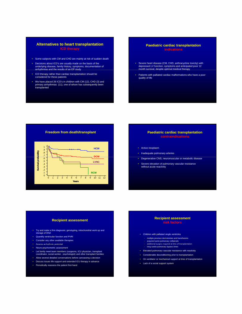
Immunosuppressive therapiesCyclosporine
• CSA enters T cells via diffusion and binds to immunophilin
• The complex binds to calcineurin and inhibits transcription of IL-2 and other cytokines
• The introduction of CSA in 1982 increased 3-year survival fromThe introduction of CSA in 1982 increased 3 year survival from 40% to 70%
• Long list of adverse effects includes nephrotoxicity, hypertension, hyperlipidaemia, type I diabetes, neurotoxicity and cholestasis
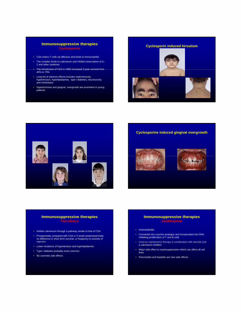
• Hypertrichosis and gingival overgrowth are prominent in young patients
Cyclosporin induced hirsutism
Cyclosporine induced gingival overgrowth
Immunosuppressive therapies Tacrolimus
• Inhibits calcineurin through a pathway similar to that of CSA
• Prospectively compared with CSA in 3 small randomised trials: no difference in short-term survival, or frequency & severity of rejectionrejection
• Lower incidence of hypertension and hyperlipidaemia
• Type I diabetes probably more common
• No cosmetic side effects
Immunosuppressive therapies Azathioprine
• Antimetabolite
• Converted into a purine analogue and incorporated into DNA, inhibiting proliferation of T and B cells
• Used as maintenance therapy in combination with steroids andUsed as maintenance therapy in combination with steroids and a calcineurin inhibitor
• Major side effect is myelosuppression which can affect all cell lines
• Pancreatitis and hepatitis are rare side effects

Immunosuppressive therapies Mycophenolate mofetil
• Noncompetitive inhibitor of de novo guanine nucleotide synthesis
• Selective inhibitor of lymphocyte proliferation with less myelosuppression than AZA
• Tested in a large, prospective randomised study: 3-year survival 88.2% compared to 81.7% for AZA
• Opportunistic infections more common
• Main side effects are gastrointestinal (nausea, vomiting and diarrhoea)
Immunosuppressive therapies Sirolimus
• Similar structure to Tacrolimus
• Disrupts a kinase which connects signals from growth factor receptors to cell nucleus, leading to growth and proliferation of T and B lymphovctes
• Also inhibits smooth muscle and endothelial cell proliferation
• Tested against AZA in a prospective randomised study: reduced acute cellular rejection and prevented graft vasculopathy at 2 years post-transplant
• No inherent nephrotoxicity; may cause thrombocytopaenia
• Role in immunosuppressive regimens is still unclear
Routine post transplant therapy
• Triple therapy with tapering steroids
• Diltiazem for antihypertensive and CSA/TAC sparing effects
• Routine pneumocystis prophylaxis with cotrimoxazole
• Pravastatin for prevention of post-transplant coronary disease in recipients >10 years
Endomyocardial biopsy
• Conventional echo parameters are insensitive markers for the presence of mild-moderate cellular rejection
• Biopsies are not a gold standard - they are subject to differences in observer interpretation and there may be little to see in someone withobserver interpretation and there may be little to see in someone with rapidly progressive rejection
• Biopsies are of low risk and often add useful information
• Children older than 12 months have a biopsy based protocol with around 12 surveillance biopsies during the first year
• Children younger than 1 year have periodic but less frequent biopsies
• Try and avoid biopsies in haemodynamically unstable patients and in very young infants
Immunosuppressive strategies
• Cyclosporine used initially for all children
• Unacceptable cosmetic side-effects: consider a change to Tacrolimus (according to EBV status)( g )
• Frequent or persisting cellular rejection: change to Tacrolimus or Mycophenolate Mofetil
• Renal dysfunction: reduce the dose of Cyclosporine/Tacrolimus or change to Sirolimus
• Coronary disease: optimise risk factors and add Sirolimus
DIAGNOSIS IN PEDIATRIC HEART TRANSPLANT RECIPIENTS (Age: < 1 Year)
29%
65%5%
1%
15%
81%2%
2%
Myopathy
Congenital
Other
ReTX
02550
75100
1988 1989 1990 1991 1992 1993 1994 1995 1996 1997 1998 1999 2000 2001 2002 2003
MyopathyCongenital
1988-1995 1/1996-6/2004
% o
f Cas
es
2005J Heart Lung Transplant 2005;24: 945-982

DIAGNOSIS IN PEDIATRIC HEART TRANSPLANT RECIPIENTS (Age: 1-10 Years)
52%
37%
5%6%52%
39%
5%
4%Myopathy
Congenital
Other
ReTX
02550
75100
1988 1989 1990 1991 1992 1993 1994 1995 1996 1997 1998 1999 2000 2001 2002 2003
Myopathy Congenital
39%1988-1995 1/1996-6/2004
% o
f Cas
es
2005J Heart Lung Transplant 2005;24: 945-982
DIAGNOSIS IN PEDIATRIC HEART TRANSPLANT RECIPIENTS (Age: 11-17 Years)
62%
27%
5%
6%
65%
25%
7%
3%
Myopathy
Congenital
Other
ReTX
02550
75100
1988 1989 1990 1991 1992 1993 1994 1995 1996 1997 1998 1999 2000 2001 2002 2003
Myopathy Congenital
6%3%1988-1995 1/1996-6/2004
% o
f Cas
es
2005J Heart Lung Transplant 2005;24: 945-982
PEDIATRIC HEART TRANSPLANTS (1/1995-6/2003)Risk Factors For 1 Year Mortality
VARIABLE N Relative
Risk P-value 95% Confidence Interval
Congenital diagnosis, on ECMO 69 4.16 <0.0001 2.66 -6.51
Congenital diagnosis, no ECMO 974 2.19 <0.0001 1.74 -2.77
ECMO, diagnosis other than congenital 68 1.9 0.0211 1.10 -3.28
N=3,014
Year of Transplant: 1995 vs. 1998 362 1.9 0.001 1.30 -2.77
Hospitalized (including ICU) 2132 1.55 0.0007 1.20 -2.00
On ventilator 448 1.35 0.0239 1.04 -1.76
Female recipient 1300 1.22 0.0409 1.01 -1.48
Donor age Cont. variable
2005J Heart Lung Transplant 2005;24: 945-982
PEDIATRIC HEART TRANSPLANTATIONKaplan-Meier Survival (1/1982-6/2003)
70
80
90
100
<1 Year (N = 1,375) 1-10 Years (N = 2,106)11-17 Years (N = 2,196) Overall (N = 5,677)
<1 year vs. 1-10 years: p = 0.0082
al (%
)
30
40
50
60
0 1 2 3 4 5 6 7 8 9 10 11 12 13 14 15 16 17 18
Years
HALF-LIFE 1-10: 13.1 years; 11-17: 11.3 years
Surv
iva
2005J Heart Lung Transplant 2005;24: 945-982
PEDIATRIC HEART TRANSPLANTATIONKaplan-Meier Survival by Era (1/1982-6/2003)
60
80
100 1982-1988 (N = 570) 1989-1993 (N = 1,704)1994-1998 (N = 1,873) 1999-6/2003 (N = 1,620)1988-2006 RCH
viva
l (%
)
0
20
40
0 1 2 3 4 5 6 7 8 9 10 11 12 13 14 15 16 17 18Years
All p-values significant at < 0.05 except comparison of 1994-1998 vs. 1999-2003
HALF-LIFE 1982-1988: 9.7 years; 1989-1993: 11.5 years;
Surv
2005J Heart Lung Transplant 2005;24: 945-982
PEDIATRIC HEART TRANSPLANT RECIPIENTS:Cause of Death (Deaths: January 1992 - June 2004)
CAUSE OF DEATH 0-30 Days (N = 335)
31 Days - 1 Year (N = 281)
>5 Years (N = 252)
CORONARY ARTERY VASCULOPATHY 3 (0.9%) 26 (9.3%) 72 (28.6%)
ACUTE REJECTION 27 (8.1%) 76 (27.0%) 32 (12.7%)
LYMPHOMA 6 (2.1%) 21 (8.3%)
INFECTION, NON-CMV 47 (14.0%) 46 (16.4%) 16 (6.3%)
PRIMARY FAILURE 58 (17.3%) 11 (3.9%) 12 (4.8%)
GRAFT FAILURE 79 (23.6%) 31 (11.0%) 49 (19.4%)
TECHNICAL 21 (6.3%) 2 (0.7%) 1 (0.4%)
OTHER 15 (4.5%) 16 (5.7%) 24 (9.5%)
MULTIPLE ORGAN FAILURE 36 (10.7%) 29 (10.3%) 6 (2.4%)
RENAL FAILURE 1 (0.3%) 4 (1.4%)
PULMONARY 24 (7.2%) 16 (5.7%) 7 (2.8%)
CEREBROVASCULAR 23 (6.9%) 7 (2.5%) 3 (1.2%)
2005J Heart Lung Transplant 2005;24: 945-982

Lymphoproliferative disease
• PTLD is the primary post-transplant malignancy in children
• Usually polymorphic, of B cell origin and EBV driven
• Incidence 9% within 7 years; 3 year 70% survival
• Options include reduction or cessation of therapy, or chemotherapy (for refractory or monomorphic disease)
• Relationship to Tacrolimus is unclear
FREEDOM FROM CORONARY ARTERY VASCULOPATHYApril 1994 - June 2004; Stratified by Age Group
80
90
100
from
CAV
50
60
70
0 1 2 3 4 5 6 7Years
<1 Year (N =574)1-10 Years (N = 862)11-17 Years (N = 838)
p = 0.0001
% F
reed
om
2005J Heart Lung Transplant 2005;24: 945-982
Late follow-up
• Regular review in a clinic setting
• Coronary angiography yearly in adolescents and 2nd yearly in younger patients
• Additional biopsies if changes in therapy low drug levels or evidence ofAdditional biopsies if changes in therapy, low drug levels or evidence of non-compliance
• Annual measurement of glomerular filtration rate
• Dental review
• Regular contact with a psychologist
Adolescent non-compliancewarning features
• Missed appointments without explanation
• Clinic attendance without parents
• Unstable social circumstances
• Low CSA levels without changes to therapyLow CSA levels without changes to therapy
• No routine for taking therapy
• Patient/family unfamiliar with drugs or doses
• Unexpected late rejection
• Previous non-compliance
Adolescent non-complianceminimising the risk
• Regular clinical review with non-invasive cardiac assessment and CSA levels
• Patient or family asked to list medications at each visit
• Pill-box
• Clinical psychologist on the team sees patients separately
• Biopsy based follow-up protocol for those with late rejection
Future directions
• Cardiac transplantation is a palliative procedure. Post-transplant survival and outcomes are acceptable and continue to gradually improve
• New immunosuppressive regimens have lowered the rates of acute rejection but have had relatively little impact on theacute rejection but have had relatively little impact on the incidence of chronic rejection.
• The ultimate goal is to induce a state of donor-specific tolerance, wherein the recipient will accept the allograft indefinitely without the need for long-term immunosuppression.
• Medical and surgical alternatives to heart transplantation should be explored and applied