Welcome IQAC at DHVI CD4 Immunophenotyping for HIV Monitoring Flow Cytometry.
FLOW CYTOMETRY IMMUNOPHENOTYPING OF HEMATOLOGICAL MALIGNANCIES:
FROM THE PAST TO THE FUTUREHow did we arrive where we are ?
Cancer Research Centre & Dpt. MedicineUniversity Hospital of Salamanca
University of Salamanca
XVII CONGRESO CHILENO DE HEMATOLOGÍA & VII CONGRESO DE MEDICINA TRANSFUSIONAL 27 AL 30 DE OCTUBRE 2010, HOTEL DE LA BAHÍA, COQUIMBO , Chile
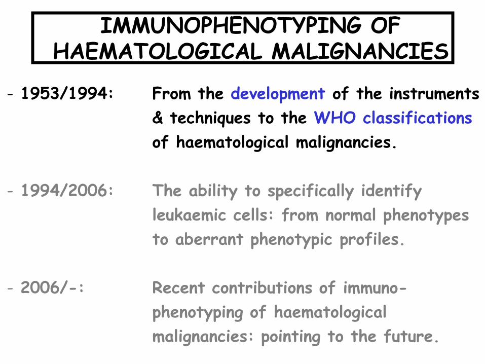
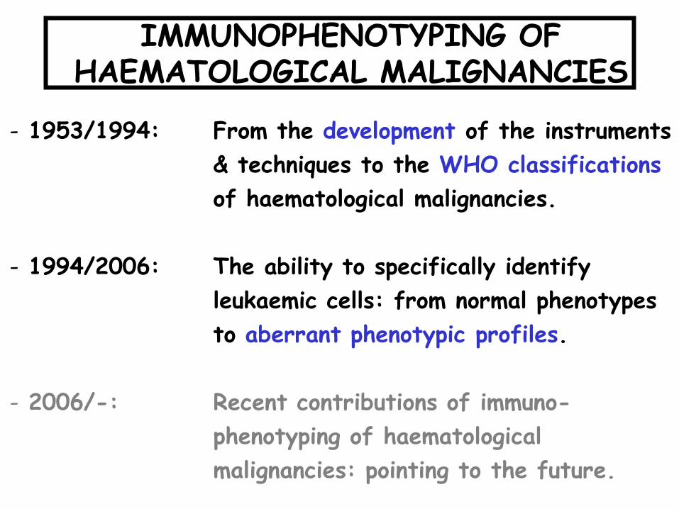
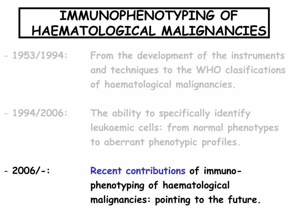
IMMUNOPHENOTYPING OFHAEMATOLOGICAL MALIGNANCIES
- 1953/1994: From the development of the instruments & techniques to the WHO classifications of haematological malignancies.
- 1994/2006: The ability to specifically identify leukaemic cells: from normal phenotypes to aberrant phenotypic profiles.
- 2006/-: Recent contributions of immuno-phenotyping of haematological malignancies: pointing to the future.
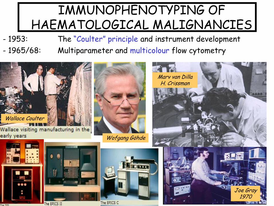
- 1953: The “Coulter” principle and instrument development- 1965/68: Multiparameter and multicolour flow cytometry
Marv van Dilla H. Crissman
Joe Gray1970
IMMUNOPHENOTYPING OFHAEMATOLOGICAL MALIGNANCIES
Wallace Coulter
Wofgang Göhde
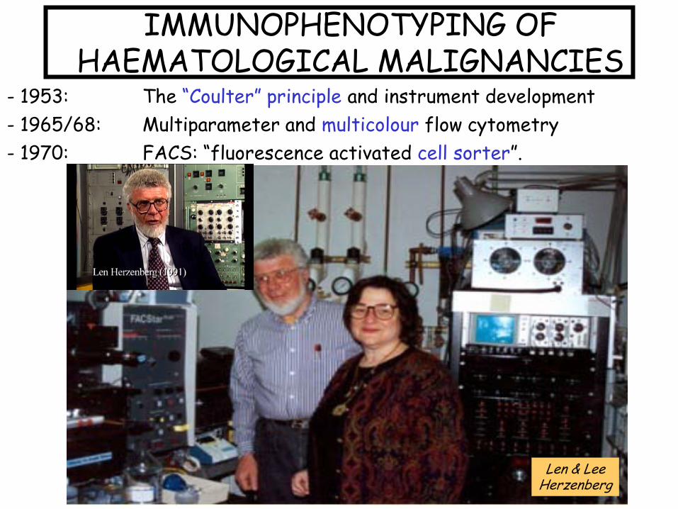
- 1953: The “Coulter” principle and instrument development- 1965/68: Multiparameter and multicolour flow cytometry- 1970: FACS: “fluorescence activated cell sorter”.
Len & LeeHerzenberg
IMMUNOPHENOTYPING OFHAEMATOLOGICAL MALIGNANCIES
¿WHERE CAN I APPLY FLOW CYTOMETRY?
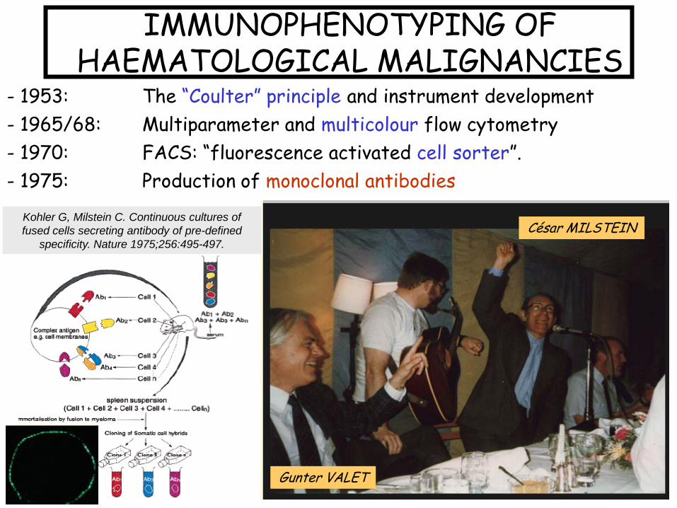
César MILSTEIN
Gunter VALET
Kohler G, Milstein C. Continuous cultures of fused cells secreting antibody of pre-defined
specificity. Nature 1975;256:495-497.
- 1953: The “Coulter” principle and instrument development- 1965/68: Multiparameter and multicolour flow cytometry- 1970: FACS: “fluorescence activated cell sorter”.- 1975: Production of monoclonal antibodies
IMMUNOPHENOTYPING OFHAEMATOLOGICAL MALIGNANCIES
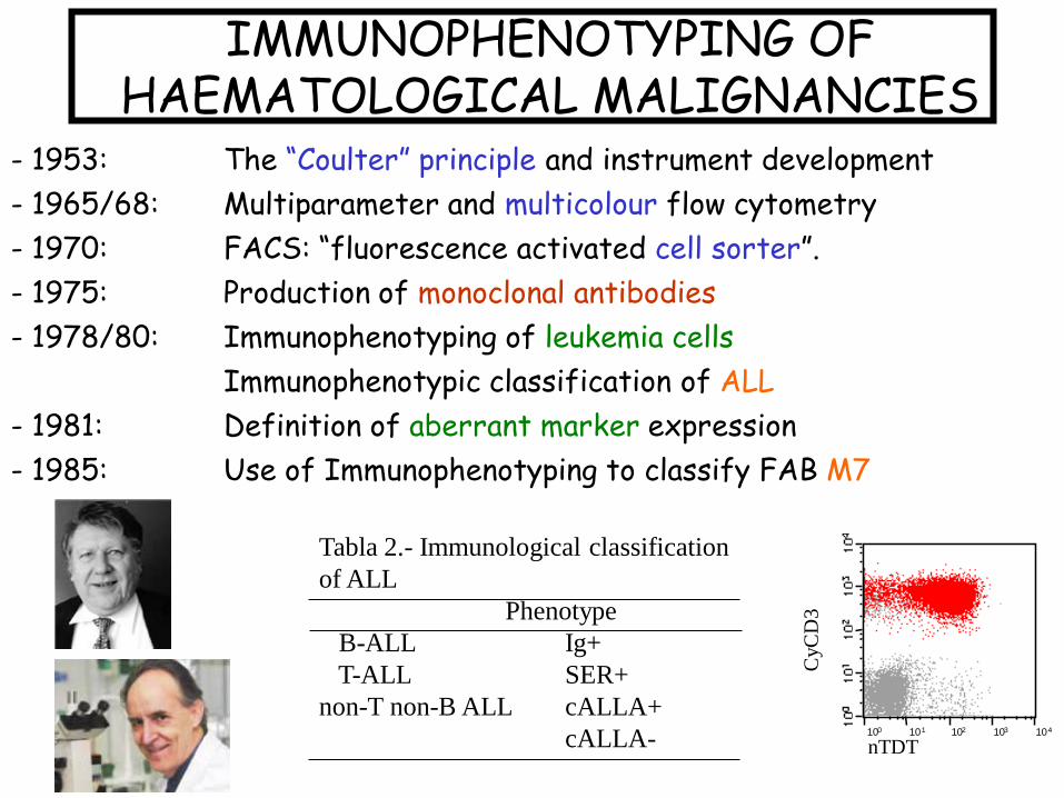
Tabla 2.- Immunological classification of ALL
PhenotypeB-ALL Ig+T-ALL SER+
non-T non-B ALL cALLA+cALLA- 10 10 10 10 100 1 2 3 4
nTDT
CyC
D3
- 1953: The “Coulter” principle and instrument development- 1965/68: Multiparameter and multicolour flow cytometry- 1970: FACS: “fluorescence activated cell sorter”.- 1975: Production of monoclonal antibodies- 1978/80: Immunophenotyping of leukemia cells
Immunophenotypic classification of ALL- 1981: Definition of aberrant marker expression- 1985: Use of Immunophenotyping to classify FAB M7
IMMUNOPHENOTYPING OFHAEMATOLOGICAL MALIGNANCIES
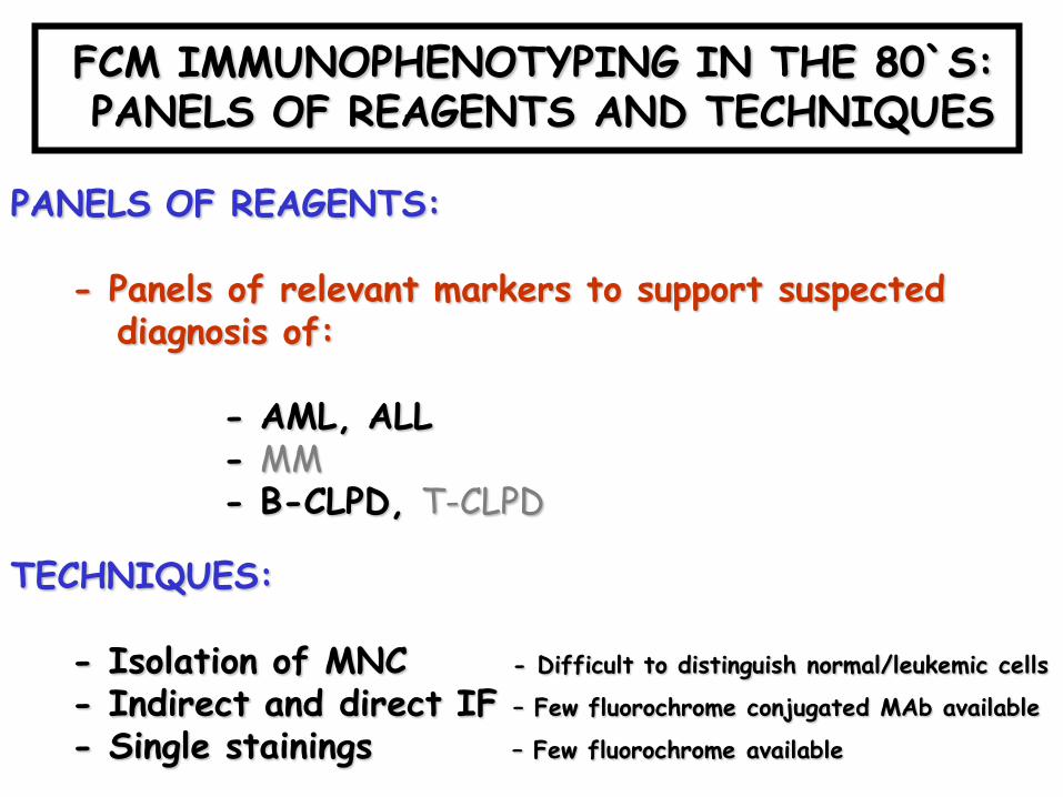
FCM IMMUNOPHENOTYPING IN THE 80`S: PANELS OF REAGENTS AND TECHNIQUES
PANELS OF REAGENTS:
- Panels of relevant markers to support suspecteddiagnosis of:
- AML, ALL- MM- B-CLPD, T-CLPD
TECHNIQUES:
- Isolation of MNC - Difficult to distinguish normal/leukemic cells
- Indirect and direct IF – Few fluorochrome conjugated MAb available
- Single stainings – Few fluorochrome available

DIAGNOSIS OF HAEMATOLOGICAL MALIGNANCIES
Clinical symptoms Laboratoryand signs findings
Morphology + cytochemistry
CMDMDS Immunophenotyping MRDMG
Acute leukemias Chronic lymphoidleukemias
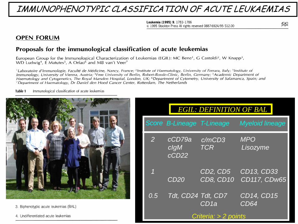
EGIL: DEFINITION OF BAL
Score
c/mCD3
B-Lineage T-Lineage Myeloid lineage
2 cCD79a MPOcIgM TCR LisozymecCD22
1 CD2, CD5 CD13, CD33CD20 CD8, CD10 CD117, CDw65
0.5 Tdt, CD24 Tdt, CD7 CD14, CD15CD1a CD64
Criteria: > 2 points
IMMUNOPHENOTYPIC CLASSIFICATION OF ACUTE LEUKAEMIAS
IMMUNOPHENOTYPING OFHAEMATOLOGICAL MALIGNANCIES
- 1953/1994: From the development of the instruments & techniques to the WHO classifications of haematological malignancies.
- 1994/2006: The ability to specifically identify leukaemic cells: from normal phenotypes to aberrant phenotypic profiles.
- 2006/-: Recent contributions of immuno-phenotyping of haematological malignancies: pointing to the future.
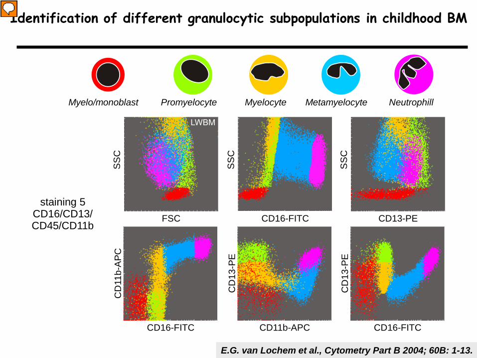
Identification of different granulocytic subpopulations in childhood BM
E.G. van Lochem et al., Cytometry Part B 2004; 60B: 1-13.
FSC
SS
C
CD11b-APC
CD
13-P
E
CD16-FITC
CD
13-P
E
CD13-PE
SS
C
staining 5CD16/CD13/CD45/CD11b
CD16-FITCS
SC
CD16-FITC
CD
11b-
APC
Myelo/monoblast Promyelocyte MetamyelocyteMyelocyte Neutrophill
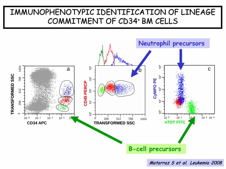
IMMUNOPHENOTYPIC IDENTIFICATION OF LINEAGE COMMITMENT OF CD34+ BM CELLS
nTDT FITC
CyM
PO P
E
c
10 10 10 10 100 1 2 3 4
TRAN
SFO
RM
ED S
SC
CD34 APC
025
651
276
810
24 a
10 10 10 10 100 1 2 3 4 1024TRANSFORMED SSC0 256 512 768
bCD
45 P
ERC
P
b
Matarraz S et al. Leukemia 2008
Neutrophil precursors
B-cell precursors

10 10 10 10 100 1 2 3 4
CD38 FITC ->
TRA
NSF
OR
MED
SSC
->
10 10 10 10 100 1 2 3 4
CD38 FITC ->
CD
138
PerC
P/C
y5 ->
CD38-FITC
CD38-FITC gated PC
T-SS
C
CD13
8-Pe
rCP/
Cy5.
5MONOCLONAL GAMMOPATHIES:
IDENTIFICATION OF CLONAL PLASMA CELLS
Clonal PC
Normal PC
CD
56
PE
9
1
D
C
CP
A
54
DC
Perez- Andres, J Biol Reg, 2004

DIAGNOSIS OF HAEMATOLOGICAL MALIGNANCIES
Clinical symptoms Laboratoryand signs findings
Morphology + cytochemistry
Cytogenetics
Immunophenotyping
Molecular biology/FISH
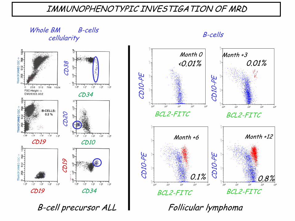
CD20
CD19
CD34
CD10
CD34
CD19
CD38
CD19
CD20
Whole BM B-cellscellularity
IMMUNOPHENOTYPIC INVESTIGATION OF MRD
Month +3
10 10 10 10 100 1 2 3 4
Month +6CD
10-P
E
BCL2-FITC10 10 10 10 100 1 2 3 4
Month +12
CD10
-PE
BCL2-FITC
0.1% 0.8%
10 10 10 10 100 1 2 3 4
13131.002cBCL2 FITC ->
CD
10 P
E ->
CD10
-PE
BCL2-FITC10 10 10 10 100 1 2 3 4
12799.002cBCL2 FITC ->
CD
10 P
E ->
CD10
-PE
BCL2-FITC
Month 0 <0.01% 0.01%
Month +3
B-cells
B-cell precursor ALL Follicular lymphoma
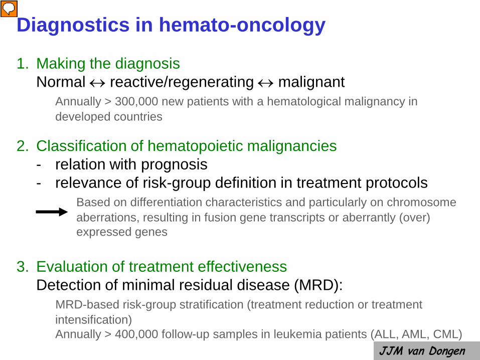
1. Making the diagnosisNormal ↔ reactive/regenerating ↔ malignant
Annually > 300,000 new patients with a hematological malignancy in developed countries
2. Classification of hematopoietic malignancies- relation with prognosis- relevance of risk-group definition in treatment protocols
Based on differentiation characteristics and particularly on chromosome aberrations, resulting in fusion gene transcripts or aberrantly (over) expressed genes
3. Evaluation of treatment effectivenessDetection of minimal residual disease (MRD):
MRD-based risk-group stratification (treatment reduction or treatment intensification)Annually > 400,000 follow-up samples in leukemia patients (ALL, AML, CML)
Diagnostics in hemato-oncology
JJM van Dongen
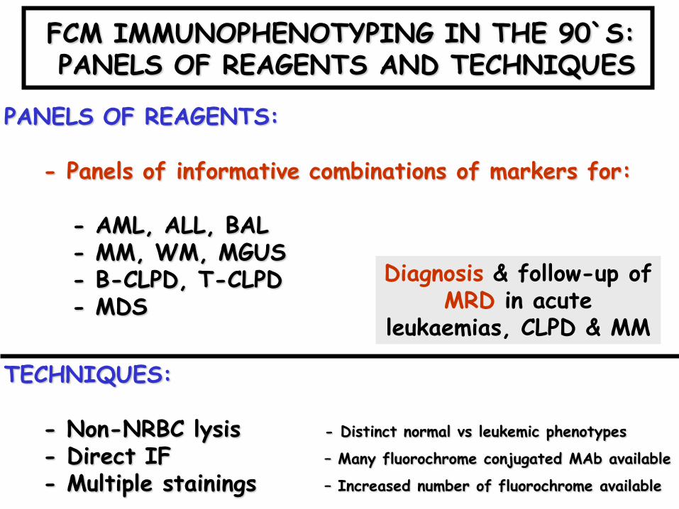
FCM IMMUNOPHENOTYPING IN THE 90`S: PANELS OF REAGENTS AND TECHNIQUES
PANELS OF REAGENTS:
- Panels of informative combinations of markers for:
- AML, ALL, BAL- MM, WM, MGUS- B-CLPD, T-CLPD- MDS
TECHNIQUES:
- Non-NRBC lysis - Distinct normal vs leukemic phenotypes
- Direct IF – Many fluorochrome conjugated MAb available
- Multiple stainings – Increased number of fluorochrome available
Diagnosis & follow-up of MRD in acute
leukaemias, CLPD & MM
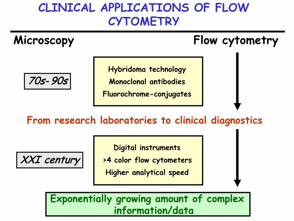
CLINICAL APPLICATIONS OF FLOW CYTOMETRY
Microscopy Flow cytometry
Hybridoma technologyMonoclonal antibodies
Fluorochrome-conjugates
From research laboratories to clinical diagnostics
Digital instruments >4 color flow cytometersHigher analytical speed
Exponentially growing amount of complex information/data
70s- 90s
XXI century
10 10 10 10 100 1 2 3 4
10578.011CD 45 PERCPCY5.5 ->
CD45 PerCPCy5.5 10 10 10 10 100 1 2 3 4
10578.007HLADR FITC ->
HLA-DR FITC 10 10 10 10 100 1 2 3 4
10578.011CD2 FITC ->
CD2 FITC
10 10 10 10 100 1 2 3 4
10578.010CD65 FITC ->
CD65 FITC 10 10 10 10 100 1 2 3 4
10578.006CD34 APC ->
CD34 FITC
10 10 10 10 100 1 2 3 4
10578.004cCD3 FITC ->
Cy-CD3 FITC 10 10 10 10 100 1 2 3 4
10578.002nTDT FITC ->
nTdT FITC 10 10 10 10 100 1 2 3 4
10578.008CD11B FITC ->
CD11b FITC 10 10 10 10 100 1 2 3 4
10578.012CD61 FITC ->
CD61 FITC
10 10 10 10 100 1 2 3 4
10578.014CD36 FITC ->
CD36 FITC
Tran
sfor
med
SSC
10 10 10 10 100 1 2 3 4
11504 L.Cel.DendrtBDCA4/CD138BDCA4 PE ->
10 10 10 10 100 1 2 3 4
11504 L.Cel.Dendrt.CD4/CD8CD4 FITC ->
CD
123
PE
CD
56 P
E
HLA
-DR
FIT
C
BDCA4 (CD304) PE
7.1
PE
CD
64 P
E
CD
117
PE
CD4 FITC
CD
8 PE
C
D7
PE
Cyt
MPO
PE
CD
13 P
E
CD
33 P
E
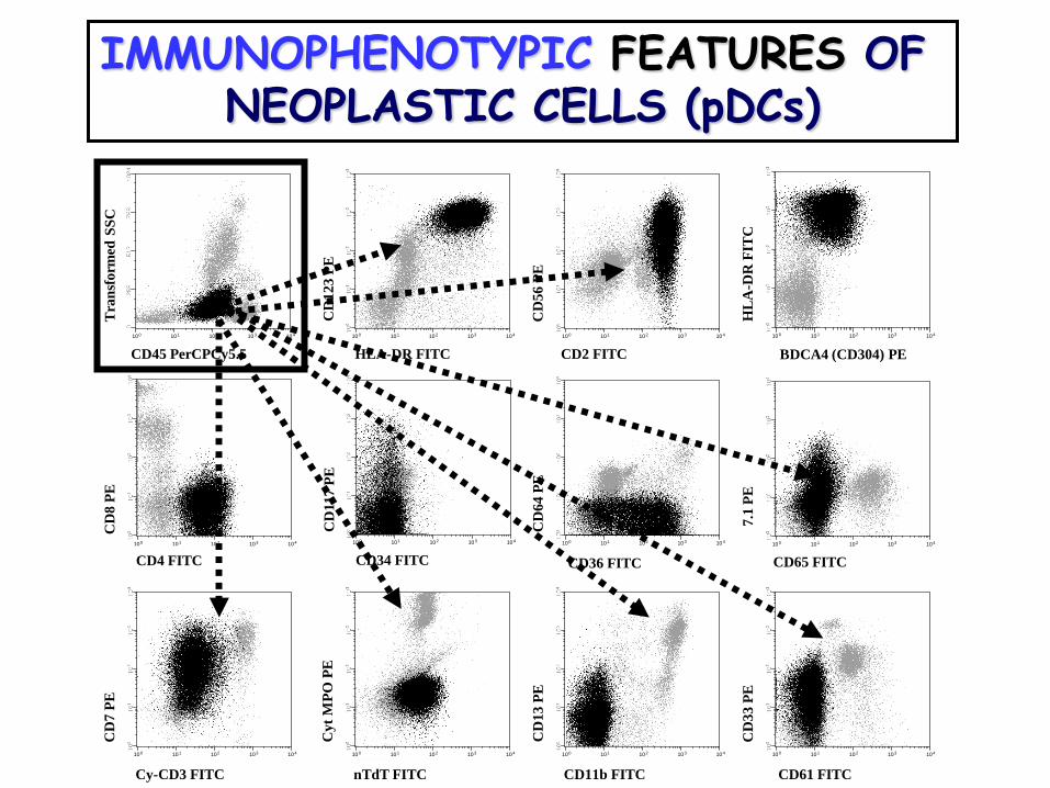
IMMUNOPHENOTYPIC FEATURES OF NEOPLASTIC CELLS (pDCs)
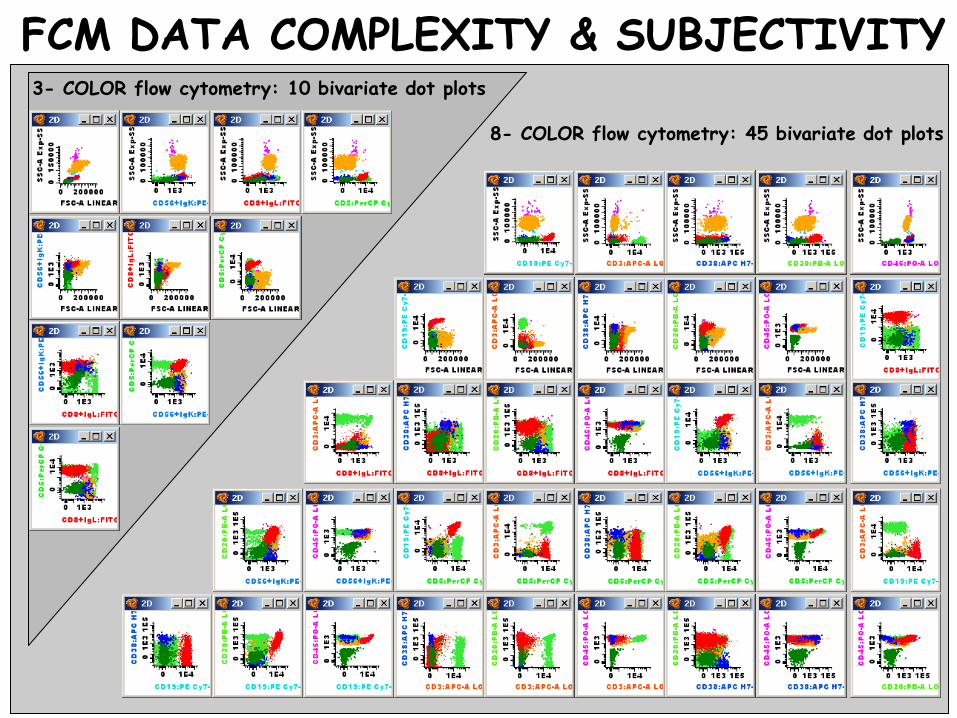
3- COLOR flow cytometry: 10 bivariate dot plots
8- COLOR flow cytometry: 45 bivariate dot plots
FCM DATA COMPLEXITY & SUBJECTIVITY

STANDARDIZATION EFFORTS FORIMMUNOPHENOTYPIC STUDIES
- CLSI (Clinical Laboratory Standards Institute):- Stetler-Stevenson et al.: Clinical flow cytometric analysis of neoplastic hematolymphoid cells; Approved guideline. CLSI document H43-A2. CLSI, 2007
- CCS (Clinical Cytometry Society):- Davis et al: 2006 Bethesda International Consensus
recommendations on the flow cytometric immunophenotypic analysis of hematolymphoid neoplasias. Clin Cytometry, 72B, 2007.
- ESCCA (European Society for Clinical Cell Analysis: www.escca.eu)
- European Leukemia Net (www.leukemia-net.org)- Consenso Latinoamericano (Clin Cytometry, 1998 y 2006)
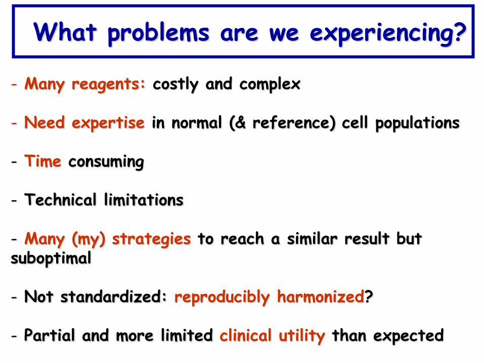
What problems are we experiencing?
- Many reagents: costly and complex
- Need expertise in normal (& reference) cell populations
- Time consuming
- Technical limitations
- Many (my) strategies to reach a similar result but suboptimal
- Not standardized: reproducibly harmonized?
- Partial and more limited clinical utility than expected
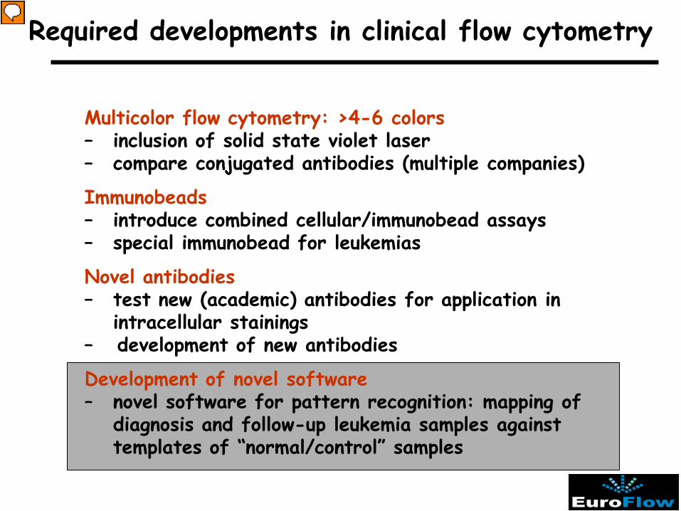
Required developments in clinical flow cytometry
Multicolor flow cytometry: >4-6 colors– inclusion of solid state violet laser– compare conjugated antibodies (multiple companies)
Immunobeads– introduce combined cellular/immunobead assays– special immunobead for leukemias
Novel antibodies– test new (academic) antibodies for application in
intracellular stainings– development of new antibodies
Development of novel software– novel software for pattern recognition: mapping of
diagnosis and follow-up leukemia samples against templates of “normal/control” samples
IMMUNOPHENOTYPING OFHAEMATOLOGICAL MALIGNANCIES
- 1953/1994: From the development of the instrumentsand techniques to the WHO clasificationsof haematological malignancies.
- 1994/2006: The ability to specifically identifyleukaemic cells: from normal phenotypesto aberrant phenotypic profiles.
- 2006/-: Recent contributions of immuno-phenotyping of haematologicalmalignancies: pointing to the future.

Technical aspects of EuroFlow protocols:instrument settings, fluorochrome choice, standardization
T. Kalina1, J. Flores-Montero2, Q. Lecrevisse2, M. Cullen3,L. Lhermitte4, L. Sedek5, A. Mendonca 6, S. Bötcher7, J. te Marvelde8, Mejstříková, O. Hrušák1,
J.J.M. van Dongen8, and A. Orfao2
1, Department of Pediatric Hematology and Oncology, Charles University, Prague, Czech Republic;2, Department of Medicine, Cancer Research Centre and Cytometry Service,
University of Salamanca, Salamanca, ES;3, St. James University Hospital, Leeds, UK;
4, Department of Hematology, Hôpital Necker, Paris, FR 5, Department of Pediatric Hematology and Oncology, Medical University of Silesia, Zabrze, PL;
6, Department of Hematology, Instituto Portugues de Oncologia , Lisbon, PT;7, 2nd Department of Medicine, University Klinik Schleswig-Holstein, Kiel, DE;
8, Department of Immunology, Erasmus MC, Rotterdam, NL;
Prepared by Thomas Kalina on behalf of theEuroFlow Consortium (EU-FP6, LSHB-CT-2006-018708)
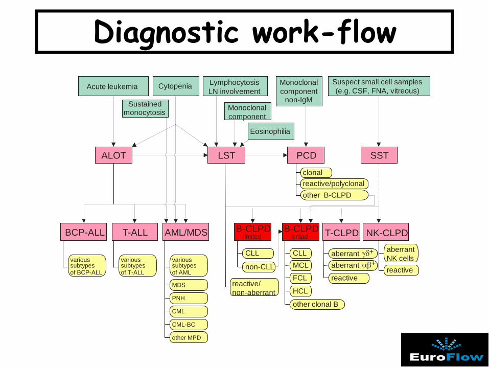
Diagnostic work-flow
Monoclonalcomponent
Monoclonalcomponent
non-IgM
ALOT LST PCD SST
BCP-ALL T-ALL AML/MDS B-CLPDlimited
B-CLPDbroad T-CLPD NK-CLPD
Sustainedmonocytosis
Eosinophilia
reactive/polyclonalother B-CLPD
reactive/non-aberrant
CLL
non-CLL
CLLMCL
FCL
HCL
other clonal B
reactive
aberrant +αβaberrant +γδ
reactive
aberrantNK cellsvarious
subtypesof BCP-ALL
various subtypesof T-ALL
various subtypesof AML
MDS
PNH
CML
CML-BC
other MPD
Acute leukemia Cytopenia LymphocytosisLN involvement
Suspect small cell samples(e.g. CSF, FNA, vitreous)
clonal
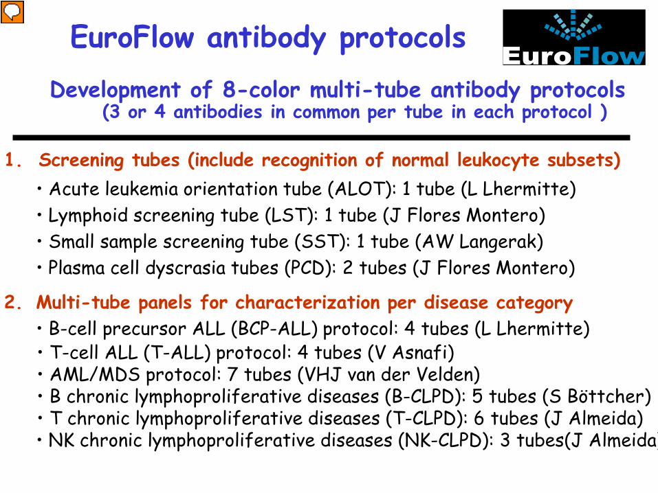
EuroFlow antibody protocolsDevelopment of 8-color multi-tube antibody protocols
(3 or 4 antibodies in common per tube in each protocol )
1. Screening tubes (include recognition of normal leukocyte subsets)• Acute leukemia orientation tube (ALOT): 1 tube (L Lhermitte)• Lymphoid screening tube (LST): 1 tube (J Flores Montero)• Small sample screening tube (SST): 1 tube (AW Langerak)• Plasma cell dyscrasia tubes (PCD): 2 tubes (J Flores Montero)
2. Multi-tube panels for characterization per disease category• B-cell precursor ALL (BCP-ALL) protocol: 4 tubes (L Lhermitte)• T-cell ALL (T-ALL) protocol: 4 tubes (V Asnafi)• AML/MDS protocol: 7 tubes (VHJ van der Velden)• B chronic lymphoproliferative diseases (B-CLPD): 5 tubes (S Böttcher)• T chronic lymphoproliferative diseases (T-CLPD): 6 tubes (J Almeida)• NK chronic lymphoproliferative diseases (NK-CLPD): 3 tubes(J Almeida)
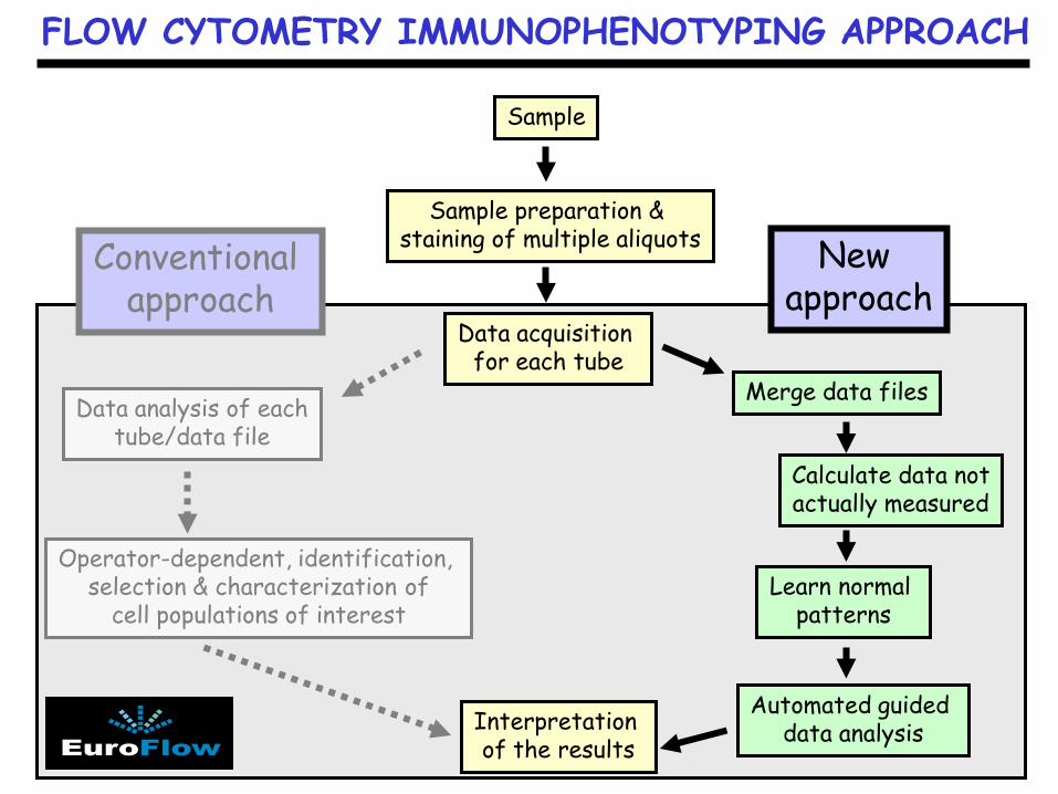
FLOW CYTOMETRY IMMUNOPHENOTYPING APPROACH
Sample
Sample preparation & staining of multiple aliquots
Data acquisition for each tube
Data analysis of eachtube/data file
Operator-dependent, identification, selection & characterization of
cell populations of interest
Interpretation of the results
Merge data files
Calculate data notactually measured
Learn normal patterns
Automated guided data analysis
New approach
Conventional approach
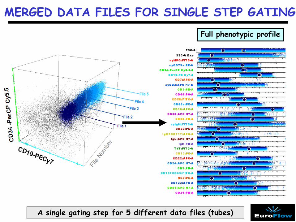
MERGED DATA FILES FOR SINGLE STEP GATING
Full phenotypic profile
A single gating step for 5 different data files (tubes)
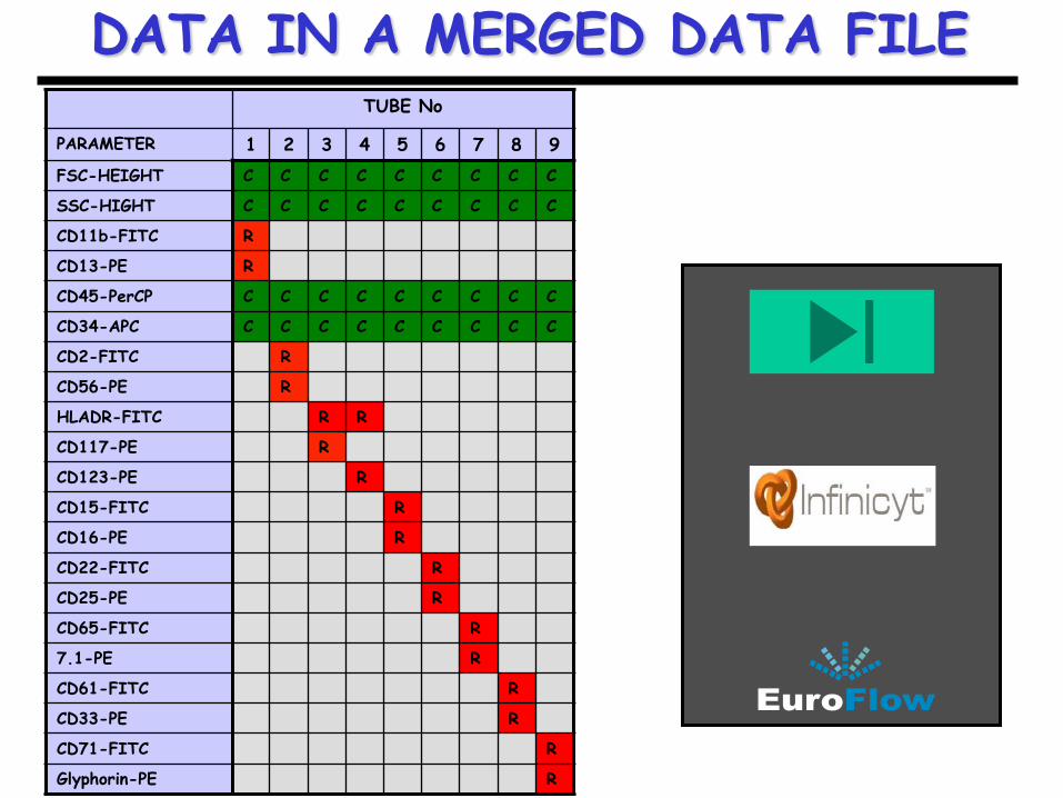
DATA IN A MERGED DATA FILETUBE No
PARAMETER 1 2 3 4 5 6 7 8 9FSC-HEIGHT C C C C C C C C C
SSC-HIGHT C C C C C C C C C
CD11b-FITC R
CD13-PE R
CD45-PerCP C C C C C C C C C
CD34-APC C C C C C C C C C
CD2-FITC R
CD56-PE R
HLADR-FITC R R
CD117-PE R
CD123-PE R
CD15-FITC R
CD16-PE R
CD22-FITC R
CD25-PE R
CD65-FITC R
7.1-PE R
CD61-FITC R
CD33-PE R
CD71-FITC R
Glyphorin-PE R
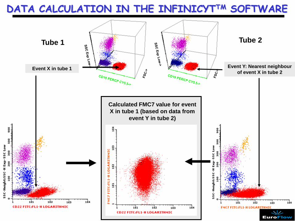
Tube 1
.
Tube 2
.Event X in tube 1 Event Y: Nearest neighbour of event X in tube 2
Calculated FMC7 value for event X in tube 1 (based on data from
event Y in tube 2)
DATA CALCULATION IN THE INFINICYTTM SOFTWARE
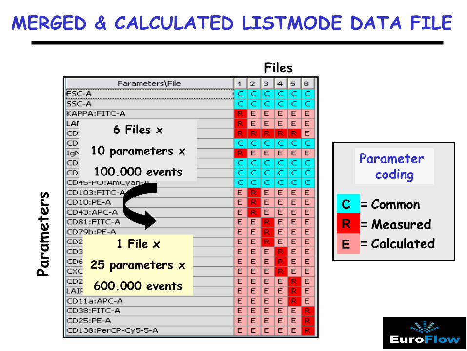
MERGED & CALCULATED LISTMODE DATA FILE
= Common= Measured= Calculated
CRE
Files
Para
met
ers
6 Files x
10 parameters x
100.000 events
1 File x
25 parameters x
600.000 events
Parameter coding
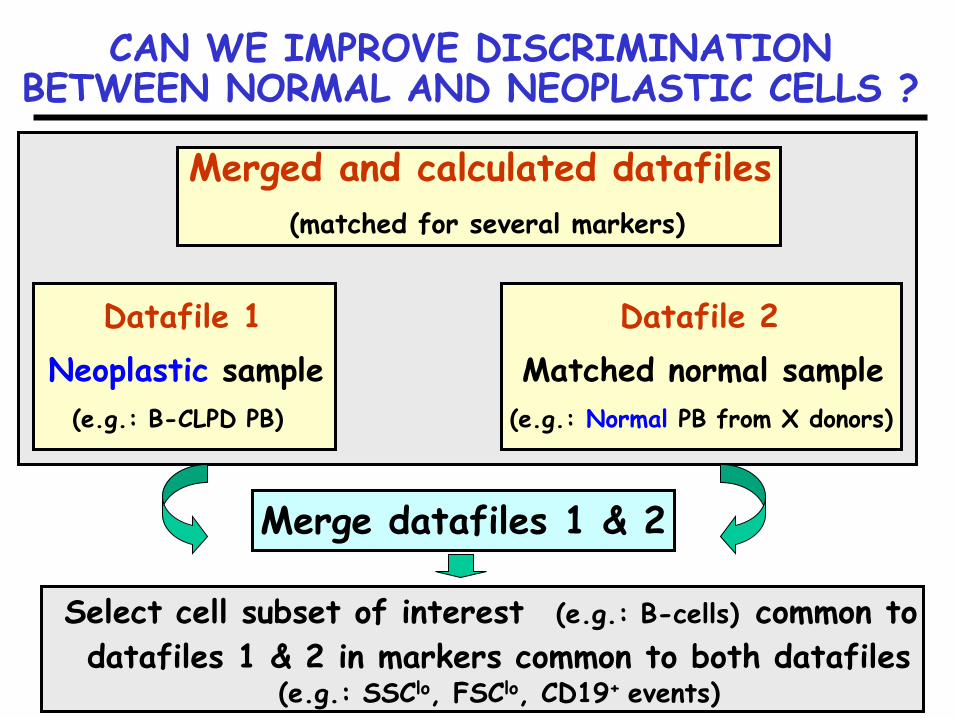
CAN WE IMPROVE DISCRIMINATION BETWEEN NORMAL AND NEOPLASTIC CELLS ?
Merged and calculated datafiles(matched for several markers)
Datafile 1 Datafile 2Neoplastic sample Matched normal sample
(e.g.: B-CLPD PB) (e.g.: Normal PB from X donors)
Merge datafiles 1 & 2
Select cell subset of interest (e.g.: B-cells) common to datafiles 1 & 2 in markers common to both datafiles
(e.g.: SSClo, FSClo, CD19+ events)
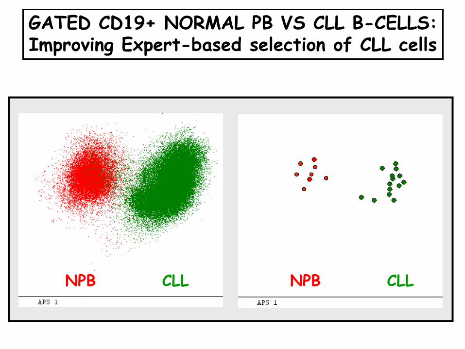
NPB CLL NPB CLL
GATED CD19+ NORMAL PB VS CLL B-CELLS:Improving Expert-based selection of CLL cells
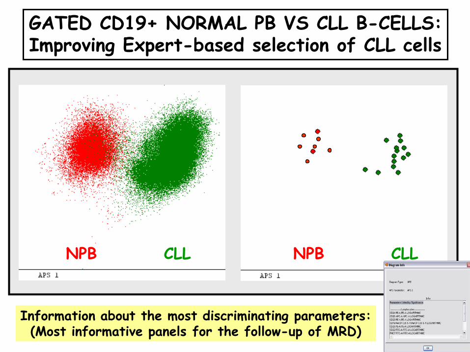
NPB CLL NPB CLL
GATED CD19+ NORMAL PB VS CLL B-CELLS:Improving Expert-based selection of CLL cells
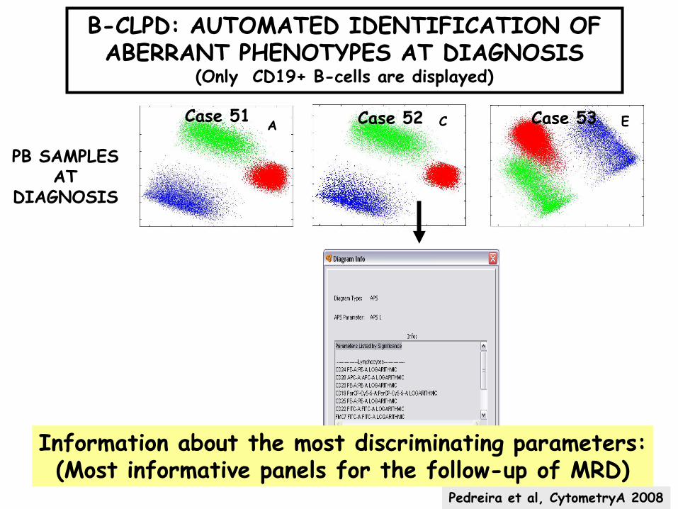
Information about the most discriminating parameters:(Most informative panels for the follow-up of MRD)
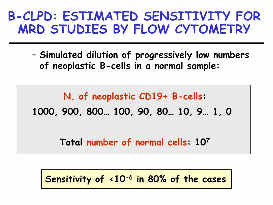
B-CLPD: ESTIMATED SENSITIVITY FOR MRD STUDIES BY FLOW CYTOMETRY
– Simulated dilution of progressively low numbers of neoplastic B-cells in a normal sample:
N. of neoplastic CD19+ B-cells:1000, 900, 800… 100, 90, 80… 10, 9… 1, 0
Total number of normal cells: 107
Sensitivity of <10-6 in 80% of the cases
PB SAMPLES AT
DIAGNOSIS
A C E
B-CLPD: AUTOMATED IDENTIFICATION OF ABERRANT PHENOTYPES AT DIAGNOSIS
(Only CD19+ B-cells are displayed)
Pedreira et al, CytometryA 2008
Information about the most discriminating parameters:(Most informative panels for the follow-up of MRD)
Case 51 Case 52 Case 53
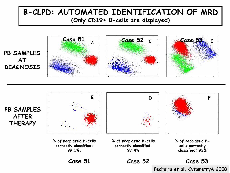
Case 51
PB SAMPLES AT
DIAGNOSIS
PB SAMPLES AFTER
THERAPY
Case 52 Case 53
% of neoplastic B-cells correctly classified:
99,1%.
% of neoplastic B-cells correctly classified:
97,4%
% of neoplastic B-cells correctly classified: 92%
A
B
C
D
E
F
B-CLPD: AUTOMATED IDENTIFICATION OF MRD (Only CD19+ B-cells are displayed)
Pedreira et al, CytometryA 2008
Caso 51 Case 52 Case 53
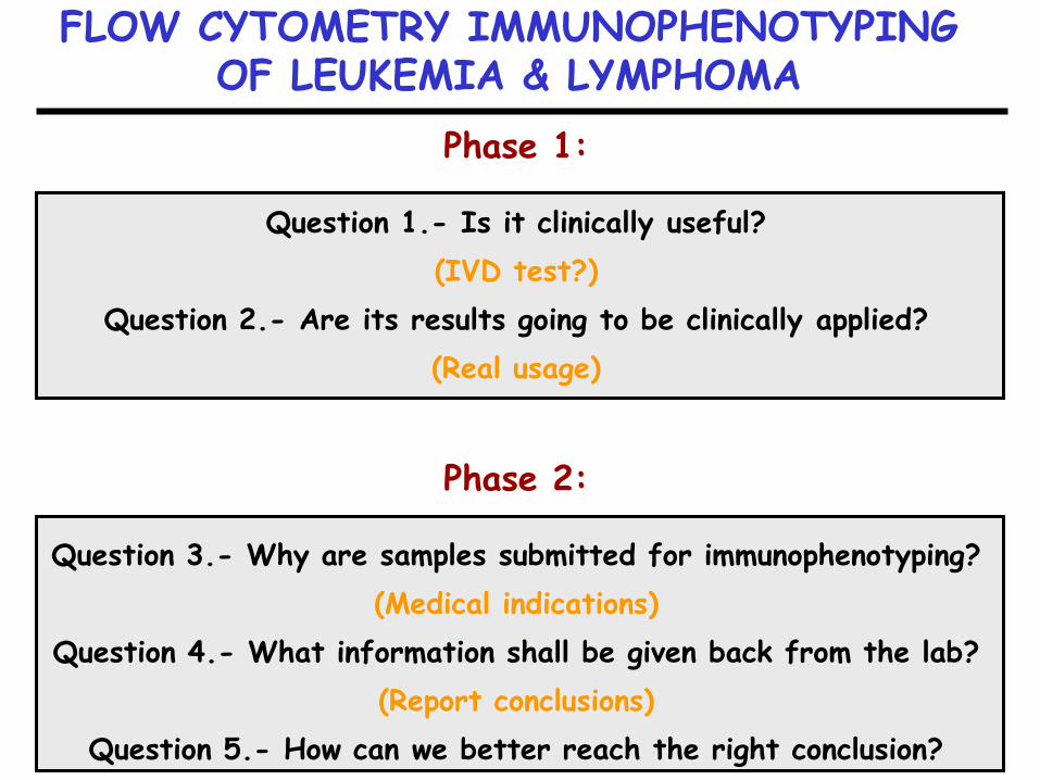
FLOW CYTOMETRY IMMUNOPHENOTYPING OF LEUKEMIA & LYMPHOMA
Phase 1:
Question 1.- Is it clinically useful? (IVD test?)
Question 2.- Are its results going to be clinically applied? (Real usage)
Phase 2:
Question 3.- Why are samples submitted for immunophenotyping?(Medical indications)
Question 4.- What information shall be given back from the lab?(Report conclusions)
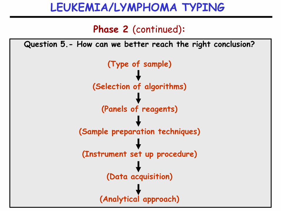
Question 5.- How can we better reach the right conclusion?
LEUKEMIA/LYMPHOMA TYPING
Phase 2 (continued):Question 5.- How can we better reach the right conclusion?
(Type of sample)
(Selection of algorithms)
(Panels of reagents)
(Sample preparation techniques)
(Instrument set up procedure)
(Data acquisition)
(Analytical approach)

FL1-H:CD8/Lambda->
APS1->
APS1->
SSC Exp Low->
A
FE
DCB
G
I
Mon
ocyt
es
Neu
trop
hils
CD
4-/C
D8-
T ly
mph
ocyt
es
B-c
ells
NK
Cel
ls
CD
4+/C
D8+
T
lym
phoc
ytes
KJ
L M
H
THANK YOU