Flavone Biotransformation by Aspergillus niger and the ... · Flavone Biotransformation by...
13
Transcript of Flavone Biotransformation by Aspergillus niger and the ... · Flavone Biotransformation by...
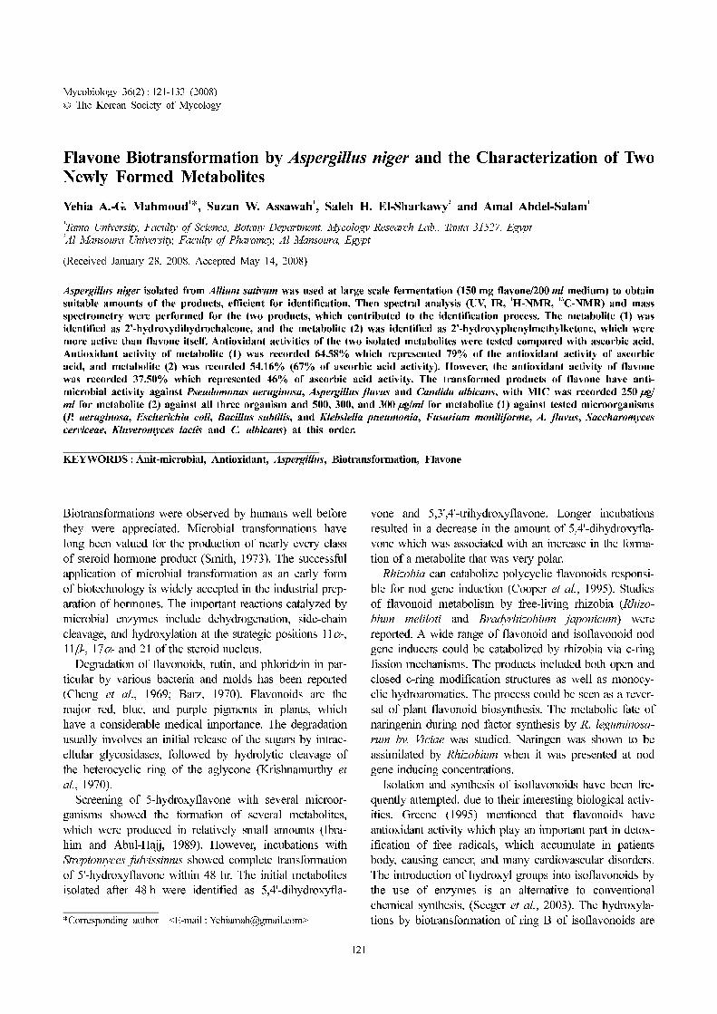
Mycobiology 36(2) : 121-133 (2008)
© The Korean Society of Mycology
121
Flavone Biotransformation by Aspergillus niger and the Characterization of TwoNewly Formed Metabolites
Yehia A.-G. Mahmoud1
*, Suzan W. Assawah1
, Saleh H. El-Sharkawy2
and Amal Abdel-Salam1
1
Tanta University, Faculty of Science, Botany Department, Mycology Research Lab., Tanta 31527, Egypt2
Al Mansoura University, Faculty of Pharamcy, Al Mansoura, Egypt
(Received January 28, 2008. Accepted May 14, 2008)
Aspergillus niger isolated from Allium sativum was used at large scale fermentation (150 mg flavone/200 ml medium) to obtain
suitable amounts of the products, efficient for identification. Then spectral analysis (UV, IR, 1
H-NMR, 13
C-NMR) and mass
spectrometry were performed for the two products, which contributed to the identification process. The metabolite (1) was
identified as 2'-hydroxydihydrochalcone, and the metabolite (2) was identified as 2'-hydroxyphenylmethylketone, which were
more active than flavone itself. Antioxidant activities of the two isolated metabolites were tested compared with ascorbic acid.
Antioxidant activity of metabolite (1) was recorded 64.58% which represented 79% of the antioxidant activity of ascorbic
acid, and metabolite (2) was recorded 54.16% (67% of ascorbic acid activity). However, the antioxidant activity of flavone
was recorded 37.50% which represented 46% of ascorbic acid activity. The transformed products of flavone have anti-
microbial activity against Pseudomonas aeruginosa, Aspergillus flavus and Candida albicans, with MIC was recorded 250 µg/
ml for metabolite (2) against all three organism and 500, 300, and 300 µg/ml for metabolite (1) against tested microorganisms
(P. aeruginosa, Escherichia coli, Bacillus subtilis, and Klebsiella pneumonia, Fusarium moniliforme, A. flavus, Saccharomyces
cerviceae, Kluveromyces lactis and C. albicans) at this order.
KEYWORDS : Anit-microbial, Antioxidant, Aspergillus, Biotransformation, Flavone
Biotransformations were observed by humans well before
they were appreciated. Microbial transformations have
long been valued for the production of nearly every class
of steroid hormone product (Smith, 1973). The successful
application of microbial transformation as an early form
of biotechnology is widely accepted in the industrial prep-
aration of hormones. The important reactions catalyzed by
microbial enzymes include dehydrogenation, side-chain
cleavage, and hydroxylation at the strategic positions 11α-,
11β-, 17α- and 21 of the steroid nucleus.
Degradation of flavonoids, rutin, and phloridzin in par-
ticular by various bacteria and molds has been reported
(Cheng et al., 1969; Barz, 1970). Flavonoids are the
major red, blue, and purple pigments in plants, which
have a considerable medical importance. The degradation
usually involves an initial release of the sugars by intrac-
ellular glycosidases, followed by hydrolytic cleavage of
the heterocyclic ring of the aglycone (Krishnamurthy et
al., 1970).
Screening of 5-hydroxyflavone with several microor-
ganisms showed the formation of several metabolites,
which were produced in relatively small amounts (Ibra-
him and Abul-Hajj, 1989). However, incubations with
Streptomyces fulvissimus showed complete transformation
of 5'-hydroxyflavone within 48 hr. The initial metabolites
isolated after 48 h were identified as 5,4'-dihydroxyfla-
vone and 5,3',4'-trihydroxyflavone. Longer incubations
resulted in a decrease in the amount of 5,4'-dihydroxyfla-
vone which was associated with an increase in the forma-
tion of a metabolite that was very polar.
Rhizobia can catabolize polycyclic flavonoids responsi-
ble for nod gene induction (Cooper et al., 1995). Studies
of flavonoid metabolism by free-living rhizobia (Rhizo-
bium meliloti and Bradyrhizobium japonicum) were
reported. A wide range of flavonoid and isoflavonoid nod
gene inducers could be catabolized by rhizobia via c-ring
fission mechanisms. The products included both open and
closed c-ring modification structures as well as monocy-
clic hydroaromatics. The process could be seen as a rever-
sal of plant flavonoid biosynthesis. The metabolic fate of
naringenin during nod factor synthesis by R. leguminosa-
rum bv. Viciae was studied. Naringen was shown to be
assimilated by Rhizobium when it was presented at nod
gene inducing concentrations.
Isolation and synthesis of isoflavonoids have been fre-
quently attempted, due to their interesting biological activ-
ities. Greene (1995) mentioned that flavonoids have
antioxidant activity which play an important part in detox-
ification of free radicals, which accumulate in patients
body, causing cancer, and many cardiovascular disorders.
The introduction of hydroxyl groups into isoflavonoids by
the use of enzymes is an alternative to conventional
chemical synthesis, (Seeger et al., 2003). The hydroxyla-
tions by biotransformation of ring B of isoflavonoids are*Corresponding author <E-mail : [email protected]>
122 Mahmoud et al.
likely to improve their antioxidant properties (Briviba et
al., 1997 and Arora et al., 1998).
Kim et al. (1998) tested the biotransformation of some
flavonoids (Rutin, hesperdin, and naringin) using intesti-
nal microflora to produce phenolic acids. The cleavage of
flavonoids by intestinal microflora makes understanding
of the mammalian metabolism of these natural products
complicated. Indeed, more than 40 mammalian metabo-
lites have been identified, many of which are hydroxy-
lated at 3 and 6 positions (Das et al., 1973).
Microbial transformation of flavones of Psidia arabica
by Cunninghamella elegans producing the 3'-glucoside
conjugates of the flavones was reported (Ibrahim et al.,
1997). Sulfations of naringenin by Cunninghamella ele-
gans to form naringenin-7-sulfate were reported (Ibrahim,
1999).
Hunter (1995) has reported anti-inflammatory and anti-
allergic effects of flavonoides on human. Because fla-
vonoid are consumed in appreciable amounts in our diet,
knowledge of their pharmacological and physiological
properties is of significant importance. Therefore, we have
planned to carry out the present study to test the possibil-
ity of getting new transformation products that might have
a medical importance to human.
Materials and Methods
Initial screening for flavone biotransformation using
some of the isolated fungi. Initial screening has been
carried out using 24 different fungal species that were iso-
lated from 44 medicinal plants at Kafr El-Sheik governor-
ate, Egypt (data not shown). The fungal genera which were
selected in initial screening for flavone transformation
were; Aspergillus, Fusarium, Alternaria, Stemphylium,
Macrosporium, Nectria, Hormodendrum, Trichothecium,
Spicaria, Microdochium, Gibberella, Macrophomina, Athe-
lia, and Cladosporium. Detection of flavone metabolites
was performed using silica gel plate coated with
(60GF254) through thin layer chromatography (TLC).
Fermentation technique. Fermentation liquid cultures
were initiated by transferring about 5 ml of fungal suspen-
sion (two-week old slants) into 50 ml sterile liquid
medium contained in 250 ml Erlenmeyer flasks placed on
a shaker operating at about 150 rpm at 27o
C for 48 hr
(stage 1).
At stage 2, cultures were obtained by transferring about
5 ml of stage-1 culture to 250 ml Erlenmeyer flasks con-
taining 50 ml of the same liquid medium at the same con-
ditions. Cultures were allowed to grow for 24 hr before
adding of substrate (flavone) dissolved in N,N-dimethyl
formamide (DMF) at a concentration of 5 mg/50 ml
medium, and continued to incubate at the same condi-
tions for 6 days. One flask was used as control in which
the culture was allowed to grow under the same condi-
tions without addition of fungal spores (Ibrahim and
Abul-Hajj, 1990).
Fermentation liquid medium was composed of 10 g
glucose, 5 g peptone, 5 g yeast extract, 5 g sodium chlo-
ride, 5 g K2HPO
4, 10 ml glycerol per 1 liter, and pH was
adjusted to 6 before autoclaving.
Detection of transformation products. Sampling of the
culture was carried out by taking 5 ml of culture suspen-
sion and then extracted with 5 ml chloroform. Then, Chlo-
roform extract was evaporated and the residue was
dissolved in a few drops of methanol, then 20 µl was spot-
ted into silica gel plate (60GF254) (Ciegler et al., 1971).
Isolation and Detection of transformation products. A
commercial applicator for spreading the prepared slurries
of silica gel (adsorbent) as thin layer to glass plates is
usually 5 × 20 cm or 20 × 20 cm. Adsorbent was applied
as aqueous slurries (5 g silica gel/20 ml dist. Water). The
thickness of the film was 250 µm. Samples which pre-
pared from the fermentation culture in the previous step
were applied. Then the applied spots were eluted with
methanol-water-acetic acid (55 : 45 : 0.5, V/V/V) and/or
chloroform-methanol (20 : 1, V/V) were being used in this
work. Chromatographic plates were usually developed
once by the ascending technique, at room temperature, to
a height of 15~18 cm. The separated bands on thin layer
of silica gel were detected using two detection methods.
The plates were dried and visualized under UV-light or
sprayed with sulphuric acid or exposed to ammonia
vapors (Horowitz, 1957).
Large scale fermentation technique and products
purification. A. niger isolated from A. sativum (data not
shown) was used as transfomant in this technique. The
same procedures mentioned above were used to obtain
large amounts of products sufficient for spectroscopic
analysis. Greater concentrations of flavone (150 mg fla-
vone/200 ml medium) were prepared to add when large
containers (flasks) were used in fermentation technique as
a way for obtaining large amount of products. A. niger
was used for large scale fermentation in order to trans-
form flavone. About 150 mg flavone/200 ml medium in
one liter flasks was used in stage 2 cultures. The fermen-
tation was terminated after 6-days starting with addition
of flavone. The cells were separated from the medium by
filtering through a cheese cloth. The cells were extracted
with ethyl acetate containing 1% methanol, while the filtrate
was extracted by chloroform containing 1% methanol.
The combined extracts were evaporated under vacuum.
Isolation of flavone metabolites. The concentrated fer-
mentation extracts were adsorbed on a suitable amount of
Flavone Biotransformation by Aspergillus niger and the Characterization of Two Newly Formed Metabolites 123
silica gel using thin layer chromatographic technique. Elu-
tion was carried out using chloroform : water : acetic acid
(55 : 45 : 0.5 V/V/V) and chloroform-methanol mixtures
(20 : 1 V/V). Then spots with similar flow rate were
scrapped and combined to dissolve in methanol which
centrifuged to separate the filtrate. Methanol was evapo-
rated to obtain the products. Identification of the obtained
products were achieved using spectroscopic analysis (UV,
IR, 1
H-NMR, 13
C-NMR) at Faculty of Science, Alexan-
dria University, and Mass spectrum at Faculty of Science,
Cairo University.
Spectroscopic analysis for identification of the isolated
metabolites UV analysis. UV analysis was carried out
with Perken Elmer Lambda 4B UV/vis spectrophotometer.
IR analysis. Activities of analyzed samples in IR were
achieved by 1340 Ratio Recording Infrared spectropho-
tometer.
NMR (Nuclear Magnetic Resonance) analysis.1
H-
NMR and 13
C-NMR measurements were carried out with
a Jeol JNM ECA 500-MHZ. A sample changer was used
to perform one-dimensional NMR scans for 1-h time peri-
ods. All of the samples were dissolved in deuterated
DMSO (DMSO-d6). The chemical shifts were expressed
as δ values (parts per million) by using the solvent as an
internal reference (DMSO-d6: δH = 16.22, δc = 77.40).
The spectral width was set at 200 ppm. Deuterated water
was used to blank water peaks.
Mass spectral analysis. PE Sciex API300 LC/MS,
single-quadrupole instrument (Toronto, Ontario, Canada)
equipped with an electron spray ion source was used for
analysis of masses of the breakdown products. All MS
analyses were carried out in the positive ionization mode.
The ring voltage was 150 V, and the mobile phase was a
gradient of 1 : 1 acetonitrite-water containing 1 mM am-
monium acetate. Spectra were analyzed and interpreted
using BioMultiview program (PE Sciex).
Biological evaluation of flavone transformation
products. Flavone and its metabolites were tested for
their antibacterial and antifungal activities. Four different
bacterial species were obtained from culture collection of
Faculty of Pharmacy, Tanta University, namely; Pseudomo-
nas aeruginosa, Bacillus subtilis, Escherichia coli 418,
and Klebsiella pneumonia. Two different fungal species;
Aspergillus flavus and Fusarium moniliforme and three
yeasts were; Saccharomyces cervisieae, Klyvromyces lac-
tis, and Candida albicans.
Standard antibacterial (antibiotic) discs containing Strep-
tomycin were obtained from El-Gomhorya Co. Tanta, Egypt,
and clotrimazole (10 mg/ml) was obtained from El-Nasr
Co. Cairo, Egypt as antifungal agent. Stock solutions of
transformation product-1 and product-2 (50 µg/ml) were
prepared for disc impregnation. Sabouraud dextrose broth
and agar were prepared. Also Nutrient broth and Nutrient
agar were prepared to grow the different microorganisms.
Biotransformation products were obtained from the
extracts which were purified as a result of transformation of
flavone using A. niger (isolated from Allium sativum). Both
Flavone transformation product (1) and Product (2) as well
as flavone as standard antimicrobial activity were evaluated.
Disc preparation. A solution of 50 µg/ml of each
metabolite was prepared for antifungal, anticandida and
antibacterial assays. Filter paper discs (4 mm diameter)
were impregnated in each solution, and then air dried at
room temperature.
Sreening for antimicrobial activity using disc diffusion
method. The stock cultures of the tested organisms were
inoculated into Sabouraud-dextrose broth for 4-days at
28o
C (for fungi) or inoculated into nutrient broth for
24 hrs at 37o
C (for bacteria). Prepared culture suspensions
were inoculated and spread on the surface of agar plates
containing sabouraud’s media for fungi and yeast, while
nutrient agar media were used for bacteria Filter paper
discs (4 mm diameter) which were impregnated in each
solution (flavone and metabolites), Streptomycin discs
(antibiotic, 30 µg/disc), and clotrimazole discs (antifun-
gal, 10 mg/ml) were distributed at the surface of the agar
plates. The plates were incubated for 24 hrs at 28o
C (for
fungi and yeast), and at 37o
C (for bacteria, then taken for
investigation. Antimicrobial activity of flavone and trans-
formed flavone against the tested microorganisms were
detected by the presence of inhibition zone around the fil-
ter paper discs. The degree of antimicrobial activity was
detected by diameter of inhibition zone (Zheng et al.,
1996).
Quantitative antimicrobial assay using minimum
inhibitory concentration (MIC). Serial concentrations
of flavone and transformed flavones (10, 5, 2.5, 1.25,
0.62 mg/ml) were prepared (Weidenborner and Jha, 1997).
Cultures which have sensitivity against transformed fla-
vone (using disc diffusion method) were incubated in 5 ml
broth tubes containing different concentrations of the
tested substances. Incubation allowed for 4-days at 28o
C
(fungi), and at 37o
C for 24 hrs (bacteria). Growth was
measured by optical density at 660 nm. Minimum inhibi-
tory concentration (MIC) of flavone and its metabolites
were detected against each microorganism which selected
after detection by disc diffusion method. Minimum inhibi-
tory concentration was the lowest concentration of the
antimicrobial substance which inhibits the growth of the
tested microorganism.

124 Mahmoud et al.
Antioxidant activity of transformation products of
flavone. A method for the screening of antioxidant
activity is according to decolourisation assay (Re et al.,
1999) that is applicable to both lipophilic and hydrophilic
antioxidants, including flavonoids. The method was based
on the evaluation of the free-radical scavenging capacity
of flavonoid, by addition of H2O
2 and peroxidase (Cano et
al., 1998), whose neutralization was easily followed by
reading the decrease in absorbance at 414 nm after the
addition of the antioxidants (flavonoids). This assay is
similar to that described by Rice-Evans and Miller (1994).
Standard solutions of 5.7 mM L-ascorbic acid (Aldrich,
Germany) in deionized water and 10 mM TROLOX
(6-hydroxy-2,5,7,8-tetramethylchroman-2-carboxylic acid,
Aldrich, Germany) in methanol were prepared. Diluted
samples in water were prepared and used on the day of
preparation, while the ascorbic acid solutions were used
within 1 hr of preparation. Fifty microlitre of diluted stan-
dard (or sample) was mixed in an Eppendorf tube with
950 µl of the free-radical solutions. These solutions were
left to react for 10 min. under continuous stirring. The
changes in absorbance were then measured at 25o
C. The
influences of both the concentration of flavonoids and
duration of reaction on the inhibition of the radical cation
absorption are taken into account. The experiments were
carried out using an improved ABTS (2',2'-azinobis-3-eth-
ylbenzothiazoline) decolourisation assay. Antioxidant activ-
ity of flavonoids was detected as compared with ascorbic
acid (as ideal antioxidant). Percentage of antioxidant activ-
ity was determined from the equation: inhibition % = Abs
(control) − Abs (test)/Abs (control) × 100.
Results
A. niger transformation of flavone has been traced in order
to get an information about the nature of the formed prod-
ucts. Three products were obtained; two of which have
been followed by spectroscopic analysis and the other one
has been neglected due to its minute amount.
Chemical characterization and identification of metab-
olite (1). Metabolite (1) was produced from transforma-
tion of flavone by A. niger. Its UV spectrum λ-max is
305.4, 278.7, 250.2 nm (Fig. 1). Infrared (KBr) showed
absorption at 3665, 2852, 1692, 1553, 1535, 1518, 1483,
1448, 1333, 875, 679 cm−1
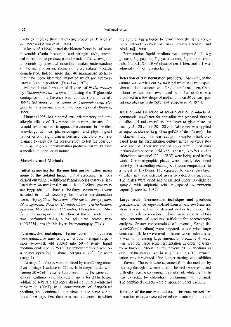
(Fig. 2). 1
H-NMR (DMSO):
4.10 (C-2), 3.68 (2H, C-3), 8.07 (C-5), 7.57 (C-6), 7.67
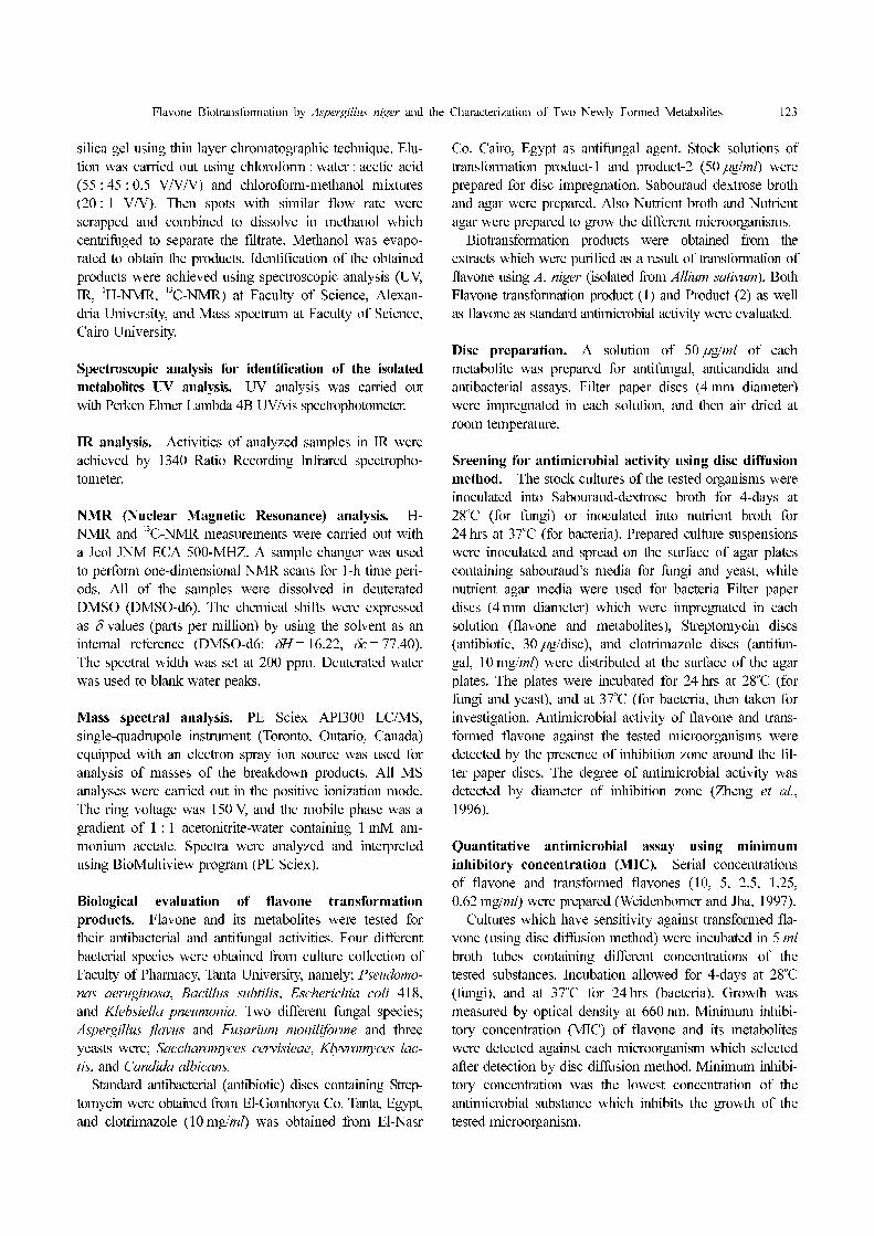
(C-7), 7.67 (C-8), 8.37 (OH proton) (Fig. 3). 13
C-NMR
(DMSO): 195.0 (C-4), 163.33 (C-9), 156.28 (C-4'),
132.40 (C-6'), 132.40 (C-2'), 132.10 (C-1'), 129.76(C-7),
129.73 (C-5), 129.70 (C-6), 126.92 (C-10), 119.12(C-3'),
119.12(C-5'), 67.97(O-CH3), 63.50 (C-3), 39.71 (C-2)
(Fig. 4). Mass spectrum (direct probe): m/e 272 (M+),
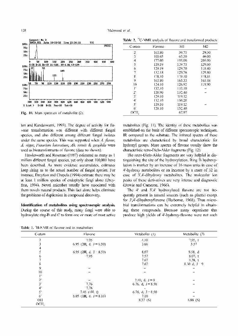
141, 116, 113, 102, 93, 66, 51 (Fig. 5).
Chemical characterization and identification of metab-
olite (2): Metabolite (2) was produced from transforma-
tion of flavone by A. niger isolated from A. sativum as
well as by other isolated fungi (data not shown). Its UV
spectrum λ max is 433, 327, 295 nm (Fig. 6). Infrared
(KBr) showed absorption at: 1709, 3639, 2851, 1552,
1536, 1517, 1413, 1333, 873, cm−1
(Fig. 7). 1
H-NMR
(DMSO), 2.95 (1
H, t, C-2), 3.57 (1
H, C-3), 8.58 (1
H, d, C-
5), 8.07 (1
H, t, C-6), 8.38 (1
H, t, C-7), 8.30 (1
H, d, C-8,
= 9 HZ), 8.88 (OH proton) (Fig. 8). 13
C-NMR: 205 (C-4),
163.88 (C-9), 129.80 (C-5), 129.80 (C-7), 118.90 (C-6),
Fig. 1. UV spectrum of metabolite (1).
Fig. 2. IR spectrum of metabolite (1).
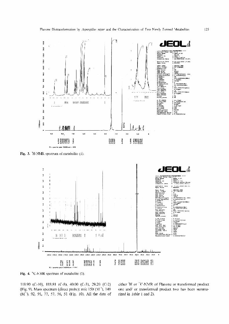
Flavone Biotransformation by Aspergillus niger and the Characterization of Two Newly Formed Metabolites 125
118.90 (C-10), 118.81 (C-8), 40.00 (C-3), 29.20 (C-2)
(Fig. 9). Mass spectrum (direct probe): m/e 150 (M+1
), 149
(M+1
), 92, 91, 77, 57, 56, 51 (Fig. 10). All the data of
either 1
H or 13
C-NMR of Flavone or transformed product
one and/ or transformed product two has been summa-
rized in Table 1 and 2).
Fig. 3.1
H-NMR spectrum of metabolite (1).
Fig. 4.13
C-NMR spectrum of metabolite (1).
126 Mahmoud et al.
Biological evaluation of the obtained metabolites.
Antimicrobial (antibacterial and antifungal) activity was
studied for starting flavone material and two of its
transformed and unidentified products (Fig. 11 and
Table 3). Four different bacterial species, two fungal
species, and three different species of yeast were used
for detection the antimicrobial activity. Flavone and its
metabolites have antibacterial activity against P. aerugi-
nosa, and antifungal activity against A. flavus, and
C. albicans. The transformed and unidentified products
have antimicrobial activity more than starting flavone.
Product 2 had the greatest antimicrobial activity when
compared with product 1 or flavone itself (Zheng et al.,
1996).
Antimicrobial activity of the obtained metabolites.
Microbes which inhibited by flavone and transformation
products were tested for minimum inhibitory concentra-
tion (Table 4). Transformation product (2) had the great-
est antimicrobial activity which inhibited the growth of P.
aeruginosa, A. flavus, and C. albicans at a concentration
of 250 µg/ml.
Antioxidant activity of flavone and transformed
flavone. Metabolite(1) and (2) were tested for their anti-
oxidant activities. Antioxidant activity of transformation
product (1) was detected as 64.58% (the greatest activ-
ity), while detected as 54.16% for product 2, and 37.5%
for flavone, and detected as 81.25% for ascorbic acid as
referred standard (Table 5).
Discussion
Flavones are a group of multi-ring hydroxyl-containing
compounds that are being studied widely for their nutri-
tional value and their use in preventive health care mea-
sures. These compounds are found in products as diverse
as Ginkgo Biloba, orange juice, and in garden herbs such
as dill, oregano and parsely.
Efficient utilization of flavonoids can lead to a positive
selection of the utilizers. Therefore it is not surprising that
flavonoid-degraders have been reported (Krishnamurthy et
al., 1970; Jeffrey et al., 1972; Sakai, 1977; Winter et al.,
1991; Rao and Cooper, 1994). Due to their rich carbon con-
tents, flavonoids have potential significance as nutritional
sources, whereas their complex structures allow them to have
diverse pharmacological and physiological effects (Middle-
Fig. 5. Mass spectrum of metabolite (1).
Fig. 7. IR spectrum of metabolite (2).
Fig. 6. UV spectrum of metabolite (2).

Flavone Biotransformation by Aspergillus niger and the Characterization of Two Newly Formed Metabolites 127
Fig. 8.1
H-NMR spectrum of metabolite (2).
Fig. 9.13
C-NMR spectrum of metabolite (2).
128 Mahmoud et al.
ton and Kandaswami, 1993). The degree of activity for fla-
vone transformation was different with different fungal
species, and also different among different fungal isolates
under the same species. This was supported when A. flavus,
A. niger, Fusarium lateratium, Alt. tenuis & geophila were
used as biotransformants of flavone (data no shown).
Hawksworth and Rossman (1987) estimated as many as 1
million different fungal species, yet only about 100,000 have
been described. As more evidence accumulates, estimates
keep rising as to the actual number of fungal species. For
instance, Dreyfuss and Chapela (1994) estimate there may be
at least 1 million species of endophytic fungi alone (Drey-
fuss, 1994). Novel microbes usually have associated with
them novels natural products. This fact alone helps eliminate
the problems of duplication in compound discovery.
Identification of metabolites using spectroscopic analysis.
During the course of this study, many fungi were able to
hydroxylate ring-B and C to form one or more of most active
metabolites (Fig. 11). The identity of these metabolites was
established on the basis of different spectroscopic techniques,
IR compared to the substrate. The infrared spectra of these
metabolites are characterized by broad characteristic for
hydroxyl groups. Mass spectra of flavone usually show the
characteristic retro-Diels-Alder fragments (Fig. 12).
The retro-Diels-Alder fragments are very helpful in dis-
tinguishing the site of the hydroxylation. Ring B hydroxy-
lation is marker by an increase of 16 mass units in case of
4'-hydroxy metabolites or an increase by a mass of 32 in
case of 3',4'-dihydroxy metabolites. The molecular ion
peaks of these derivatives are very intense and diagnostic
(Bowie and Cameron, 1966).
The 4' and 3',4' hydroxylated flavone are not fre-
quently present in natural sources (such as plants) except
for 3',4'-dihydroxyflavone (Harborne, 1968). Thus micro-
bial transformations can be extremely helpful in obtain-
ing these compounds. Because many organisms that
produce high yields of 4'-hydroxy-flavone were not such
Fig. 10. Mass spectrum of metabolite (2).
Table 1.1
H-NMR of flavone and its metabolites
C-atom Flavone Metabolite (1) Metabolite (2)
2 7.95 4.10 2.95, t3 6.95 (2H, d, J = 8.50) 3.68 3.574 − − −
5 6.95 (2H, d, J = 8.50) 8.07 8.58, d6 7.95 7.57 8.07, t7 − 7.67 8.38, t8 − 7.67 8.30 d, J = 99 − − −
10 − − −
1' − − −
2' − 7.10, d, J = 8 −
3' 7.76 6.76, d, J = 8.50 −
4' 7.76 - −
5' 7.45 (1H, t) 6.76, d, J = 8.50 −
6' 8.05 (1H, d, J = 8.16) 7.10 −
OH − 8.37 (S) 8.88 (S)OCH
3−
Table 2.13
C-NMR analysis of flavone and transformed products
C-atom Flavone M1 M2
2 163.80 039.71 029.303 105.05 063.50 040.004 177.00 195.00 205.005 129.19 129.73 129.006 129.19 129.70 118.407 132.18 129.76 129.808 118.10 129.10 118.819 163.80 163.33 163.8810 124.10 126.92 118.901' 132.10 132.10 −
2' 128.90 132.40 −
3' 129.10 119.12 −
4' 132.10 156.28 −
5' 129.10 119.12 −
6' 128.10 132.40 −
OCH3
− 067.97 −

Flavone Biotransformation by Aspergillus niger and the Characterization of Two Newly Formed Metabolites 129
good producers of 3',4'-dihydroxyflavone, a different
enzyme system seems to be involved in the conversion to
3',4'-dihydroxylated metabolite than that which causes the
initial formation of 4'-hydroxylation product.
The site of aromatic hydroxylation by fungi may be pre-
dicted, as is the case in mammalian systems, by the rules of
electronic substitution reactions. While the electron rich
rings are readily hydroxylated (Smith and Rosazza, 1975).
These findings may be used to explain the predominance of
ring B hydroxylation in flavonoids. Recent studies have
showed that mammals such as rats and guinea pigs metabo-
lize flavone to 4'-hydroflavone and 3',4'-dihydroyflavone
(Svardal et al., 1981). These results are essentially similar
to those obtained by other microbial systems.
Microbial cleavage of flavone by A. niger. Several
microorganisms were found to be capable of cleaving ring
C of flavone. The two ring-C cleavage products were
Fig. 11. Antimicrobial activities of flavone and the two
produced metabolites. Where A; Antimicrobia activity
of Pseudomonas aeruginosa. B; Antimicrobia activity
of Candida albicans. C; Antimicrobia activity of A.
flavus.
Table 3. Antimicrobial activity of flavone and some derivatives assayed by disc diffusion method (Inhibition zone diameter, mm)
(Weidenborner and Jha, 1997)
Microorganism Metabolite (1) Metabolite (2) FlavoneStandard antifungal
(Clotrimazole)
Standard antibacterial
(Streptomycin)
Pseudomonas aeruginosa 09.0 ± 2.0 009.6 ± 1.52 06.6 ± 0.57 − 08.0 ± 1.0
Escherichia coli ND ND ND − 15.30 ± 2.50
Bacillus subtilus ND ND ND − 015.0 ± 1.15
Klebsiella pneumonia ND ND ND − 07.0 ± 1.0
Aspergillus flavus 21.60 ± 2.08 26.60 ± 1.50 12.30 ± 1.15 0.026 ± 1.70 −
Fusarium moniliforme ND ND ND 08.30 ± 1.15 −
Saccharomyces cerviceae ND ND ND 10.60 ± 2.08 −
Kluvromyces lactis ND ND ND 10.60 ± 1.00 −
Candida albicans 14.0 ± 2.0 15.60 ± 2.00 6.60 ± 0.6 0.22 ± 2.0 −
Where; ND means Not Determined.
Table 4. Minimum inhibition concentration (MIC, µg/ml) of flavone and its transformed products by tube dilution method
Microorganism Metabolite (1) Metabolite (2) FlavoneStreptomycin sulphate
(standard antibacterial)
Clotrimazole
(standard antifungal)
Pseudomonas aeruginosa 300 250 500 − −
Aspergillus flavus 300 250 500 − 062
Candida albicans 400 250 500 − 125
*Reading is the average of three replica.
Table 5. Antioxidant assays of transformed flavone by (ABTS)
method
Compounds
ABTS
Abs(control) − Abs(test)/Abs(control) × 100
Absorbance of samples % inhibition
Control (ABTS) 0.48 0.0Ascorbic acid 0.09 81.25Metabilite (1) 0.17 64.58Metabolite (2) 0.22 54.16Flavone 0.30 37.50

130 Mahmoud et al.
identifies as metabolite (1) and metabolite (2).
Formation of Metabolite (1): Biotransformation of fla-
vone by A. niger isolated from Allium sativum gave
metabolite (1).
Infrared spectrum of metabolite (1) showed absorption
band at 3665 Cm−1
indicating the presence of hydroxyl
group (s) (Fig. 2). The identification of this product was
based on chemical ionization mass spectrometry (Fig. 5),
which indicated that M+
at 272 (53.4%), C4H
3 base peak
51 (100%), M+
-(C=O), 244 (13.1%).
13
C-NMR (off resonance and completely decoupled)
showed methyl (67-97) and carbonyl (195) beside for
hydroxyl groups (163.33) (Fig. 9). 1
H-NMR showed the
presence of methyl group at 4.86 ppm (Fig. 8). The pres-
ence of aromatic hydroxyl group at (8.37 ppm) was
detected. 1
H-NMR showed aliphatic OH at 3.28 ppm. This
metabolite is produced by most microbial isolates at the
early stage of fermentation, which might suggest an inter-
mediate towards formation of the metabolite (2).
Formation of Metabolite (2): A. niger isolated from
Allium Sativum was found to transform flavone to metab-
olite (2) with percentage increasing rate of transformation
reaching up to more than 75%.
Several microbes such as Rhizobia, Agrobacterium.
Pseudomonas, Bacillus, and Rhodococcus spp. that exist
in the rhizosphere are known to participate in the break-
down or degradation of flavonoids (Barz, 1970; Barz et
al., 1970). Although some mechanisms have been pro-
posed, the pathways were not elucidated. A survey of fla-
vonoid-degrading rhizobial strains revealed that flavonoids
were generally cleaved via c-ring fission (Rao et al.,
1991; Rao and Cooper, 1994). An early study on fla-
vonoid degradation by a soil Pseudomonads indicated the
presence of oxygenase based on the oxidation products
and proposed that the degradation proceeds via protocate-
chuate production (Shultz et al., 1974). The anaerobic
degradation of flavonoids by the intestinal microflora, in
comparison to the aerobic pathways, has been well docu-
mented, and several reports describe the reduction and
dehydroxylation reactions leading to phloroglucinol for-
mation (Gorny and Schink, 1994; Schneider et al., 1999;
Schneider and Blaut, 2000).
Gajendiran and Mahadevan (1988) demonstrated that
Rhizobium could degrade the flavan-3-ol catechin, a com-
ponent of condensed tannin in tannery effluent, but there
are no reports of Rhizobia cleaving the flavone-type ring
structure. Rao et al. (1991) reported biotransformation of
the pentahydroxy flavone quercetin by Rhizobium loti and
Bradyrhizobium strains. Structural characteristics suggest
that protocatechuic acid originated from the B-ring of
quercetin, while phloroglucinol was derived from the A-
ring. Some intestinal Clostridia and a Eubacterium spe-
cies can also degrade quercetin via c-ring cleavage
(Krumholtz and Bryant, 1986; Winter et al., 1989), but
the end products are not the same, suggesting that both
the pattern of ring fission and the ensuing metabolic path-
ways are different from those in rhizobia.
Flavone degradation has also been reported in Pseudomo-
nas species (Schultz et al., 1974) but with fission initiat-
ing in the A-ring via hydroxylation at C-8. Other bacterial
transformations of quercetin include sulfation (Koizumi et
al., 1990) and glucosidation (Rao and Weisner, 1981).
Metabolite (2) infrared spectrum showed a broad band
at 3639 cm−1
indicating a hydroxylated metabolite and
another band at 873 cm−1
for ortho disubstituted benzene
(Fig. 12).
High resolution electron impact mass spectrometry
(Fig. 11) showed a molecular ion at (149, 56.9%) (M+
)
Fig. 12. Microbial hydroxylation of flavone.
Fig. 13. Retro-Diels-Alder fragmentation mechanism of flavone.
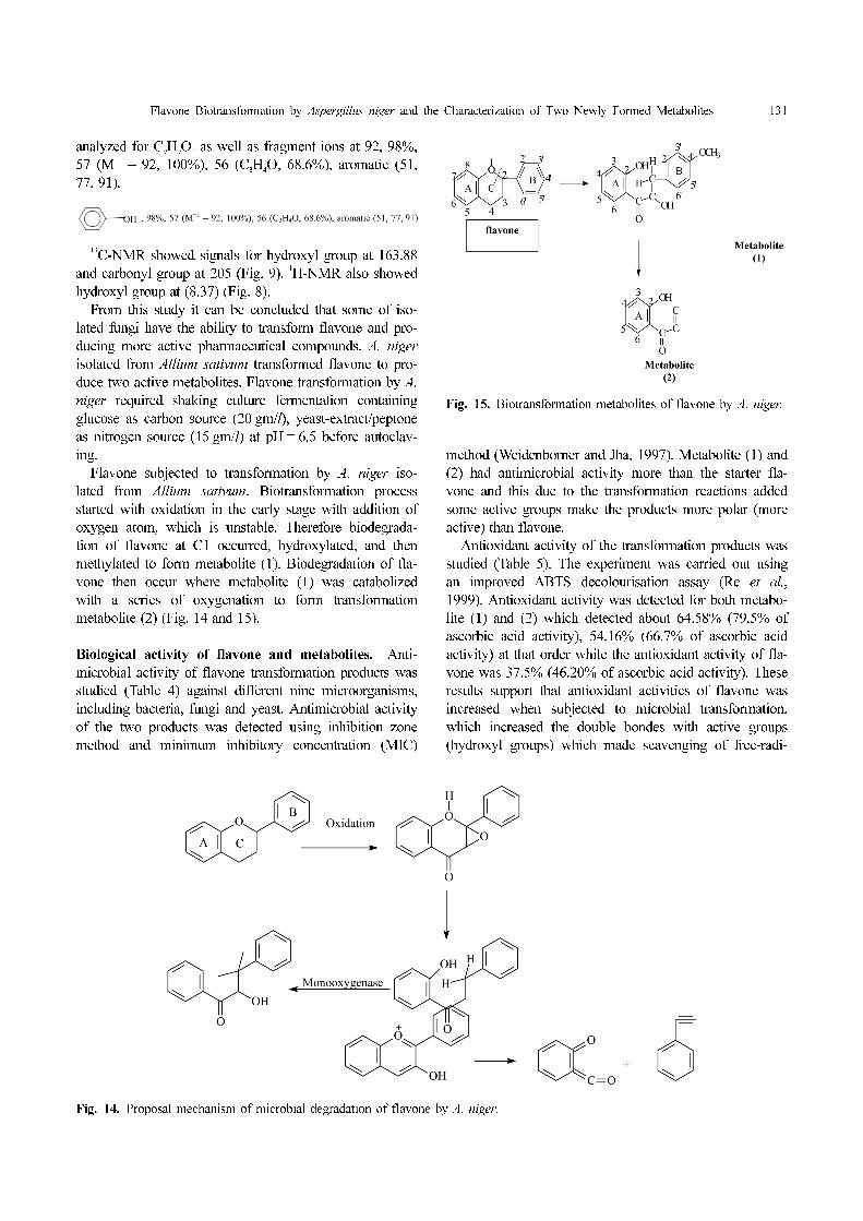
Flavone Biotransformation by Aspergillus niger and the Characterization of Two Newly Formed Metabolites 131
analyzed for C9H
9O
2 as well as fragment ions at 92, 98%,
57 (M+1
– 92, 100%), 56 (C3H
4O, 68.6%), aromatic (51,
77, 91).
13
C-NMR showed signals for hydroxyl group at 163.88
and carbonyl group at 205 (Fig. 9). 1
H-NMR also showed
hydroxyl group at (8.37) (Fig. 8).
From this study it can be concluded that some of iso-
lated fungi have the ability to transform flavone and pro-
ducing more active pharmaceutical compounds. A. niger
isolated from Allium sativum transformed flavone to pro-
duce two active metabolites. Flavone transformation by A.
niger required shaking culture fermentation containing
glucose as carbon source (20 gm/l), yeast-extract/peptone
as nitrogen source (15 gm/l) at pH = 6.5 before autoclav-
ing.
Flavone subjected to transformation by A. niger iso-
lated from Allium sativum. Biotransformation process
started with oxidation in the early stage with addition of
oxygen atom, which is unstable. Therefore biodegrada-
tion of flavone at C1 occurred, hydroxylated, and then
methylated to form metabolite (1). Biodegradation of fla-
vone then occur where metabolite (1) was catabolized
with a series of oxygenation to form transformation
metabolite (2) (Fig. 14 and 15).
Biological activity of flavone and metabolites. Anti-
microbial activity of flavone transformation products was
studied (Table 4) against different nine microorganisms,
including bacteria, fungi and yeast. Antimicrobial activity
of the two products was detected using inhibition zone
method and minimum inhibitory concentration (MIC)
method (Weidenborner and Jha, 1997). Metabolite (1) and
(2) had antimicrobial activity more than the starter fla-
vone and this due to the transformation reactions added
some active groups make the products more polar (more
active) than flavone.
Antioxidant activity of the transformation products was
studied (Table 5). The experiment was carried out using
an improved ABTS decolourisation assay (Re et al.,
1999). Antioxidant activity was detected for both metabo-
lite (1) and (2) which detected about 64.58% (79.5% of
ascorbic acid activity), 54.16% (66.7% of ascorbic acid
activity) at that order while the antioxidant activity of fla-
vone was 37.5% (46.20% of ascorbic acid activity). These
results support that antioxidant activities of flavone was
increased when subjected to microbial transformation,
which increased the double bondes with active groups
(hydroxyl groups) which made scavenging of free-radi-
Fig. 14. Proposal mechanism of microbial degradation of flavone by A. niger.
Fig. 15. Biotransformation metabolites of flavone by A. niger.
132 Mahmoud et al.
cals. The absence of this double bond significantly
reduces the antioxidant activity of the flavones. Salah et
al. (1995) illustrate that, epicatechin, which also lacks this
double bond, has an antioxidant activity which is only
53% of quercetin’s. Epicatechin can increase its antioxi-
dant activity with the addition of another hydroxyl group
on the B ring (forming epigallocatechin), and further with
the addition of gallic acid (with its three hydroxyl groups,
forming epigallocatechin gallate) on the C ring, to the
point where it is equivalent to quercetin.
References
Arora, A., Nair, M. G. and Strasburg, G. M. 1998. Antioxidant
activities of isoflavones and their biological metabolites in a
liposomal system. Arch. Biochem. Biophys. 356:133-141.
Kim, D. H., Jung, E. A., Sohng, I. S., Han, J. A., Kim, T. H. and
Han, M. J. 1998. Intestinal bacterial metabolism of flavonoids
and its some biological activities. Arch. Pharm. Res. 21:17-23.
Barz, W. 1970. Isolation of rhizosphere bacterium capable of
degrading flavonoids. Phytochemistry 9:1745-1949.
Barz, W., Adamek, C. and Berlin, J. 1970. Ion of formation and
daidzein in Cicer arietinum and Phaseolus aureus. Phytochem-
istry 9:1735-1744.
Bowie, J. H. and Cameron, D. W. 1966. Electron impact studies.
II Mass spectra of quercetagetin derivatives. Australian J.
Chem. 19:1627-1635.
Briviba, K., Sepulveda-Boza, S., Zilliken, F. and Sies, H. 1997.
Isoflavonoids as inhibitors of lipid peroxidation and quenchers
of singlet oxygen. In: Flavonoids in health and disease, pp.
295-302. Eds. C. A. Rice-Evans and L. Packer. Marcel Dek-
ker, Inc., New York, N.Y.
Cano, A., Hernandez-Ruiz, J., Garcia-Canovas, F., Acosta, M. and
Arnao, M. B. 1998. An end-point method for estimation of the
total antioxidant activity in plant material. Phytochem. Anal.
9:196-202.
Cheng, K. J., Jones, G. A., Simpson, F. J. and Bryant, M. P. 1969.
Isolation and identification of rumen bacteria capable of anaer-
obic rutin degradation. Can. J. Microbiol. 15:1365-1371.
Ciegler Alex, Lloyd, A., Lindernfelser and George Nelson, E. N.
1971. Microbial transformation of flavonoids. Agr. Res. Ser-
vice. Peoria, Illinois, Appl. Microbiol. 22:974-979.
Cooper, J. E., Rao, J. R., Evertaert, E., Cooman, L-de., De-
cooman-L. and Tikhonovich, I. A. 1995. Metabolism of fla-
vonoids by rhizobia. Provorov-N.A., Romanov-V.I. and New-
ton-W.E., Proceedings of the 10th International Congress On
Nitrogen Fixation, St. Petersburg, Russia, 287-292.
Das, N. P., Scott, K. N. and Duncan, J. H. 1973. Identification of
flavanone metabolites in the rat urine by combined GC-MS.
Biochem. J. 136:903-909.
Gajendiran, N. and Mahadevan, A. 1988. Utilization of catechin
by Rhizobium sp. Plant Soil 108:263-266.
Gorny, N. and Schink, B. 1994. Anaerobic degradation of cate-
chol by Desulfobacterium sp. strain Cat2 proceeds via carboxy-
lation to protocatechuate. Appl. Environ. Microbiol. 60:3396-
3400.
Greene, L. S. 1995. Asthma and oxidant stress: nutritional, envi-
ronmental, and genetic risk factors. J. Am. Coll. Nutr. 14:317-
324.
Harborne, J. B. 1968. Comparative biochemistry of flavonoids-
VII. Correlations between flavonoid pigmentation and system-
atics in the family Primulaceae. Phytochem. 7:1215-1230.
Horowitz, R. M. 1957. Detection of flavanones by reduction with
sodium borohydride. J. Org. Chem. 22:1733-1734. Weiden-
borner, M. and Jha, H. C. 1997. Antifungal spectrum of fla-
vone and flavanone tested against 34 different fungi.
Mycological-Research. 101:733-736.
Ibrahim A. R. S. 1999. Sulfation of naringenin by cunning-
hamella elegans. Egypt phytochemistry 53:209-212.
Hunter, T. 1995. Protein kinases and phosphatases: The Yin and
Yang of protein phosphorylation and signaling. Cell 80:225-
236.
Ibrahim, A. R. S. and Abul-Haji, Y. J. 1989. Aromatic hydroxyla-
tion and sulfation of 5'-hydroxyflavone by Streptomyces fulvis-
simus. Appl. Environ. Microbiol. 55:3140-3142.
Ibrahim, A. R. S. and Abul-Hajj, Y. J. 1990. Microbiological
transformation of (1) flavonone and (±) isoflavonone. J. Nat.
Prod. 53:644-656.
Ibrahim, A. R. S., Galal, A. M., Mossa, J. S. and El-Feraly, F. S.
1997. Glucose-conjugation of the flavones of Psidia arabica
by cunninghamell elegans. Phytochemsity 46:1193- 1195.
Koizumi, M., Shimuzi, M. and Kobashi, K. 1990. Enzymic sulfa-
tion of quercetin by arylsulfotransferase from a human intesti-
nal bacterium. Chem. Pharm. Bull. Tokyo 38:794-796.
Krishnamurthy, H. G., Cheng, K. J., Jones, G. A., Simpson, F. J.
and Watkin, J. E. 1970. Identification of products produced by
the anaerobic degradation of rutin and related flvonoids by
Butyrivibrio spp. C. Can. J. Microbiol. 16:759-767.
Rao, K. V. and Weisner, N. T. 1981. Microbial transformation of
quercetin by Bacillus cereus. Appl. Environ. Microbiol. 42:450-
452.
Rao, R. J. and Cooper, J. E. 1994. Rhizobia catabolize nod gene-
inducing flavonoids via C-ring fission mechanisms. J. Bacte-
riol. 176:5409-5413.
Rao, R. J., Sharma, N. D., Hamilton, J. T. G., Boyd, D. R. and
Cooper, J. E. 1991. Biotransformation of the pentahydroxy fla-
vone quercetin by Rhizobium loti and Bradyrhizobium strains
(Lotus). Appl. Environ. Microbiol. 57:1563-1565.
Krumholz, L. R. and Bryant, M. P. 1986. Eubacterium oxi-
doreducens sp. nov. requiring H2 or formate to degrade gallate,
pyrogallol, phloroglucinol and quercetin. Arch. Microbiol. 144:
8-14.
Re, R., Pellegrini, N., Proteggente, A., Pannala, A., Yang, M.
and Rice-Evans, C. 1999. Antioxidant activity applying an
improved ABTS radical cation decolorization assay. Free
Radic. Biol. 26:1231-1237.
Rice-Evans, C. A. and Miller, N. J. 1994. Total antioxidant status
in plasma and body fluids. Methods Enzymol. 234:279-293.
Salah, N., Miller, N. and Paganga, G. 1995. Polyphenolic fla-
vanols as scavengers of aqueous phase radicals and as chain-
breaking antioxidants. Arch. Biochem. Biophys 322:339-346.
Schneider, H. and Blaut, M. 2000. Anaerobic degradation of fla-
vonoids by Eubacterium ramulus. Arch. Microbiol. 173:71-75.
Schneider, H., Schwiertz, A., Collins, M. D. and Blaut, M. 1999.
Anaerobic transformation of quercetin-3-glucosidase by bacte-
ria from human intestinal tract. Arch. Microbiol. 171:81-91.
Seeger, M., Gonzalez, M., Camara, B., Munoz, L., Ponce, E.,
Mejias, L., Mascayano, C., Vasquez, Y. and Sepulveda-Boza,
S. 2003. Biotransformation of natural and synthetic isofla-
vonoids by two recombinant microbial enzymes. Faculty of
Flavone Biotransformation by Aspergillus niger and the Characterization of Two Newly Formed Metabolites 133
Medical Science. University of Santiago, Santiago, Chile. App.
and Environ. Microbiol. 69:5045-5050.
Shultz, E., Engle, F. E. and Wood, J. M. 1974. New oxygenases
in the degradation of flavones and flavonones by Pseudomo-
nas putida. Biochemistry 13:1768-1776.
Smith, L. L. 1973. Microbiological reactions with steroids. Spec.
Period. Rep. Terpenoids Steroids 4:394-530.
Smith, R. V. and Rosazza, J. P. 1975. Microbial models of mam-
malian metabolism. J. Pharm. Sci. 64:1737-1759.
Svardal, A., Buset, H. and Scheline, R. R. 1981. Disposition of
(2-14C) flavone in the rat. Acta Pharmaceutica Suecica. 18:55-
62.
Weidenborner, M. and Jha, H. C. 1997. Antifungal spectrum of
flavone and flavanone tested against 34 different fungi. Myco-
logical-Research 101:733-736.
Winter, J., Moore, L. H., Dowell, V. R. and Bokkenheuser, V. D.
1989. C-ring cleavage of flavonoids by intestinal bacteria.
Appl. Environ. Microbiol. 55:1203-1208.
Zheng, W. F., Tan, R. X., Yang, L. and Liu, Z. L. 1996. Two fla-
vones from Artemsia giraldii and their antimicrobial activity.
Planta. Medica. 62:160-162.