FlavinylationandAssemblyofSuccinateDehydrogenaseAre ......
11
Flavinylation and Assembly of Succinate Dehydrogenase Are Dependent on the C-terminal Tail of the Flavoprotein Subunit * Received for publication, July 27, 2012, and in revised form, October 4, 2012 Published, JBC Papers in Press, October 7, 2012, DOI 10.1074/jbc.M112.405704 Hyung J. Kim 1,2 , Mi-Young Jeong 1 , Un Na, and Dennis R. Winge 3 From the Departments of Medicine and Biochemistry, University of Utah Health Sciences Center, Salt Lake City, Utah 84132 Background: Succinate dehydrogenase (SDH) requires a covalent addition of FAD for catalytic function. Results: Mutational analyses of Sdh1 implicate C-terminal region Arg residues involvement in covalent flavinylation and SDH assembly. Conclusion: SDH assembly is dependent on FAD binding to Sdh1 but not covalent binding. Significance: These results document the basis for the SDH deficiency and pathology seen with mutations in human Sdh1. The enzymatic function of succinate dehydrogenase (SDH) is dependent on covalent attachment of FAD on the 70-kDa fla- voprotein subunit Sdh1. We show presently that flavinylation of the Sdh1 subunit of succinate dehydrogenase is dependent on a set of two spatially close C-terminal arginine residues that are distant from the FAD binding site. Mutation of Arg 582 in yeast Sdh1 precludes flavinylation as well as assembly of the tetra- meric enzyme complex. Mutation of Arg 638 compromises SDH function only when present in combination with a Cys 630 sub- stitution. Mutations of either Arg 582 or Arg 638 /Cys 630 do not markedly destabilize the Sdh1 polypeptide; however, the steady- state level of Sdh5 is markedly attenuated in the Sdh1 mutant cells. With each mutant Sdh1, second-site Sdh1 suppressor mutations were recovered in Sdh1 permitting flavinylation, sta- bilization of Sdh5 and SDH tetramer assembly. SDH assembly appears to require FAD binding but not necessarily covalent FAD attachment. The Arg residues may be important not only for Sdh5 association but also in the recruitment and/or guidance of FAD and or succinate to the substrate site for the flavinylation reaction. The impaired assembly of SDH with the C-terminal Sdh1 mutants suggests that FAD binding is important to stabi- lize the Sdh1 conformation enabling association with Sdh2 and the membrane anchor subunits. Succinate dehydrogenase (SDH), 4 also known as succinate: quinone oxidoreductase, is a citric acid cycle enzyme that links directly to the aerobic respiratory chain. The enzyme catalyzes the FAD-dependent oxidation of succinate to fumarate coupled with the reduction of ubiquinone to ubiquinol. SDH is a hetero- tetrameric integral membrane protein complex. The eukary- otic enzyme is embedded within the mitochondrial inner mem- brane by a hydrophobic module associated with the catalytic module protruding into the mitochondrial matrix (1). The hydrophilic catalytic module consists of Sdh1 and Sdh2 sub- units and the electron transfer cofactors. Succinate oxidation occurs in the FAD-containing Sdh1 with the abstracted elec- trons from the reaction shuttled via three iron-sulfur centers in Sdh2 to the ubiquinone reduction site (Q P -proximal) at the interface of Sdh2 and the membrane anchor (1– 4). The Sdh3/ Sdh4 membrane domain contains a bound heme b moiety at the subunit interface of Sdh3 and Sdh4 with each providing one of the two axial His ligands, although the role of the heme in eukaryotic SDH is unresolved (5). The FAD of Sdh1 is covalently attached at an active site His residue (2). This covalent bond increases the FAD redox poten- tial by 60 mV to permit succinate oxidation (6). SDH is the major mitochondrial protein containing a covalent bound fla- vin (7). Sdh1 containing a H90S substitution is enzymatically inactive in succinate oxidation but assembles into a tetrameric complex that exhibits fumarate reductase activity (8). Fumarate reductase activity in SDH does not require covalent flavinyla- tion. SDH is related to the bacterial fumarate reductase and both enzymes can catalyze succinate oxidation and fumarate reduction with different efficiencies (9). Flavinylation of Sdh1 was found to occur after import into the matrix and to be influ- enced by the presence of the iron/sulfur cluster subunit Sdh2 but largely independent of the membrane anchor (10). The presence of citric acid intermediates stimulated the flavinyla- tion process (10). The covalent addition of FAD was proposed to be autocata- lytic (7), but recently, a dedicated assembly factor Sdh5 was identified that is required for covalent flavinylation (11). The role of Sdh5 in Sdh1 flavinylation was discovered by the inter- action of the two proteins and the demonstration that sdh5 yeasts were devoid of SDH activity and lacked flavinylated Sdh1. The direct role of Sdh5 in Sdh1 flavinylation was shown by the enhanced covalent addition of FAD to recombinant Sdh1 expressed in bacteria in the presence of Sdh5. However, the * This work was supported by Grant ES03817 from the NIEHS, National Insti- tutes of Health (to D. R. W.), funding from the Huntsman Cancer Institute (P30 CA042014), and support from the National Institutes of Health (R24DK092784-01). 1 Both authors contributed equally to this work. 2 Supported by Training Grant T32 DK007115 from the National Institutes of Health. 3 To whom correspondence should be addressed. Tel.: 801-585-5103; Fax: 801-585-3432; E-mail: [email protected]. 4 The abbreviations used are: SDH, succinate dehydrogenase; BN-PAGE, blue native PAGE; FAD, flavin adenine dinucleotide; DDm, n-dodecyl- -D-maltoside. THE JOURNAL OF BIOLOGICAL CHEMISTRY VOL. 287, NO. 48, pp. 40670 –40679, November 23, 2012 © 2012 by The American Society for Biochemistry and Molecular Biology, Inc. Published in the U.S.A. 40670 JOURNAL OF BIOLOGICAL CHEMISTRY VOLUME 287 • NUMBER 48 • NOVEMBER 23, 2012 by guest on April 7, 2019 http://www.jbc.org/ Downloaded from
Transcript of FlavinylationandAssemblyofSuccinateDehydrogenaseAre ......

Flavinylation and Assembly of Succinate Dehydrogenase AreDependent on the C-terminal Tail of the FlavoproteinSubunit*
Received for publication, July 27, 2012, and in revised form, October 4, 2012 Published, JBC Papers in Press, October 7, 2012, DOI 10.1074/jbc.M112.405704
Hyung J. Kim1,2, Mi-Young Jeong1, Un Na, and Dennis R. Winge3
From the Departments of Medicine and Biochemistry, University of Utah Health Sciences Center, Salt Lake City, Utah 84132
Background: Succinate dehydrogenase (SDH) requires a covalent addition of FAD for catalytic function.Results:Mutational analyses of Sdh1 implicate C-terminal region Arg residues involvement in covalent flavinylation and SDHassembly.Conclusion: SDH assembly is dependent on FAD binding to Sdh1 but not covalent binding.Significance: These results document the basis for the SDH deficiency and pathology seen with mutations in human Sdh1.
The enzymatic function of succinate dehydrogenase (SDH) isdependent on covalent attachment of FAD on the �70-kDa fla-voprotein subunit Sdh1.We showpresently that flavinylation ofthe Sdh1 subunit of succinate dehydrogenase is dependent on aset of two spatially close C-terminal arginine residues that aredistant from the FAD binding site. Mutation of Arg582 in yeastSdh1 precludes flavinylation as well as assembly of the tetra-meric enzyme complex. Mutation of Arg638 compromises SDHfunction only when present in combination with a Cys630 sub-stitution. Mutations of either Arg582 or Arg638/Cys630 do notmarkedly destabilize the Sdh1polypeptide; however, the steady-state level of Sdh5 is markedly attenuated in the Sdh1 mutantcells. With each mutant Sdh1, second-site Sdh1 suppressormutations were recovered in Sdh1 permitting flavinylation, sta-bilization of Sdh5 and SDH tetramer assembly. SDH assemblyappears to require FAD binding but not necessarily covalentFAD attachment. The Arg residues may be important not onlyfor Sdh5 associationbut also in the recruitment and/or guidanceof FADandor succinate to the substrate site for the flavinylationreaction. The impaired assembly of SDH with the C-terminalSdh1 mutants suggests that FAD binding is important to stabi-lize the Sdh1 conformation enabling association with Sdh2 andthe membrane anchor subunits.
Succinate dehydrogenase (SDH),4 also known as succinate:quinone oxidoreductase, is a citric acid cycle enzyme that linksdirectly to the aerobic respiratory chain. The enzyme catalyzesthe FAD-dependent oxidation of succinate to fumarate coupledwith the reduction of ubiquinone to ubiquinol. SDH is a hetero-
tetrameric integral membrane protein complex. The eukary-otic enzyme is embeddedwithin themitochondrial innermem-brane by a hydrophobic module associated with the catalyticmodule protruding into the mitochondrial matrix (1). Thehydrophilic catalytic module consists of Sdh1 and Sdh2 sub-units and the electron transfer cofactors. Succinate oxidationoccurs in the FAD-containing Sdh1 with the abstracted elec-trons from the reaction shuttled via three iron-sulfur centers inSdh2 to the ubiquinone reduction site (QP -proximal) at theinterface of Sdh2 and the membrane anchor (1–4). The Sdh3/Sdh4membrane domain contains a boundheme bmoiety at thesubunit interface of Sdh3 and Sdh4 with each providing one ofthe two axial His ligands, although the role of the heme ineukaryotic SDH is unresolved (5).The FAD of Sdh1 is covalently attached at an active site His
residue (2). This covalent bond increases the FAD redox poten-tial by �60 mV to permit succinate oxidation (6). SDH is themajor mitochondrial protein containing a covalent bound fla-vin (7). Sdh1 containing a H90S substitution is enzymaticallyinactive in succinate oxidation but assembles into a tetramericcomplex that exhibits fumarate reductase activity (8). Fumaratereductase activity in SDH does not require covalent flavinyla-tion. SDH is related to the bacterial fumarate reductase andboth enzymes can catalyze succinate oxidation and fumaratereduction with different efficiencies (9). Flavinylation of Sdh1was found to occur after import into thematrix and to be influ-enced by the presence of the iron/sulfur cluster subunit Sdh2but largely independent of the membrane anchor (10). Thepresence of citric acid intermediates stimulated the flavinyla-tion process (10).The covalent addition of FAD was proposed to be autocata-
lytic (7), but recently, a dedicated assembly factor Sdh5 wasidentified that is required for covalent flavinylation (11). Therole of Sdh5 in Sdh1 flavinylation was discovered by the inter-action of the two proteins and the demonstration that sdh5�yeasts were devoid of SDH activity and lacked flavinylatedSdh1. The direct role of Sdh5 in Sdh1 flavinylation was shownby the enhanced covalent addition of FAD to recombinant Sdh1expressed in bacteria in the presence of Sdh5. However, the
* This work was supported by Grant ES03817 from the NIEHS, National Insti-tutes of Health (to D. R. W.), funding from the Huntsman Cancer Institute(P30 CA042014), and support from the National Institutes of Health(R24DK092784-01).
1 Both authors contributed equally to this work.2 Supported by Training Grant T32 DK007115 from the National Institutes of
Health.3 To whom correspondence should be addressed. Tel.: 801-585-5103; Fax:
801-585-3432; E-mail: [email protected] The abbreviations used are: SDH, succinate dehydrogenase; BN-PAGE,
blue native PAGE; FAD, flavin adenine dinucleotide; DDm, n-dodecyl-�-D-maltoside.
THE JOURNAL OF BIOLOGICAL CHEMISTRY VOL. 287, NO. 48, pp. 40670 –40679, November 23, 2012© 2012 by The American Society for Biochemistry and Molecular Biology, Inc. Published in the U.S.A.
40670 JOURNAL OF BIOLOGICAL CHEMISTRY VOLUME 287 • NUMBER 48 • NOVEMBER 23, 2012
by guest on April 7, 2019
http://ww
w.jbc.org/
Dow
nloaded from

mechanism by which the 22-kDa Sdh5 protein mediates flavi-nylation of Sdh1 remains unresolved.The human Sdh5 ortholog, SDHAF2, was shown to have a
corresponding role in covalent flavinylation in human cells(11). Mutations in SDHAF2 are associated with a neuroendro-crine tumor designated as paraganglioma. Mutations in theSDH structural genes are also detected in a range of tumors,including paraganglioma, pheochromocytomas, gastrointesti-nal stromal tumors, and renal cell carcinoma (12). Recently, thebacterial ortholog of Sdh5 was identified in the bacterium Ser-ratia. The factor designated SdhE was shown to interact withthe flavoprotein (SdhA) and independently associate with FAD(13). This is the first suggestion that the assembly factor maymediate Sdh1 flavinylation by presenting bound FAD.Whereassdh5� cells exhibited a specific defect in SDH, the bacterialsdhE mutant showed a more pleiotropic defect, suggestingSdhE may flavinylate other flavoproteins (13).SDH is one of many flavoproteins with a covalently bound
cofactor. SDH has an 8�-N3-histidyl-FAD linkage, but otherflavoproteins use cysteinyl-FAD or tyrosyl-FAD linkages (14).Two large families of covalent flavoproteins are the glucoseoxidase/methanol oxidase family and the vanillyl-alcohol oxi-dase family. Within the vanillyl-alcohol oxidase family, FADlinkages to histidine, cysteine, and tyrosine are known,although no ancillary enzyme is known to catalyze the covalentaddition. Covalent flavinylation with FAD is known to proceednonenzymatically, albeit slowly.In the present study, we identify essential arginines in the
C-terminal tail in yeast Sdh1 that are required for flavinylationand SDH assembly. The impaired assembly of the tetramericenzyme with these Sdh1 mutants correlates with the lack ofFAD binding.
MATERIALS AND METHODS
Yeast Strains and Vectors—All Saccharomyces cerevisiaestrains used in this study were derivatives of Trp303 (Mataura3-1 ade2-1 trp1-1 his3-11,15 leu2-3 112). Deletion strainssdh1�, sdh2�, sdh3�, and sdh4�, in addition to plasmidpRS414 Sdh2-His6Myc2, were kindly provided byDr. JaredRut-ter at the University of Utah. Additional deletion strains (flx�,sdh5�, sdh3�sdh4� double) were constructed by homologousrecombination using either the KanMX4 or the HIS3MX6 dis-ruption cassettes (15). The C-terminal mutants of SDH1 alongwith SDH1 WT under the control of its own promoter andCYC1 terminator were expressed in sdh1� strain or subclonedinto integrating pRS405 vectors for chromosomal expression.The integrating constructs were further digested with AflII andintegrated at the leu2-3,112 locus of sdh1� to be able to expresswild type and mutants SDH1 chromosomally. All integratedstrains were confirmed by PCR analysis of the locus. The C-ter-minal point mutations were introduced by QuikChangemutagenesis PCR system (Agilent Technology). All mutationswere confirmed by DNA sequencing. Yeast strains were trans-formed using lithium acetate. Strains were grown in syntheticcomplete medium lacking the amino acid(s) to maintain plas-mid selection with either 2% galactose or 2% glycerol/lactate asthe carbon source.
For carbon swap cultures overnight, 50-ml glucose-growncultures were used to inoculate 1 liter ofmedium containing 2%galactose as the carbon source. Cells were grown to an A600 nm� 0.4 to 0.5 and harvested in a sterile manner. The cell pelletwas resuspended in fresh medium containing 2% glycerol/lac-tate as the carbon source and grown for �10 h (�3–4 dou-blings) before harvesting.Mitochondria Isolation—Intact mitochondria were isolated
using previously described methods of Glick and Pon (16) andDiekert et al. (17). For HPLC experiments, isolated mitochon-dria were further purified using ultracentrifugation through aHistodenz (Sigma Aldrich) step gradient (14 and 22%). Totalmitochondrial protein was quantified using either the Bradford(18) or the bicinchoninic acid assays (19).Immunoblotting and Blue-native PAGE—Steady-state levels
of mitochondrial proteins were analyzed using the NuPAGEBis-Tris gel system (Invitrogen) usingMES as the buffer system.Proteins were subsequently transferred to nitrocellulose mem-brane and probed using the indicated primary antibodies andvisualized using enhanced chemiluminescence (ECL) reagentswith horseradish peroxidase-conjugated secondary antibodies.Primary antibodies were obtained from the following: anti-Sdh1, Sdh2, Sdh3, and Sdh5 were generated in this study (21stCentury Biochemicals). Anti-HA, anti-Myc, and anti-porinwere purchased from Rockland, Roche Applied Science, andMolecular Probes, respectively. Anti-F1 ATP synthase was agenerous gift from Alex Tzagoloff.Analysis of yeastmitochondrial nativemembrane complexes
was performed using the native PAGE gel system (Invitrogen)that is based on the blue-native polyacrylamide gel electropho-resis (BN-PAGE) technique developed by Schägger and vonJagow (20). Solubilized mitochondria (1% digitonin for 20–40�g of mitochondrial protein) were separated on either 3–12%or 4–16% Bis-Tris native gels, transferred to PVDFmembrane,and analyzed by immunoblotting.Hemin-agarose Pulldown Assays and Immunoprecipitation—
Purified mitochondria (0.75 to 1 mg of total protein) were sol-ubilized using either 0.1% DDM or 1% digitonin in PBS bufferand clarified at 20,000 � g for 5 min. For hemin-agarose pull-down assay, the clarified lysates were incubatedwith 20 to 40�lof hemin-agarose beads (Sigma), which had been prereducedusing DTT. Binding was performed for 2 h or overnight at 4 °C.Beads were subsequently washed (15min � 4) with PBS buffer,0.1% DDM or 1% digitonin, � DTT, and eluted with 2� SDS-PAGE loading buffer. For immunoprecipitation, the clarifiedlysates were incubated with anti-HA or anti-Myc beads for 2 hor overnight at 4 °C, washed four times with PBS buffer � 0.1%DDM or 1% digitonin, and eluted with 2� SDS-PAGE loadingbuffer. The clarified lysate, final wash, and eluate fraction wereanalyzed by SDS-PAGE and immunoblotting. Bands fromCoo-massie-stained SDS-PAGE gel from hemin-agarose pulldownassay were also excised for identification using MS analysis ofthe tryptic digests.SDH Activity Assay—SDH activities in isolated yeast mito-
chondria were measured spectrophotometrically at 22 °C fol-lowing the succinate-dependent, phenazine methosulfate-me-diated reduction of either 2,6-dichlorophenolindophenol or
SDH Flavinylation
NOVEMBER 23, 2012 • VOLUME 287 • NUMBER 48 JOURNAL OF BIOLOGICAL CHEMISTRY 40671
by guest on April 7, 2019
http://ww
w.jbc.org/
Dow
nloaded from

cytochrome c as the terminal electron acceptor described forintact mitochondria (21).Analysis of Sdh1-bound FAD and Total Mitochondrial FAD
Levels—Levels of covalently attached FAD to Sdh1 were ana-lyzed as described previously (11, 22). Mitochondrial proteinswere resolved on SDS-PAGE, and the gel was placed in a 10%acetic acid solution for 20 min to oxidize flavin. The FAD bandwas visualized upon exposure to UV light using a Bio-Rad GelDoc transilluminator.Freely soluble mitochondrial FAD levels were analyzed as
described previously (23). Purifiedmitochondria (500 �g) werepelleted at 20,000 � g for 10 min and resuspended in 500 �l ofdeionized water and boiled at 105 °C for 10–15 min to precip-itate proteins. The resulting yellowish supernatant was clarifiedat 20,000 � g for 5 min and analyzed on a Waters Sunfire C18HPLC column (5 �m 4.6 � 150 mm) equilibrated with 15%MeOH in 10 mM ammonium acetate, pH 6.4. Flavin was elutedusing a linear MeOH gradient to 75% in 10 mM ammonium
acetate, pH 6.4, for 25 min and monitored at 450 nm. Elutiontimes were compared with FAD and FMN flavin mononucle-otide standards (Sigma). The concentration of FAD for thepeak area was determined using an extinction coefficient of11,300 M�1 cm�1 at 450 nm (9).
RESULTS
Adsorption of Sdh1 on Heme Agarose—The focus on SDHflavinylation initiated from a goal to isolate heme-binding pro-teins within the mitochondria using affinity purification with aheme-agarose matrix. Detergent-solubilized mitochondriallysates were chromatographed on heme-agarose affinity beadseither in the oxidized (Fe3�; hemin) or reduced (Fe2�; heme)state (Fig. 1A). Proteins adsorbed on the heme matrix wereeluted by SDS treatment and analyzed by SDS-PAGE. Withreduced heme-agarose, a single major band was apparent withCoomassie staining that was identified as Sdh1 by mass spec-trometry. The 67-kDa band was further verified to be Sdh1 by
FIGURE 1. Subunit 1 of SDH (Sdh1) binds hemin in vitro, and truncation of its last 13 residues abrogates this heme binding and renders the cellsrespiratory defective. A, Coomassie-stained SDS-PAGE gel of hemin-agarose purified Sdh1 from DDM-solubilized mitochondria � DTT (lane 4 versus 5) and inthe presence of increasing concentrations of free hemin as a competitor to the hemin-agarose beads (lanes 6 – 8). Activated agarose beads conjugated to asix-carbon linker (minus hemin) failed to capture Sdh1 (lane 9) std, protein standards; wash, final wash of the resin concentrated 10-fold. B, the last 13 residuesof Sdh1 is a featureless strand (highlighted in red) located on the surface of the protein and away from the buried FAD cofactor (colored sticks). Sdh1 is shownin light beige, Sdh2 is shown in light gray, and its iron-sulfur clusters are shown as dark gray spheres. PyMOL was used to generate the figure from Protein DataBank code 2H88 (avian). C, WT and sdh1� strains transformed with vectors (Vec) encoding full-length and truncated Sdh1 were spotted on fermentable(glucose) and non-fermentable (glycerol/lactate) medium with serial dilution and incubated at 30 °C. Cells with truncation of the last 13 residues in Sdh1 (ClipK628) failed to grow on respiratory medium. D, SDS-PAGE and immunoblot analysis of mitochondria isolated from strains in B. Covalent flavinylation of Sdh1is lost in cells with truncation of the last 13 residues in Sdh1 (Clip K629) as analyzed by UV illumination of SDS-PAGE gel. Immunoblots show that steady-statelevels of Sdh2 and Sdh3 are also affected by C-terminal truncation. E, immunoblot analysis of hemin-agarose purification of Sdh1 using mitochondria isolatedfrom strains indicated in B shows that heme binding is lost in the Sdh1 C-terminal truncate (Clip K629). Flavo, flavoprotein. KanMX is the deletion cassette.
SDH Flavinylation
40672 JOURNAL OF BIOLOGICAL CHEMISTRY VOLUME 287 • NUMBER 48 • NOVEMBER 23, 2012
by guest on April 7, 2019
http://ww
w.jbc.org/
Dow
nloaded from

immunoblotting using Sdh1 antisera. Subunits Sdh2 and Sdh3were also detected with their respective antiseras but in lessabundance compared with Sdh1 (data not shown). The adsorp-tion of Sdh1 on heme-agarose was competed by preincubationof the lysate with free heme, and no Sdh1 was retained on acti-vated agarose beads lacking the heme moiety.The retention of Sdh1 on heme-agarose suggested Sdh1may
possess a heme-bindingmotif. Inspection of the yeast Sdh1pro-tein sequence showed a single CP motif in the C-terminal tailfound in a subset of heme-binding proteins (Fig. 1B). ThisC-terminal tail strand lies on the surface of Sdh1 and is locatedfar from the buried FAD cofactor. Deletion of a C-terminal13-residue segment in Sdh1 generated a truncate (designatedClipK628) that was stably expressed, although cells harboringthis truncate failed to propagate on non-fermentable carbonsources (Fig. 1C). The presence of the Sdh1 truncate abrogatedassembly of the tetrameric SDH enzyme and steady-state levelsof Sdh2 and Sdh3 were markedly attenuated (Fig. 1D). Thetruncated Sdh1 was not flavinylated as shown by the lack of aFAD fluorescence band after SDS-PAGE (10). Chromatogra-phy of detergent lysates on the mutant cells on heme-agarosedid not result in any retention of the Sdh1 truncate on thecolumn (Fig. 1E).
C-terminal Segment of Sdh1 Is Critical for Flavinylation—Site-directed mutagenesis was carried out to map the residuesresponsible for flavinylation and heme-agarose binding.C630A,V633S, or a double P634A,P635Amutation in Sdh1 hadno effect of glycerol/lactate growth (Fig. 2A). Furthermore, theC630A mutant in the CP motif did not attenuate SDH activityor compromise steady-state protein levels (Fig. 2B). Heme-aga-rose binding was also unaffected (Fig. 2C). However, cells har-boring a mutant allele of Sdh1 with a R638A substitution werecompromised in glycerol/lactate growth (Fig. 2A) and SDHactivity (Fig. 2D). Flavinylation of the mutant Sdh1 was attenu-ated by �60% relative to the WT subunit. Assembly of themutant Sdh1 into the tetrameric complex was not impaired asseen by BN-PAGE (Fig. 2E). The R638A mutant Sdh1 retainedthe ability to associate with the heme-agarose matrix (Fig. 2F).A double C630A,R638A mutant revealed a more impaired
phenotype than the single R638A mutant. The double mutantin Sdh1 failed to support SDH assembly of the tetrameric com-plex (Fig. 2E), resulting in a marked drop in steady-state levelsof Sdh2 and Sdh3 (Fig. 2D). The mutant Sdh1 exhibited noflavinylation, although the mutant Sdh1 was stably expressed.Furthermore, the double mutant failed to associate with theheme-agarose matrix (Fig. 2F).
FIGURE 2. Double mutation of C630A and R638A residues leads to loss of respiratory growth and assembly of the SDH complex and loss of hemebinding to Sdh1. A, growth test of strains with chromosomally integrated SDH1 C-terminal point mutants. Cells possessing R638A substitutions showedgrowth impairment on fermentable medium with the double C630A,R638A mutant showing the greatest growth defect. B, upper panel: SDH activities ofisolated mitochondria from sdh1� strains transformed with vectors (vec) encoding WT and C630A Sdh1 mutant. Activities expressed as a percentage of WT (n �3 � S.D.). Lower panels, corresponding immunoblot analysis of mitochondria isolated from above strains showing steady-state levels of covalent flavinylation(UV illumination) and SDH subunits. C, immunoblot analysis of hemin-agarose purification of Sdh1 using mitochondria isolated from strains indicated in B. D,upper panel: SDH activities of isolated mitochondria from strains expressing chromosomally integrated SDH1 C-terminal point mutants expressed as a per-centage of WT (n � 3 � S.D.). Lower panel: steady-state of flavinylated-Sdh1 and SDH complex subunits in corresponding mitochondria of upper panel. Theflavoprotein (Flavo) % WT is a relative comparison to WT based on band density as quantified by ImageJ software. E, BN-PAGE immunoblots of isolatedmitochondria from B. 20 �g of isolated mitochondria were solubilized in 1% digitonin and separated on 3–12% native PAGE. Native protein complexes weretransferred onto PVDF membrane and probed with antibodies to Sdh1 and F1 (loading control). F, immunoblot analysis of hemin-agarose purification of Sdh1.DDM-solubilized mitochondria lysates in B shows that heme binding is lost in the double C630A,R638A mutant.
SDH Flavinylation
NOVEMBER 23, 2012 • VOLUME 287 • NUMBER 48 JOURNAL OF BIOLOGICAL CHEMISTRY 40673
by guest on April 7, 2019
http://ww
w.jbc.org/
Dow
nloaded from

Sdh1-Sdh5 Interaction in the Sdh1 C-terminal Mutants—The diminished flavinylation in the C-terminal Sdh1 mutantsraised the possibility that the mutations attenuated the knowninteraction of Sdh1 and Sdh5 that is important in Sdh1 flaviny-lation. The cellular level of Sdh5 has been observed to bedependent on Sdh1 (11) (i.e. Sdh5 is destabilized in cells lackingSdh1). Steady-state levels of Sdh5 were tested in sdh1� cellsexpressing Sdh1 mutant alleles (Fig. 3A). A significant diminu-tion in Sdh5 levels was apparent in cells with the doubleC630A,R638A Sdh1 mutant.The interaction of Sdh1 and Sdh5 is apparent by co-immu-
noprecipitation. Mitochondria from cells containingHA-tagged Sdh5 and wild-type Sdh1 were solubilized with dig-itonin and incubatedwith anti-HA beads. The immunoprecipi-tation of Sdh5 led to the co-purification of Sdh1 in the WT asexpected (Fig. 3B). This was also true with Sdh1 containing thesingle R638Amutation. Attempts to test binding of Sdh5 to thedouble C630A,R638A Sdh1 mutant were compromised by theattenuated steady-state levels of Sdh5 in the Sdh1 mutant cells.Although the IP of Sdh5-HA failed to show any Sdh1, thereduction in input Sdh5 makes the absence of Sdh1 in the co-immunoprecipitate difficult to interpret.We evaluated whether the impaired SDH function observed
in sdh1� cells expressing Sdh1 mutant alleles was suppressedby overexpressing SDH5 (Fig. 4A). Expression of SDH5 from alow-copy vector in the Sdh1 mutant cells did not significantlyincrease total Sdh5 levels as measured by immunoblotting withantisera to Sdh5 (data not shown). However, the expression ofSDH5 gave a modest improvement in respiratory growth (Fig.4A) as well as SDH activity (Fig. 4B) in cells harboring theR638A mutant Sdh1. Sdh1 flavinylation was also partially ele-vated by overexpression of Sdh5. The respiratory growthenhancement was not further increased by overexpression ofSDH5 using a high-copy vector (data not shown). In contrast,no rescue was observed in growth or SDH activity of cells con-taining the double C630A,R638A Sdh1 mutant. The effect ofoverexpression of Sdh5 in cells with the Sdh1 double mutantactually led to a diminution in SDH activity rather than anyrestoration.
Isolation of Intragenic SDH1 Suppressors—In the absence of amore marked rescue of the respiratory defect of R638A Sdh1mutant cells by overexpression of SDH5, we isolated geneticsuppressors in the mutant cells to gain possible insight into themechanism underlying the point mutants. In addition to focus-ing on the R638A mutant, we analyzed a Sdh1 mutant in asecond conserved arginine residue (Arg582) that is spatiallyadjacent toArg638. The corresponding residue in humanSDHAis Arg589 and a R589W mutation was reported in a patientafflicted with paraganglioma (24). We generated R582W andR582A mutations in yeast Sdh1 and observed that both substi-tutions resulted in lack respiratory growth (Fig. 5A).Plating sdh1� cells expressing either R638A, R589A, or
R589W Sdh1 from high-copy plasmids at high density on glyc-erol/lactate medium resulted in the appearance of papillae thatcontained mutants proficient in respiratory growth. The sup-pressors failed to respire in each casewhen the plasmid express-ing the Sdh1 point mutation was shed, suggesting intragenicsuppression. Rescue of the plasmid in Escherichia coli andretransformation in parent sdh1� cells resulted in glycerol/lac-tate growth.DNA sequencing of the R638Amutant SDH1 gene revealed a
second-sitemutation resulting in aG70V substitution. The res-piratory competency of the G70V,R638A Sdh1 mutant isshown in Fig. 5A. Gly70 is located 35 Å from Arg638 but is closeto the FAD. It is also on the opposite face from the His90 thatforms the covalent bond (Fig. 5B). SDH activity was restored inthese second-site mutant cells (Fig. 6A).The cultures used for the studies in Fig. 6 were propagated
using a carbon swap protocol in which cultures initially grownin glucose to anA600 nm � 0.5 were switched to glycerol/lactatefor the last 10 h of the experiment.Under these conditions, eventhe sdh1� cells maintained viability during the last phase ofgrowth. Propagation of sdh1� cells with the R638A Sdh1mutant on galactose medium rather than the carbon swap pro-tocol resulted inmarkedly reduced SDH activity as seen in Figs.2D and 4B; however, the presence of the G70V,R638A Sdh1suppressor mutant did not exhibit enhanced SDH activity. Theabundance of wild-type and mutant SDH complexes is mark-edly enhanced in glycerol/lactate medium relative to galactosemedium (data not shown).The genetic suppressor screen carried out with the R582A
Sdh1 mutant cells resulted in the recovery of a second site sup-pressor with aM599R Sdh1 substitution in addition to the orig-inal R582A mutation (Fig. 5A). Met599 is spatially close toArg582 and Arg638 (Fig. 5, C andD). The double R582A,M599RSdh1 mutant was catalytically active unlike the R582A singlemutant (Fig. 6A). Steady-state levels of Sdh1, Sdh2, and Sdh3were restored. The suppressor was able to partially assemble,unlike the R582A single mutant, into a tetrameric SDH com-plex that could be visualized upon extended exposure of theblue-native immunoblot (Fig. 6B). In addition, the doublemutant was competent to bind the heme-agarose matrix(Fig. 6D). The suppressor mutation resulted in a stabilizedSdh5 polypeptide.A second site suppressor was also found for the non-func-
tional R582W Sdh1 mutant (Fig. 5A). The suppressor mutantconsisted of a conversion of the Trp to a Cys residue (Fig. 5A).
FIGURE 3. The steady-state level of Sdh5 is diminished in theC630A,R638A double mutant. A, steady-state immunoblot analysis ofendogenous Sdh5 from isolated mitochondria from strains expressing chro-mosomally integrated SDH1 C-terminal mutants. B, immunoprecipitation ofHA-tagged Sdh5. Isolated mitochondria from cells expressing exogenous HA-tagged Sdh5 on top of the strains in A were solubilized in digitonin and sub-ject to IP. The Sdh1-Sdh5 interaction was analyzed by SDS-PAGE immunoblotanalysis of the HA-agarose purification eluate and probed with antibodies toHA (Sdh5) and Sdh1.
SDH Flavinylation
40674 JOURNAL OF BIOLOGICAL CHEMISTRY VOLUME 287 • NUMBER 48 • NOVEMBER 23, 2012
by guest on April 7, 2019
http://ww
w.jbc.org/
Dow
nloaded from
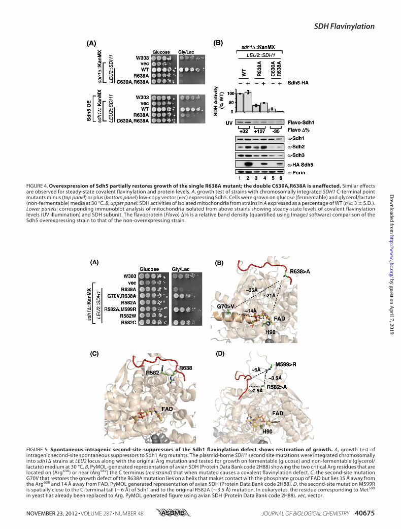
FIGURE 4. Overexpression of Sdh5 partially restores growth of the single R638A mutant; the double C630A,R638A is unaffected. Similar effectsare observed for steady-state covalent flavinylation and protein levels. A, growth test of strains with chromosomally integrated SDH1 C-terminal pointmutants minus (top panel) or plus (bottom panel) low-copy vector (vec) expressing Sdh5. Cells were grown on glucose (fermentable) and glycerol/lactate(non-fermentable) media at 30 °C. B, upper panel: SDH activities of isolated mitochondria from strains in A expressed as a percentage of WT (n � 3 � S.D.).Lower panels: corresponding immunoblot analysis of mitochondria isolated from above strains showing steady-state levels of covalent flavinylationlevels (UV illumination) and SDH subunit. The flavoprotein (Flavo) �% is a relative band density (quantified using ImageJ software) comparison of theSdh5 overexpressing strain to that of the non-overexpressing strain.
FIGURE 5. Spontaneous intragenic second-site suppressors of the Sdh1 flavinylation defect shows restoration of growth. A, growth test ofintragenic second-site spontaneous suppressors to Sdh1 Arg mutants. The plasmid-borne SDH1 second site mutations were integrated chromosomallyinto sdh1� strains at LEU2 locus along with the original Arg mutation and tested for growth on fermentable (glucose) and non-fermentable (glycerol/lactate) medium at 30 °C. B, PyMOL-generated representation of avian SDH (Protein Data Bank code 2H88) showing the two critical Arg residues that arelocated on (Arg638) or near (Arg582) the C terminus (red strand) that when mutated causes a covalent flavinylation defect. C, the second-site mutationG70V that restores the growth defect of the R638A mutation lies on a helix that makes contact with the phosphate group of FAD but lies 35 Å away fromthe Arg638 and 14 Å away from FAD. PyMOL generated representation of avian SDH (Protein Data Bank code 2H88). D, the second-site mutation M599Ris spatially close to the C-terminal tail (�6 Å) of Sdh1 and to the original R582A (�3.5 Å) mutation. In eukaryotes, the residue corresponding to Met599
in yeast has already been replaced to Arg. PyMOL generated figure using avian SDH (Protein Data Bank code 2H88). vec, vector.
SDH Flavinylation
NOVEMBER 23, 2012 • VOLUME 287 • NUMBER 48 JOURNAL OF BIOLOGICAL CHEMISTRY 40675
by guest on April 7, 2019
http://ww
w.jbc.org/
Dow
nloaded from
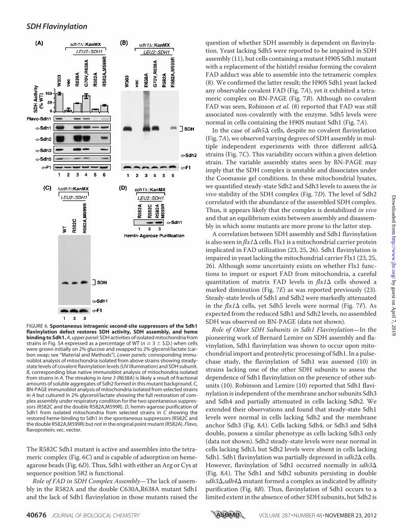
The R582C Sdh1 mutant is active and assembles into the tetra-meric complex (Fig. 6C) and is capable of adsorption on heme-agarose beads (Fig. 6D). Thus, Sdh1with either anArg or Cys atsequence position 582 is functional.Role of FAD in SDH Complex Assembly—The lack of assem-
bly in the R582A and the double C630A,R638A mutant Sdh1and the lack of Sdh1 flavinylation in those mutants raised the
question of whether SDH assembly is dependent on flavinyla-tion. Yeast lacking Sdh5 were reported to be impaired in SDHassembly (11), but cells containing amutantH90S Sdh1mutantwith a replacement of the histidyl residue forming the covalentFAD adduct was able to assemble into the tetrameric complex(8). We confirmed the latter result; the H90S Sdh1 yeast lackedany observable covalent FAD (Fig. 7A), yet it exhibited a tetra-meric complex on BN-PAGE (Fig. 7B). Although no covalentFAD was seen, Robinson et al. (8) reported that FAD was stillassociated non-covalently with the enzyme. Sdh5 levels werenormal in cells containing the H90S mutant Sdh1 (Fig. 7A).In the case of sdh5� cells, despite no covalent flavinylation
(Fig. 7A), we observed varying degrees of SDH assembly inmul-tiple independent experiments with three different sdh5�strains (Fig. 7C). This variability occurs within a given deletionstrain. The variable assembly states seen by BN-PAGE mayimply that the SDH complex is unstable and dissociates underthe Coomassie gel conditions. In these mitochondrial lysates,we quantified steady-state Sdh2 and Sdh3 levels to assess the invivo stability of the SDH complex (Fig. 7D). The level of Sdh2correlated with the abundance of the assembled SDH complex.Thus, it appears likely that the complex is destabilized in vivoand that an equilibrium exists between assembly and disassem-bly in which some mutants are more prone to the latter step.A correlation between SDH assembly and Sdh1 flavinylation
is also seen in flx1� cells. Flx1 is amitochondrial carrier proteinimplicated in FAD utilization (23, 25, 26). Sdh1 flavinylation isimpaired in yeast lacking themitochondrial carrier Flx1 (23, 25,26). Although some uncertainty exists on whether Flx1 func-tions to import or export FAD from mitochondria, a carefulquantitation of matrix FAD levels in flx1� cells showed amarked diminution (Fig. 7E) as was reported previously (23).Steady-state levels of Sdh1 and Sdh2 were markedly attenuatedin the flx1� cells, yet Sdh5 levels were normal (Fig. 7F). Asexpected from the reduced Sdh1 and Sdh2 levels, no assembledSDH was observed on BN-PAGE (data not shown).Role of Other SDH Subunits in Sdh1 Flavinylation—In the
pioneering work of Bernard Lemire on SDH assembly and fla-vinylation, Sdh1 flavinylation was shown to occur upon mito-chondrial import andproteolytic processing of Sdh1. In a pulse-chase study, the flavinylation of Sdh1 was assessed (10) instrains lacking one of the other SDH subunits to assess thedependence of Sdh1 flavinylation on the presence of other sub-units (10). Robinson and Lemire (10) reported that Sdh1 flavi-nylation is independent of themembrane anchor subunits Sdh3and Sdh4 and partially attenuated in cells lacking Sdh2. Weextended their observations and found that steady-state Sdh1levels were normal in cells lacking Sdh2 and the membraneanchor Sdh3 (Fig. 8A). Cells lacking Sdh4, or Sdh3 and Sdh4double, possess a similar phenotype as cells lacking Sdh3 only(data not shown). Sdh2 steady-state levels were near normal incells lacking Sdh3, but Sdh2 levels were absent in cells lackingSdh1. Sdh1 flavinylation was partially depressed in sdh2� cells.However, flavinylation of Sdh1 occurred normally in sdh3�(Fig. 8A). The Sdh1 and Sdh2 subunits persisting in doublesdh3�,sdh4� mutant formed a complex as indicated by affinitypurification (Fig. 8B). Thus, flavinylation of Sdh1 occurs to alimited extent in the absence of other SDH subunits, but Sdh2 is
FIGURE 6. Spontaneous intragenic second-site suppressors of the Sdh1flavinylation defect restores SDH activity, SDH assembly, and hemebinding to Sdh1. A, upper panel: SDH activities of isolated mitochondria fromstrains in Fig. 5A expressed as a percentage of WT (n � 3 � S.D.) when cellswere grown initially on 2% glucose and swapped to 2% glycerol/lactate (car-bon swap; see “Material and Methods”). Lower panels: corresponding immu-noblot analysis of mitochondria isolated from above strains showing steady-state levels of covalent flavinylation levels (UV illumination) and SDH subunit.B, corresponding blue native immunoblot analysis of mitochondria isolatedfrom strains in A. The streaking in lane 3 (R638A) is likely a result of fractionalamounts of soluble aggregates of Sdh2 formed in this mutant background. C,BN-PAGE immunoblot analysis of mitochondria isolated from selected strainsin A but cultured in 2% glycerol/lactate showing the full restoration of com-plex assembly under respiratory condition for the two spontaneous suppres-sors (R582C and the double R582A,M599R). D, hemin-agarose purification ofSdh1 from isolated mitochondria from selected strains in C showing therestored heme-binding to Sdh1 in the spontaneous suppressors (R582C andthe double R582A,M599R) but not in the original point mutant (R582A). Flavo,flavoprotein; vec, vector.
SDH Flavinylation
40676 JOURNAL OF BIOLOGICAL CHEMISTRY VOLUME 287 • NUMBER 48 • NOVEMBER 23, 2012
by guest on April 7, 2019
http://ww
w.jbc.org/
Dow
nloaded from

important for efficient covalent FAD binding. Interestingly, theSdh5 steady-state levels were elevated in sdh2� or sdh3� cells(Fig. 8A).
DISCUSSION
We show presently that flavinylation of the Sdh1 subunit ofsuccinate dehydrogenase is dependent on a set of two spatiallyclose Arg residues near the C terminus, which are distant (�20
Å) fromtheFADbindingsitebutarecritical in flavinylation.Theseresidues are also important for the assemblyof Sdh1 into the tetra-meric enzyme complex. Mutant Sdh1 proteins with either aR582A or double C630A,R638A substitution are neither flaviny-lated nor assembled into the SDH complex (Table 1).With each mutant Sdh1, second-site Sdh1 suppressor muta-
tions were recovered in Sdh1 permitting both flavinylation andSDH assembly. In the case of the single R638A mutation, thesecond site suppressor was a G70V substitution, whereas theR582A second site suppressor was a M599R substitution. Thepresence of the Arg at residue 599 restores a positively chargedresidue in proximity to residue position 582. It is of interest thatthe corresponding residue toMet599 in humans andmetazoansis Arg.In the human SDHA (Sdh1 equivalent), the Arg residue cor-
responding to yeast Arg582 is Arg589. Substitution of this Arg589
to a Trp (R589W) has been reported in a patient afflicted withparaganglioma (24). Yeast harboring a corresponding R582Wmutant Sdh1 are compromised in SDH assembly and flavinyla-tion, but a reversion mutant of R582C restores both Sdh1 flavi-nylation and SDH assembly.
FIGURE 7. Covalent flavinylation of Sdh1 is not required for assembly of SDH as indicated by sdh5� and H90S Sdh1 mutants. However, Sdh1-FADbinding is likely required for stability of Sdh1 and SDH assembly. A, steady-state immunoblot analysis of isolated mitochondria from sdh1� strains expressingeither wild type Sdh1 or H90S substitution (lanes 1 and 2) and sdh5� cells transformed with the indicated vectors (vec; lanes 3 and 4). B, corresponding BN-PAGEimmunoblot analysis of mitochondria from A showing the assembly of SDH even in the absence of covalent flavinylation. C, BN-PAGE immunoblot analysis ofmitochondria from WT and three different sdh5� cells showing the assembly of SDH even in the absence of covalent flavinylation. D, steady-state immunoblotanalysis of isolated mitochondria from C. E, HPLC analysis of FAD levels in purified mitochondria from WT and flx� strains showing the �50% decrease in FADlevels in the flx� strain. F, steady-state SDS-PAGE UV and immunoblot analysis of isolated mitochondria from strains in E. Note the dramatic decrease in Sdh1and the absence of Sdh2 in the flx� strain likely resulting from the decrease in mitochondrial FAD levels. Abs., absorbance. prot, total mitochondrial protein.
FIGURE 8. Sdh1 is flavinylated in the absence of the membrane anchorsubunits and forms a stable dimeric complex with Sdh2. A, steady-stateimmunoblot and Sdh1 flavinylation analysis (UV illumination). B, nickel-nitri-lotriacetic acid purification of Sdh2-His6Myc2 in mitochondria isolated fromsdh3�sdh4� cells. UB, unbound; E, eluate; Flavo, flavoprotein.
SDH Flavinylation
NOVEMBER 23, 2012 • VOLUME 287 • NUMBER 48 JOURNAL OF BIOLOGICAL CHEMISTRY 40677
by guest on April 7, 2019
http://ww
w.jbc.org/
Dow
nloaded from

One major question emerging from the present studies con-cerns the role of the C-terminal Arg residues in Sdh1 flavinyla-tion. These mutations do not appear to destabilize the Sdh1polypeptide; rather, they only impair Sdh1 maturation. Threecandidate roles for the C-terminal Arg residues may be envi-sioned. One candidate link involves the binding of Sdh5 and itsimportance in Sdh1 flavinylation. Sdh5 levels are dependent onthe presence of Sdh1. The reduced steady-state levels of Sdh5 inthe Sdh1 mutants may reflect attenuated binding. However,cells harboring the mutant Sdh1 alleles are markedly impairedin SDH assembly, whereas sdh5� cells are only partially atten-uated in SDH stability as seen by the variable levels of theassembled SDH complex in our series of isolates. Thus, thephenotypes observed with either the R582A or doubleC630A,R638Amutants appear distinct from that of sdh5� cells.A second scenario is that the flavinylation may occur in a
nascent conformation of Sdh1 that is somewhat distinct fromthe final mature conformation. In this scenario, the C-terminalArg residuesmay be in juxtaposition for FAD flavinylation. TheG70V second site suppressor mutation in the SDH-deficientR638A Sdh1 mutant is consistent with this postulate of a dis-tinct nascent conformation inwhich these two residues are nowin closer proximity. However, two observations argue againstthis model. The observation that citric acid cycle intermediatescan stimulate the flavinylation process (10) suggests that a fla-vinylation-competent conformation may have a preformednative-like substrate binding site. The dependence of Sdh2 onefficient Sdh1 flavinylation suggests that the flavinylation-com-petent conformation of Sdh1 must be quite similar to the finalmature fold enabling Sdh2 association. In the flavoproteinvanilly-alcohol oxidase containing a histidyl-linked FAD, struc-tural similarity between the holo- and the apo-forms indicatesthat FAD and substrate bind to a folded, highly preorganizedcofactor/active site cavity, followed by autocatalytic covalentflavinylation (27).A third candidate role for the C-terminal Arg residues may
relate to FAD binding. Although the C-terminal Arg residuesare distantly removed from the FAD or substrate site in themature Sdh1 structure, the Arg residues may be important inrecruitment and/or guidance of FAD and or succinate to thesubstrate site. Asmentioned, flavinylation of Sdh1 is dependenton succinate (10).Another conserved Arg residue (human Arg408) stabilizes
succinate or inhibitor binding through two hydrogen bonds.Mutations in the human gene yielding a R408C substitutionwas reported in a patient with a late onset neurodegenerativedisease (28). Engineering the corresponding mutation in the
E. coli enzyme resulted in impaired covalent flavinylation andthe absence of membrane-associated enzyme (28).The impaired assembly of SDH with the C-terminal Sdh1
mutants suggests that FAD binding is important to stabilize theSdh1 conformation enabling association with Sdh2 and themembrane anchor subunits. To address the role of FAD bind-ing in SDH assembly, we utilized a yeast flx1� deletion strain.This mutant was reported to have attenuated levels of matrixFAD levels (23), and we confirmed this observation. Cells lack-ing Flx1 are known to be deficient in two FAD-containingenzymes SDH and lipoamide dehydrogenase (22).We show themutant cells are also impaired in SDH assembly and stability ofSdh2. The impaired SDH assembly in the FAD-deficient flx1�cells suggests that FAD binding is important for Sdh1 matura-tion enabling assembly of the tetrameric enzyme. Cells contain-ing theH90S Sdh1mutant that precludes covalent flavinylationassemble into the SDH complex consistent with a non-covalentassociation of FAD as was reported previously (8).The covalent addition of FAD to Sdh1 likely occurs in a spe-
cific folded conformation of Sdh1 that brings a set of aminoacids in juxtaposition for the autocatalytic addition. Sdh5 aswell as succinate as a substrate are proposed to stabilize theflavinylation-competent conformation of Sdh1 for the reaction.The conserved C-terminal Arg residues (Arg582 and Arg638)could contribute to FAD recruitment and or its binding prior toformation of the covalent attachment. The G70V second sitesuppressormutation in the SDH-deficient R638A Sdh1mutantmay merely partially deform the Sdh1 conformation allowingFAD binding in the absence of Arg638. The C-terminal Arg res-idues may also have a secondary role in the binding of Sdh5.The propensity of Sdh1 to adsorb onto heme-agarose beads
may relate to either a hydrophobic pocket that fortuitouslyaccommodates hemewith no physiological consequence of thisbinding. Alternatively, the presence of the positively chargedArg residues in the C-terminal segment could interact electro-statically with the dianionic propionate groups of heme, facili-tating the association of Sdh1 with heme-agarose. This interac-tion may be analogous to the possible dianionic succinate orphosphates of FAD. Thus, in this scenario, heme is actingmerely as a dianionicmimic of FAD or succinate. This notion issupported by the fact that protoporphyrin IX (heme lacking theiron center, but still possessing the dianionic propionates) cancompete for heme binding to Sdh1 (results not shown). Thisobservation argues that the iron center is not important forbinding to Sdh1. Thus, the increased affinity of Sdh1 to heme inthe presence of a reductant may bemore related to the reducedstate of Sdh1 than the heme iron.
TABLE 1Molecular properties of strains with mutations or deletions that result in the loss of covalent flavinylation in Sdh1The double C630A,R638A and the single R582W mutations at or near the C terminus lead to loss of SDH assembly as well as binding to heme-agarose beads. However,mutations or deletions that maintain heme-agarose binding also maintain SDH assembly. ND, not determined.
StrainHemebinding
SDHassembly
Sdh2steady-state
Sdh5steady-state
Sdh1-Sdh5interaction
H90S Yes Yes Down Stable Attenuatedsdh5� Yes Yes DownClipK No No Absent ND NDC630A,R638A No No Absent Unstable AttenuatedR582A No No Absent Unstable Attenuated
SDH Flavinylation
40678 JOURNAL OF BIOLOGICAL CHEMISTRY VOLUME 287 • NUMBER 48 • NOVEMBER 23, 2012
by guest on April 7, 2019
http://ww
w.jbc.org/
Dow
nloaded from

A third less likely scenario is that heme has an effector role inthe flavinylation reaction. We have no evidence that SDH bio-genesis requires heme for Sdh1 maturation. Because heme isessential for cell survival and important in yeast for Hap1-me-diated gene expression of mitochondrial proteins, the investi-gation of a role of heme in Sdh1 maturation is challenging andwill be the topic of future studies.REFERENCES1. Sun, F., Huo, X., Zhai, Y., Wang, A., Xu, J., Su, D., Bartlam,M., and Rao, Z.
(2005) Crystal structure of mitochondrial respiratory membrane proteincomplex II. Cell 121, 1043–1057
2. Hägerhäll, C. (1997) Succinate: quinone oxidoreductases. Variations on aconserved theme. Biochim. Biophys. Acta 1320, 107–141
3. Oyedotun, K. S., and Lemire, B. D. (2001) The quinone-binding sites of theSaccharomyces cerevisiae succinate-ubiquinone oxidoreductase. J. Biol.Chem. 276, 16936–16943
4. Silkin, Y., Oyedotun, K. S., and Lemire, B. D. (2007) The role of Sdh4Tyr-89 in ubiquinone reduction by the Saccharomyces cerevisiae succi-nate dehydrogenase. Biochim. Biophys. Acta 1767, 143–150
5. Maklashina, E., Rajagukguk, S., McIntire, W. S., and Cecchini, G. (2010)Mutation of the heme axial ligand of Escherichia coli succinate-quinonereductase: implications for heme ligation in mitochondrial complex IIfrom yeast. Biochim. Biophys. Acta 1797, 747–754
6. Tomasiak, T.M.,Maklashina, E., Cecchini, G., and Iverson, T.M. (2008) Athreonine on the active site loop controls transition state formation inEscherichia coli respiratory complex II. J. Biol. Chem. 283, 15460–15468
7. Lemire, B. D., and Oyedotun, K. S. (2002) The Saccharomyces cerevisiaemitochondrial succinate:ubiquinone oxidoreductase. Biochim. Biophys.Acta 1553, 102–116
8. Robinson, K. M., Rothery, R. A., Weiner, J. H., and Lemire, B. D. (1994)The covalent attachment of FAD to the flavoprotein of Saccharomycescerevisiae succinate dehydrogenase is not necessary for import and assem-bly into mitochondria. Eur. J. Biochem. 222, 983–990
9. Hägerhäll, C., Sled, V., Hederstedt, L., and Ohnishi, T. (1995) The tri-nuclear iron-sulfur cluster S3 in Bacillus subtilis succinate:menaquinonereductase; effects of a mutation in the putative cluster ligation motif onenzyme activity and EPR properties. Biochim. Biophys. Acta 1229,356–362
10. Robinson, K.M., and Lemire, B. D. (1996) Covalent attachment of FAD tothe yeast succinate dehydrogenase flavoprotein requires import into mi-tochondria, presequence removal, and folding. J. Biol. Chem. 271,4055–4060
11. Hao, H. X., Khalimonchuk, O., Schraders, M., Dephoure, N., Bayley, J. P.,Kunst, H., Devilee, P., Cremers, C.W., Schiffman, J. D., Bentz, B. G., Gygi,S. P.,Winge, D. R., Kremer, H., and Rutter, J. (2009) SDH5, a gene requiredfor flavination of succinate dehydrogenase, is mutated in paraganglioma.Science 325, 1139–1142
12. Rutter, J.,Winge, D. R., and Schiffman, J. D. (2010) Succinate dehydrogen-ase - Assembly, regulation, and role in human disease.Mitochondrion 10,393–401
13. McNeil, M. B., Clulow, J. S.,Wilf, N.M., Salmond, G. P., and Fineran, P. C.(2012) SdhE is a conserved protein required for the flavinylation of succi-nate dehydrogenase in bacteria. J. Biol. Chem. 287, 18418–18428.
14. Heuts, D. P., Scrutton, N. S., McIntire, W. S., and Fraaije, M. W. (2009)What’s in a covalent bond?On the role and formation of covalently boundflavin cofactors. FEBS J. 276, 3405–3427
15. Longtine, M. S., McKenzie, A., 3rd, Demarini, D. J., Shah, N. G.,Wach, A.,Brachat, A., Philippsen, P., and Pringle, J. R. (1998) Additional modules forversatile and economical PCR-based gene deletion and modification inSaccharomyces cerevisiae. Yeast 14, 953–961
16. Glick, B. S., and Pon, L. A. (1995) Isolation of highly purifiedmitochondriafrom Saccharomyces cerevisiae.Methods Enzymol. 260, 213–223
17. Diekert, K., De Kroon, A. I., Kispal, G., and Lill, R. (2001) Isolation andsubfractionation of mitochondria from the yeast Saccharomyces cerevi-siae.Methods Cell Biol. 65, 37–51
18. Bradford, M. M. (1976) A rapid and sensitive method for the quantitationof microgram quantities of protein utilizing the principle of protein-dyebinding. Anal. Biochem. 72, 248–254
19. Smith, P. K., Krohn, R. I., Hermanson, G. T., Mallia, A. K., Gartner, F. H.,Provenzano, M. D., Fujimoto, E. K., Goeke, N. M., Olson, B. J., and Klenk,D. C. (1985) Measurement of protein using bicinchoninic acid. Anal.Biochem. 150, 76–85
20. Schägger, H., and von Jagow, G. (1991) Blue native electrophoresis forisolation of membrane protein complexes in enzymatically active form.Anal. Biochem. 199, 223–231
21. Robinson, K. M., and Lemire, B. D. (1995) Flavinylation of succinate:ubiquinone oxidoreductase from Saccharomyces cerevisiae. Methods En-zymol. 260, 34–51
22. Bafunno, V., Giancaspero, T. A., Brizio, C., Bufano, D., Passarella, S., Boles,E., and Barile, M. (2004) Riboflavin uptake and FAD synthesis in Saccha-romyces cerevisiaemitochondria: involvement of the Flx1p carrier in FADexport. J. Biol. Chem. 279, 95–102
23. Tzagoloff, A., Jang, J., Glerum, D.M., andWu,M. (1996) FLX1 codes for acarrier protein involved in maintaining a proper balance of flavin nucleo-tides in yeast mitochondria. J. Biol. Chem. 271, 7392–7397
24. Burnichon, N., Brière, J. J., Libé, R., Vescovo, L., Rivière, J., Tissier, F.,Jouanno, E., Jeunemaitre, X., Bénit, P., Tzagoloff, A., Rustin, P., Bertherat,J., Favier, J., and Gimenez-Roqueplo, A. P. (2010) SDHA is a tumor sup-pressor gene causing paraganglioma. Hum. Mol. Genet. 19, 3011–3020
25. Wu,M., Repetto, B., Glerum, D. M., and Tzagoloff, A. (1995) Cloning andcharacterization of FAD1, the structural gene for flavin adenine dinucle-otide synthetase of Saccharomyces cerevisiae.Mol. Cell. Biol. 15, 264–271
26. Giancaspero, T. A., Wait, R., Boles, E., and Barile, M. (2008) Succinatedehydrogenase flavoprotein subunit expression in Saccharomycescerevisiae–involvement of themitochondrial FAD transporter, Flx1. FEBSJ. 275, 1103–1117
27. Fraaije, M. W., van Den Heuvel, R. H., van Berkel, W. J., and Mattevi, A.(2000) Structural analysis of flavinylation in vanillyl-alcohol oxidase.J. Biol. Chem. 275, 38654–38658
28. Birch-Machin,M. A., Taylor, R.W., Cochran, B., Ackrell, B. A., and Turn-bull, D. M. (2000) Late-onset optic atrophy, ataxia, and myopathy associ-ated with a mutation of a complex II gene. Ann. Neurol. 48, 330–335
SDH Flavinylation
NOVEMBER 23, 2012 • VOLUME 287 • NUMBER 48 JOURNAL OF BIOLOGICAL CHEMISTRY 40679
by guest on April 7, 2019
http://ww
w.jbc.org/
Dow
nloaded from

Hyung J. Kim, Mi-Young Jeong, Un Na and Dennis R. WingeC-terminal Tail of the Flavoprotein Subunit
Flavinylation and Assembly of Succinate Dehydrogenase Are Dependent on the
doi: 10.1074/jbc.M112.405704 originally published online October 7, 20122012, 287:40670-40679.J. Biol. Chem.
10.1074/jbc.M112.405704Access the most updated version of this article at doi:
Alerts:
When a correction for this article is posted•
When this article is cited•
to choose from all of JBC's e-mail alertsClick here
http://www.jbc.org/content/287/48/40670.full.html#ref-list-1
This article cites 28 references, 9 of which can be accessed free at
by guest on April 7, 2019
http://ww
w.jbc.org/
Dow
nloaded from