BiosynthesisandTranslocationofUnsulfatedAcyltrehaloses in ......
15
Biosynthesis and Translocation of Unsulfated Acyltrehaloses in Mycobacterium tuberculosis * Received for publication, May 14, 2014, and in revised form, August 12, 2014 Published, JBC Papers in Press, August 14, 2014, DOI 10.1074/jbc.M114.581199 Juan Manuel Belardinelli ‡ , Gérald Larrouy-Maumus § , Victoria Jones ‡ , Luiz Pedro Sorio de Carvalho § , Michael R. McNeil ‡ , and Mary Jackson ‡1 From the ‡ Mycobacteria Research Laboratories, Department of Microbiology, Immunology and Pathology, Colorado State University, Fort Collins, Colorado 80523-1682 and the § Division of Mycobacterial Research, Medical Research Council National Institute for Medical Research, London NW7 1AA, United Kingdom Background: The biogenesis of 2,3-diacyltrehaloses (DAT) and penta-acyltrehaloses (PAT) found in the outer membrane of Mycobacterium tuberculosis is ill defined. Results: DAT synthesis is cytosolic. Chp2-mediated transesterification reactions between DAT substrates yield PAT on the periplasmic face of the membrane. Conclusion: DAT and PAT biosynthesis is topologically split across the membrane. Significance: DAT and PAT biosynthesis and transport are coupled and dependent on the MmpL10 transporter. A number of species-specific polymethyl-branched fatty acid- containing trehalose esters populate the outer membrane of Mycobacterium tuberculosis. Among them, 2,3-diacyltrehaloses (DAT) and penta-acyltrehaloses (PAT) not only play a structural role in the cell envelope but also contribute to the ability of M. tuberculosis to multiply and persist in the infected host, pro- moting the intracellular survival of the bacterium and modulat- ing host immune responses. The nature of the machinery, topol- ogy, and sequential order of the reactions leading to the biosynthesis, assembly, and export of these complex glycolipids to the cell surface are the object of the present study. Our genetic and biochemical evidence corroborates a model wherein the biosynthesis and translocation of DAT and PAT to the periplas- mic space are coupled and topologically split across the plasma membrane. The formation of DAT occurs on the cytosolic face of the plasma membrane through the action of PapA3, FadD21, and Pks3/4; that of PAT occurs on the periplasmic face via trans- esterification reactions between DAT substrates catalyzed by the acyltransferase Chp2 (Rv1184c). The integral membrane transporter MmpL10 is essential for DAT to reach the cell sur- face, and its presence in the membrane is required for Chp2 to be active. Disruption of mmpL10 or chp2 leads to an important build-up of DAT inside the cells and to the formation of a novel form of unsulfated acyltrehalose esterified with polymethyl- branched fatty acids normally found in sulfolipids that is trans- located to the cell surface. It is now generally accepted that the organisms belonging to the corynebacteria-mycobacteria-nocardia group possess a complex cell envelope made of an inner plasma membrane, the peptidoglycan-arabinogalactan complex, and a pseudo-outer membrane also referred to as “mycomembrane” (1–5). In the interest of simplicity, the term “outer membrane” will here be used to designate the mycomembrane, and “periplasm” will be used to define the space located between the mycobacterial inner and outer membranes. Lipids and glycolipids of unusual structures populate the outer membrane of mycobacteria, governing various aspects of their physiology and pathogenicity (1–2, 5). Among them, the acyltrehaloses produced by Mycobacterium tuberculosis, which include sulfolipids (SL), 2 diacyltrehaloses (DAT), and penta- (or poly-) acyltrehaloses (PAT), have in common a basic structure consisting of a trehalose moiety esterified with one middle-chain saturated fatty acid (palmitic or stearic acid) at the 2-position and up to four polymethyl-branched long-chain fatty acids at the 3-, 6-, 2-, 4-, or 6-position (Fig. 1). The polymethyl-branched fatty acids found in SL are known as the (C31–C46) phthioceranic and hydroxyphthioceranic acids, whereas (C21–C28) mycosanoic, mycolipenic, and mycolipanolic acids esterify trehalose in the case of DAT and PAT. In addition, the trehalose moiety of SL is sulfated at the 2-position, a modification not found in the unsulfated DAT and PAT. DAT and PAT are unique to M. tuberculosis complex species, and SL are exclusively found in the human pathogen, M. tuberculosis. In vitro studies using purified SL, DAT, and PAT indicate that they are biologically active molecules capable of modulat- ing a number of cell functions and host immune responses (6 –18). M. tuberculosis knock-out mutants deficient in their synthesis, however, failed to show any consistent virulence phe- notype in animal and cellular models of tuberculosis infection (18 –27) unless simultaneously impaired in their ability to syn- thesize phthiocerol dimycocerosates (PDIM) (28), suggestive of partially redundant functions among polymethyl-branched * This work was supported, in whole or in part, by National Institutes of Health, NIAID, Grant AI063054. This work was supported by funds from the Medical Research Council (MRC) to L.P.S.d.C. (MC_UP_A253_1111). The MRC Biomedical NMR Centre, NIMR, Mill Hill, is funded by MRC Grant-in-aid U117533887. Author’s Choice—Final version full access. 1 To whom correspondence should be addressed: Dept. of Microbiology, Immu- nology and Pathology, Colorado State University, Fort Collins, CO 80523-1682. Tel.: 970-491-3582; Fax: 970-491-1815; E-mail: [email protected]. 2 The abbreviations used are: SL, sulfolipid(s); DAT, 2,3-diacyltrehalose(s); PAT, penta-acyltrehaloses; PDIM, phthiocerol dimycocerosates; THL, tetrahydrolipstatin. THE JOURNAL OF BIOLOGICAL CHEMISTRY VOL. 289, NO. 40, pp. 27952–27965, October 3, 2014 Author’s Choice © 2014 by The American Society for Biochemistry and Molecular Biology, Inc. Published in the U.S.A. 27952 JOURNAL OF BIOLOGICAL CHEMISTRY VOLUME 289 • NUMBER 40 • OCTOBER 3, 2014 at Imperial College London on May 19, 2015 http://www.jbc.org/ Downloaded from
-
Upload
truonghanh -
Category
Documents
-
view
217 -
download
0
Transcript of BiosynthesisandTranslocationofUnsulfatedAcyltrehaloses in ......

Biosynthesis and Translocation of Unsulfated Acyltrehalosesin Mycobacterium tuberculosis*
Received for publication, May 14, 2014, and in revised form, August 12, 2014 Published, JBC Papers in Press, August 14, 2014, DOI 10.1074/jbc.M114.581199
Juan Manuel Belardinelli‡, Gérald Larrouy-Maumus§, Victoria Jones‡, Luiz Pedro Sorio de Carvalho§,Michael R. McNeil‡, and Mary Jackson‡1
From the ‡Mycobacteria Research Laboratories, Department of Microbiology, Immunology and Pathology, Colorado StateUniversity, Fort Collins, Colorado 80523-1682 and the §Division of Mycobacterial Research, Medical Research Council NationalInstitute for Medical Research, London NW7 1AA, United Kingdom
Background: The biogenesis of 2,3-diacyltrehaloses (DAT) and penta-acyltrehaloses (PAT) found in the outer membraneof Mycobacterium tuberculosis is ill defined.Results: DAT synthesis is cytosolic. Chp2-mediated transesterification reactions between DAT substrates yield PAT on theperiplasmic face of the membrane.Conclusion: DAT and PAT biosynthesis is topologically split across the membrane.Significance: DAT and PAT biosynthesis and transport are coupled and dependent on the MmpL10 transporter.
A number of species-specific polymethyl-branched fatty acid-containing trehalose esters populate the outer membrane ofMycobacterium tuberculosis. Among them, 2,3-diacyltrehaloses(DAT) and penta-acyltrehaloses (PAT) not only play a structuralrole in the cell envelope but also contribute to the ability ofM. tuberculosis to multiply and persist in the infected host, pro-moting the intracellular survival of the bacterium and modulat-ing host immune responses. The nature of the machinery, topol-ogy, and sequential order of the reactions leading to thebiosynthesis, assembly, and export of these complex glycolipidsto the cell surface are the object of the present study. Our geneticand biochemical evidence corroborates a model wherein thebiosynthesis and translocation of DAT and PAT to the periplas-mic space are coupled and topologically split across the plasmamembrane. The formation of DAT occurs on the cytosolic faceof the plasma membrane through the action of PapA3, FadD21,and Pks3/4; that of PAT occurs on the periplasmic face via trans-esterification reactions between DAT substrates catalyzed bythe acyltransferase Chp2 (Rv1184c). The integral membranetransporter MmpL10 is essential for DAT to reach the cell sur-face, and its presence in the membrane is required for Chp2 tobe active. Disruption of mmpL10 or chp2 leads to an importantbuild-up of DAT inside the cells and to the formation of a novelform of unsulfated acyltrehalose esterified with polymethyl-branched fatty acids normally found in sulfolipids that is trans-located to the cell surface.
It is now generally accepted that the organisms belonging tothe corynebacteria-mycobacteria-nocardia group possess a
complex cell envelope made of an inner plasma membrane, thepeptidoglycan-arabinogalactan complex, and a pseudo-outermembrane also referred to as “mycomembrane” (1–5). In theinterest of simplicity, the term “outer membrane” will here beused to designate the mycomembrane, and “periplasm” will beused to define the space located between the mycobacterialinner and outer membranes.
Lipids and glycolipids of unusual structures populate theouter membrane of mycobacteria, governing various aspects oftheir physiology and pathogenicity (1–2, 5). Among them, theacyltrehaloses produced by Mycobacterium tuberculosis, whichinclude sulfolipids (SL),2 diacyltrehaloses (DAT), and penta- (orpoly-) acyltrehaloses (PAT), have in common a basic structureconsisting of a trehalose moiety esterified with one middle-chainsaturated fatty acid (palmitic or stearic acid) at the 2-position andup to four polymethyl-branched long-chain fatty acids at the 3-, 6-,2�-, 4�-, or 6�-position (Fig. 1). The polymethyl-branched fattyacids found in SL are known as the (C31–C46) phthioceranic andhydroxyphthioceranic acids, whereas (C21–C28) mycosanoic,mycolipenic, and mycolipanolic acids esterify trehalose in the caseof DAT and PAT. In addition, the trehalose moiety of SL is sulfatedat the 2�-position, a modification not found in the unsulfated DATand PAT. DAT and PAT are unique to M. tuberculosis complexspecies, and SL are exclusively found in the human pathogen,M. tuberculosis.
In vitro studies using purified SL, DAT, and PAT indicatethat they are biologically active molecules capable of modulat-ing a number of cell functions and host immune responses(6 –18). M. tuberculosis knock-out mutants deficient in theirsynthesis, however, failed to show any consistent virulence phe-notype in animal and cellular models of tuberculosis infection(18 –27) unless simultaneously impaired in their ability to syn-thesize phthiocerol dimycocerosates (PDIM) (28), suggestive ofpartially redundant functions among polymethyl-branched
* This work was supported, in whole or in part, by National Institutes ofHealth, NIAID, Grant AI063054. This work was supported by funds from theMedical Research Council (MRC) to L.P.S.d.C. (MC_UP_A253_1111). TheMRC Biomedical NMR Centre, NIMR, Mill Hill, is funded by MRC Grant-in-aidU117533887.Author’s Choice—Final version full access.
1 To whom correspondence should be addressed: Dept. of Microbiology, Immu-nology and Pathology, Colorado State University, Fort Collins, CO 80523-1682.Tel.: 970-491-3582; Fax: 970-491-1815; E-mail: [email protected].
2 The abbreviations used are: SL, sulfolipid(s); DAT, 2,3-diacyltrehalose(s);PAT, penta-acyltrehaloses; PDIM, phthiocerol dimycocerosates; THL,tetrahydrolipstatin.
THE JOURNAL OF BIOLOGICAL CHEMISTRY VOL. 289, NO. 40, pp. 27952–27965, October 3, 2014Author’s Choice © 2014 by The American Society for Biochemistry and Molecular Biology, Inc. Published in the U.S.A.
27952 JOURNAL OF BIOLOGICAL CHEMISTRY VOLUME 289 • NUMBER 40 • OCTOBER 3, 2014
at Imperial C
ollege London on M
ay 19, 2015http://w
ww
.jbc.org/D
ownloaded from

fatty acid-containing lipids. Recent evidence indicates that oneof these functions is to alleviate the propionate-mediated stressundergone by the bacilli during growth on host cholesterol as amajor carbon source (29 –30). The contribution of these lipidsto blocking the phagosome acidification of infected macro-phages further suggests that their presence at the cell surfacemay promote the intracellular survival of M. tuberculosis (28).
Similarities in the genetic organization of the SL and DAT/PAT biosynthetic gene clusters is suggestive of conservedmechanisms of assembly and export for both families of acyl-trehaloses. To this date, however, only two genes of the DAT/PAT biosynthetic cluster have been characterized (Fig. 1).pks3/4 encodes the polyketide synthase responsible for theelongation of mycosanoic and mycolipenic acids (31), whereaspapA3 encodes the acyltransferase that catalyzes the sequentialesterification of the 2- followed by the 3-position of trehalose,leading to the formation of DAT (32). Disruption of pks3/4 andpapA3 in M. tuberculosis yields mutants devoid of DAT andPAT. By analogy to the better studied SL biosynthetic pathway(2, 33), we hypothesized that FadD21 is the fatty acid AMPligase that provides the activated fatty acid starter unit toPks3/4, whereas Chp2 (Rv1184c) transfers the remaining threemycolipenoyl groups onto DAT to form PAT, and MmpL10 isan inner membrane RND (resistance, nodulation, and division)
transporter required for the translocation of DAT and/or PATto the cell surface. This work was undertaken with the goals ofnot only establishing the involvement of FadD21, Chp2, andMmpL10 in DAT and PAT biosynthesis but also addressing anumber of outstanding questions related to the assembly andexport of these lipids including: (i) the sequential order of thereactions leading to the synthesis and export of DAT and PAT;(ii) the topology of the pathway; (iii) the determination ofwhether a multiprotein complex coupling biosynthesis andexport may be involved; and (iv) the requirement of MmpL10for the translocation of DAT, PAT, or both substrates to the cellsurface. Our results corroborate a model wherein the biosyn-thesis and translocation of DAT and PAT are coupled and topo-logically split across the plasma membrane, with the formationof DAT occurring on the cytosolic side of the plasma mem-brane and that of PAT occurring on the periplasmic face viaChp2-mediated transesterification reactions between DATsubstrates.
EXPERIMENTAL PROCEDURES
Bacterial Strains and Growth Conditions—M. tuberculosismc26206 (an avirulent �panCD�leuCD mutant of M. tubercu-losis H37Rv; kind gift from Dr. W. R. Jacobs Jr., Albert EinsteinCollege of Medicine, New York) and Mycobacterium smegma-
FIGURE 1. DAT and PAT structures and biosynthetic gene cluster. Genes associated with DAT and PAT biosynthesis and export are clustered on theM. tuberculosis H37Rv chromosome. In the forms of DAT and PAT represented here, trehalose is esterified with palmitic acid and multimethyl-branchedmycolipenic acids.
Biosynthesis and Export of DAT and PAT in M. tuberculosis
OCTOBER 3, 2014 • VOLUME 289 • NUMBER 40 JOURNAL OF BIOLOGICAL CHEMISTRY 27953
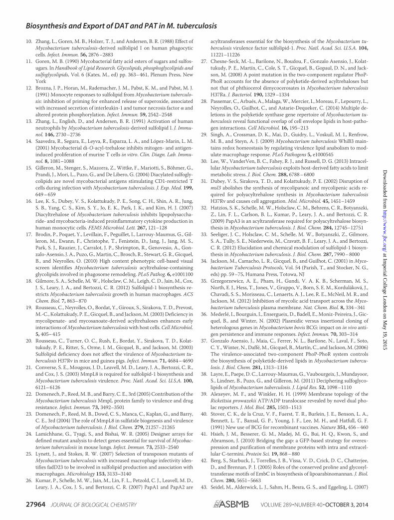
at Imperial C
ollege London on M
ay 19, 2015http://w
ww
.jbc.org/D
ownloaded from

tis mc2155 were grown in Middlebrook 7H9 broth with 10%oleic acid-albumin-dextrose-catalase supplement (BD Biosci-ences), 0.5% glycerol, and 0.05% Tween 80 or on Middlebrook7H11 agar supplemented with 10% oleic acid-albumin-dex-trose-catalase (BD Biosciences) and 0.5% glycerol. All mediaused to grow M. tuberculosis mc26206 were supplemented with0.2% casamino acids, 48 �g/ml pantothenate, and 20 �g/mlL-leucine. Escherichia coli DH5�, the strain used for cloning,was grown in Luria-Bertani (LB) broth or agar (BD Biosci-ences). Kanamycin (20 –50 �g/ml), hygromycin (50 –150�g/ml), ampicillin (100 �g/ml), and 2% sucrose were added tothe culture media when needed.
Construction of M. tuberculosis Mutants and ComplementedMutant Strains—The construction of fadD21, mmpL10, andchp2 (Rv1184c) deletion mutants of M. tuberculosis mc26206involved replacing the corresponding entire ORFs by the kana-mycin resistance cassette from pUC4K (GE Healthcare) follow-ing standard allelic replacement strategies with pPR27-xylE, areplicative plasmid harboring a temperature-sensitive origin ofreplication, the counterselectable marker sacB, and the coloredmarker xylE (34). Details of the plasmid constructs are availableupon request. Complementation constructs for fadD21 andchp2 consist of the full-size genes, PCR-amplified fromM. tuberculosis mc26206 genomic DNA and expressed undercontrol of the phsp60 promoter from the replicative plasmidpMVGH1 (35). The complementation construct used in thecase of the mmpL10 mutant, pNIP40b-mmpL10, consists of themmpL10 gene expressed from its own promoter in the integra-tive plasmid pNIP40b (36).
Metabolic Labeling—M. tuberculosis cultures grown to mid-exponential phase (A600 � 0.5– 0.6) were added 0.5 �Ci/ml[1-14C]propionate (specific activity, 55 Ci/mol, AmericanRadiolabeled Chemicals, Inc.) and labeled for 24 h at 37 °C withshaking. Cell pellets were washed twice with phosphate-buff-ered saline prior to lipid extraction.
Lipid Extraction and Analyses—Surface-exposed lipids extractedwith water-saturated butanol and cell pellet-associated lipidsextracted with chloroform and methanol were analyzed by one-and two-dimensional thin layer chromatography (TLC) follow-ing procedures described earlier (35, 37). Radiolabeled prod-ucts were visualized using a PhosphorImager (Typhoon, GEHealthcare). SL-1, DAT, PAT, and lipid AT-X were purified bypreparative TLC or as described elsewhere (32, 38). Total, ace-tone-soluble, and purified lipids were suspended in chloro-form/methanol (8:2, v/v) and directly deposited onto a steeltarget for analysis by MALDI-TOF MS (microflex LRF (BrukerDaltonics, Billerica, MA) or Ultraflex (Bruker, Bremen, Ger-many)). Spectra were acquired in reflectron mode and mass-assigned through external calibration. 2-(4-Hydroxyphenyla-zo)benzoic acid or 2,5-dihydroxybenzoic acid matrix (Sigma)was used at a concentration of �10 mg/ml in ethanol/water(1:1, v/v). In a typical experiment, 1 �l of glycolipid (5–10 �g) inchloroform/methanol (8:2, v/v) and 1 �l of the matrix solutionwere mixed with a micropipette directly on the target. MALDI-TOF/MS/MS was performed using the LIFT method. One-di-mensional 1H and two-dimensional 1H-1H COSY and 1H-13CHSQC NMR spectroscopy were carried out on a Bruker 600-and 800-MHz NMR spectrometer, equipped with a 5-mm tri-
ple resonance probe head and z axis pulsed field gradients.AT-X was dissolved in CDCl3-CD3OD (8:2, v/v) and analyzedin 200 5-mm 535 PP NMR tubes at 295 K. Proton chemicalshifts are expressed in ppm downfield from the signal of chlo-roform (�H/TMS 7.26 and �C/TMS 77.7).
GC/MS Analysis of Fatty Acyl Groups—The glycolipid AT-Xwas treated with 3 M HCl in CH3OH (Supelco) overnight at80 °C to both release the fatty acyl groups from AT-X and formtheir methyl esters. The sample was then dried and dissolved in50 �l of N,O-bis(trimethylsilyl) trifluoroacetamide (Sigma-Al-drich) and heated at 60 °C for 10 min prior to injection forGC/MS to form the trimethylsilyl ethers of any hydroxylgroups. Samples were injected directly from the silylating rea-gent. Analyses were carried out using a CP 3800 gas chromato-graph (Varian) equipped with an MS320 mass spectrometer inthe electron impact mode and scanning from m/z 50 to 800 over0.5 s. Helium was used as the carrier gas with a flow rate of 1ml/min. The samples were run on a DB 5 column (10 m �0.20-mm inner diameter). The injector (splitless mode) was setfor 250 °C. The oven temperature was held at 50 °C for 1 min,programmed at 30 °C/min to 130 °C, and then programmed at10 °C/min to 330 °C, followed by a 10-min hold. The data anal-yses were carried out on a Varian WS data station.
Topology of Chp2 in E. coli and M. smegmatis—A gene fusionapproach combining the alkaline phosphatase gene (phoA) andthe �-fragment of the �-galactosidase (lacZ�) was used toestablish the topology of the catalytic domains of Chp2 inE. coli. To this end, the full-length chp2 gene fused at its 3� endin frame with the dual phoA-lacZ� reporter cassette frompMA632 (39) was inserted at the HindIII site of pUC19, yieldingpUC-[chp2-phoA-lacZ]. Control plasmids harbored either noinsert or the only phoA-lacZ� reporter cassette expressed fromthe lacZ promoter of pUC19. E. coli DH5� transformed withthese plasmids was plated on dual indicator plates containing80 �g/ml 5-bromo-4-chloro-3-indolyl phosphate, 100 �g/ml6-chloro-3-indolyl-�-D-galactoside (Red-Gal), 1 mM isopropyl1-thio-�-D-galactopyranoside, and 80 mM K2HPO4 (pH 7.0) toassess concomitantly PhoA and �-galactosidase activities (39).
To establish the subcellular localization of the catalytic sitesof Chp2 and Chp1 in mycobacteria, the mycobacterial expres-sion plasmids pJB(�) and JB(�) were engineered in-house frompMV261 (40) and the E. coli expression plasmids pWARF(�)and pWARF (�) (41) to allow for the mycobacterial expressionof proteins C-terminally fused to the green fluorescent pro-tein (GFP).3 Briefly, in pJB(�), a single transmembrane domainfrom glycophorin A is added between the C-terminal fusionpoint of the protein of interest and the GFP to convert mem-brane proteins with extracellular C-terminal fusions to proteinswith intracellular C-terminal fusions. Because GFP fluorescesin the cytoplasm but not in the periplasm, a high fluorescencesignal in the pJB(�) version and background fluorescence in thepJB(�) version are indicative of the C-terminal fusion of theprotein being cytoplasmic. Opposite fluorescence intensitiesindicate, on the contrary, that the C-terminal fusion of the pro-tein is localized in the periplasm (41). Fusions between the
3 J. M. Belardinelli and M. Jackson, manuscript in preparation.
Biosynthesis and Export of DAT and PAT in M. tuberculosis
27954 JOURNAL OF BIOLOGICAL CHEMISTRY VOLUME 289 • NUMBER 40 • OCTOBER 3, 2014
at Imperial C
ollege London on M
ay 19, 2015http://w
ww
.jbc.org/D
ownloaded from

C-terminal ends of Chp2 or Chp1 and GFP were generated inpJB(�) and pJB(�) and used to transform M. smegmatis. Con-trol pJB(�) and pJB(�) plasmids harbor GFP fusions with theC-terminal ends of EmbC and PimA. EmbC is a decaprenylphosphate arabinose-dependent arabinosyltransferase whoseC-terminal domain is periplasmic (42– 43). PimA is a cytoplas-mic GDP-mannose-dependent mannosyltransferase (44). Cul-tures of transformants grown to log phase, washed twice withPBS, and resuspended in 100 �l of the same buffer were trans-ferred to black 96-well plates with transparent bottoms (Corn-ing, Inc.), and their fluorescence was determined using a 2030MultiLabel Reader Victor X5 plate reader (PerkinElmer LifeSciences) at excitation and emission wavelengths of 485 and535 nm, respectively. The fluorescence value of each samplewas normalized to the A600 of the culture.
Biochemical Characterization of Chp2—The Chp2 proteindevoid of its N-terminal transmembrane domain was producedin E. coli. To this end, the chp2 gene was amplified fromgenomic M. tuberculosis H37Rv genomic DNA by standard
PCR using primers Chp2Fw (5�-AAGCCATATGGCGTAC-CCGTGGGCTCCTGGG-3�) and Chp2Rv (5�-TTTGCTC-GAGGGCAGTCGATCGTACGCTAGTTA-3�), digested withNdeI and XhoI, and cloned into the expression plasmid pET14b(Novagen), yielding pET14b-chp2. Following a 3– 4-h induc-tion with 1 mM isopropyl 1-thio-�-D-galactopyranoside at 37 °Cin LB-ampicillin broth, E. coli BL21(DE3) cells transformedwith pET14b-chp2 were harvested, washed, and resuspended inlysis buffer consisting of 100 mM potassium phosphate (pH 7.2)and 5 mM imidazole. Cells were disrupted by sonication and theclarified lysate was incubated with HIS-Select� High Flow (HF)nickel affinity gel (Sigma) for 1 h at 4 °C. The gel was then washedsix times with lysis buffer, and the protein was eluted with anincreasing gradient of 25–250 mM imidazole in lysis buffer. Theelution fractions were concentrated using a Vivaspin� 6 centrifu-gal device (Viva Products) prior to use in enzyme assays.
In Vitro PAT Synthesis—The assay used to analyze the activ-ity of the catalytic domain of Chp2 in vitro consisted of incu-bating 14C-labeled DAT (�2000 cpm) with 15 �g of purified
FIGURE 2. Disruption of the fadD21, chp2 (Rv1184c), and mmpL10 genes of M. tuberculosis H37Rv mc26206. 1–3 candidate mutants obtained for each ofthe three genes were analyzed by PCR. The expected sizes of the PCR fragments for the wild-type parent strain are indicated in the schematic representationof the DAT/PAT locus. A 1.2-kb kanamycin-resistance cassette replaces the ORFs in each of the knock-out mutants. Thus, sizes are 3.87 kb for the wild-typeparent strain and 3.12 kb for the knock-out mutants in the case of fadD21; 3.06 kb for the wild-type parent and 3.44 kb for the knock-out mutants in the caseof chp2; and 5.0 kb for the wild-type parent and 3.5 kb for the knock-out mutants in the case of mmpL10. MWM, molecular weight marker. wt, wild-type parentstrain.
Biosynthesis and Export of DAT and PAT in M. tuberculosis
OCTOBER 3, 2014 • VOLUME 289 • NUMBER 40 JOURNAL OF BIOLOGICAL CHEMISTRY 27955
at Imperial C
ollege London on M
ay 19, 2015http://w
ww
.jbc.org/D
ownloaded from

Chp2 catalytic domain in 1 ml of reaction buffer (100 mM potas-sium phosphate (pH 7.2) and 1 mM DTT). The lipase inhibitortetrahydrolipstatin (THL) (40 �g/ml) was added to some reac-tion mixtures. 14C-Labeled DAT was purified by preparativeTLC from the [1-14C]propionate-derived lipids of the M. tuber-culosis mmpL10 knock-out mutant. Reaction mixtures wereincubated overnight at room temperature, and the products ofthe reactions extracted with chloroform/methanol (33) wereanalyzed by TLC. Assays with [14C]C16:0 (10 �M; 55 mCi/mmol; American Radiochemicals Inc.) and 10 �M CoA used
whole cell lysates prepared from the E. coli control and chp2-expressing strains (600 �g of total proteins) to generate [14C]C16:0-CoA in situ and non-radiolabeled DAT (0, 1, or 10 �M) as theacceptor substrate. The reactions were performed, and the prod-ucts of the reaction were analyzed as described above.
RESULTS
Construction of fadD21, chp2, and mmpL10 DeletionMutants of M. tuberculosis H37Rv—The involvement ofFadD21, Chp2, and MmpL10 in the biosynthesis of DAT and
FIGURE 3. Effects of knocking out fadD21, chp2, and mmpL10 on DAT and PAT biosynthesis and export in M. tuberculosis. Thin layer chromatograms ofsurface-exposed and cell pellet-associated lipids derived from [1-14C]propionate-labeled wild-type, mutant, and complemented mutant strains. 10,000 cpmwere loaded per lane. TLC plates were developed in CHCl3/CH3OH/H2O (90:10:1, v/v/v) for DAT, SL-1, and AT-X analysis (A) or in petroleum ether/acetone (92:8,v/v) for PAT analysis and revealed by phosphorimaging (B). C, thin layer chromatogram of total lipids derived from [1-14C]propionate-labeled wild-type,mmpL10 mutant, and complemented mmpL10 mutant strains showing the absence of PDIM in the mutant and complemented mutant strains. The TLC platewas developed in petroleum ether/diethyl ether (95:5, v/v).
Biosynthesis and Export of DAT and PAT in M. tuberculosis
27956 JOURNAL OF BIOLOGICAL CHEMISTRY VOLUME 289 • NUMBER 40 • OCTOBER 3, 2014
at Imperial C
ollege London on M
ay 19, 2015http://w
ww
.jbc.org/D
ownloaded from

PAT was assessed by generating deletion mutants in the aviru-lent auxotrophic (�panCD�leuCD) M. tuberculosis H37Rvstrain mc26206 (Fig. 2) and comparing the surface-exposed andintracellular lipid profiles of the mutants with that of their wild-type parent. Complemented mutant strains expressing wild-type copies of fadD21, chp2, and mmpL10 from replicative orintegrative expression plasmids were generated by transform-ing the corresponding knock-out mutants with pMVGH1-fadD21, pMVGH1-chp2, and pNIP40b-mmpL10, as describedunder “Experimental Procedures.” Wild-type, mutant, andcomplemented mutant strains were metabolically labeled with[1-14C]propionate, which preferentially incorporates in themethyl-branched fatty acid-containing lipids (37) to facilitatethe detection of biosynthetic intermediates and end products ofthe SL, DAT, and PAT pathways.
Disruption of fadD21 Results in the Loss of DAT and PAT—FadD21 belongs to a family of fatty acyl AMP ligases whose roleis to activate long-chain fatty acids as acyl adenylates, which arethen transferred to polyketide synthases for further chainextension (45). Consistent with the likely requirement offadD21 for the elongation of mycosanoic and mycolipenic acidsby Pks3/4, disruption of this gene in M. tuberculosis H37Rvresulted in the complete loss of DAT and PAT production thatwas restored in the complemented mutant strain (Figs. 3 (A andB) and 4A). The identity of the missing lipids was confirmed byMALDI-MS analysis of the corresponding compounds purified
by preparative TLC from the wild-type parent strain (data notshown).
Involvement of Chp2 in the Biosynthesis of PAT from DAT—The closest homolog of Chp2 is the acyltransferase encoded bychp1 (Rv3822) in the SL biosynthetic cluster (41% sequenceidentity), which catalyzes the regioselective trans-esterificationof two diacylated sulfolipid substrates on the cytosolic face ofthe plasma membrane to afford SL-I, the final tetraacylatedproduct of the SL biosynthetic pathway (33). Analysis of thesurface-exposed and cell pellet-associated lipids produced bythe chp2 null mutant (Fig. 3B) revealed an absence of PAT in themutant strain concomitant with the accumulation of DAT inboth lipid fractions (Figs. 3B and 4 (B and D). That the disrup-tion of chp2 was responsible for this phenotype was supportedby the restoration of PAT synthesis in the complementedmutant. The lipid profile of Mtb�chp2 is thus suggestive of theinvolvement of Chp2 in the acylation of DAT with one or moremethyl-branched fatty acid products of Pks3/4. Chp2, however,is clearly dispensable for the translocation of DAT to the cellsurface.
To gain further insight into the function of Chp2 and deter-mine the number of sequential acylations that this enzyme maycatalyze, a recombinant form of Chp2 devoid of the N-terminaltransmembrane domain was produced in E. coli (Fig. 5A), puri-fied, and used in enzyme assays where 14C-labeled DAT servedboth as the donor and acceptor substrates. A 14C-labeled lipid
FIGURE 4. MALDI-MS analysis of lipids extracted from the wild-type, knock-out mutant, and complemented mutant strains. Total lipids extracted fromwild-type M. tuberculosis H37Rv mc26206; the fadD21, chp2 (Rv1184), and mmpL10 knock-out mutants; and the complemented mutant strains were precipi-tated with acetone and subjected to MALDI-MS analysis in the positive ion mode as described under “Experimental Procedures.” Peaks observed are m/z2153.5, corresponding to sodium-cationized PAT, and m/z 993.7 and 1021.8, corresponding to sodium-cationized DAT. DAT accumulate in the cell pellet-associated lipids of the mmpL10 and chp2 mutant strains as well as in the surface-exposed lipids of the chp2 knock-out mutant. No DAT or PAT were detectedin the fadD21 mutant. A–C correspond to surface-exposed lipids and focus on the PAT content of the strains; D and E correspond to cell-associated lipids andfocus on the DAT content of the strains.
Biosynthesis and Export of DAT and PAT in M. tuberculosis
OCTOBER 3, 2014 • VOLUME 289 • NUMBER 40 JOURNAL OF BIOLOGICAL CHEMISTRY 27957
at Imperial C
ollege London on M
ay 19, 2015http://w
ww
.jbc.org/D
ownloaded from

product displaying the TLC migration properties of PAT wasformed in the reaction mixtures containing both the catalyticdomain of Chp2 and 14C-labeled DAT (Fig. 5B). Attempts touse [14C]C16:0-CoA as an acyl donor in similar reactions wherecell-free extracts prepared from the same E. coli control andchp2-expressing strains served as enzyme sources failed toreveal any transfer of [14C]C16:0 onto DAT, suggesting thatChp2 is not able to use this acyl donor (data not shown). PATsynthesis in vitro was inhibited by the addition of THL to thereaction mixture (Fig. 5B), consistent with the partial inhibitionof PAT synthesis observed in THL-treated M. tuberculosis cells(Fig. 6A). The inhibitory effect of THL on PAT synthesis inwhole cells (65 and 74% inhibition after 24 h of exposure to 10and 40 �g/ml of the compound, respectively) was, however, lesspronounced than that on SL-I synthesis (96 and 98% inhibitionafter 24 h of exposure to 10 and 40 �g/ml of the compound,respectively), indicating that THL is a more potent inhibitor ofChp1 (33) than Chp2.
The Elaboration of PAT from DAT Occurs on the PeriplasmicFace of the Plasma Membrane—Similar to Chp1 (Rv3822),Chp2 is a 359-amino acid-long protein with a single predictedN-terminal transmembrane domain (residues 5–27) and an�/�-hydrolase fold C-terminal domain (residues 28 –359) har-boring a cutinase-like motif (33) (Fig. 7, A and B). That Chp2associates with the membrane was confirmed by expressing aC-terminal GFP-tagged form of this protein in M. smegmatisand probing its localization by fluorescence detection uponsubcellular fractionation (Fig. 7C). To determine whether thecatalytic C-terminal domain of Chp2 faced the cytosolic orperiplasmic face of the membrane, a construct, pUC-[chp2-phoA-lacZ], was first generated in which the C-terminal end ofChp2 was fused to a dual phoA-lacZ� reporter cassette.Because the alkaline phosphatase encoded by phoA is onlyactive in the periplasm and the �-galactosidase (�-gal) encodedby lacZ is only functional in the cytosol, active PhoA and inac-tive �-gal indicate a periplasmic location of the fusion, whereas
FIGURE 5. Chp2 catalyzes the formation of PAT from DAT. A, Coomassie Blue-stained SDS-PAGE showing the recombinant Chp2 protein devoid of N-ter-minal transmembrane domain purified from E. coli; 1.5 �g of protein was loaded on the gel. B, 15 �g of purified recombinant Chp2 protein was incubated with14C-labeled DAT (2000 cpm) in the presence or absence of THL (40 �g/ml). The reaction products were analyzed by one- and two-dimensional TLC and revealedby phosphorimaging. One-dimensional TLC plates were developed in CHCl3/CH3OH/H2O (90:10:1, v/v/v). Two-dimensional TLC plates were developed threetimes in petroleum ether/acetone (92:8, v/v) in the first dimension and once in toluene/acetone (95:5, v/v) in the second dimension. A 14C-labeled lipid productwith the migration characteristics of PAT is formed when Chp2 is incubated with DAT in the absence of THL. C, transesterification reactions between DATsubstrates catalyzed by Chp2.
Biosynthesis and Export of DAT and PAT in M. tuberculosis
27958 JOURNAL OF BIOLOGICAL CHEMISTRY VOLUME 289 • NUMBER 40 • OCTOBER 3, 2014
at Imperial C
ollege London on M
ay 19, 2015http://w
ww
.jbc.org/D
ownloaded from

reversed enzyme activities point to a cytoplasmic location of thefusion. Transformation of E. coli DH5� with this construct andplating of the transformants on dual indicator plates containingthe substrates for both reporter enzymes (Red-Gal and 5-bro-mo-4-chloro-3-indolyl phosphate; see “Experimental Proce-dures”) yielded blue colonies indicative of PhoA activity (Fig.7D). Transformation of a control plasmid (pUC-[phoA-lacZ])in which the phoA-lacZ� reporter cassette was directly placedunder control of the lacZ promoter in pUC19 to allow for thecytosolic production of �-gal yielded, in contrast, the expectedred/purple colonies indicative of �-gal activity (Fig. 7D). Resultsthus clearly pointed to the catalytic site of Chp2 being on theperiplasmic side of the plasma membrane when expressed inE. coli.
To further confirm that the catalytic domain of Chp2mapped to the periplasmic face of the plasma membrane whenexpressed in a mycobacterial host, the full-length chp2 gene wasnext fused at its 3�-end in frame with gfp in pJB(�) and JB(�),yielding plasmids pJB(�)chp2 and pJB(�)chp2 (see “Experi-mental Procedures”). Because GFP is folded and active only inthe cytosol, a high fluorescence signal in M. smegmatis pJB(�)chp2 transformants and background fluorescence in pJB(�)chp2 transformants would indicate that the C-terminal cat-alytic domain of Chp2 is cytoplasmic. Opposite fluorescenceintensities would indicate, on the contrary, that this domain islocalized in the periplasm (41). Determination of the fluores-cence intensities of three independent M. smegmatis pJB(�)chp2 and M. smegmatis pJB(�)chp2 transformantsclearly pointed to the periplasmic location of the catalyticdomain of Chp2 (Fig. 7E). In-frame C-terminal fusions of theChp1 protein with GFP in the same plasmids and analysis ofthe fluorescence intensities of M. smegmatis pJB(�)chp1and pJB(�)chp1 transformants confirmed the cytosolic loca-tion of the catalytic domain of this enzyme in mycobacteria(Fig. 7E) (33). Control C-terminal GFP fusions of theM. tuberculosis EmbC and PimA proteins using the sameplasmids confirmed the periplasmic location of the C-termi-
nal end of the first enzyme and the cytosolic location of theC-terminal end of the latter (42– 44). It follows that, in con-trast to SL biosynthesis wherein the fully acylated SL-I prod-uct is elaborated in the cytosol, PAT are elaborated fromDAT on the periplasmic face of the plasma membrane.
Involvement of mmpL10 in the Biosynthesis of PAT and theTransport of DAT to the Cell Surface—Similar to the situationwith chp2, deletion of mmpL10 in M. tuberculosis H37Rvmc26206 (Fig. 2) led to a mutant devoid of PAT, which accu-mulated important amounts of DAT (Figs. 3A and 4 (C and E)).In contrast to the chp2 mutant, however, DAT only accumu-lated inside the mmpL10 mutant cells, and no trace of DAT wasfound at the cell surface (Figs. 3A and 4 (C and E)). Comple-mentation of Mtb�mmpL10 with a wild-type copy of mmpL10expressed from pNIP40b-mmpL10 restored the production ofPAT and export of both DAT and PAT in the mutant strain.Thus, MmpL10 is both required for the export of DAT to thecell surface and the formation of PAT in M. tuberculosis. Inci-dentally, in the process of disrupting mmpL10, the mutant alsolost the ability to produce PDIM (Fig. 2C). This loss of PDIMhas been shown to occur spontaneously in the process of gen-erating M. tuberculosis knock-out mutants (23) and is notrelated to MmpL10, as evidenced by the absence of PDIM in thecomplemented mutant strain.
Whether MmpL10 participates in the export of PAT inaddition to DAT to the cell surface could not be concludedfrom these experiments due to the absence of PAT synthesisin the mmpL10 knock-out mutant. The requirement ofMmpL10 for PAT synthesis is reminiscent of the situationdescribed previously in the SL biosynthetic pathway,wherein MmpL8 needs to be present for Chp1 to elaboratethe diacylated sulfolipid precursor (SL1278) into SL-I (21, 23,33). A role of MmpL proteins in targeting other enzymes andtransporters of the same pathway to the plasma membraneto couple biosynthesis and export was proposed to accountfor this requirement (33, 46).
FIGURE 6. Inhibition of SL-I, PAT, and AT-X synthesis by the lipase inhibitor THL in whole M. tuberculosis cells. Thin layer chromatograms of surface-exposed and cell pellet-associated lipids derived from [1-14C]propionate-labeled wild-type (A) and mmpL10 knock-out mutant (B) strains either untreated ortreated with THL (10 and 40 �g/ml). The same volume of samples was loaded per lane. TLC plates were developed in CHCl3/CH3OH/H2O (90:10:1, v/v/v) (DAT,SL-1, and AT-X analysis) or three times in petroleum ether/acetone (92:8, v/v) (PAT analysis) and revealed by phosphorimaging.
Biosynthesis and Export of DAT and PAT in M. tuberculosis
OCTOBER 3, 2014 • VOLUME 289 • NUMBER 40 JOURNAL OF BIOLOGICAL CHEMISTRY 27959
at Imperial C
ollege London on M
ay 19, 2015http://w
ww
.jbc.org/D
ownloaded from

Evidence of Cross-talk between the SL and DAT/PAT Biosyn-thetic Pathways—Noticeable in the mmpL10 and chp2 deletionmutants was the appearance of a novel [1-14C]propionate-la-beled compound (AT-X) at the cell surface of the cells (Fig. 3A).This compound was not detected in cell pellet lipids and wasnot found in the wild-type M. tuberculosis strain or in the com-plemented mmpL10 and chp2 mutants. AT-X was prepared innon-radioactive form and analyzed for its constituent fatty acylgroups by formation of the corresponding fatty acyl methylesters, trimethylsilyl ethers. The molecular or M-15 ionsformed during GC/MS analysis of these compounds revealedthe presence of saturated C16 and C18 fatty acyl groups (ratio of1:2); C-25, -26, and -27 unsaturated fatty acyl groups presumedfrom precedent to be mycolipenoyl groups (ratio of 1:0.3:2.7);and monohydroxy C-34, 37, 40, 43, and 47 fatty acyl groups,
presumed from precedent to be hydroxyphthioceranoylgroups, in a ratio of 2:1:14:2:4. The location of the hydroxylgroup in the hydroxyphthioceranoyl groups was both 16 and 18carbons from the end of the chain, as shown by fragment ions atm/z 313 and 341. MALDI analysis of purified AT-X revealedseries of [M � Na�] ions separated by 14 units with the majorpeak at m/z � 1613.6 (isotope-averaged mass) (Fig. 8B). Thiscorresponded to a triacylated trehalose esterified with stearoyl,C27 mycolipenoyl, and C43 hydroxyphthioceranoyl residues;the high and lower molecular weight ions are readily inter-preted using other combinations of the fatty acyl groups. TheNMR spectrum of AT-X (Fig. 8A and Table 1) showed that thestructure of this lipid corresponds to a trehalose esterified ononly one of the glucosyl units (system II; Table 1) with fatty acylgroups in positions 2 and 3. Most importantly, no downfield
FIGURE 7. Subcellular localization and topology of Chp2. A, topology of Chp1 (33) and topology of Chp2 as predicted by HMMTOP version 2.1. B, primarysequence alignment of Chp2 and Chp1 showing the N-terminal transmembrane domain (green highlight) and putative catalytic triads (Ser-141/Asp-226/His-248 in Chp2) (yellow highlight) (33) of these enzymes. Conserved residues are indicated with an asterisk. C, subcellular localization of Chp2. Membrane (CM),cytosol (Cyt), and cell wall (CW) fractions were prepared as described (55) from an M. smegmatis pJB(�)chp2 transformant expressing a C-terminal GFP-taggedform of Chp2, run on an SDS-polyacrylamide gel (2 �g of protein/lane), and analyzed for the presence of Chp2 by in-gel fluorescence (�ex � 485 nm, �em � 525nm). Chp2 localizes to the cell membrane. D, topology of Chp2 in E. coli. Plating of E. coli DH5�/pUC-[chp2-phoA-lacZ] transformants (expressing chp2 fused atits C-terminal end to a dual phoA-lacZ� reporter cassette) on dual indicator plates containing the substrates for both �-gal and PhoA yielded blue coloniesindicative of PhoA activity. Transformation of the control plasmid pUC-[phoA-lacZ] yielded the expected red/purple colonies indicative of �-gal activity. Thecatalytic site of Chp2 expressed in E. coli is thus on the periplasmic side of the plasma membrane. E, topology of Chp2 and Chp1 in M. smegmatis. The full-lengthchp2 and chp1 genes were fused at their 3�-ends in frame with gfp in pJB(�) and JB(�), as described under “Experimental Procedures.” Fluorescence intensitieswere normalized to the A600 of the cultures. Fluorescence intensities of M. smegmatis pJB(�)chp2, pJB(�)chp2, pJB(�)chp1, and pJB(�)chp1 transformantsconfirmed the periplasmic location of the catalytic domain of Chp2 and the cytosolic location of the catalytic domain of Chp1. Control pJB(�) and JB(�)plasmids confirmed the periplasmic location of the C-terminal domain of EmbC and the cytosolic location of the C-terminal end of PimA.
Biosynthesis and Export of DAT and PAT in M. tuberculosis
27960 JOURNAL OF BIOLOGICAL CHEMISTRY VOLUME 289 • NUMBER 40 • OCTOBER 3, 2014
at Imperial C
ollege London on M
ay 19, 2015http://w
ww
.jbc.org/D
ownloaded from

shift of any additional ring protons on either glucosyl residuewas found, suggesting the presence of one acyl on the hydroxylgroup of the hydroxyphthioceranoyl residue (Table 1). HMBCNMR analysis allowed the carbonyl groups of the three differ-ent fatty acyl chains to be identified due to differences in thehydrogens of the � and � carbons. In particular, the carbonyl of
the mycolipenic acid was clearly coupled to the vinyl proton onC-3 of the fatty acyl group and then to H-3 of the system 2glucosyl residue (Table 1). It then must follow that the stearoylchain is attached to the hydroxyl group of the hydroxyphthio-ceranoyl residue, which is in turn attached to O-2 of the system2 glucosyl residue. The MS/MS spectrum (Fig. 8C) of the sodi-
FIGURE 8. Structural characterization of compound AT-X. A, two-dimensional 1H-13C HMBC NMR spectrum of AT-X presenting coupling resonances to thecarbonyl group region. The assignments of the various signals are reported in Table 1. B, the MALDI-TOF spectrum of AT-X. At this mass range, the individualisotope peaks are merged; hence, the mass number refers to the average (not monoisotopic) mass, and the mass inaccuracy is plus or minus an atomic massunit or so. The masses are consistent with the sodiated molecular ion adduct for trehalose with three related fatty acyl groups as described under “Results.” C,the MS/MS spectrum of m/z 1613.5. All ions are sodiated. Masses are average, and the accuracy is as in B. D, a structure of AT-X consistent with the NMR andMS/MS data. A rationalization of the MS/MS data is shown. All ions are sodiated.
Biosynthesis and Export of DAT and PAT in M. tuberculosis
OCTOBER 3, 2014 • VOLUME 289 • NUMBER 40 JOURNAL OF BIOLOGICAL CHEMISTRY 27961
at Imperial C
ollege London on M
ay 19, 2015http://w
ww
.jbc.org/D
ownloaded from

ated ion at m/z � 1613.6 yielded fragment ions correspondingto the loss of an unsubstituted glucosyl residue (m/z � 1449)and release of sodiated unsubstituted glucose residue (m/z �203 and the ion corresponding to the loss of water from it atm/z � 185), indicating that all of the fatty acyl groups are in oneglucosyl residue. Other fragments consistent with this arrange-ment are shown in Fig. 8C as interpreted in Fig. 8D. This led usto propose the structure of AT-X shown in Fig. 8D.
Thus, for the first time, structural analyses revealed the exist-ence in M. tuberculosis of an unsulfated acyltrehalose display-ing mixed characteristics of SL and DAT/PAT with the unusualcharacteristic of a fatty acyl group esterified to the OH of the
hydroxyphthioceranoyl residue. Its export to the cell surfacecould suggest that its formation is a response of the mmpL10and chp2 mutants to the significant and potentially toxicbuild-up of DAT in the plasma membrane. Its finding at the cellsurface of the mmpL10 knock-out mutant further indicates thatits translocation is independent from MmpL10. Whether itsexport proceeds through the SL translocation machineryremains to be determined but may be envisaged given the pres-ence in AT-X of a hydroxyphthioceranyl chain esterifying thetrehalose, similar to the situation in SL-I precursors. Analysis ofthe surface-exposed lipids extracted from [1-14C]propionate-labeled cultures of Mtb�mmpL10 either treated with THL or
FIGURE 9. Proposed DAT/PAT, sulfolipid, and phthiocerol dimycocerosate biosynthetic pathways. Left, DAT and PAT biosynthetic pathway. The acyl-transferase PapA3 initiates DAT and PAT biosynthesis on the cytosolic face of the plasma membrane by transferring a palmitoyl group to the 2-position of oneof the glucosyl residues of trehalose to form trehalose 2-palmitate. PapA3 next transfers a mycolipenoyl group, synthesized by the polyketide synthase Pks3/4,to the 3-position of trehalose 2-palmitate to yield DAT. FadD21 is the fatty acyl AMP ligase that provides the activated fatty acyl starter unit to Pks3/4. DAT isthen flipped across the plasma membrane either by an as yet unknown flippase or by MmpL10 and further elaborated with mycosanoyl, mycolipenoyl, and/ormycolipanolyl chains by Chp2 on the periplasmic face of the plasma membrane to form the penta-acylated PAT. DAT serves both as the donor and acceptorsubstrate in these Chp2-mediated transesterification reactions. DAT and possibly PAT are taken up by MmpL10 and/or by other as yet unknown periplasmicand outer membrane proteins from the outer leaflet of the plasma membrane and exported to the cell surface. The enzymes and transporters involved in theelongation, assembly, and export of sulfolipids (middle) and PDIM (right) and their localization in the bacterium are represented (for a recent review, see Ref. 2).PpsA-E is a type 1 polyketide synthase responsible for the formation of the phthiocerol; Mas is mycocerosic acid synthase; TesA is a type II thioesterase thoughtto be involved in the release of phthiocerol from PpsE; PapA5 is an acyltransferase responsible for the transfer of mycocerosic acids to phthiocerol to form PDIM;FadD23, FadD26, and FadD28 are long-chain fatty acyl-AMP ligases; Stf0 is a sulfotransferase; and PapA2 and PapA1 are acyltransferases responsible for thetransfer of the first (palmitoyl or stearyl) and second ((hydroxy)phthioceranoyl) acyl chains, respectively, onto trehalose 2-sulfate to form the diacylatedsulfolipid, SL1278. MmpL8 participates in the export of SL-I to the cell surface. MmpL7 participates in the export of PDIM. DrrABC and LppX are an ABC transporterand a periplasmic lipoprotein, respectively, required for PDIM to reach the cell surface. Sap is an integral membrane protein thought to facilitate the translo-cation of SL-I to the cell surface. The precise extent of sulfolipid and PDIM translocation mediated by MmpL7, MmpL8, Sap, LppX, and DrrABC has not yet beendefined. Note that in the case of both sulfolipids and PDIM, the biosynthetic end products are formed on the cytoplasmic side of the plasma membrane priorto export to the periplasm and outer membrane, whereas the Chp2-mediated elaboration of PAT from DAT occurs on the periplasmic side of the membrane.
TABLE 1Diagnostic 1H and 13C NMR chemical shifts of AT-X measured at 295 K in CDCl3-CD3OD (8:2, v/v)Assignments were made using two-dimensional 1H-1H COSY and 1H-13C HSQC NMR spectroscopy. The superscript letters refer to resonances shown in Fig. 8A.
Chemical shift (ppm)
System I System II Esterified fattyacyl OH
C�O chemicalshift
C�O3 bondconnectivity1H 13C 1H 13C
H1/C1 5.138e 93.97 5.304d 91.07H2 3.499 5.036f
H3 3.783 5.526b
H4 3.419 3.631H5 3.619 4.199H6 3.706 3.752H6� 3.817 3.861CH2CH(OAcyl)CH2 (hydroxyphthioceranoyl) 4.371g
C�O (stearoyl) 177.79 1.829; 2.698; 4.371g
C�O (hydroxyphthioceranoyl) 173.37 1.573; 5.036f
C�O (mycolipenoyl) 168.28 6.598a; 5.526b
Biosynthesis and Export of DAT and PAT in M. tuberculosis
27962 JOURNAL OF BIOLOGICAL CHEMISTRY VOLUME 289 • NUMBER 40 • OCTOBER 3, 2014
at Imperial C
ollege London on M
ay 19, 2015http://w
ww
.jbc.org/D
ownloaded from

untreated showed a significant and THL concentration-depen-dent decrease in AT-X production in the treated cells (78 and91% inhibition after 24 h of exposure to 10 and 40 �g/ml of thecompound, respectively), indicating that the acyltransferase(s)responsible for the formation of this acyltrehalose is susceptibleto the effect of THL (Fig. 6B).
Consistent with earlier findings that the loss of production ofDAT and PAT does not result in significant changes in thenature or abundance of related acyltrehaloses (28, 32), nochanges in SL-I or any other known SL precursors were other-wise observed in any of the three DAT/PAT deletion mutants(data not shown).
DISCUSSION
Altogether, the results presented herein are consistent withthe DAT and PAT biosynthetic model presented in Fig. 9. DATis formed in the cytosol upon sequential acylation of trehalosewith a palmitoyl or stearoyl group and a fatty acyl product ofPks3/4 by PapA3 (32). DAT is then flipped across the plasmamembrane either by MmpL10 or by an as yet unknown flippaseand further elaborated with mycosanoyl, mycolipenoyl, and/ormycolipanolyl chains through Chp2-mediated trans-esterifica-tion reactions between DAT substrates on the periplasmic faceof the plasma membrane to yield the penta-acylated PAT. Sucha trans-esterification mechanism has precedent in M. tubercu-losis and, in fact, seems to be a recurring theme in the biosyn-thesis of mycobacterial acyltrehaloses. Indeed, the three majormycoloyltransferases of M. tuberculosis known as the antigens85A, 85B, and 85C catalyze the formation of trehalose dimyco-late between two molecules of trehalose monomycolate on theperiplasmic face of the plasma membrane (47). Likewise, Chp1catalyzes trans-esterification reactions between two diacylatedsulfolipid precursors (SL1278) on the cytoplasmic face of themembrane to yield SL-I, the final product of the sulfolipid bio-synthetic pathway (33) (Fig. 9). DAT and possibly PAT aretaken up by MmpL10 and/or by other as yet unknown periplas-mic and outer membrane proteins from the outer leaflet of theplasma membrane and exported to the cell surface. The factthat PAT is synthesized on the periplasmic side of the plasmamembrane calls into question the extent of (glyco)lipid trans-location mediated by MmpL proteins. The localization ofMmpL proteins in the plasma membrane could indeed suggestan involvement of these transporters in the translocation of(glyco)lipids either across the plasma membrane (“flippase”activity) or from the outer leaflet of the plasma membrane tothe periplasm or outer membrane (intermembrane transport)or in both processes. The involvement of MmpL10 in the flip-ping of DAT across the plasma membrane would be consistentwith the inability of the mmpL10 null mutant to synthesizePAT. Alternatively, MmpL10 may mediate the intermembranetranslocation of DAT (and possibly PAT), and the absence ofPAT in the mmpL10 knock-out be due to the failure of Chp2 toelaborate PAT in the absence of a functional MmpL10 protein,similar to the situation reported earlier for the sulfolipid bio-synthetic pathway, where the elaboration of the fully acylatedSL-I by Chp1 is potentiated by the presence of the MmpL8transporter (33). In the case of an intermembrane transport,MmpL10 and possibly other mycobacterial MmpL proteins
would take up their substrates from the outer leaflet of theplasma membrane and therefore resemble the classical Gram-negative RND transporters, which are known to pump out sub-strates from the periplasm rather than across the plasma mem-brane (48). An important correlate of this scenario is that,similar to Gram-negative RND transporters (49), MmpLs arelikely to require the assistance of “flippases” and, possibly, addi-tional periplasmic adapters, lipoproteins, and/or outer mem-brane proteins to deliver their substrates to or in the vicinity ofthe outer membrane. That MmpL-dependent translocationmachineries involve such additional components is in factalready supported by a number of studies on the export of sul-folipids (33) (Fig. 9), phthiocerol dimycocerosates (50, 51) (Fig.9), glycopeptidolipids (52, 53), and siderophores (54). Clearly,the precise definition of the compositions and export mecha-nisms of these MmpL-dependent translocation machineriesawait further investigations.
Interestingly, the formation of AT-X under conditions whereDAT builds up in the plasma membrane highlights for the firsttime the existence of a cross-talk between the SL and DAT/PATbiosynthetic pathways. Independent from their interest in deci-phering the biogenesis of unsulfated acyltrehaloses in M. tuber-culosis, the set of recombinant strains described in this study,including those deficient in DAT and PAT translocation to thecell surface and those accumulating DAT in addition to a newlydescribed acyltrehalose (AT-X), provide new opportunities forfuture studies aimed at understanding the role of these glyco-lipids in M. tuberculosis pathogenesis.
Acknowledgments—We are grateful to Dr. W. R. Jacobs Jr. (AlbertEinstein College of Medicine, New York) for the kind gift of M. tuber-culosis H37Rv mc26206.
REFERENCES1. Daffé, M., and Draper, P. (1998) The envelope layers of mycobacteria with
reference to their pathogenicity. Adv. Microb. Physiol. 39, 131–2032. Daffé, M., Crick, D. C., and Jackson M. (2014) Genetics of capsular polysac-
charides and cell envelope (glyco)lipids. Microbiol. Spectrum 10.1128/microbiolspec.MGM2-0021-2013
3. Hoffmann, C., Leis, A., Niederweis, M., Plitzko, J. M., and Engelhardt, H.(2008) Disclosure of the mycobacterial outer membrane: cryo-electrontomography and vitreous sections reveal the lipid bilayer structure. Proc.Natl. Acad. Sci. U.S.A. 105, 3963–3967
4. Zuber, B., Chami, M., Houssin, C., Dubochet, J., Griffiths, G., and Daffé,M. (2008) Direct visualization of the outer membrane of mycobacteria andcorynebacteria in their native state. J. Bacteriol. 190, 5672–5680
5. Bansal-Mutalik, R., and Nikaido, H. (2014) Mycobacterial outer mem-brane is a lipid bilayer and the inner membrane is unusually rich in diacylphosphatidylinositol dimannosides. Proc. Natl. Acad. Sci. U.S.A. 111,4958 – 4963
6. Husseini, H., and Elberg, S. (1952) Cellular reactions to phthienoic acidand related branched-chain acids. Am. Rev. Tuberc. 65, 655– 672
7. Kato, M., and Goren, M. B. (1974) Synergistic action of cord factor andmycobacterial sulfatides on mitochondria. Infect. Immun. 10, 733–741
8. Goren, M. B., D’Arcy Hart, P., Young, M. R., and Armstrong, J. A. (1976)Prevention of phagosome-lysosome fusion in cultured macrophages bysulfatides of Mycobacterium tuberculosis. Proc. Natl. Acad. Sci. U.S.A. 73,2510 –2514
9. Pabst, M. J., Gross, J. M., Brozna, J. P., and Goren, M. B. (1988) Inhibitionof macrophage priming by sulfatide from Mycobacterium tuberculosis.J. Immunol. 140, 634 – 640
Biosynthesis and Export of DAT and PAT in M. tuberculosis
OCTOBER 3, 2014 • VOLUME 289 • NUMBER 40 JOURNAL OF BIOLOGICAL CHEMISTRY 27963
at Imperial C
ollege London on M
ay 19, 2015http://w
ww
.jbc.org/D
ownloaded from
10. Zhang, L., Goren, M. B., Holzer, T. J., and Andersen, B. R. (1988) Effect ofMycobacterium tuberculosis-derived sulfolipid I on human phagocyticcells. Infect. Immun. 56, 2876 –2883
11. Goren, M. B. (1990) Mycobacterial fatty acid esters of sugars and sulfos-ugars. In Handbook of Lipid Research. Glycolipids, phosphoglycolipids andsulfoglycolipids, Vol. 6 (Kates, M., ed) pp. 363– 461, Plenum Press, NewYork
12. Brozna, J. P., Horan, M., Rademacher, J. M., Pabst, K. M., and Pabst, M. J.(1991) Monocyte responses to sulfolipid from Mycobacterium tuberculo-sis: inhibition of priming for enhanced release of superoxide, associatedwith increased secretion of interleukin-1 and tumor necrosis factor � andaltered protein phosphorylation. Infect. Immun. 59, 2542–2548
13. Zhang, L., English, D., and Andersen, B. R. (1991) Activation of humanneutrophils by Mycobacterium tuberculosis-derived sulfolipid I. J. Immu-nol. 146, 2730 –2736
14. Saavedra, R., Segura, E., Leyva, R., Esparza, L. A., and Lopez-Marin, L. M.(2001) Mycobacterial di-O-acyl-trehalose inhibits mitogen- and antigen-induced proliferation of murine T cells in vitro. Clin. Diagn. Lab. Immu-nol. 8, 1081–1088
15. Gilleron, M., Stenger, S., Mazorra, Z., Wittke, F., Mariotti, S., Böhmer, G.,Prandi, J., Mori, L., Puzo, G., and De Libero, G. (2004) Diacylated sulfogly-colipids are novel mycobacterial antigens stimulating CD1-restricted Tcells during infection with Mycobacterium tuberculosis. J. Exp. Med. 199,649 – 659
16. Lee, K. S., Dubey, V. S., Kolattukudy, P. E., Song, C. H., Shin, A. R., Jung,S. B., Yang, C. S., Kim, S. Y., Jo, E. K., Park, J. K., and Kim, H. J. (2007)Diacyltrehalose of Mycobacterium tuberculosis inhibits lipopolysaccha-ride- and mycobacteria-induced proinflammatory cytokine production inhuman monocytic cells. FEMS Microbiol. Lett. 267, 121–128
17. Brodin, P., Poquet, Y., Levillain, F., Peguillet, I., Larrouy-Maumus, G., Gil-leron, M., Ewann, F., Christophe, T., Fenistein, D., Jang, J., Jang, M. S.,Park, S. J., Rauzier, J., Carralot, J. P., Shrimpton, R., Genovesio, A., Gon-zalo-Asensio, J. A., Puzo, G., Martin, C., Brosch, R., Stewart, G. R., Gicquel,B., and Neyrolles, O. (2010) High content phenotypic cell-based visualscreen identifies Mycobacterium tuberculosis acyltrehalose-containingglycolipids involved in phagosome remodeling. PLoS Pathog. 6, e1001100
18. Gilmore, S. A., Schelle, M. W., Holsclaw, C. M., Leigh, C. D., Jain, M., Cox,J. S., Leary, J. A., and Bertozzi, C. R. (2012) Sulfolipid-1 biosynthesis re-stricts Mycobacterium tuberculosis growth in human macrophages. ACSChem. Biol. 7, 863– 870
19. Rousseau, C., Neyrolles, O., Bordat, Y., Giroux, S., Sirakova, T. D., Prevost,M.-C., Kolattukudy, P. E., Gicquel, B., and Jackson, M. (2003) Deficiency inmycolipenate- and mycosanoate-derived acyltrehaloses enhances earlyinteractions of Mycobacterium tuberculosis with host cells. Cell Microbiol.5, 405– 415
20. Rousseau, C., Turner, O. C., Rush, E., Bordat, Y., Sirakova, T. D., Kolat-tukudy, P. E., Ritter, S., Orme, I. M., Gicquel, B., and Jackson, M. (2003)Sulfolipid deficiency does not affect the virulence of Mycobacterium tu-berculosis H37Rv in mice and guinea pigs. Infect. Immun. 71, 4684 – 4690
21. Converse, S. E., Mougous, J. D., Leavell, M. D., Leary, J. A., Bertozzi, C. R.,and Cox, J. S. (2003) MmpL8 is required for sulfolipid-1 biosynthesis andMycobacterium tuberculosis virulence. Proc. Natl. Acad. Sci. U.S.A. 100,6121– 6126
22. Domenech, P., Reed, M. B., and Barry, C. E., 3rd (2005) Contribution of theMycobacterium tuberculosis MmpL protein family to virulence and drugresistance. Infect. Immun. 73, 3492–3501
23. Domenech, P., Reed, M. B., Dowd, C. S., Manca, C., Kaplan, G., and Barry,C. E., 3rd (2004) The role of MmpL8 in sulfatide biogenesis and virulenceof Mycobacterium tuberculosis. J. Biol. Chem. 279, 21257–21265
24. Lamichhane, G., Tyagi, S., and Bishai, W. R. (2005) Designer arrays fordefined mutant analysis to detect genes essential for survival of Mycobac-terium tuberculosis in mouse lungs. Infect. Immun. 73, 2533–2540
25. Lynett, J., and Stokes, R. W. (2007) Selection of transposon mutants ofMycobacterium tuberculosis with increased macrophage infectivity iden-tifies fadD23 to be involved in sulfolipid production and association withmacrophages. Microbiology 153, 3133–3140
26. Kumar, P., Schelle, M. W., Jain, M., Lin, F. L., Petzold, C. J., Leavell, M. D.,Leary, J. A., Cox, J. S., and Bertozzi, C. R. (2007) PapA1 and PapA2 are
acyltransferases essential for the biosynthesis of the Mycobacterium tu-berculosis virulence factor sulfolipid-1. Proc. Natl. Acad. Sci. U.S.A. 104,11221–11226
27. Chesne-Seck, M.-L., Barilone, N., Boudou, F., Gonzalo Asensio, J., Kolat-tukudy, P. E., Martın, C., Cole, S. T., Gicquel, B., Gopaul, D. N., and Jack-son, M. (2008) A point mutation in the two-component regulator PhoP-PhoR accounts for the absence of polyketide-derived acyltrehaloses butnot that of phthiocerol dimycocerosates in Mycobacterium tuberculosisH37Ra. J. Bacteriol. 190, 1329 –1334
28. Passemar, C., Arbues, A., Malaga, W., Mercier, I., Moreau, F., Lepourry, L.,Neyrolles, O., Guilhot, C., and Astarie-Dequeker, C. (2014) Multiple de-letions in the polyketide synthase gene repertoire of Mycobacterium tu-berculosis reveal functional overlap of cell envelope lipids in host-patho-gen interactions. Cell Microbiol. 16, 195–213
29. Singh, A., Crossman, D. K., Mai, D., Guidry, L., Voskuil, M. I., Renfrow,M. B., and Steyn, A. J. (2009) Mycobacterium tuberculosis WhiB3 main-tains redox homeostasis by regulating virulence lipid anabolism to mod-ulate macrophage response. PLoS Pathogens 5, e1000545
30. Lee, W., VanderVen, B. C., Fahey, R. J., and Russell, D. G. (2013) Intracel-lular Mycobacterium tuberculosis exploits host-derived fatty acids to limitmetabolic stress. J. Biol. Chem. 288, 6788 – 6800
31. Dubey, V. S., Sirakova, T. D., and Kolattukudy, P. E. (2002) Disruption ofmsl3 abolishes the synthesis of mycolipanoic and mycolipenic acids re-quired for polyacyltrehalose synthesis in Mycobacterium tuberculosisH37Rv and causes cell aggregation. Mol. Microbiol. 45, 1451–1459
32. Hatzios, S. K., Schelle, M. W., Holsclaw, C. M., Behrens, C. R., Botyanszki,Z., Lin, F. L., Carlson, B. L., Kumar, P., Leary, J. A., and Bertozzi, C. R.(2009) PapA3 is an acyltransferase required for polyacyltrehalose biosyn-thesis in Mycobacterium tuberculosis. J. Biol. Chem. 284, 12745–12751
33. Seeliger, J. C., Holsclaw, C. M., Schelle, M. W., Botyanszki, Z., Gilmore,S. A., Tully, S. E., Niederweis, M., Cravatt, B. F., Leary, J. A., and Bertozzi,C. R. (2012) Elucidation and chemical modulation of sulfolipid-1 biosyn-thesis in Mycobacterium tuberculosis. J. Biol. Chem. 287, 7990 – 8000
34. Jackson, M., Camacho, L. R., Gicquel, B., and Guilhot, C. (2001) in Myco-bacterium Tuberculosis Protocols, Vol. 54 (Parish, T., and Stocker, N. G.,eds) pp. 59 –75, Humana Press, Totowa, NJ
35. Grzegorzewicz, A. E., Pham, H., Gundi, V. A. K. B., Scherman, M. S.,North, E. J., Hess, T., Jones, V., Gruppo, V., Born, S. E. M., Korduláková, J.,Chavadi, S. S., Morisseau, C., Lenaerts, A. J., Lee, R. E., McNeil, M. R., andJackson, M. (2012) Inhibition of mycolic acid transport across the Myco-bacterium tuberculosis plasma membrane. Nat. Chem. Biol. 8, 334 –341
36. Mederlé, I., Bourguin, I., Ensergueix, D., Badell, E., Moniz-Peireira, J., Gic-quel, B., and Winter, N. (2002) Plasmidic versus insertional cloning ofheterologous genes in Mycobacterium bovis BCG: impact on in vivo anti-gen persistence and immune responses. Infect. Immun. 70, 303–314
37. Gonzalo Asensio, J., Maia, C., Ferrer, N. L., Barilone, N., Laval, F., Soto,C. Y., Winter, N., Daffé, M., Gicquel, B., Martın, C., and Jackson, M. (2006)The virulence-associated two-component PhoP-PhoR system controlsthe biosynthesis of polyketide-derived lipids in Mycobacterium tubercu-losis. J. Biol. Chem. 281, 1313–1316
38. Layre, E., Paepe, D. C., Larrouy-Maumus, G., Vaubourgeix, J., Mundayoor,S., Lindner, B., Puzo, G., and Gilleron, M. (2011) Deciphering sulfoglyco-lipids of Mycobacterium tuberculosis. J. Lipid Res. 52, 1098 –1110
39. Alexeyev, M. F., and Winkler, H. H. (1999) Membrane topology of theRickettsia prowazekii ATP/ADP translocase revealed by novel dual pho-lac reporters. J. Mol. Biol. 285, 1503–1513
40. Stover, C. K., de la Cruz, V. F., Fuerst, T. R., Burlein, J. E., Benson, L. A.,Bennett, L. T., Bansal, G. P., Young, J. F., Lee, M. H., and Hatfull, G. F.(1991) New use of BCG for recombinant vaccines. Nature 351, 456 – 460
41. Hsieh, J. M., Besserer, G. M., Madej, M. G., Bui, H. Q., Kwon, S., andAbramson, J. (2010) Bridging the gap: a GFP-based strategy for overex-pression and purification of membrane proteins with intra and extracel-lular C-termini. Protein Sci. 19, 868 – 880
42. Berg, S., Starbuck, J., Torrelles, J. B., Vissa, V. D., Crick, D. C., Chatterjee,D., and Brennan, P. J. (2005) Roles of the conserved proline and glycosyl-transferase motifs of EmbC in biosynthesis of lipoarabinomannan. J. Biol.Chem. 280, 5651–5663
43. Seidel, M., Alderwick, L. J., Sahm, H., Besra, G. S., and Eggeling, L. (2007)
Biosynthesis and Export of DAT and PAT in M. tuberculosis
27964 JOURNAL OF BIOLOGICAL CHEMISTRY VOLUME 289 • NUMBER 40 • OCTOBER 3, 2014
at Imperial C
ollege London on M
ay 19, 2015http://w
ww
.jbc.org/D
ownloaded from

Topology and mutational analysis of the single Emb arabinofuranosyl-transferase of Corynebacterium glutamicum as a model of Emb proteins ofMycobacterium tuberculosis. Glycobiology 17, 210 –219
44. Guerin, M. E., Schaeffer, F., Chaffotte, A., Gest, P., Giganti, D., Kordulák-ová, J., van der Woerd, M., Jackson, M., and Alzari, P. M. (2009) Substrate-induced conformational changes in the essential peripheral membrane-associated mannosyltransferase PimA from mycobacteria: implicationsfor catalysis. J. Biol. Chem. 284, 21613–21625
45. Trivedi, O. A., Arora, P., Sridharan, V., Tickoo, R., Mohanty, D., andGokhale, R. S. (2004) Enzymic activation and transfer of fatty acids asacyl-adenylates in mycobacteria. Nature 428, 441– 445
46. Jain, M., and Cox, J. S. (2005) Interaction between polyketide synthase andtransporter suggests coupled synthesis and export of virulence lipid inM. tuberculosis. PLoS Pathog. 1, e2
47. Belisle, J. T., Vissa, V. D., Sievert, T., Takayama, K., Brennan, P. J., andBesra, G. S. (1997) Role of the major antigen of Mycobacterium tubercu-losis in the cell wall biogenesis. Science 276, 1420 –1422
48. Murakami, S. (2008) Multidrug efflux transporter, AcrB: the pumpingmechanism. Curr. Opin. Struct. Biol. 18, 459 – 465
49. Zgurskaya, H. I., and Nikaido, H. (1999) Bypassing the periplasm: recon-stitution of the AcrAB multidrug efflux pump of Escherichia coli. Proc.Natl. Acad. Sci. U.S.A. 96, 7190 –7195
50. Camacho, L. R., Constant, P., Raynaud, C., Laneelle, M. A., Triccas, J. A.,Gicquel, B., Daffe, M., and Guilhot, C. (2001) Analysis of the phthiocerol
dimycocerosate locus of Mycobacterium tuberculosis: evidence that thislipid is involved in the cell wall permeability barrier. J. Biol. Chem. 276,19845–19854
51. Sulzenbacher, G., Canaan, S., Bordat, Y., Neyrolles, O., Stadthagen, G.,Roig-Zamboni, V., Rauzier, J., Maurin, D., Laval, F., Daffé, M., Cambillau,C., Gicquel, B., Bourne, Y., and Jackson, M. (2006) LppX is a lipoproteinrequired for the translocation of phthiocerol dimycocerosates to the sur-face of Mycobacterium tuberculosis. EMBO J. 25, 1436 –1444
52. Sonden, B., Kocıncova, D., Deshayes, C., Euphrasie, D., Rhayat, L., Laval,F., Frehel, C., Daffe, M., Etienne, G., and Reyrat, J.-M. (2005) Gap, a my-cobacterial specific integral membrane protein, is required for glycolipidtransport to the cell surface. Mol. Microbiol. 58, 426 – 440
53. Deshayes, C., Bach, H., Euphrasie, D., Attarian, R., Coureuil, M., Souga-koff, W., Laval, F., Av-Gay, Y., Daffé, M., Etienne, G., and Reyrat, J. M.(2010) MmpS4 promotes glycopeptidolipids biosynthesis and export inMycobacterium smegmatis. Mol. Microbiol. 78, 989 –1003
54. Wells, R. M., Jones, C. M., Xi, Z., Speer, A., Danilchanka, O., Doornbos,K. S., Sun, P., Wu, F., Tian, C., and Niederweis, M. (2013) Discovery of asiderophore export system essential for virulence of Mycobacterium tu-berculosis. PLoS Pathog. 9, e1003120
55. Rezwan, M., Lanéelle, M. A., Sander, P., and Daffé, M. (2007) Breakingdown the wall: fractionation of mycobacteria. J. Microbiol. Methods 68,32–39
Biosynthesis and Export of DAT and PAT in M. tuberculosis
OCTOBER 3, 2014 • VOLUME 289 • NUMBER 40 JOURNAL OF BIOLOGICAL CHEMISTRY 27965
at Imperial C
ollege London on M
ay 19, 2015http://w
ww
.jbc.org/D
ownloaded from

Mary JacksonSorio de Carvalho, Michael R. McNeil andLarrouy-Maumus, Victoria Jones, Luiz Pedro Juan Manuel Belardinelli, Gérald Mycobacterium tuberculosisUnsulfated Acyltrehaloses in Biosynthesis and Translocation ofLipids:
doi: 10.1074/jbc.M114.581199 originally published online August 14, 20142014, 289:27952-27965.J. Biol. Chem.
10.1074/jbc.M114.581199Access the most updated version of this article at doi:
.JBC Affinity SitesFind articles, minireviews, Reflections and Classics on similar topics on the
Alerts:
When a correction for this article is posted•
When this article is cited•
to choose from all of JBC's e-mail alertsClick here
http://www.jbc.org/content/289/40/27952.full.html#ref-list-1
This article cites 55 references, 32 of which can be accessed free at
at Imperial C
ollege London on M
ay 19, 2015http://w
ww
.jbc.org/D
ownloaded from