AmyloidFeaturesandNeuronalToxicityofMaturePrion ... ·...
13
Amyloid Features and Neuronal Toxicity of Mature Prion Fibrils Are Highly Sensitive to High Pressure * □ S Received for publication, October 8, 2010, and in revised form, February 24, 2011 Published, JBC Papers in Press, February 25, 2011, DOI 10.1074/jbc.M110.192872 Driss El Moustaine ‡§¶1 , Veronique Perrier ‡§¶ , Isabelle Acquatella-Tran Van Ba ‡§¶ , Filip Meersman , Valeriy G. Ostapchenko**, Ilia V. Baskakov**, Reinhard Lange ‡§¶ , and Joan Torrent ‡§¶2 From the ‡ University of Montpellier 2 and § INSERM, U710, Montpellier F-34095, France, ¶ Ecole Pratique des Hautes E ´ tudes, Paris F-75007, France, the Department of Chemistry, Katholieke Universiteit Leuven, Leuven B-3001, Belgium, and the **Center for Biomedical Engineering and Technology, Department of Anatomy and Neurobiology, University of Maryland School of Medicine, Baltimore, Maryland 21201 Prion proteins (PrP) can aggregate into toxic and possibly infectious amyloid fibrils. This particular macrostructure con- fers on them an extreme and still unexplained stability. To pro- vide mechanistic insights into this self-assembly process, we used high pressure as a thermodynamic tool for perturbing the structure of mature amyloid fibrils that were prepared from recombinant full-length mouse PrP. Application of high pres- sure led to irreversible loss of several specific amyloid features, such as thioflavin T and 8-anilino-1-naphthalene sulfonate binding, alteration of the characteristic proteinase K digestion pattern, and a significant decrease in the -sheet structure and cytotoxicity of amyloid fibrils. Partial disaggregation of the mature fibrils into monomeric soluble PrP was observed. The remaining amyloid fibrils underwent a change in secondary structure that led to morphologically different fibrils composed of a reduced number of proto-filaments. The kinetics of these reactions was studied by recording the pressure-induced disso- ciation of thioflavin T from the amyloid fibrils. Analysis of the pressure and temperature dependence of the relaxation rates revealed partly unstructured and hydrated kinetic transition states and highlighted the importance of collapsing and hydrat- ing inter- and intramolecular cavities to overcome the high free energy barrier that stabilizes amyloid fibrils. Amyloid fibrils are filamentous polypeptide aggregates that are associated with devastating disorders such as Alzheimer and Parkinson diseases, type II diabetes, and prion (proteina- ceous infectious particle) diseases, including Creutzfeldt-Jakob and mad cow disease (1, 2). There is increasing evidence that under appropriate conditions the amyloid state is accessible also to many other proteins that are not related to diseases, suggesting that the amyloid state is a generic structural feature, which might be adopted by any polypeptide chain (3– 6). The development of in vitro model systems together with various biophysical and biochemical techniques (7–9) has improved the knowledge on the physicochemical basis of amy- loid formation as well as on their structural and biochemical properties. It is now well recognized that a common property of amyloid fibrils is the extensive stacking of intermolecular -strands that are arranged perpendicularly to the fibril axis and stabilized by a dense network of non-covalent interactions (10 –13). These fibrils consist of a variable number and arrange- ment of thin assemblies called proto-filaments that give rise to different fibril morphologies of diameters between 5 and 30 nanometers, both in in vitro preparations or in tissues (14 –22). Fibrils and their precursors are generally cytotoxic (23, 24) and are, thus, thought to be responsible for the neurodegeneration that is associated with many amyloid diseases. Yet the fundamental parameters that govern the protein aggregation process and dictate fibril stability/clearance are not well known. As the study of protein folding has been greatly advanced by examining its reverse process (i.e. protein unfold- ing), the assessment of amyloid fibril disassembly could be val- uable not only for determining the parameters that define their formation and stability but also for designing medical/biotech- nological strategies to prevent or delay the formation of protein aggregates or to favor the clearance of amyloid deposits. Fibrils, although very stable in vitro, can undergo a continuous “molec- ular recycling” (25) and are susceptible to revert to monomeric forms in vivo upon a decrease in the levels of fresh amyloido- genic precursor (26 –29). Dissociation of amyloid fibrils can be triggered by the addition of highly concentrated chemical dena- turants such as urea or guanidinium hydrochloride or of tri- fluoroethanol and the use of high temperature (30 –34). Although not as well studied, the application of elevated pres- sure has also been used to dissociate non-mature amyloid fibrils and proto-fibrils of several proteins (35– 42). We, thus, hypothesized that under high pressure mature fibrillar PrP structures should dissociate and eventually unfold. To prove this hypothesis, a high pressure and particularly the pressure-jump technique (43– 46) was applied to study the acti- vation energy parameters and structural changes involved in mature amyloid fibril disassembly by using the recombinant prion protein (PrP) 3 as a model amyloidogenic protein. A rapid * This work was supported, in whole or in part, by National Institutes of Health Grant NS045585 (to I. V. B.). □ S The on-line version of this article (available at http://www.jbc.org) contains supplemental Figs. S1–S4. 1 Recipient of Grant FDT20080914144 from the Foundation pour la Recher- che Medicale. 2 To whom correspondence should be addressed: INSERM U710, CC 105, Uni- versite ´ Montpellier 2, Place Euge ` ne Bataillon, F-34095 Montpellier Ce ´ dex 5, France. Tel.: 33-467-14-93-47; Fax: 33-467-14-33-86; E-mail: joan.torrent@ inserm.fr. 3 The abbreviations used are: PrP, prion protein; ANS, 8-anilino-1-naphtha- lene sulfonate; ThT, thioflavin T; PK, proteinase K; MPa, megapascals. THE JOURNAL OF BIOLOGICAL CHEMISTRY VOL. 286, NO. 15, pp. 13448 –13459, April 15, 2011 © 2011 by The American Society for Biochemistry and Molecular Biology, Inc. Printed in the U.S.A. 13448 JOURNAL OF BIOLOGICAL CHEMISTRY VOLUME 286 • NUMBER 15 • APRIL 15, 2011 by guest on June 15, 2018 http://www.jbc.org/ Downloaded from
Transcript of AmyloidFeaturesandNeuronalToxicityofMaturePrion ... ·...

Amyloid Features and Neuronal Toxicity of Mature PrionFibrils Are Highly Sensitive to High Pressure*□S
Received for publication, October 8, 2010, and in revised form, February 24, 2011 Published, JBC Papers in Press, February 25, 2011, DOI 10.1074/jbc.M110.192872
Driss El Moustaine‡§¶1, Veronique Perrier‡§¶, Isabelle Acquatella-Tran Van Ba‡§¶, Filip Meersman�,Valeriy G. Ostapchenko**, Ilia V. Baskakov**, Reinhard Lange‡§¶, and Joan Torrent‡§¶2
From the ‡University of Montpellier 2 and §INSERM, U710, Montpellier F-34095, France, ¶Ecole Pratique des Hautes Etudes,Paris F-75007, France, the �Department of Chemistry, Katholieke Universiteit Leuven, Leuven B-3001, Belgium, and the **Center forBiomedical Engineering and Technology, Department of Anatomy and Neurobiology, University of Maryland School of Medicine,Baltimore, Maryland 21201
Prion proteins (PrP) can aggregate into toxic and possiblyinfectious amyloid fibrils. This particular macrostructure con-fers on them an extreme and still unexplained stability. To pro-vide mechanistic insights into this self-assembly process, weused high pressure as a thermodynamic tool for perturbing thestructure of mature amyloid fibrils that were prepared fromrecombinant full-length mouse PrP. Application of high pres-sure led to irreversible loss of several specific amyloid features,such as thioflavin T and 8-anilino-1-naphthalene sulfonatebinding, alteration of the characteristic proteinase K digestionpattern, and a significant decrease in the �-sheet structure andcytotoxicity of amyloid fibrils. Partial disaggregation of themature fibrils into monomeric soluble PrP was observed. Theremaining amyloid fibrils underwent a change in secondarystructure that led to morphologically different fibrils composedof a reduced number of proto-filaments. The kinetics of thesereactions was studied by recording the pressure-induced disso-ciation of thioflavin T from the amyloid fibrils. Analysis of thepressure and temperature dependence of the relaxation ratesrevealed partly unstructured and hydrated kinetic transitionstates and highlighted the importance of collapsing and hydrat-ing inter- and intramolecular cavities to overcome the high freeenergy barrier that stabilizes amyloid fibrils.
Amyloid fibrils are filamentous polypeptide aggregates thatare associated with devastating disorders such as Alzheimerand Parkinson diseases, type II diabetes, and prion (proteina-ceous infectious particle) diseases, including Creutzfeldt-Jakoband mad cow disease (1, 2). There is increasing evidence thatunder appropriate conditions the amyloid state is accessiblealso to many other proteins that are not related to diseases,suggesting that the amyloid state is a generic structural feature,which might be adopted by any polypeptide chain (3–6).
The development of in vitro model systems together withvarious biophysical and biochemical techniques (7–9) hasimproved the knowledge on the physicochemical basis of amy-loid formation as well as on their structural and biochemicalproperties. It is nowwell recognized that a commonproperty ofamyloid fibrils is the extensive stacking of intermolecular�-strands that are arranged perpendicularly to the fibril axisand stabilized by a dense network of non-covalent interactions(10–13). These fibrils consist of a variable number and arrange-ment of thin assemblies called proto-filaments that give rise todifferent fibril morphologies of diameters between 5 and 30nanometers, both in in vitro preparations or in tissues (14–22).Fibrils and their precursors are generally cytotoxic (23, 24) andare, thus, thought to be responsible for the neurodegenerationthat is associated with many amyloid diseases.Yet the fundamental parameters that govern the protein
aggregation process and dictate fibril stability/clearance are notwell known. As the study of protein folding has been greatlyadvanced by examining its reverse process (i.e. protein unfold-ing), the assessment of amyloid fibril disassembly could be val-uable not only for determining the parameters that define theirformation and stability but also for designing medical/biotech-nological strategies to prevent or delay the formation of proteinaggregates or to favor the clearance of amyloid deposits. Fibrils,although very stable in vitro, can undergo a continuous “molec-ular recycling” (25) and are susceptible to revert to monomericforms in vivo upon a decrease in the levels of fresh amyloido-genic precursor (26–29). Dissociation of amyloid fibrils can betriggered by the addition of highly concentrated chemical dena-turants such as urea or guanidinium hydrochloride or of tri-fluoroethanol and the use of high temperature (30–34).Although not as well studied, the application of elevated pres-sure has also been used to dissociate non-mature amyloid fibrilsand proto-fibrils of several proteins (35–42).We, thus, hypothesized that under high pressure mature
fibrillar PrP structures should dissociate and eventually unfold.To prove this hypothesis, a high pressure and particularly thepressure-jump technique (43–46)was applied to study the acti-vation energy parameters and structural changes involved inmature amyloid fibril disassembly by using the recombinantprion protein (PrP)3 as a model amyloidogenic protein. A rapid
* This work was supported, in whole or in part, by National Institutes of HealthGrant NS045585 (to I. V. B.).
□S The on-line version of this article (available at http://www.jbc.org) containssupplemental Figs. S1–S4.
1 Recipient of Grant FDT20080914144 from the Foundation pour la Recher-che Medicale.
2 To whom correspondence should be addressed: INSERM U710, CC 105, Uni-versite Montpellier 2, Place Eugene Bataillon, F-34095 Montpellier Cedex 5,France. Tel.: 33-467-14-93-47; Fax: 33-467-14-33-86; E-mail: [email protected].
3 The abbreviations used are: PrP, prion protein; ANS, 8-anilino-1-naphtha-lene sulfonate; ThT, thioflavin T; PK, proteinase K; MPa, megapascals.
THE JOURNAL OF BIOLOGICAL CHEMISTRY VOL. 286, NO. 15, pp. 13448 –13459, April 15, 2011© 2011 by The American Society for Biochemistry and Molecular Biology, Inc. Printed in the U.S.A.
13448 JOURNAL OF BIOLOGICAL CHEMISTRY VOLUME 286 • NUMBER 15 • APRIL 15, 2011
by guest on June 15, 2018http://w
ww
.jbc.org/D
ownloaded from

increase in pressure forced PrP amyloid fibrils to change irre-versibly to a new, less cytotoxic state that was still partly fibrillarbut lacked the typical structural features of amyloids. The anal-ysis of the relaxation kinetics toward this new state gave infor-mation about the reaction mechanism and the transition stateensemble. The pressure-jump technique proved to be advanta-geous as 1) it does not require the introduction of a chemicalreagent into the sample, 2) pressure propagates nearly instan-taneously and homogeneously through the sample, and 3) theactivation volumes of the reactions can be measured, thus pro-viding structural (volumetric) information about the kinetictransition state, which cannot be obtained with other experi-mental approaches.
EXPERIMENTAL PROCEDURES
Protein Expression and Purification—The gene encodingmPrP23–230 (murine full-length recombinant prion protein)was cloned into the pET22b(�) vector (Invitrogen) andexpressed in Escherichia coli BL21(DE3) cells after isopropylthio-�-D-galactoside induction. Recombinant PrP accumulatedas inclusion bodies. After lysis, sonication, and solubilization ofthe inclusion bodies by guanidine hydrochloride, purificationof PrP was performed essentially as described previously (47)using a nickel-Sepharose column. Refolding of the protein wasachieved on the column by heterogeneous phase renaturationsimultaneously with purification. Purified PrPwas recovered inthe desired buffer by elution through a G25 desalting column.The final protein concentrationwasmeasured by absorbance at280 nm using an extinction coefficient of 63,495 M�1cm�1.Purified PrP was stored lyophilized.Formation of Amyloid Fibrils—To form amyloid fibrils, PrP
stock solutions were prepared immediately before use by resus-pending lyophilized PrP in 50mMMES, pH 6.0. The stock solu-tion was diluted to a final protein concentration of 0.5 mgml�1
with MES, pH 6.0 (final concentration, 50 mM) and guanidineHCl (final concentration, 2 M). Fibrillation reactions were per-formed in 1.5-ml conical plastic tubes in a total reaction volumeof 0.6 ml at 37 °C with continuous shaking at 600 rpm using aTitramax 100 plate shaker (Heidolph). Fibril formation wasmonitored using a ThT binding assay (7). Fibrils were dialyzedin 10 mM sodium acetate, pH 5.0.Fluorescence Measurements under High Pressure—Fluores-
cence measurements were carried out using an Aminco Bow-man Series 2 fluorescence-spectrophotometer (SLM Aminco)modified to accommodate a thermostated high pressure opticalcell that allowedmeasurements up to 700MPa. Dialyzed fibrilswere diluted in the same buffer to a final protein concentrationof 0.125mgml�1 and placed in 5-mmdiameter quartz cuvettesclosed at the top with flexible polyethylene film that was kept inplace by a rubber O-ring. Pressure-jumps consisted of rapid(within 30 s) changes from atmospheric pressure to a range offinal pressures of 330–600 MPa. The pressure was then main-tained for�90min. A pressurization cycle was then completedby decompression of the sample to atmospheric pressure.Protein disaggregation was followed by monitoring the
changes in light-scattering intensity at 300 nm (4 nm slits). ThT(10�M final concentration) was also used as a probe tomeasurethe extrinsic fluorescence. ThT fluorescence intensity was
recorded at 482 nm (16 nm slit) and excited at 385 nm using a4-nm slit.After each pressure-jump the relaxation profiles of the amy-
loid structural reaction were fitted to double exponentialdecays, according to Equation 1,
I�t� � I0 � A�1 � e�kobs(1)t� � B�1 � e�kobs(2)t� (Eq. 1)
where I(t) and I0 are the fluorescence intensities at time t and attime 0, A and B are the phase amplitudes, and kobs is the meas-ured apparent rate constant at the final pressure p.The thermodynamic apparent activation parameters �H*
and �S* were determined by fitting NAkBln(kobs/kobs0 ) �f(1/T)to Equation 2,
NAkBln�kobs
kobs0 � � �S* � �H*
1
T(Eq. 2)
where kobs� 1/� is the observed rate, kobs0 is the pre-exponentialfactor for the observed rate corrected for the change inmediumviscosity due to change in pressure, NA is Avogadro’s number,kB is the Boltzmann factor, andT is the absolute temperature inkelvin. From theory, kobs0 � 1.33kBTcNA(1/�), where c is themolar concentration of PrP protein, and � is the medium vis-cosity at a given pressure (48).The change in apparent activation volumeof the kinetic tran-
sition state was determined by fitting RTln(kobs/kobs0 ) � �G* toEquation 3,
RTln�kobs
kobs0 � � �G*0 � �V*P (Eq. 3)
where �G*0 is the apparent activation free energy change atatmospheric pressure at a given temperature T.Fluorescence Measurements at Atmospheric Pressure—Fluo-
rescence measurements at atmospheric pressure were per-formed at 20 °C using a FluoroMax-2 fluorimeter (Jobin Yvon-Spex) and a 10 � 2-mm path length rectangular cuvette.Aliquots of soluble PrP, PrP fibrils, and PrP fibrils after pressuretreatment were incubated with either 10 �M ThT for 1 min or50 �M 8-anilino-1-naphthalene sulfonate (ANS) at room tem-perature for 10 min before monitoring the fluorescence. ForANS spectra, excitation was at 385 nm. ThT emission spectrawere recorded after excitation at 450 nm. The excitation andemission slits widths were 4 nm. Each emission spectrum wasthe average of three scans.Epifluorescence Microscopy—Sample preparation was car-
ried out as described (7). Briefly, fibrils were diluted to 0.1 �M
using the same buffer and stained with ThT (10 �M) for 3 min.Samples were analyzed with an inverted microscope (ZeissAxiovert 200M). The emission was isolated from Rayleigh andRaman-shifted light by a GFP filter, with excitation at 455–495nm, and emission at 505–555 nm. Digital images were acquiredusing an AxiocamMRm camera (Zeiss).Attenuated Total Reflectance-Fourier Transform Infrared
Spectroscopy (FTIR) at Atmospheric Pressure—Infrared spectrawere measured using a Bruker Tensor 27 FTIR instrument(Bruker Optics). Both untreated and pressure-treated sampleswere concentrated in 5 mM NaOAc, pD 5.0, to 1.5 mg ml�1
Pressure Affects Prion Amyloid Fibril Integrity
APRIL 15, 2011 • VOLUME 286 • NUMBER 15 JOURNAL OF BIOLOGICAL CHEMISTRY 13449
by guest on June 15, 2018http://w
ww
.jbc.org/D
ownloaded from

usingNanosep centrifugal devices with a 3-kDa cutoff. For eachconcentrated sample, 20 �l were loaded into BioATR II cell,and 512 scans were collected at 2 cm�1 resolution under con-stant purgingwith nitrogen. Average spectrawere corrected forbuffer and water vapor. Absorption bands were resolved byFourier self-deconvolution using Lorentz parameters of 20cm�1 bandwidth and a noise suppression factor of 0.3. Secondderivatives of deconvoluted spectra were calculated using13-point Savitzky-Golay smoothing.FTIR Spectroscopy at High Pressure—PrP fibrils were con-
centrated by centrifugation. The fibril pellet was washed twicewith 50 mM sodium acetate buffer, pH 5.0, and resuspended inthe same buffer to a final protein concentration of 50 mgml�1.The in situ pressure experiments were performed with a dia-mond anvil cell (DAC) using barium sulfate as an internal pres-sure calibrant (49). The pressure was raised to 540MPa (within2min), and subsequently IR spectra were acquired every 5min.Infrared spectra were recorded on a Bruker IFS66 FTIR spec-trometer equipped with a liquid nitrogen-cooled mercury cad-mium telluride detector at a nominal resolution of 2 cm�1. Eachspectrum was the average of 256 interferograms. The samplecompartment was continuously purged with dry air to mini-mize the spectral contribution of atmospheric water.Atomic Force Microscopy—Samples of PrP fibrils before and
after treatmentwith pressurewere 100� dilutedwith ultrapurewater to �5 �g ml�1. 5 �l of each sample were left on a freshlycleaved piece of mica at room temperature on the bench untildry. To analyze all the ingredients of fibril solutions, the wash-ing step was omitted. Atomic force microscopy imaging wasperformed using a Pico LE system (Agilent Technologies). Theatomic force microscopy scanner equipped with a silicon can-tilever PPP-NCH (Nanosensors)was operated in tappingmode.Images (512 � 512 pixels) were collected at a scan rate of 1.5lines s�1.Transmission ElectronMicroscopy—Samples were deposited
onto Formvar carbon-coated grids, negatively stained withfreshly filtered 2% uranyl acetate, dried, and viewed in a JEOL1200EX2 electron microscope at an accelerating voltage of 80kV.SDS-PAGE in Denaturing and Non-denaturing Conditions—
To study whether monomeric soluble PrP was present beforeand after pressure treatment of PrP fibrils, sampleswere treatedwith denaturing (60mMTris-HCl, 2% SDS, 5% �-mercaptoeth-anol, 2.25 M urea, heating for 15 min at 90 °C) and non-dena-turing buffer (same buffer but without SDS, �-mercaptoetha-nol, and urea; no heating). 12% SDS-PAGE gels (Criterion XTprecast gel, Bio-Rad)were used for sample analysis. To estimatethe amount of soluble protein, fibrils and insoluble aggregateswere eliminated by centrifugation at 20,800 � g for 45 min.Annealing of Fibrils and Proteinase K Digestion Assay—Tris-
HCl buffer, pH 7.5, and Triton X-100 were added to untreatedand pressure-treated PrP fibrils to final concentrations of 100mM and 0.1%, respectively. Aliquots (8 �l, 0.1 mg ml�1 of PrP)were placed in 0.5-ml conical plastic tubes, incubated at 80 °Cfor 15 min, and cooled down. Fibrils were treated with protein-ase K (proteinase K to PrP ratio of 1:100) at 37 °C for 1h. Diges-tion was stopped by adding PMSF. Samples were heated at
95 °C for 10 min and analyzed on 12% SDS-PAGE gels (Crite-rion XT precast gel, Bio-Rad) followed by silver staining.Primary Neuronal Cells—Primary cell cultures of neurons
were derived from the cerebral cortex of 17.5 E rat embryos.Cells were dissociated by enzymatic incubation in 0.05% tryp-sin-EDTA and by mechanical dissociation. Cells were resus-pended in Neurobasal medium with 2% B27, 0.25% 200 mM
glutamine, 1% Glutamax, and 1% penicillin/streptomycin andseeded to a density of 50,000 cells per dish on glass coverslips in24-well plates previously coated with 10 �gml�1 poly-D-lysine.Two days later, recombinant PrP isoforms (soluble, fibrils, orpressure-treated fibrils) were added at a final concentration of 1�M. Separation of pressure-treated fibrils from soluble specieswas performed by centrifugation at 20,800 � g for 45 min. Thepelleted fibrils, resuspended in 10 mM sodium acetate pH 5.0,and the supernatant were added to the cultured cells at a finalconcentration of 1 �M protein. After 72 h of treatment, thecytotoxic effect of each PrP conformer was analyzed by fixingcells in 4% paraformaldehyde solution and staining withHoechst 33258. Apoptotic cells were identified by the charac-teristic nuclear bright blue fluorescence due to condensed orfragmented chromatin. Neurons were identified with primarymouse anti-�-3 tubulin antibodies at 1:400 (Sigma). Secondaryantibodies were labeled with Cy3 and diluted 1:400 (JacksonImmunoResearch). Digital images were captured from 5 ran-dom fields for each sample (�500 cells total) using an Axiovert200M Zeiss inverted microscope. Neuronal cell death wasdetermined by counting the bright neurons (including thosewith fragmented or condensed nuclei) and expressed as a per-centage of the total neuronal cell number compared with con-trol cultures. Statistical analysis (Student’s t test) was carriedout on the results of three independent experiments.
RESULTS
Pressure-induced Amyloid Fibril Dissociation—By applyingthe method (manual set up) developed by the group ofBaskakov and co-workers (7, 50), full-length mouse recombi-nant PrP were converted into amyloid fibrils under partiallydenaturing solvent conditions (2 M guanidine HCl, pH 6.0) andcontinuous shaking as described under “Experimental Proce-dures.” Mature amyloid fibrils were then dialyzed against 10mM sodium acetate, pH 5.0.
To evaluate the effects of pressure on amyloid fibrils, 600MPa (at 25 °C) was applied using the pressure-jump techniqueto PrP fibrils. The light-scattering intensity substantiallydecreased within �80 min (Fig. 1), and the initial intensity wasnot recovered upon depressurization, suggesting that pressur-ization led to irreversible, partial dissociation of PrP fibrils. Theamount of resolubilized PrP rescued from the fibrillar formafter the pressure treatment was about 30% that of the entireprotein sample, as judged from the absorbance at 280 nm of thesupernatants obtained after removing fibrils and insolubleaggregates by centrifugation. In accordance with the light-scat-tering results, the remaining insoluble fraction appeared to bestable, as an additional cycle of compression/decompressiondid not affect the balance between aggregated and solubleproteins.
Pressure Affects Prion Amyloid Fibril Integrity
13450 JOURNAL OF BIOLOGICAL CHEMISTRY VOLUME 286 • NUMBER 15 • APRIL 15, 2011
by guest on June 15, 2018http://w
ww
.jbc.org/D
ownloaded from

Analysis of ThT and ANS Binding to Pressure-treated Amy-loid Fibrils—Moreover, after a cycle of compression (90 min at600 MPa, 25 °C) and decompression to atmospheric pressure,the ThT fluorescence emission spectrum of amyloid fibrils wascomparable with that of native PrP (Fig. 2A), demonstrating acomplete and irreversible loss of their amyloid ThT bindingcapacity. In line with this observation, after decompression,amyloid fibrils could not be detected by fluorescence micros-copy (Fig. 2B).Similar results were obtained with the fluorescent dye ANS.
Indeed, ANS binding to pressure-treated fibrils resulted only in
aminor increase of the fluorescence yield and did not show anyevidence of a blue shift in the fluorescence spectrum. Con-versely, upon binding to PrP fibrils, the ANS fluorescence spec-trumwas enhanced and blue-shifted in comparison to theweakfluorescence yield observed after binding to native, soluble PrP(supplemental Fig. S1).Pressure-jump-induced Amyloid Structural Kinetics—To
better understand the structural changes induced by pressure,we then measured the kinetics of amyloid fibril structuralchanges by monitoring ThT fluorescence after fast increases(within 30 s) of pressure to 600 MPa. Each pressure-jump pro-duced double exponential kinetics that consisted of a relativelyfast (relaxation time, �fast� 4.2min) and a slower decay (�slow�27.8 min), each encompassing about half of the total amplitude
FIGURE 1. Pressure-induced amyloid fibril dissociation after a suddenincrease of pressure to 600 MPa. The extent of structural changes wasrecorded as a decrease in light scattering intensity. The arrow denotes thepoint in the kinetics when a second cycle of compression (600 MPa)/decom-pression was carried out. The temperature was 25 °C. a.u., arbitrary units.
FIGURE 2. Pressure-induced irreversible loss of ThT binding to amyloidfibrils. A, shown are ThT fluorescence emission spectra of native monomericPrP protein (dashed and dotted line), untreated amyloid fibrils (solid line), andafter a cycle of compression (600 MPa)/decompression at 25 °C (dashed line).B, shown are epifluorescence microscopy images of untreated PrP fibrils (leftpanel) and after a cycle of compression (600 MPa)/decompression at 25 °C(right panel). Fibrils were stained using ThT. a.u., arbitrary units.
FIGURE 3. Kinetics of the pressure-induced dissociation of ThT from theamyloid fibrils. A, shown are residuals of the pressure-induced relaxationkinetics after fitting the data to a single and double exponential equation.Temperature (B) and pressure (C) dependence of the pressure-induced amy-loid structural kinetics is shown. Pressure-jumps (from 0.1 to 600 MPa) werecarried out at different temperatures; 15 °C (solid line), 25 °C (long dashed line),35 °C (short dashed line), and 45 °C (dashed and dotted line). Pressure-jumps (at25 °C) were performed to obtain different final pressures from 10 to 600 MPa(short and long dashed line), to 540 MPa (dashed and dotted line), to 470 MPa(short dashed line), to 400 MPa (long dashed line), and to 330 MPa (solid line).Insets show the pressure and temperature dependence of the observed relax-ation times (�) calculated from the fast phase (solid circles) and from the slowphase (open circles). Solid lines are linear fits to the data.
Pressure Affects Prion Amyloid Fibril Integrity
APRIL 15, 2011 • VOLUME 286 • NUMBER 15 JOURNAL OF BIOLOGICAL CHEMISTRY 13451
by guest on June 15, 2018http://w
ww
.jbc.org/D
ownloaded from

(Fig. 3). As shown in Fig. 3A, fitting the data to a mono-expo-nential decay was not satisfactory. Because both processes wereirreversible and the amplitude of the fluorescence reduction didnot vary significantly as a function of the pressure, we assumedthat the individual rate constants for the backward reaction(k�1),
Amyloid state ¢O¡k1
k�1
Dissociated state
Reaction 1
associated to the fast and slow phases were negligibly small,such that the relaxation time, � � (1/kobs), predominantlyreflected the inverse of the individual rate constant for the for-ward reaction � � (1/k1).Temperature Dependence of the Relaxation Kinetics—The
observed pressure-induced structural reactions acceleratedwhen the temperature at which pressure-jumps were appliedincreased from 15 to 45 °C. Within our experimental tempera-ture range, the Arrhenius plots of the relaxation times for thefast and slow phases were linear (Fig. 3B), with positive appar-ent activation energies of 72 3 and 64 8 KJ mol�1, respec-tively. These values suggest that the pressure-induced reactioncould be ascribed to a chemical transformation process (47).Moreover, when the known contribution to binding energies ofhydrogen bonds was taken into account (51), these apparentactivation energy values pointed to significant conformationalremodeling in the kinetic transition state.Then, the free energy barriers associated with the pressure-
induced process were estimated from the temperature depend-ence of the observed rate constants. To this aim, the relativecontribution of apparent activation enthalpy and entropy to therate constants was calculated using Equation 2 (see “Experi-mental Procedures”).The resulting apparent activation parameters summarized in
Table 1 showed that the pressure-induced reaction kinetics wascontrolled by competition between apparent activationenthalpy and entropy. Furthermore, the pressure-inducedstructural transition involved a high energy barrier, �G*, thataccounted for the strong stability of amyloid fibrils at atmo-spheric pressure and physiological temperature.Pressure Dependence of the Relaxation Kinetics—We per-
formed pressure-jumps of different magnitude within a broadpressure range, with final pressures ranging from 330 to 600MPa.The resulting relaxationkinetics of the amyloid structural changeswere faster at high pressure, and the plots of relaxation times as a
function of pressure were linear (Fig. 3C). The thermodynamicapparent activation parameters derived from the analysis areshown in Table 1.The negative apparent activation volumes, �V* � �18 2
and�24 2mlmol�1, for the fast and slow phase respectively,indicate that the fibrils in their ground state weremore volumi-nous than in their kinetic transition state. Although the pres-sure-induced conformational change of mature fibrils wasapparently prohibited by relatively large apparent activationfree energies (�G*), these energetic barriers were compensatedby negative contributions from �V*, leading to an accelerationof the reaction at increasing pressure.Analysis of the Pressure-induced Structural Changes—To
assess whether the soluble fraction produced by pressure-in-duced reactions contained monomeric PrP, samples (i.e. nativemonomeric PrP, PrP fibrils, PrP fibrils incubated at 25 °C atatmospheric pressure for 90 min, and PrP fibrils after a cycle ofcompression/decompression at 25 °C) were separated on SDS-PAGE gels under denaturing and non-denaturing conditions.Although, as expected, under denaturing conditions mono-mers were detected in all samples, under non-denaturing con-ditions a substantial amount of monomeric PrP was observedonly after pressure treatment (Fig. 4).We then evaluated whether pressure affected the generic
physical properties of amyloid fibrils by using different proce-dures. First, changes in the protein secondary structure thataccompany PrP fibrillation and fibril pressurization wereassessed by FTIR spectral analysis of the amide I band (Fig. 5).The major contribution of the soluble native protein corre-sponded to the �-helical form (1653 cm�1), conforming to itsstructural prevalence (52). Amyloid fibrils showed strong peaksat 1630 and 1616 cm�1, which are characteristic of intermolec-ular �-sheet structures, and a peak at 1662 cm�1 that can beassigned to loop components with possible contributions from�-turns and �-helices. In contrast, pressure-treated samplesshowed an intermediate spectrum as the relative intensity ofthe band at 1630 cm�1 decreased and that of the 1653 cm�1
FIGURE 4. Pressure-induced partial disaggregation of amyloid fibrils intomonomeric soluble PrP. Resolubilization was followed by silver staining ofSDS-PAGE gels in denaturing and non-denaturing conditions. PrP sampleswere prepared in denaturing (60 mM Tris, 2% SDS, and 5% �-mercaptoetha-nol, 2.25 M urea; heating for 15 min at 90 °C) or non-denaturing sample buffer(no SDS/�-mercaptoethanol/urea/heating). Under native conditions, onlynon-fibrillar PrP enters the PAGE. Lane 1, PrP fibrils. Lane 2, fibrils after a cycleof compression (600 MPa)/decompression at 25 °C. SB, sample buffer.
TABLE 1Thermodynamic activation parameters
Fast phase Slow phase
Temperature-dependent kinetics�H* (kJ mol�1) 54.4 2.5 48.5 3.3�S* (J mol�1 K�1) 54.9 8.1 21.4 0.0T�S*298K(kJ mol�1) 16.4 2.4 6.4 0.0�G*298K (kJ mol�1) 38.0 3.4 42.1 3.3
Pressure-dependent kinetics�G*0298K (kJ mol�1) 48.1 0.8 54.6 0.8�V* (ml mol�1) �18.3 1.7 �23.8 1.8�G*298K (kJ mol�1) 37.1 1.3 40.3 1.3
Pressure Affects Prion Amyloid Fibril Integrity
13452 JOURNAL OF BIOLOGICAL CHEMISTRY VOLUME 286 • NUMBER 15 • APRIL 15, 2011
by guest on June 15, 2018http://w
ww
.jbc.org/D
ownloaded from
band increased, reflecting a partial loss of �-sheet structurewith a concomitant gain in �-helical structure. As we used anattenuated total reflectance cell, which registers infraredabsorption only in the thin lowest layer of the sample, these dataconcern predominantly structural features characteristic of PrPfibrils and much less of soluble PrP. Pressurization did not,however, lead to new spectral bands thatwere not present in theoriginal native or fibrillar forms (supplemental Fig. S2). Thiswas also confirmed by far UV CD analysis (results not shown).Hence, the observed changes in the secondary structure pre-sumably reflect the pressure-induced partial dissociation offibrils into soluble native PrP together with a structural reorga-nization of PrP subunits in fibrils.We next measured the time course of the secondary struc-
tural changes after a pressure-jump from atmospheric pressureto 570MPa. In agreement with the abovementioned results, anexponential decrease in band intensities assigned to �-sheetstructure was observed, indicating that fibrils under pressureexhibit a lower content of �-sheet. In addition, we noticed aconcomitant increase in band intensity around 1641 cm�1,indicating a conformational transition to a more disorderedstructure (supplemental Fig. S3).Then the PK digestion assay was used to assess the presence
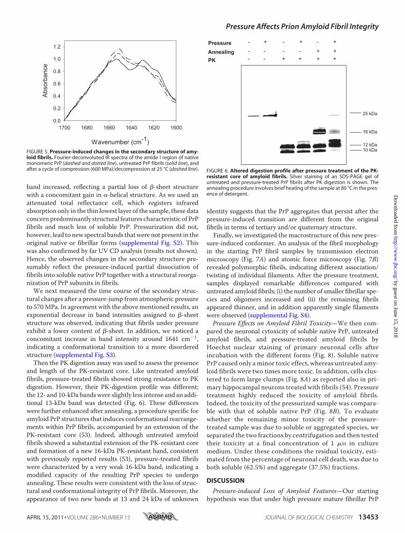
and length of the PK-resistant core. Like untreated amyloidfibrils, pressure-treated fibrils showed strong resistance to PKdigestion. However, their PK-digestion profile was different;the 12- and 10-kDa bandswere slightly less intense and an addi-tional 13-kDa band was detected (Fig. 6). These differenceswere further enhanced after annealing, a procedure specific foramyloid PrP structures that induces conformational rearrange-ments within PrP fibrils, accompanied by an extension of thePK-resistant core (53). Indeed, although untreated amyloidfibrils showed a substantial extension of the PK-resistant coreand formation of a new 16-kDa PK-resistant band, consistentwith previously reported results (53), pressure-treated fibrilswere characterized by a very weak 16-kDa band, indicating amodified capacity of the resulting PrP species to undergoannealing. These results were consistent with the loss of struc-tural and conformational integrity of PrP fibrils. Moreover, theappearance of two new bands at 13 and 24 kDa of unknown
identity suggests that the PrP aggregates that persist after thepressure-induced transition are different from the originalfibrils in terms of tertiary and/or quaternary structure.Finally, we investigated the macrostructure of this new pres-
sure-induced conformer. An analysis of the fibril morphologyin the starting PrP fibril samples by transmission electronmicroscopy (Fig. 7A) and atomic force microscopy (Fig. 7B)revealed polymorphic fibrils, indicating different association/twisting of individual filaments. After the pressure treatment,samples displayed remarkable differences compared withuntreated amyloid fibrils; (i) the number of smaller fibrillar spe-cies and oligomers increased and (ii) the remaining fibrilsappeared thinner, and in addition apparently single filamentswere observed (supplemental Fig. S4).Pressure Effects on Amyloid Fibril Toxicity—We then com-
pared the neuronal cytoxicity of soluble native PrP, untreatedamyloid fibrils, and pressure-treated amyloid fibrils byHoechst nuclear staining of primary neuronal cells afterincubation with the different forms (Fig. 8). Soluble nativePrP caused only aminor toxic effect, whereas untreated amy-loid fibrils were two times more toxic. In addition, cells clus-tered to form large clumps (Fig. 8A) as reported also in pri-mary hippocampal neurons treated with fibrils (54). Pressuretreatment highly reduced the toxicity of amyloid fibrils.Indeed, the toxicity of the pressurized sample was compara-ble with that of soluble native PrP (Fig. 8B). To evaluatewhether the remaining minor toxicity of the pressure-treated sample was due to soluble or aggregated species, weseparated the two fractions by centrifugation and then testedtheir toxicity at a final concentration of 1 �M in culturemedium. Under these conditions the residual toxicity, esti-mated from the percentage of neuronal cell death, was due toboth soluble (62.5%) and aggregate (37.5%) fractions.
DISCUSSION
Pressure-induced Loss of Amyloid Features—Our startinghypothesis was that under high pressure mature fibrillar PrP
FIGURE 5. Pressure-induced changes in the secondary structure of amy-loid fibrils. Fourier-deconvoluted IR spectra of the amide I region of nativemonomeric PrP (dashed and dotted line), untreated PrP fibrils (solid line), andafter a cycle of compression (600 MPa)/decompression at 25 °C (dashed line). FIGURE 6. Altered digestion profile after pressure treatment of the PK-
resistant core of amyloid fibrils. Silver staining of an SDS-PAGE gel ofuntreated and pressure-treated PrP fibrils after PK digestion is shown. Theannealing procedure involves brief heating of the sample at 80 °C in the pres-ence of detergent.
Pressure Affects Prion Amyloid Fibril Integrity
APRIL 15, 2011 • VOLUME 286 • NUMBER 15 JOURNAL OF BIOLOGICAL CHEMISTRY 13453
by guest on June 15, 2018http://w
ww
.jbc.org/D
ownloaded from

structures should dissociate and eventually unfold. The presentresults show that the pressure-induced effects on mature prionamyloid fibrils are more complex than expected. About 30% ofamyloid fibrils becamedissociated into soluble species, whereasthe remaining fibrils stayed in fibrillar form but lost some of theamyloid-specific features.High pressure refolding of temperature-induced �-sheet-
rich aggregates of a mammalian prion protein has been previ-ously reported (55, 56). However, to our knowledge, none of the�-sheet-rich misfolded conformers was described to displayamyloid fibril structure. In line with these reported effects ofpressure on PrP aggregates, our in vitro results provide the firstevidence that undermild physicochemical conditions (ambienttemperature, absence of extraneous chemical agents) the use ofhigh pressure can irreversible change the PrP fibril structureand unlock PrP from amyloid fibrils. It is, therefore, reasonableto think that unlocking PrP from the aggregate state could alsoaccount for the clearance process of amyloids observed in vivo(26–29). Because previous reports show that amyloid fibrilsprepared from full-length recombinant mammalian prion pro-
tein are highly toxic to cultured cells and primary neurons (54)and display infectivity (57), our results may also explain thedecreased resistance to proteolytic digestion and the reductionof the infectivity titer of brain homogenates, processed meat,and isolated pathogenic isoform, PrPSc (58–60) upon pressuretreatment. Nevertheless, caution should be taken in extrapolat-ing these findings, as the mechanisms linking PrP structuraltransformation to infectivity and neuropathological changescharacteristic of prion diseases remain enigmatic and underdebate (61).After pressurization amyloid fibrils seem to form a new, not
yet described PrP structural species in which the fibrillar mac-rostructure is similar to that of the original amyloid fibrils butthe secondary, tertiary, and quaternary structures are signifi-cantly different, as judged from the lack of ANS (tertiary struc-ture) and ThT binding (quaternary structure), the strongdecrease in �-sheet content, and relative increase of �-helices(secondary structure) in comparison to untreated fibrils. Thestructural changes are further confirmed by the different PKdigestion patterns of treated and untreated fibrils. Presumably,
FIGURE 7. Macrostructure of the new pressure-induced conformer. Negative-stained transmission electron micrographs (A) and atomic force microscopyimages (B) of PrP fibrils collected before (left panels) and after a cycle of compression (600 MPa)/decompression at 25 °C. Arrowheads show small oligomers andindividual filaments.
Pressure Affects Prion Amyloid Fibril Integrity
13454 JOURNAL OF BIOLOGICAL CHEMISTRY VOLUME 286 • NUMBER 15 • APRIL 15, 2011
by guest on June 15, 2018http://w
ww
.jbc.org/D
ownloaded from

as a result of pressure-induced conformational rearrangementswithin the fibrillar substructure, the PrP central region (previ-ously described as encompassing residues �90–140) (53)adopts an altered conformation with a more tightly packedstructure that in turn may account for the modified annealingbehavior. We, thus, suggest that pressurized fibrils should beconsidered as a new alternative conformation that can bereached by a protein under certain physicochemical conditions.Indeed, previous insight into unexplored conformational statesof the recombinant cellular PrP isoform was obtained using
high pressure (55, 56, 62–66). The results revealed that thesePrP states are distinguished by their volumetric properties (i.e.hydration and packing).How to explain these pressure-induced fibril structural
changes? Due to the pressure dependence of the chemicalpotential under equilibrium or micro-equilibrium conditions,pressure influences chemical reactions that are characterizedby a net volume change (67). For proteins, these volumechanges arise from contributions of internal cavities and inter-actions with hydrationwater (68). Our findings suggest that the
FIGURE 8. Pressure alters cytotoxicity of amyloid fibrils in primary neurons. A, photomicrographs of representative microscopic fields of cells stained withHoechst 33258 and �3-tubulin showing the cytotoxic effect of the different PrP isoforms: soluble monomeric protein (upper panel; Soluble), fibrils (middlepanel), and fibrils after a cycle of compression (600 MPa)/decompression at 25 °C (lower panel; P-treated fibrils). Scale bar, 20 �m. Neurons were cultured for 2days and then incubated with the different PrP isoforms (final concentration, 1 �M). B, neuronal cell death quantification is shown. Each set of data is the meanvalue S.E. (in percentage) of three experiments; five independent microscopic fields were counted for each experiment. Asterisks indicate significantdifferences after Student’s t test. ***, p � 0.001, untreated fibrils versus pressure-treated fibrils; untreated fibrils versus soluble protein. ns, non-significantdifference between soluble protein and pressure-treated fibrils.
Pressure Affects Prion Amyloid Fibril Integrity
APRIL 15, 2011 • VOLUME 286 • NUMBER 15 JOURNAL OF BIOLOGICAL CHEMISTRY 13455
by guest on June 15, 2018http://w
ww
.jbc.org/D
ownloaded from

packing defects and hydrophobic pockets in mature PrP fibril-lar structures disappear upon pressure treatment and that thesum of the dissociated prion protein conformers and the newfibrillar species occupy a smaller volume than the initial amy-loid fibrils, in agreement with previous volumetric measure-ments of amyloid fibrils (35, 40, 69, 70). Moreover, severalrecent reports indicate the existence in amyloid fibrils of welldefinedThTbinding sites (71) that formcavities of about 8–9Åin diameter. The absence of ThT fluorescence in pressurizedfibrils can be explained by the collapse of these cavities, leadingto ThT expulsion. Similarly, loss of ANS binding can beexplained by the fact that ANS binds to intramolecular hydro-phobic pockets, which might disappear in the pressurizedfibrillar species.Apparent Activation Energy Parameters—The kinetic data
on the pressure-induced amyloid structural changes obtainedwith experiments performed at different temperatures andpressures permitted us to gain insight into the kinetic transitionstates of the reaction paths.Particularly, the strong reduction in volume (�V* between
�18 and �24 ml mol�1) suggests that the kinetic transitionstate is significantly more hydrated than the initial amyloidstate. This might arise from the hydration of cavities or voids(due to their collapse) and from the polar alignment of initiallybulk water around charged residues (electrostriction) atmolec-ular surfaces that become exposed in the kinetic transitionstate.Water is indeed expected to occupy a 15% smaller volumewhen transferred from bulk to the first protein hydration shell(72).The kinetic transition and initial amyloid states also differ
significantly in both apparent activation enthalpy andentropy. Several factors may contribute to the increasedapparent activation enthalpy of the kinetic transition states,such as the disruption of atomic packing interactions and theunfavorable energy of solvation of newly exposed surfaces.The significant increase in entropy suggests also a transitionfrom a more ordered, bound structure, to a more disordered,loose structure. The indication of partial protein unfoldingin the course of the activation process, which is suggested byour results, is conceptually attractive as it may help to under-stand why otherwise stable amyloid fibrils dissociate partlyunder high pressure.Multiple Steps—The ruggedness and complexity of the amy-
loid energy landscape, including a wide range of different con-formational stages and the multitude of available pathways,render the interpretation of the kinetic data hypothetical. Nev-ertheless, the existence of two kinetic rate constants for thepressure-induced structural changes of PrP fibrils generallyconcurs with previous kinetic data revealing that �-amyloidfibril association/dissociation occurs in multiple steps (3) andcould imply two different degrees of association/dissociationbetween monomers and the fibril structure. However, we can-not exclude the possibility of two consecutive and dynamicallywell separated processes.The heterogeneity of amyloid fibrils is further supported by
the finding that their pressurization leads to partial dissociationand to a new fibrillar species that does not dissociate whensubjected to a second pressurization cycle. These results can be
interpreted by assuming that before pressure treatment twopopulations of PrP fibrils coexist in the sample; that is, a “par-tially locked” (reversibly bound monomers) form that can bedissociated and a “totally locked” form (irreversibly boundmonomers) that cannot be dissociated. These two conformersare currently thought to be different states occurring duringamyloidogenesis (73–76). Yet the structural differencesbetween these two initial and co-existing populations of fibrilsappear to be subtle. Indeed, the apparent activation parametersof both kinetic phases are rather similar. It is, therefore, likelythat the kinetic transition states of both phases reflect a partiallydisordered or unfolded form of the amyloid fibrils. Dependingon whether the transition state was attained by pressurizingpartially locked or totally locked amyloid fibrils, it relaxes bydissociating or by forming a new stable fibrillar conformer,respectively.Cytotoxicity—The issue of reversibility of amyloid formation
may be of significance in assessing ways of stopping and revers-ing toxic and/or infectious species for therapeutic purposes invivo, presuming that reversing amyloid deposition does notresult in an increased production of toxic species. Indeed,although several reports suggest that fibrils themselves possesstoxicity (54, 77–79), oligomeric species are considered to be theprimary cytotoxic species. Here we show that pressure treat-ment of amyloid fibrils results in partial structural changes anddissociation concomitantly with a strongly reduced cytotoxic-ity when compared with initial PrP fibrils. These data supportthe finding that the aggregation state of recombinant PrP iscrucial for cytotoxicity and, even under certain physicochemi-cal conditions, for encoding prion infectivity (57, 80, 81).Intriguingly, the soluble fraction of the pressure-treated fibrilsaccounts for almost two-thirds of its cytotoxic properties. Thestructural details of this low but persisting cytotoxicity remainto be elucidated.AModel of the Effects of Pressure on Amyloid Fibrils—On the
basis of structural studies of PrP fibrils (82) and a single-fibrilimmunoconformational study (83), PrP protofilaments can beconsidered as composed of an outer and an inner layer of�-strands characterized by different degrees of compactnessand packing. Based on this model, the pressure-induced struc-tural changes of mature amyloid fibrils can be schematized asfollows.In one population of initial PrP fibrils, high pressure converts
the outer layer into a random coil conformation (supplementalFigs. S2 and S3), whereas the inner layer remains intact (sup-plemental Fig. S2) (Fig. 9A). The higher susceptibility of the�-sheeted outer layer results from poorly optimized side-chainpacking interactions that give rise to void volumes. As pressureis released, this structurally altered region tends to refold intoits native secondary structure, which for PrP is likely to be�-helical, as suggested also by the changes in the second deriv-atives of the FTIR spectra (supplemental Fig. S2). The unfoldingand the subsequent alternative refolding of the fibrillar outerlayer disrupt ThT and ANS binding and lead to the alteredproteolytic cleavage after PK treatment. In addition, the signif-icant thinning of the remaining fibrils observed upon pressur-ization (supplemental Fig. S4) supports the hypothesis that bysuppressing intermolecular interactions between the outer
Pressure Affects Prion Amyloid Fibril Integrity
13456 JOURNAL OF BIOLOGICAL CHEMISTRY VOLUME 286 • NUMBER 15 • APRIL 15, 2011
by guest on June 15, 2018http://w
ww
.jbc.org/D
ownloaded from

�-strands, pressure promotes lateral disassembly or untwistingof bundles of two or more proto-filaments. The resulting newfibrillar structures, which are insensitive to further pressuretreatment, therefore consist of well packed amyloid fibrils with-out structured hydrophobic or �-sheeted domains at the pro-tein surface, where compounds such as ANS or ThT can bind.In another population of initial PrP fibrils, the partial dis-
aggregation of the sample into native PrP monomers (Fig.9B) could be attributed to the presence of proto-filamentsthat are characterized by a lower degree of packing in theinner layer and by internal cavities and hydrophobic pockets.Under such conditions, the PrP assembly, being highly sen-sitive to pressure, could easily attain new conformationalcoordinates, leading to unfolded PrP that, as observed in thepresent work, appears to refold into the native conformationafter pressure release.Conclusion—This work highlights the value of studying
pressure-induced destruction of amyloid features and disso-ciation of prion fibrils. Further development of this approachshould allow us to gain fundamental knowledge on the solu-bilization and degradation mechanisms of large proteina-ceous deposits and on the biological effects of prions. More-over, because amyloid fibrils are associated with a number ofdiseases, such as Alzheimer disease, type 2 diabetes, senileamyloidosis, and dialysis-related amyloidosis, we envisagethat the ability to study pressure-induced PrP amyloid fibrilstructural changes could be transferred to other specificamyloidogenic proteins. Furthermore, the finding that pres-
sure can be used to convert an amyloid to a non-amyloidfibril conformer might be interesting for the study of pro-teins that are forming non-amyloid fibrils, such as lithos-tathine (84, 85).
Acknowledgment—We are grateful for the expert advice of Dr. R.Sabate.
REFERENCES1. Fandrich, M. (2007) Cell. Mol. Life Sci. 64, 2066–20782. Sipe, J. D., and Cohen, A. S. (2000) J. Struct. Biol. 130, 88–983. Dobson, C. M. (2003) Nature 426, 884–8904. Fandrich, M., and Dobson, C. M. (2002) EMBO J. 21, 5682–56905. Jimenez, J. L., Guijarro, J. I., Orlova, E., Zurdo, J., Dobson, C.M., Sunde,M.,
and Saibil, H. R. (1999) EMBO J. 18, 815–8216. Pertinhez, T. A., Bouchard, M., Tomlinson, E. J., Wain, R., Ferguson, S. J.,
Dobson, C. M., and Smith, L. J. (2001) FEBS Lett. 495, 184–1867. Breydo, L., Makarava, N., and Baskakov, I. V. (2008) Methods Mol. Biol.
459, 105–1158. Fernandez-Busquets, X., de Groot, N. S., Fernandez, D., and Ventura, S.
(2008) Curr. Med. Chem. 15, 1336–13499. Langkilde, A. E., and Vestergaard, B. (2009) FEBS Lett. 583, 2600–260910. Chamberlain, A. K., MacPhee, C. E., Zurdo, J., Morozova-Roche, L. A.,
Hill, H. A., Dobson, C.M., and Davis, J. J. (2000) Biophys. J. 79, 3282–329311. Chiti, F., and Dobson, C. M. (2006) Annu. Rev. Biochem. 75, 333–36612. Harper, J. D., Lieber, C. M., and Lansbury, P. T., Jr. (1997) Chem. Biol. 4,
951–95913. Jimenez, J. L., Nettleton, E. J., Bouchard, M., Robinson, C. V., Dobson,
C. M., and Saibil, H. R. (2002) Proc. Natl. Acad. Sci. U.S.A. 99, 9196–920114. Bauer, H. H., Aebi, U., Haner, M., Hermann, R., Muller, M., and Merkle,
FIGURE 9. Schematic representation of the effects of high pressure on mature PrP amyloid fibrils. We considered that each PrP proto-filament consistedof an outer and an inner layer of �-strands. This diagram illustrates the proposed model of pressure-induced structural changes, which depends on thethermodynamic state (partially locked or locked) adopted by PrP proteins in the proto-filament. A, after pressure treatment, fibrils composed of totally lockedproto-filaments convert into morphologically different fibrils characterized by an irreversible loss of several amyloid specific features. B, partially lockedproto-filaments, characterized by a lower degree of packing, disaggregate into native monomers after a pressure-induced unfolding-refolding process. Kineticdata reveal that both pathways proceed via a mechanistically similar unfolding process that implies a disordered and hydrated kinetic transition state. Furtherdetails are specified under “Discussion.”
Pressure Affects Prion Amyloid Fibril Integrity
APRIL 15, 2011 • VOLUME 286 • NUMBER 15 JOURNAL OF BIOLOGICAL CHEMISTRY 13457
by guest on June 15, 2018http://w
ww
.jbc.org/D
ownloaded from

H. P. (1995) J. Struct. Biol. 115, 1–1515. Fandrich, M., Meinhardt, J., and Grigorieff, N. (2009) Prion 3, 89–9316. Goldsbury, C. S., Cooper, G. J., Goldie, K. N., Muller, S. A., Saafi, E. L.,
Gruijters, W. T., Misur, M. P., Engel, A., Aebi, U., and Kistler, J. (1997) J.Struct. Biol. 119, 17–27
17. Jimenez, J. L., Tennent, G., Pepys, M., and Saibil, H. R. (2001) J. Mol. Biol.311, 241–247
18. Madine, J., Jack, E., Stockley, P. G., Radford, S. E., Serpell, L. C., andMiddleton, D. A. (2008) J. Am. Chem. Soc. 130, 14990–15001
19. Meinhardt, J., Sachse, C., Hortschansky, P., Grigorieff, N., and Fandrich,M. (2009) J. Mol. Biol. 386, 869–877
20. Paravastu, A. K., Leapman, R. D., Yau, W. M., and Tycko, R. (2008) Proc.Natl. Acad. Sci. U.S.A. 105, 18349–18354
21. Pedersen, J. S., Dikov, D., Flink, J. L., Hjuler, H. A., Christiansen, G., andOtzen, D. E. (2006) J. Mol. Biol. 355, 501–523
22. Petkova, A. T., Leapman, R. D., Guo, Z., Yau, W. M., Mattson, M. P., andTycko, R. (2005) Science 307, 262–265
23. Bucciantini, M., Giannoni, E., Chiti, F., Baroni, F., Formigli, L., Zurdo, J.,Taddei, N., Ramponi, G., Dobson, C. M., and Stefani, M. (2002) Nature416, 507–511
24. Conway, K. A., Lee, S. J., Rochet, J. C., Ding, T. T., Williamson, R. E., andLansbury, P. T., Jr. (2000) Proc. Natl. Acad. Sci. U.S.A. 97, 571–576
25. Carulla, N., Caddy, G. L., Hall, D. R., Zurdo, J., Gairí, M., Feliz, M., Giralt,E., Robinson, C. V., and Dobson, C. M. (2005) Nature 436, 554–558
26. Hawkins, P. N., Richardson, S., MacSweeney, J. E., King, A. D., Vigushin,D. M., Lavender, J. P., and Pepys, M. B. (1993) Q. J. Med. 86, 365–374
27. Hawkins, P. N., Richardson, S., Vigushin, D. M., David, J., Kelsey, C. R.,Gray, R. E., Hall, M. A., Woo, P., Lavender, J. P., and Pepys, M. B. (1993)Arthritis Rheum. 36, 842–851
28. Rydh, A., Suhr, O., Hietala, S. O., Ahlstrom, K. R., Pepys,M. B., andHawk-ins, P. N. (1998) Eur. J. Nucl. Med. 25, 709–713
29. Tan, S. Y., Irish, A., Winearls, C. G., Brown, E. A., Gower, P. E., Clutter-buck, E. J., Madhoo, S., Lavender, J. P., Pepys, M. B., and Hawkins, P. N.(1996) Kidney Int. 50, 282–289
30. Calamai,M., Canale, C., Relini, A., Stefani,M., Chiti, F., andDobson, C.M.(2005) J. Mol. Biol. 346, 603–616
31. Callahan, M. A., Xiong, L., and Caughey, B. (2001) J. Biol. Chem. 276,28022–28028
32. Loksztejn, A., and Dzwolak, W. (2009) Biochemistry 48, 4846–485133. MacPhee, C. E., and Dobson, C. M. (2000) J. Mol. Biol. 297, 1203–121534. Morel, B., Varela, L., and Conejero-Lara, F. (2010) J. Phys. Chem. B. 114,
4010–401935. Abdul Latif, A. R., Kono, R., Tachibana, H., and Akasaka, K. (2007) Bio-
phys. J. 92, 323–32936. Chatani, E., Kato, M., Kawai, T., Naiki, H., and Goto, Y. (2005) J. Mol. Biol.
352, 941–95137. Dirix, C.,Meersman, F.,MacPhee, C. E., Dobson, C.M., andHeremans, K.
(2005) J. Mol. Biol. 347, 903–90938. Foguel, D., Suarez,M. C., Ferrao-Gonzales, A. D., Porto, T. C., Palmieri, L.,
Einsiedler, C. M., Andrade, L. R., Lashuel, H. A., Lansbury, P. T., Kelly,J. W., and Silva, J. L. (2003) Proc. Natl. Acad. Sci. U.S.A. 100, 9831–9836
39. Grudzielanek, S., Smirnovas, V., and Winter, R. (2006) J. Mol. Biol. 356,497–509
40. Kamatari, Y. O., Yokoyama, S., Tachibana, H., and Akasaka, K. (2005) J.Mol. Biol. 349, 916–921
41. Meersman, F., and Dobson, C. M. (2006) Biochim. Biophys. Acta 1764,452–460
42. Mishra, R., and Winter, R. (2008) Angew. Chem. Int. Ed. Engl. 47,6518–6521
43. Font, J., Torrent, J., Ribo, M., Laurents, D. V., Balny, C., Vilanova, M., andLange, R. (2006) Biophys. J. 91, 2264–2274
44. Torrent, J., Font, J., Herberhold, H., Marchal, S., Ribo, M., Ruan, K., Win-ter, R., Vilanova, M., and Lange, R. (2006) Biochim. Biophys. Acta 1764,489–496
45. Vidugiris, G. J., Markley, J. L., and Royer, C. A. (1995) Biochemistry 34,4909–4912
46. Woenckhaus, J., Kohling, R., Thiyagarajan, P., Littrell, K. C., Seifert, S.,Royer, C. A., and Winter, R. (2001) Biophys. J. 80, 1518–1523
47. Rezaei, H.,Marc, D., Choiset, Y., Takahashi,M., Hui BonHoa, G., Haertle,T., Grosclaude, J., and Debey, P. (2000) Eur. J. Biochem. 267, 2833–2839
48. Forst, P.,Werner, F., and Delgado, A. (2000) Rheologica Acta 39, 566–57349. Meersman, F., and Heremans, K. (2003) Biophys. Chem. 104, 297–30450. Bocharova, O. V., Breydo, L., Salnikov, V. V., Gill, A. C., and Baskakov, I. V.
(2005) Protein Sci. 14, 1222–123251. Pace, C. N. (2009) Nat. Struct. Mol. Biol. 16, 681–68252. James, T. L., Liu, H., Ulyanov, N. B., Farr-Jones, S., Zhang, H., Donne,
D. G., Kaneko, K., Groth, D., Mehlhorn, I., Prusiner, S. B., and Cohen, F. E.(1997) Proc. Natl. Acad. Sci. U.S.A. 94, 10086–10091
53. Bocharova, O. V., Makarava, N., Breydo, L., Anderson,M., Salnikov, V. V.,and Baskakov, I. V. (2006) J. Biol. Chem. 281, 2373–2379
54. Novitskaya, V., Bocharova, O. V., Bronstein, I., and Baskakov, I. V. (2006)J. Biol. Chem. 281, 13828–13836
55. Cordeiro, Y., Kraineva, J., Ravindra, R., Lima, L. M., Gomes, M. P., Foguel,D., Winter, R., and Silva, J. L. (2004) J. Biol. Chem. 279, 32354–32359
56. Torrent, J., Alvarez-Martinez,M.T., Heitz, F., Liautard, J. P., Balny, C., andLange, R. (2003) Biochemistry 42, 1318–1325
57. Makarava, N., Kovacs, G. G., Bocharova, O., Savtchenko, R., Alexeeva, I.,Budka, H., Rohwer, R. G., and Baskakov, I. V. (2010) Acta Neuropathol.119, 177–187
58. Brown, P., Meyer, R., Cardone, F., and Pocchiari, M. (2003) Proc. Natl.Acad. Sci. U.S.A. 100, 6093–6097
59. Fernandez García, A., Heindl, P., Voigt, H., Buttner, M., Wienhold, D.,Butz, P., Starke, J., Tauscher, B., and Pfaff, E. (2004) J. Gen. Virol. 85,261–264
60. Heindl, P., FernandezGarcía, A., Buttner,M., Voigt,H., Butz, P., Tauscher,B., and Pfaff, E. (2005) Braz. J. Med. Biol. Res. 38, 1223–1231
61. Aguzzi, A., Sigurdson, C., and Heikenwaelder, M. (2008) Annu. Rev.Pathol. 3, 11–40
62. Alvarez-Martinez, M. T., Torrent, J., Lange, R., Verdier, J. M., Balny, C.,and Liautard, J. P. (2003) Biochim. Biophys. Acta 1645, 228–240
63. El Moustaine, D., Perrier, V., Smeller, L., Lange, R., and Torrent, J. (2008)FEBS J. 275, 2021–2031
64. Silva, J. L., Vieira, T. C., Gomes, M. P., Bom, A. P., Lima, L. M., Freitas,M. S., Ishimaru, D., Cordeiro, Y., and Foguel, D. (2010)Acc. Chem. Res. 43,271–279
65. Torrent, J., Alvarez-Martinez, M. T., Harricane, M. C., Heitz, F., Liautard,J. P., Balny, C., and Lange, R. (2004) Biochemistry 43, 7162–7170
66. Torrent, J., Alvarez-Martinez, M. T., Liautard, J. P., Balny, C., and Lange,R. (2005) Protein Sci. 14, 956–967
67. Van Eldik, R., Asano, T., and Le Noble, W. J. (1989) Chem. Rev. 89,549–688
68. Silva, J. L., Foguel, D., and Royer, C. A. (2001) Trends Biochem. Sci. 26,612–618
69. Akasaka, K., Latif, A. R., Nakamura, A., Matsuo, K., Tachibana, H., andGekko, K. (2007) Biochemistry 46, 10444–10450
70. Niraula, T. N., Konno, T., Li, H., Yamada, H., Akasaka, K., and Tachibana,H. (2004) Proc. Natl. Acad. Sci. U.S.A. 101, 4089–4093
71. Biancalana, M., and Koide, S. (2010) Biochim. Biophys. Acta 1804,1405–1412
72. Merzel, F., and Smith, J. C. (2002) Proc. Natl. Acad. Sci. U.S.A. 99,5378–5383
73. Esler, W. P., Stimson, E. R., Jennings, J. M., Vinters, H. V., Ghilardi, J. R.,Lee, J. P., Mantyh, P. W., and Maggio, J. E. (2000) Biochemistry 39,6288–6295
74. Gobbi, M., Colombo, L., Morbin, M., Mazzoleni, G., Accardo, E., Vanoni,M., Del Favero, E., Cantu, L., Kirschner, D. A.,Manzoni, C., Beeg,M., Ceci,P., Ubezio, P., Forloni, G., Tagliavini, F., and Salmona, M. (2006) J. Biol.Chem. 281, 843–849
75. Maggio, J. E., Stimson, E. R., Ghilardi, J. R., Allen, C. J., Dahl, C. E., Whit-comb, D. C., Vigna, S. R., Vinters, H. V., Labenski, M. E., and Mantyh,P. W. (1992) Proc. Natl. Acad. Sci. U.S.A. 89, 5462–5466
76. Takeda, T., and Klimov, D. K. (2008) Biophys. J. 95, 1758–177277. Gharibyan, A. L., Zamotin, V., Yanamandra, K., Moskaleva, O. S., Margu-
lis, B. A., Kostanyan, I. A., and Morozova-Roche, L. A. (2007) J. Mol. Biol.365, 1337–1349
78. Meyer-Luehmann,M., Spires-Jones, T. L., Prada, C., Garcia-Alloza,M., de
Pressure Affects Prion Amyloid Fibril Integrity
13458 JOURNAL OF BIOLOGICAL CHEMISTRY VOLUME 286 • NUMBER 15 • APRIL 15, 2011
by guest on June 15, 2018http://w
ww
.jbc.org/D
ownloaded from

Calignon, A., Rozkalne, A., Koenigsknecht-Talboo, J., Holtzman, D. M.,Bacskai, B. J., and Hyman, B. T. (2008) Nature 451, 720–724
79. Pieri, L., Bucciantini, M., Nosi, D., Formigli, L., Savistchenko, J., Melki, R.,and Stefani, M. (2006) J. Biol. Chem. 281, 15337–15344
80. Legname, G., Baskakov, I. V., Nguyen, H. O., Riesner, D., Cohen, F. E.,DeArmond, S. J., and Prusiner, S. B. (2004) Science 305, 673–676
81. Wang, F.,Wang, X., Yuan, C. G., andMa, J. (2010) Science 327, 1132–113582. Ostapchenko, V. G., Sawaya, M. R., Makarava, N., Savtchenko, R.,
Nilsson, K. P., Eisenberg, D., and Baskakov, I. V. (2010) J. Mol. Biol.
400, 908–92183. Novitskaya, V.,Makarava, N., Bellon, A., Bocharova, O. V., Bronstein, I. B.,
Williamson, R. A., and Baskakov, I. V. (2006) J. Biol. Chem. 281,15536–15545
84. Laurine, E., Gregoire, C., Fandrich, M., Engemann, S., Marchal, S., Thion,L., Mohr,M., Monsarrat, B., Michel, B., Dobson, C.M.,Wanker, E., Erard,M., and Verdier, J. M. (2003) J. Biol. Chem. 278, 51770–51778
85. Milhiet, P. E., Yamamoto, D., Berthoumieu, O., Dosset, P., Le Grimellec,C., Verdier, J. M., Marchal, S., and Ando, T. (2010) PLoS One 5, e13240
Pressure Affects Prion Amyloid Fibril Integrity
APRIL 15, 2011 • VOLUME 286 • NUMBER 15 JOURNAL OF BIOLOGICAL CHEMISTRY 13459
by guest on June 15, 2018http://w
ww
.jbc.org/D
ownloaded from

TorrentMeersman, Valeriy G. Ostapchenko, Ilia V. Baskakov, Reinhard Lange and Joan Driss El Moustaine, Veronique Perrier, Isabelle Acquatella-Tran Van Ba, Filip
Sensitive to High PressureAmyloid Features and Neuronal Toxicity of Mature Prion Fibrils Are Highly
doi: 10.1074/jbc.M110.192872 originally published online February 25, 20112011, 286:13448-13459.J. Biol. Chem.
10.1074/jbc.M110.192872Access the most updated version of this article at doi:
Alerts:
When a correction for this article is posted•
When this article is cited•
to choose from all of JBC's e-mail alertsClick here
Supplemental material:
http://www.jbc.org/content/suppl/2011/02/25/M110.192872.DC1
http://www.jbc.org/content/286/15/13448.full.html#ref-list-1
This article cites 85 references, 22 of which can be accessed free at
by guest on June 15, 2018http://w
ww
.jbc.org/D
ownloaded from