
Flagellar pocket restructuring through the Leishmania life ...

F L A G E L L A R M O T I O N A N D F I N E
S T R U C T U R E O F T H E F L A G E L L A R
A P P A R A T U S I N CHLAMYDOMONAS
D A V I D L. R I N G O
From The Cell Research Institute, The University of Texas, Austin. The author's present address is The Department of Biology, Yale University, New Haven, Connecticut
A B S T R A C T
The biflagellate alga Chlamydomonas reinhar&" was studied with the light and electron micro- scopes to determine the behavior of flagella in the living cell and the structure of the basal apparatus of the flagella. During normal forward swimming the flagella beat synchronously in the same plane, as in the human swimmer's breast stroke. The form of beat is like that of cilia. Occasionally cells swim backward with the flagella undulating and trailing the cell. Thus the same flagellar apparatus produces two types of motion. The central pair of fibers of both flagella appear to lie in the same plane, which coincides with the plane of beat. The two basal bodies lie in a V configuration and are joined at the top by a striated fiber and at the bottom by two smaller fibers. From the area between the basal bodies four bands of microtubules, each containing four tubules, radiate in an X-shaped pattern, diverge, and pass under the cell membrane. Details of the complex arrangement of tubules near the basal bodies are described. I t seems probable that the connecting fibers and the micro- tubules play structural roles and thereby maintain the alignment of the flagellar apparatus. The relation of striated fibers and microtubules to cilia and flagella is reviewed, particularly in phytoflagellates and protozoa. Structures observed in the transitional region between the basal body and flagellar shaft are described and their occurrence is reviewed. Details of structure of the flagellar shaft and flagellar tip are described, and the latter is reviewed in detail.
I N T R O D U C T I O N
Cells do work in many ways to move themselves, their internal constituents, and their environment. Contraction of muscle cells, beating of cilia and flagella, protoplasmic streaming, and ameboid motion are but a few of the known examples of this phenomenon. Cilia and flagella were first de- scribed by Leeuwenhoek in the 17th ccntury, and his rccord of one such observation, of a biflagellate alga which he saw in 1702, is still relevant to the work to be presented here: "The i r bodies seemed to be composed of particles that represented an oval figure; and therewithal they had [two] short thin instruments [i.e., flagella] which stuck out a
little way from the round contour, and wherewith they performed the motions of rolling around and going forward." 1
In recent years the application of the electron microscope to biological materials has resulted in a wealth of information on cilia and flagella (11, 24, 27, 86, 88, 108, 112), but the mechanism of their motion remains a subject for speculation.
1 Quoted lrom the translation of Dobell (21), who identifies the organism as Haematococcus and indicates that Leeuwenhoek probably saw the smaller-celled Chlamydornonas in the same sample of water from his gutter.
543
Dow
nloaded from http://rupress.org/jcb/article-pdf/33/3/543/1383807/543.pdf by guest on 31 January 2022

That the same morphological pat tern of 9 + 2
units can produce a variety of motions, even in a
single cell, is a simple paradox which speaks
against an easy morphological solution to the
question of flagellar motion, but this same paradox
leads us to probe more deeply into the structure of
the organelle, since such a high degree of organiza-
tion demands some relation between form and
function. The present study attempts to approach
the problem on several levels: (a) the function of
flagella of living cells, (b) the structure of the
flagellum and its basal apparatus, and (c) correla-
tions between structure and function, when they
can be observed. A fourth level, the molecular
architecture of flagellar fibers, is the subject of a
separate paper (91).
The organism under study was the biflagellate
alga Chlamydomonas, a eukaryotic cell whose small
size and relative ease of culture under controlled
conditions recommends it for study. The genetics
and physiology of this organism have been in-
vestigated fairly extensively (see reviews by Levine
and Ebersold, reference 59, and Sager, reference
101), and the basic pat tern of its ultrastructure is
known from the work of Sager and Palade (103,
104) and the related studies of Ris and Plaut (92)
and Lang (53-55). In addition, the preliminary
work of Gibbs et al. (39) has given good indication
of the complexity of the flagellar apparatus in
Chlamydornonas.
M A T E R I A L S A N D M E T H O D S
Culture Techniques
Chlamydomonas reinhardi Dangeard, strain 21gr, from the laboratory of Dr. Ruth Sager, was grown in synchronous culture (10, 48) using a cycle of 15 hr light and 9 hr dark. Cells were removed for fixation during the light period, when the cells are motile and synthesis and growth occur. 2 Cultures were grown at 25°C in Medium I of Sager and Granick (102) and were bubbled with air to which approximately 1 CO2 was added.
2 Vegetative cells from nonsynchronous cultures, gametes, and zygotes soon after gamctc fusion were also studied to some extent, representing all the motile stages of the life-cycle of the organism, and showed no obvious departures from the general pattern of flagcllar structure to be described in synchronous vegetative cells.
Light Microscopy The mode of swimming and motion of flagella of
C. reinhardi were studied by observing cells in viscous media. Either gelatin or agar was added to the me- dium to slow the frequency of flagellar beat to about one per second; cells were then observed at room temperature by phase-contrast microscopy. In other experiments, cells swimming actively in normal medium were photographed with phase-contrast or interference-contrast optics using electronic flash illumination of 0.3-0.5 msec duration.
Electron Microscopy Cells were fixed in 2% glutaraldehyde (100),
washed, and then postfixed in 1% OsO4, with 0.05 ~ collidine buffer (9), pH 7.5, as the wash solution and as the vehicle for both fixatives. Material was embedded by conventional techniques in Epon- Araldite mixture No. 1 of Mollenhaucr (80) or in Luft's Epon (63). Sections were cut with a diamond knife on an LKB microtome and examined with an RCA EMU-3F electron microscope.
Information on the structure of the basal apparatus was derived to some extent from serial sections but largely from single sections of random orientation. In order to construct a three-dimensional model of the structure of the basal apparatus from random sections, it was necessary to know the orientation of each cell with respect to the section in which it ap- peared, so that information from many orientations could be correlated. Since the major structures of the C. reinhardi cell (the chloroplast, pyrenoid, nucleus, contractile vacuole, and flagella) have a fairly con- stant orientation with respect to one another (Fig. 1), it was possible to use these organelles as morphological markers, and, from their appearance in a micrograph, to determine at what level and angle the section had passed through the cell.
Terminology The terminology associated with motility structures
is both confusing and inconsistent, since many of the terms commonly used apply to a single structure while some have more than one meaning. In general I have tried to follow the terminology of Gibbons (37, 38) and Sleigh (112) whenever possible, to introduce as few new terms as possible, and to give preference to names which are more or less self-explanatory..
For purposes of discussion, the flagellum is divided into four portions: (a) the tip, the region at the end of the flagellum, characterized by an alteration and disorganization of the normal 9 + 2 pattern of the flagellar fibers; (b) the main shaft of the flagellum, which shows a constant 9 + 2 pattern in cross-section; (c) the transitional rcgion, between the shaft and the basal body, where the central fibcrs end and the
544 THE JOCRNAL OF CELL BIOLOGY " VOLUME 38, 1967
Dow
nloaded from http://rupress.org/jcb/article-pdf/33/3/543/1383807/543.pdf by guest on 31 January 2022

FmuaE 1 General view of a longitudinal section of the Chlamydomonas reinhardi cell showing the two flagella a t the anterior end, with a striated fiber joining the basal bodies of the flagella. The nucleus lies near the center of the cell, surrounded by the single chloroplast which contains a pyrenoid (lower end) and many starch grains (light areas). Golgi regions lie near the nucleus but are not well preserved in this fixation. X 19,000.
545
Dow
nloaded from http://rupress.org/jcb/article-pdf/33/3/543/1383807/543.pdf by guest on 31 January 2022

flagellum shows alterations in its cross-sectional pattern; and (d) the basal body, the portion of the flagellum ending in the cytoplasm, where the nine peripheral fibers show a triple structure in cross- section (37).
The terms "anterior" and "posterior" are used to refer to the whole cell, the anterior end being the one which bears the flagella. "Proximal" and "distal" refer to points along the length of the flagellum; the proximal end of the flagellum corresponds to the end of the basal body lying deepest in the cytoplasm. "Below" and "above" will occasionally be used to indicate a proximal or distal direction, respectively.
The flagellar apparatus of C. reinhardi is sufficiently complicated that verbal description of the structures is difficult, and reference should be made to the explanatory drawings which accompany the text (Figs. 27, 28, and 30).
R E S U L T S
Swimming
The motion of flagella of cells in viscous media is diagrammed in Fig. 2, while Figs. 3-10 show the same patterns in high-speed flash photographs of cells swimming freely in normal medium. In normal forward swimming, cells travel with the anterior (flagella-bearing) end toward the direc- tion of motion. At the beginning of the power stroke the flagella are straight and extended forward. The flagellum sweeps backward, remain- ing fairly straight and bending near the base. The return stroke begins as the power stroke is com- pleted, with a wave of bending passing along the
FIGURE
Power stroke
Return stroke
Drawings indicating the position of C. reinhardi flagella during forward swimming, as deter- mined by observing cells in viscous media. Beating is synchronous and resembles that of cilia.
flagellum from base to tip; this restores the flagel- lum to its original position.
The positional relationship of the flagella is normally bilaterally symmetrical, the synchrony of motion being analogous to that of a human swimmer's breast stroke, but synchrony may be disturbed for brief periods. The beat of the flagel- lum is planar, and both flagella beat in the same plane, which also coincides with the central longitudinal axis of the cell (Fig. 10). Chlamydomonas reinhardi cells show little or no rotation as they swim; the appearance of rapid vibration of the forward-swimming cell is due to the alternate acceleration and deceleration of the cell dur ingthe power and return strokes.
Another type of swimming was occasionally observed in which cells reverse their direction and swim backward for short periods. In this case the flagella-bearing end of the cell faces away from the direction of motion; the flagella are extended and lie near one another (Figs. 11 and 12). Waves of bending, probably traveling from base to tip, pass along the flagella and exert a pushing force. Beating appears to be synchronous, and Fig. 11 indicates that the planar relationship of flagellar beating may be the same in backward swimming as in forward swimming. In backward swimming the cells travel smoothly, without the vibration seen in forward swimming, although the velocity is less. When cells were exposed to high intensity electronic-flash illumination (60 watt-seconds, 0.5 msec) during photography, a high percentage of the ceils stopped their forward motion, swam backward for about a second, and then resumed forward swimming.
Structure of the Basal Apparatus
GENERAL MORPHOLOGY
The two flagella are inserted in the cell through specialized regions of the cell wall. Their basal bodies lie in a V configuration and are joined at the top by a striated fiber and at the bottom by two smaller fibers (Figs. 13 and 30). From the area be- tween the basal bodies four bands of microtubules radiate in an X-shaped pattern, diverge, and pass under the cell membrane (Fig. 14). All of these structures will be described in detail below.
MICROTUBULE SYSTEM
The microtubule bands, seen near the basal bodies, each consist of four tubules of about 30
546 THE JOURNAL OF CELL BIOLOGY • VOLUME 33, 1967
Dow
nloaded from http://rupress.org/jcb/article-pdf/33/3/543/1383807/543.pdf by guest on 31 January 2022

FIGURES B-I~ Light micrographs of cells swimming in normal medium. Figs. B-8 show flagella in vari- ous positions during forward swimming. The cell in Fig. 9 shows slightly disturbed synchrony. Fig. 10 shows a cell whose plane of beat was perpendicular to the plane of focus of the microscope when it was photographed during the return stroke, indicating the relationship between the plane of beat of the flagella and the longitudinal axis of the cell. Figs. 11 and 1~ show the pattern of flagellar motion in backward swimming. In all photographs the direction of motion of the cells is toward the top of the page. High-speed electronic flash illumination; Figs. 3-11, interference contrast; Fig. 1~, phase contrast. )< 1,000 (approximate).
mg diameter in a 3-over-1 pattern (Figs. 19-21). As the bands radiate from the basal bodies, they pass near the cell membrane as a flat sheet, still with four tubular components (Figs. 18, 25, and 26). Once the tubules reach their position near the cell membrane, single tubules presumably diverge from the pattern; this leaves bands of three (Fig. 17) or two tubules (Fig. 16) which are frequently seen at the anterior end of the cell in four low ridges of cytoplasm which radiate from the basal- body region (Figs. 16-18). Individual tubules proceed for different distances just beneath the cell membrane. In a transverse section of a cell at the level of the nucleus, eight to ten tubules are often seen clearly in cross-section spaced around the periphery of the cell (Fig. 15), and all 16 tubales probably persist at least this far. The tubules are quite close to the cell membrane and often are surrounded by a region free of ribosomes. A few tubules are present in cross-section at the level of the pyrenoid (near the posterior end of the cell), so that the system of tubules encloses the whole cell to a certain extent. A diagrammatic interpretation of this pattern is given in Fig. 28.
The arrangement of the tubules near the basal bodies is quite complex. As noted above, the four bands, of four tubules each, approach the area between the basal bodies in a 3-over-1 configura- tion (Figs. 19-21). One of the tubules ends, prob- ably the single one on the bottom of the 3-over-1 pattern, to leave a band of three tubules (Fig. 22). Still closer to the midpoint between the basal bodies another tubule ends. Above the remaining pair of tubules a flat plate of electron-opaque material is present (Fig. 23). In the very center of the area between the basal bodies no tubules re- main; only the dense plate remains which lies just below the distal striated fiber (Figs. 1 and 13). Thus the tubule bands are not continuous with one another, and no direct connection between the tubules and any part of the basal apparatus has been demonstrated by the procedures used.
A possible interpretation of the different tubule patterns observed is given in the drawing in Fig. 27. This diagram indicates that the tubule of the triad which ends before the final pair is the one nearest to the center of the basal-body complex, and that it moves slightly upward to end just at the
DAVID L. I~INGO FIagellar Apparatus in Chlamydomonas 547
Dow
nloaded from http://rupress.org/jcb/article-pdf/33/3/543/1383807/543.pdf by guest on 31 January 2022
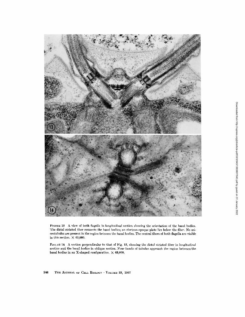
FIGURE 13 A view of both flagella in longitudinal section showing the orientation of the basal bodies. The distal striated fiber connects the basal bodies; an electron-opaque plate lies below the fiber. No mi- crotubules are present in the region between the basal bodies. The central fibers of both flagella are visible in this section. × 63,000.
FIGURE 14 A section perpendicular to that of Fig. 13, showing the distal striated fiber in longitudinal section and the basal bodies in oblique section. Four bands of tubules approach the region between the basal bodies in an X-shaped configuration. × 63,000.
548 TH~ JOURNAL OF CELL BIOLOGY " VOLUME 33, 1967
Dow
nloaded from http://rupress.org/jcb/article-pdf/33/3/543/1383807/543.pdf by guest on 31 January 2022

Provide 15 Transverse section of a cell intersecting the nucleus and nucleolus (lower left). Six micro- tubules are present just beneath the cell membrane (arrows). X ~8,500.
FIGUREs 16 and 17 Groups of two and three microtubules in cross-section near the anterior end of the cell. Contractile vacuole regions are at the lower left in Fig. 16 and the lower right in Fig. 17. X 55,500.
DAVID L. RINGO Flagellar Apparatus in Chlamydomonas 549
Dow
nloaded from http://rupress.org/jcb/article-pdf/33/3/543/1383807/543.pdf by guest on 31 January 2022

FmvnE 18 Band of four microtubules a t the anterior end of a cell. Other single tubules are also present. The contractile vacuole region is near the center of the picture. )< 59,000.
FIGURES 19 and ~0 Successive serial sections through the anterior region of a cell, showing one micro- tubule band (3-over-1 configuration) in cross-section as it approaches the flagellar base (flagellum is seen at left in grazing section). The other tubule band approaching the basal bodies from that side is sectioned obliquely (B in Fig. 19, right-hand arrow in Fig. 20). A transitional fiber is shown in cross-section in Fig. 19 (TR) and connecting to the flagellar fiber in Fig. o~0 (left-hand arrow). )< 59~000.
550 THE ~OURNAL OF CELL BIOLOGY • VOLUME 33, 1967
Dow
nloaded from http://rupress.org/jcb/article-pdf/33/3/543/1383807/543.pdf by guest on 31 January 2022

FIGURES !~1--~8 Microtubule patterns near the flagellar bases. Fig. ~1 shows the 8-over-1 configuration slightly closer to the basal bodies than in Figs. 19 and ~0. The other tubule band approaches obliquely from the left. Fig. £~ shows the band of three tubules very near the basal body. The other band of tu- bules is at the left. Fig. ~8 shows a band of two tubules in the region between the basal bodies. Below the tubules is the proximal connecting fiber; above the tubules is the electron-opaque plate associated with tile tubule ends, and immediately above that is the distal connecting fiber. )< 55,500.
FIGURE ~4 This section has grazed the proximal ends of both basal bodies, which appear as crescent- shaped areas. The two proximal striated fibers are shown connecting the basal bodies at this level. X 45,000.
level of the dense plate (Figs. 25 and 26). This reconstruct ion also accounts for the fact tha t only one set of tubules is visible in cross-section in a given cell (Figs. 19-23). T h e arched pa th of the tubule bands, as they proceed from the basal-body region to the cell surface (Fig. 25), explains why the tubule bands only appear for relatively short
distances when encountered in longi tudinal section (Figs. 14, 26, and 41). I t is possible tha t there are addi t ional microtubules in the anter ior region of the cell which do not form a par t of the 16-member tubule network (see Fig. 18), bu t it is also possible tha t such views, seen infrequently, represent a stage in the replication of the 16 tubule system.
DAVID L. RINGO Flagellar Apparatus in Chlamydomonas 551
Dow
nloaded from http://rupress.org/jcb/article-pdf/33/3/543/1383807/543.pdf by guest on 31 January 2022

FmVRE ~5 Two mierotubule bands in longitudinal section, showing their form as they approach the cell membrane. The proximal end of one basal body is present in oblique section. The larger electron- opaque area is the distal connecting fiber. Immediately below it is the electron-opaque plate. One tubule ends at the level of the plate. X 48,000.
FIGURE ~6 A transverse section of the anterior end of a cell at a level just below the distal striated fiber, showing two of the microtubule bands approaching the central region. )< 55,000.
STRIATED FIBERS
The distal str iated fiber connects the two basal bodies at a level jus t below the top of the basal body (see Fig. 30). T h e fiber, seen in "ver t ica l" longi tudinal section in Figs. 1 and 13, in "hor i -
zonta l" longi tudinal section in Fig. 14, and in
cross-section in Fig. 29, is abou t 300 m ~ long, 250
m # wide, and 75 m # thick. T h e pa t te rn of cross-
banding, seen in properly or iented longi tudinal
sections, is complex and bi lateral ly symmetrical .
552 T H E JOURNAL OF CELL BIOLOGY • VOLUME 83, 1967
Dow
nloaded from http://rupress.org/jcb/article-pdf/33/3/543/1383807/543.pdf by guest on 31 January 2022

A pair of dark lines lies in the center, wi th a l ight line and another pair of dark lines on ei ther side; the spacing of the central pair of striations is nar- rower than tha t of the outer pairs. Dimensions of the striation pa t te rn are given in Fig. 31. Fine fi laments runn ing the length of the striated fiber are occasionally observed (Figs. 13 and 14), al- though this is not shown clearly by the present fixation.
Two smaller striated fibers, the proximal con- nect ing fibers, link the basal bodies at their proxi- mal ends. The long axis of these fibers is in the same direct ion as tha t of the distal striated fiber, but they are connected at the outside edge of the basal body on ei ther side, and thus have a distance of abou t one basal body d iameter separat ing them (see Figs. 28 and 30). Both of the proximal fibers are present in Fig. 24, where only the very tips of the basal body have been cut by the section. In Fig. 29 they are seen in cross-section as two ill- defined, roughly t r iangular pieces of mater ia l lying below the distal fiber. One of the pa i r of fibers is
/ FmunE ~8 A schematic drawing of tile general form of the cell's microtubule system. Tile emerging flagella are represented by truncated cylinders, and the relative sizes of the microtubules and basal apparatus are not to scale. Details of how the tubules end in the posterior region of the cell are not known.
FmURE ~7 A diagram illustrating the orientation of microtubules as they approach the basal bodies. The view is from above the distal striated fiber looking in a proximal direction, with the position of the fiber indi- cated by dashed lines and the dense plate below it rep- resented by the central grey area. The basal bodies aloe represented by two circles. In each tubule band, one tubule ends at the level of the dense plate, while two others pass below it. Only one of the bands is drawn in detail, with all four tubules shown and cross-sections indicated along its length. In the other three bands only three of the four tubules are shown.
visible in Figs. 23, 25, and 40. T h e exact n u m b e r and spacing of the cross-striations are not clear from these micrographs.
Between the proximal pair of fibers and the distal connect ing f b e r lies the f lat tened plate which is associated with the ends of the microtubule bands. In addi t ion to views in cells sectioned in the longi- tud ina l p lane of the basal bodies (Figs. 1 and 13), Fig. 29 shows the plane lying jus t between the basal bodies. F rom these and other views of known or ienta t ion (Figs. 23, 25, and 26), the approxi- ma te size and shape of the plate can be deduced. I t measures 25 m # thick and is 80-100 m # long in bo th its longi tudinal axes (see Fig. 27).
BXSAL BODY
The basal bodies are identical in s tructure and are otherwise unexcept ional ; they are composed of nine tr iplet fibers with connect ions between the A and C subfibers of adjacent triplets (Figs. 32 and 39). The basal body is 220 m~ in d iameter and abou t 400 m# long, with no structures appa ren t in the lumen. At the proximal end of the basal body a th in f i lament runs f rom the A subfiber of each tr iplet to a central hub , and thus form the cart-
D.J, VlD L. RINGO Flagellar Apparatus in Chlamydomonas 553
Dow
nloaded from http://rupress.org/jcb/article-pdf/33/3/543/1383807/543.pdf by guest on 31 January 2022

FIGVRE 29 Longitudinal section of a portion of a cell made perpendicular to that in Fig. 1. The section has passed exactly between the two basal bodies, intersecting the connecting fibers in cross-section. The prominent electron-opaque area is the distal connecting fiber (arrow). Below it lies the plate associated with the microtubule system, and below that lie two irregular shapes which represent the proximal con- necting fibers. )< 22,000.
wheel pat tern . The extent of the cartwheel struc- ture along the length of the basal body is very slight, since it is not visible in longi tudinal section and is only rarely encountered in cross-section (Fig. 40).
I t is obvious from sections like tha t in Fig. 40 tha t the two basal bodies do not lie in exactly the same plane, and tha t while the basal bodies lie opposite one ano ther at their tops they are skewed by a distance of somewhat less than one basal- body d iameter at their bottoms. In sections where one basal body appears in exact longi tudinal sec- tion, the o ther one is slightly oblique (Figs. 1 and 13), and other indicat ions of the slight tilt of the basal bodies with respect to one ano ther can be seen in Figs. 14, 26, and 41.
At the distal end of the basal body, t ransi t ional filaments radiate from the area of the B subfiber of the triplets (Fig. 38). Fig. 37 shows the point of
doublet- to-tr iplet t ransi t ion in the per ipheral fibers and the appearance of the t ransi t ional filaments. One of these filaments is seen in cross- section in Figs. 19 and 20.
TRANSITIONAL REGION
J u s t distal to the level of the doublet- to- t r iplet t ransi t ion, the cell m e m b r a n e approaches the per ipheral flagellar fibers to form the flagellar m e m b r a n e (Figs. 36 and 37). At about this level connect ions between the center of the double t fibers and the flagellar m e m b r a n e are often ob- served (Fig. 36). Only the nine per ipheral doublets are visible, wi th no structures present in the central area of the flagellum.
At a slightly h igher level, s e e n in cross-section, the stellate structure of Lang (54) is visible as a n ine-pointed star formed by th in fi laments about 50 A wide which make V-shaped connect ions
554 THE JOURNAL OF CELL BIOLOGY • VOLUME 38, 1967
Dow
nloaded from http://rupress.org/jcb/article-pdf/33/3/543/1383807/543.pdf by guest on 31 January 2022

/ &
" '"" "' 09®. FmVRE 30 A schematic drawing of an idealized longitudinal section through both basal bodies. The tilt of the basal bodies with respect to one another is not shown, and the position of the two proximal connecting fibers, which would be out of the plane of the drawing, is indicated by a dashed line. The four regions of the flagellum are designated, and 10 typical cross-sections are shown for the numbered points marked along the length of one flagellum.
FmURn 31 A drawing showing the approximate di- mensions of the striation pattern of the distal connect- ing fiber.
between one of the subfibers of alternate peripheral fibers (Figs. 34 and 35). Two classes of star pat- terns are observed: one (Fig. 35) in which the star-forming fibers are clearly defined over their whole length and in which the apex of the V-shaped connection is visible; and another (Fig. 34) in which electron-opaque material present at the apex of the V forms a dark ring 80-90 m~ in diameter at the center of the star pattern. In longitudinal sections of flagella, the area of the star pattern appears as the profile of two cylinders about 85 m# in diameter; this area corresponds to the ring at the center of the star. The proximal cylinder, about 50 m# long, appears to be open at both ends, while the distal cylinder is about 100 m# long and appears to be closed by a diaphragm at its proximal end (Fig. 32). A distance of about 25 m# separates the two cylinders. The star pat-
tern lacking the central ring (Fig. 35) probably represents the gap between the two cylinders. The diaphragm closing the distal cylinder has not been observed in cross-section.
The thin filaments forming the star are seen alongside the cylinders in favorable sections as lines or dots (see Fig. 32). About 10 or 12 of these are visible on either side in the best views, and, if Manton 's (70) conclusion, that the star-forming filaments spiral as they connect alternate periph- eral fibers and that they take two turns around the flagellum to complete the nine-pointed star, is correct, then in C. reinhardi the star-forming fila- ments would take about five or six turns along the length of the cylinders.
At certain levels of the star pattern, amorphous material is present between each peripheral doublet and the flagellar membrane. This is seen in both Figs. 34 and 35. In longitudinal sections this material appears as four wedge-shaped dense areas, two on either side of the flagellum, at levels near the space between the two cylinders (see Fig. 32 and the diagram in Fig. 30). They extend from the flagellar membrane to the peripheral fibers and overlap the latter to some extent. It is probably this material which was interpreted as a transverse diaphragm in C. moewusii by Gibbs et al. (39).
The distal end of the upper cylinder of the star
DAVID L. RINGO Flagellar Apparatus in Chlamydomonas 555
Dow
nloaded from http://rupress.org/jcb/article-pdf/33/3/543/1383807/543.pdf by guest on 31 January 2022

pat te rn coincides with the point of appearance of the two central fibers of the flagellum, a l though there does not seem to be a direct connect ion between the cylinder and the pair of fibers. At the level where the central fibers begin, the per ipheral fibers are joined together by th in connections (Fig. 33). I t is not known over what extent of the flagel- lum these connect ions occur, bu t they have only been observed at areas where the flagellum is within the collar of the cell wall.
The collar in the cell wall forms a cylinder sur- rounding the flagellum for a distance of abou t 0.8 # between the point where it leaves the cytoplasm and the point where it exits from the cell wall. T h e collar appears to be somewhat different in organi- zat ion from the rest of the wall. In general the cell wall has the appearance of an electron-opaque outer line, a denser middle line, and an amorphous inner layer (see Fig. 18). T he collar does not show this layering (see Figs. 1, 13, 19, 20 and 32), and in cross-section the collar can be seen to be formed by an inner array of over a h u n d r e d 50-75 A filaments sur rounded by amorphous mater ia l (Fig. 33). Lang (53) has noted a similar pa t t e rn in the flagellar channel of the colonial green alga golvulina.
ADDITIONAL BASAL BODIES
In addi t ion to the normal complement of the basal appara tus of the flagella, an extra basal body is occasionally observed in cross-section lying near
the normal pair (Fig. 41). The th i rd basal body occurs to one side of the normal basal bodies in the V formed by the two tubule bands approach ing the basal appara tus from tha t side. In addi t ion to this fixed position, the extra basal body seems always to have its longi tudinal axis in the same direct ion as one of the normal basal bodies, so tha t the tubules of the normal and extra basal bodies are bo th seen in exact cross-section in the same cell. The extra basal bodies observed have shown the normal triplet structure, with connec- tions between the A and C subfibers, and show a cartwheel pa t te rn at their lower end. Two extra basal bodies have not been observed in the same cell in the l imited sample studied, and they have
not been seen in longi tudinal section. Therefore
it is not known how their length compares with
tha t of the normal basal body.
In o ther organisms wi th a small n u m b e r of
flagella, where it is possible to observe the whole
complement of basal bodies in a single section,
" ex t r a " basal bodies have occasionally been seen.
M a n t o n reported one extra basal body in a small uniflagellate alga (67) and two extra ones in a
biflagellate (71). In all cases the extra basal bodies
seem to have some definite spatial relat ionship to
the cell's normal complement , and p robab ly
represent newly formed basal bodies. A similar
relat ionship has been repor ted in a ciliate by
Bradbury and Pitelka (13).
FmuREs 32-40 Electron mierographs of the transitional region and basal body. Figs. 38-40 represent a series of cross-sections beginning in the transitional region and progress- ing to the proximal end of the basal body.
FIGURE 32 A longitudinal section of a flagellum from the point where it enters the collar of the cell wall to the proximal end of the basal body. A portion of the distal connecting fiber is shown (electron-opaque area to right of basal body), and below it are three tubules of the microtubule band. The dots along the inner left side of the basal body probably represent the A-C subfiber connections of the basal body triplets seen in cross-section. X 92,000.
FIGm~E 33 Cross-section of a flagellum within the collar of the cell wall. Connections between the doublets are present, and the regular structure of the collar is shown. X 120,000.
FmVRE 34 Cross-section in the transitional region showing the stellate pattern. The electron-opaque circle in the center of the pattern corresponds to the cylinder seen in longitudinal section in Fig. 32. X 120,000.
Fmu~E 35 Another form of the stellate pattern, probably representing the region be- tween the two cylinders (see text). X 120,000.
556 THE JOURNAL OF CELL BIOLOGY • VOLUME 33, 1967
Dow
nloaded from http://rupress.org/jcb/article-pdf/33/3/543/1383807/543.pdf by guest on 31 January 2022

The Flagellum
FLAGELLAR ~/~2~TR I X
In longitudinal views of flagella, the area between the peripheral and central fibers contains
material which often appears to have an orderly structure. The density of this matrix material can best be realized by comparing the normal shaft of the flagellum to the flagellar tip, which lacks the matrix material (Fig. 43). The structure
DAwD L. RINGO FlageUar Apparatus in Chlamydomonas 557
Dow
nloaded from http://rupress.org/jcb/article-pdf/33/3/543/1383807/543.pdf by guest on 31 January 2022

FIGURE 36 Cross-section in the transitional region below the stellate pattern, showing connections between doublets and the flagellar membrane. X 100,000.
FIGURE 37 The point of doublet-to-triplet transition between the flagellum and basal body, showing transitional fibers. X 100,000.
FIGVRE 38 Cross-section at the distal end of the basal body, showing transitional fibers connecting to the B subfiber of the triplets. >( 100,000.
FIGURE 39 Cross-section of a basal body, showing the normal arrangement of triplets and A-C sub- fiber connections. X 100,000.
FIGURE 40 Longitudinal section through one flagellum and basal body and cross-section of the very proximal end of the other basal body. One of the proximal connecting fibers is shown, along with the cart- wheel pattern at the end of the basal body and the relationship of the basal bodies at their lower ends. X 80,000.
of the matrix seems to be in the form of lateral connections (probably filaments of about 50 A diameter) between the central and peripheral fibers. These connections are usually spaced at intervals of approximately 150 A along the length of the flagellum. They run perpendicular to the main axis of the flagellar fibers (Fig. 43) or are slightly inclined (Fig. 44). Unfortunately these
connections are not clearly visible in cross-sec- tions of flagella (see Fig. 48), and it has not been possible to examine enough longitudinal sections of flagella, which show the connections in sufficient detail, to draw any conclusions about their three- dimensional arrangement. These connections probably correspond to the "spokes" described by Afzelius (1) and the "radial links" of Gibbons
558 THE JOURNAL OF CELL BXOLOGY • VOLUME 33, 1967
Dow
nloaded from http://rupress.org/jcb/article-pdf/33/3/543/1383807/543.pdf by guest on 31 January 2022

(36-38), which have been reported in a variety of materials (14, 56, 110) and studied most thor- oughly by Andr6 (7).
ADDITIONAL COMPONENTS OF THE FLAGELLUM
The arms of the peripheral fibers, described by Afzelius (1), Gibbons and Grimstone (38), and others, can be seen occasionally on the fibers of flagella in C. reinhardi, but arms are not strikingly visible on each of the peripheral fibers (Fig. 48). In general it is at least possible to tell, in a given cross-section, in what over-all direction the arms are pointing. But the arms seem to be present more as diffuse areas on one side of the fiber rather than as distinct protrusions. Arms or arm material seem to be completely lacking in the transitional (Figs. 33-36) and the tip regions (Figs. 49-55).
Electron-opaque regions are occasionally ob- served against the flagellar membrane (Figs. 44, 48, 49, and 53) as are connections between the peripheral fibers and the flagellar membrane (Fig. 44). Neither the secondary fibers nor the central sheath described by Gibbons and Grimstone (38) have been observed in the present study.
ORIENTATION OF THE CENTRAL PAIR
In all cases where the orientation of the central pairs of both flagella of a single cell could be
determined, they were found to lie in approxi- mately the same plane, one which coincides with the plane of beat of the flagella in forward swim- ming. Thus a median longitudinal section through the flagella and both basal bodies intersects all four. central fibers (Fig. 13). In Fig. 1 the section misses the central pair of the right-hand flagellum, but intersects both central fibers of the left-hand one. Figs. 45--47 show the same orientation of flagella seen in cross-section over about the first 0.8/z of their length, i.e., while they are within the collar of the cell wall. In the three examples, a greater variation is seen as the flagella are sec- tioned in a more distal plane. Pairs of flagellar cross-sections outside the cell wall could not be positively identified as originating from the same cell without the use of elaborate serial-sectioning procedures.
FLAGELLAR TIP
Over about the last micron of its length, the flagellum forms a blunt or slightly pointed tip
and shows a change in its internal structure (Figs. 43 and 49-57). As the flagellar fibers approach the tip, the matr ix material is lost, first partially, then
completely (Fig. 43). The peripheral doublet fibers become single, and these single fibers end
FIGURE 41 The normal pair of basal bodies are at the center of the picture, one in cross-section and the other above it in oblique section showing the entering flagellum. An additional basal body appears in cross-section to tile right of the normal pair (see text). Microtubules can be seen approaching the central region between the basal bodies. X 58,000.
DAVID L. RINGO Flagellar Apparatus in Chlamydomonas 559
Dow
nloaded from http://rupress.org/jcb/article-pdf/33/3/543/1383807/543.pdf by guest on 31 January 2022

FIGURE 4~ Electron mierograph of a shadowed flagellum showing its blunt end and hairlike projections. Ceils were prepared by freeze-drying and shadowed with platinum-carbon. )< 9.3,000.
FIGUEE 43 Longitudinal section through the tip of a flagellum. The matrix between the central and peripheral fibers shows an ordered structure in some areas, while the matrix material is absent a t the very tip. The central fibers persist to the end of the flagellum, with a sheet of material intercalated between the central pair, seen here in longitudinal section as a widened area of greater electron density. )< 68,000.
FIGURE 44 Longitudinal section of a flagellum showing connections between the central and peripheral fibers. Electron-opaque material next to the flagellar membrane and connections between the peripheral fibers and the membrane are also visible in this figure. × 150,000.
560 THE JOUaNAL OF CELL BIOLOGY • VOLUME 33~ 1967
Dow
nloaded from http://rupress.org/jcb/article-pdf/33/3/543/1383807/543.pdf by guest on 31 January 2022

FIGURES 45--47 Three sections which show the emerging flagella of single cells, with the orientation of the central pair of fibers indicated. A line, here vertical, between the two flagella would define the plane of flagellar beat in forward swimming. In Fig. 45 the flagella are sectioned fairly near the ends of the central pair, while those in Fig. 47 are almost outside the cell wall. Fig. 46 represents an intermediate position. X 40,000.
one by one, with the lumen of the tubule becoming electron-opaque just before it ends (Figs. 51-55). The level at which the fibers become single and at which they end varies among individual fibers, apparently at random. For example, in Figs. 49, 51, and 52 some of the single fibers are ending while others are still double. The single peripheral fibers usually retain their circular orientation in the tip region. The number of single fibers de- creases as the end of the flagellum is reached, until only the central fibers remain. The central pair persists almost to the very end (Figs. 55-57), and its members maintain an orderly relation to one another. Near the end of the flagellum a sheet of material is intercalated between the central fibers. This tip sheet is seen in longitudinal section in Fig. 43 and in cross-section in Figs. 49, 51, and 55-57. Evidently its length varies from flagellum to flagellum, since it is present in Fig. 49 where doublets are still present, but not in Fig. 54 where the remaining seven tubules are all single. Tip sheets as much as 0.5 ~ long have been measured in longitudinal sections.
FLAGELIaAR SHEATH
The flagellar sheath is a layer of material, ex- ternal to the flagellar membrane, which covers the
flagellum over most of its length. I t is visible in longitudinal and transverse sections of flagella as an electron-opaque line spaced about 20 m# from the flagellar membrane (Figs. 43, 44, and 48). The sheath begins at the base of the flagellar shaft at about the level of the star pattern (see Figs. 32-34) and continues to the tip of the flagellum (Figs. 43 and 48-56). The surface of the sheath shows no defined ultrastructure when seen in grazing longi- tudinal section, and the sheath material is evi- dently poorly preserved when cells are fixed in OsO4 alone. Shadowed preparations of whole flagella show fine hairlike projections extending from the flagellar surface (Fig. 42). These have been seen occasionally in thin sections but have not been studied in any detail, and their relation to the sheath is not known.
D I S C U S S I O N
Motion of Flagella
In an early study of swimming in microor- ganisms, Ulehla (117) examined Chlamydomonas braunii using dark-field il lumination. More re- cently Lewin (60, 61) has described the swimming of C. moew~ii using dark-field and phase-contrast microscopy and stroboscopic illumination. Both
DAVID L, RIN~o Flagellar Apparatus in Cldamydomonas 561
Dow
nloaded from http://rupress.org/jcb/article-pdf/33/3/543/1383807/543.pdf by guest on 31 January 2022

FIGURE 48-57 Sections representing the progressive changes in appearance of the flagellum in cross- section from the normal shaft through the tip region. )< 100,000.
FmURE 48 Normal cross-section of a flagellum, typical of the entire length of the flagellum between the transitional region and the tip region (see diagram in Fig. 30).
FIGURE 49-55 Cross-sections in the tip region of the flagellum showing the transition from doublets to singlets and the ending of singlets. The pattern of both changes appears to be random. Tip sheet ma- terial is visible in Figs. 49, 51, and 55.
FlOraE 56 and 57 Sections through the very ends of flagella.
562 THE JOURNAL OF CELL BIOLOGY • VOLUME 33, 1967
Dow
nloaded from http://rupress.org/jcb/article-pdf/33/3/543/1383807/543.pdf by guest on 31 January 2022
these authors described forward and backward swimming, and the data presented here are in good general agreement with their accounts, except for the fact that both the organisms they studied swim in spiral paths while C. reinhardi does not spiral. Lowndes (62) was unable to photograph swimming cells of a large unidentified species of Chlamydomonas using high-speed motion picture apparatus. Instead he examined cells which he took to be "in a more or less morbid state." But from his description of them, lying on the bottom of a watch glass, having ceased to swim, with their flagella spread out and the cells vibrating, it appears that he observed normal cells whose flagella had stuck to the glass surface, as commonly occurs when cells are observed on glass slides. Lowndes' diagram of swimming in Chlamydomonas seems to have been based mainly on theoretical considerations and observations of other organ- isms, and does not agree with the data presented here or with those of the authors cited above.
J a h n and Bovee (45) have noted that the beat of Chlamydomonas flagella in forward swimming is similar to that typical of many cilia, with a power stroke in which bending occurs at the base and a return stroke in which a wave is propagated from base to tip (41, 112). Other examples of cilia-like beat of flagella are known, but of particular inter- est are the reports of various tr ichomonad flagel- lates (12, 46, 112) where, depending on the genus, the three or four anterior flagella beat synchro- nously in a cilia-like manner to drive the cell forward, in a fashion completely analogous to the motion of Chlamydomonas flagella in forward swim- ming.
It is interesting that both the observed types of flagellar motion in C. reinhardi involve a degree of synchrony. Ciliary coordination has been the subject of much study, but Pitelka and Child (88) conclude that at present neither fibrillar systems nor viscous coupling, the two most frequently invoked explanations, can completely account for the phenomena of ciliary coordination as observed in the ciliates. In the case of backward swimming in Chlamydomonas, where the flagella lie close to- gether, viscous coupling probably offers a good explanation for synchrony. In forward swimming, however, where the two flagella beat in opposite directions and exert their major force on opposite sides of the cell, it is not clear whether hydro- dynamic coupling could account for the synchrony observed. That synchrony may become disturbed
in older cultures, as Lewin (60) reports, indicates at least some physiological effect.
There is as yet no firm evidence to implicate any of the morphological entities observed, e.g. striated fibers or microtubules, in the coordination of flagella in Chlamydomonas, and, indeed, any such morphological explanation faces the problem of accounting for at least two distinctive types of behavior in the same flagellar apparatus. It should be noted, however, that if the initiation of the flagellar beat in Chlamydomonas somehow originates in or is conveyed by the distal striated fiber, this structure is in a unique position to provide co- ordination between two flagella which beat in opposite directions at the same time, since it con- tacts the basal bodies on opposite sides, i.e. contacts each of them at the same point in relation to its direction of beat.
When isolated Chlamydomonas flagella are acti- vated by A T P (15) they show an undulatory mo- tion which is very similar to the motion of flagella in backward swimming. One might conclude from this that the property of undulatory beating is inherent in the flagella themselves (17), that the flagella are normally constrained to undergo syn- chronous cilia-like beating by some mechanism in the cell, and that backward swimming is the result of a temporary breakdown of this constraint. The mechanism of bending of cilia and flagella sug- gested by Brokaw (17), however, might account for the two types of motion by a simple difference in the way in which bending is initiated. In this connection, it is interesting to compare the flagellar motion of Polytoma, as studied by Brokaw (16), with that of Chlamydomonas. The two organisms are quite closely related, and the structure of their flagellar apparatus is probably very similar, at least in the structure of the transitional region and in the presence of a striated fiber connecting the two basal bodies (54, 55). Polytoma cells normally swim forward, but also show brief periods of back- ward swimming ("shock reaction"). In both types of swimming the flagella exhibit propagated un- dulatory beating normally characteristic of flagella. In forward swimming the flagella are directed backward along the sides of the cell, while in back- ward swimming they are directed forward as in Chlamydornonas. Thus forward swimming is ac- complished in Polytoma by flagella-like beating and in Chlamydomonas by cilia-like beating, with the net effect being almost identical. In both organ- isms backward swimming is accomplished by
DAVID L. I~INGO Flagellar Apparatus in Ctdamydomonas 563
Dow
nloaded from http://rupress.org/jcb/article-pdf/33/3/543/1383807/543.pdf by guest on 31 January 2022

flagella-like beating. This comparison suggests, as does Brokaw's model (17), that the basic differ- ences between ciliary and flagellar motion may be slight.
The Central Pair
Fawcett and Porter (32) were the first to observe the uniform orientation of central fibers in fields of epithelial cilia and to indicate that the direction of ciliary beat is probably perpendicular to the plane of the central fibers. Gibbons (37), studying mussel gill cilia, and Afzelius (2), studying cilia of ctenophore swimming plates, found similar uni- formity of orientation and showed clearly that the central fibers lie in a plane perpendicular to the plane of ciliary motion. Since these studies, it has been generally assumed that cilia, or flagella with a planar beat, move in a plane perpendicular to that of the central pair (27, 28). For that reason it is surprising that the orientation of the central pairs in Chlamydomonas flagella is opposite what would have been expected. These results remain unexplained and difficult to interpret. Some varia- tion has been observed in other organisms, how- ever. Satir (106) showed that nonbeating gill cilia had a uniform orientation of central fibers and gave pictures similar to those of Fawcett and Porter (32) and Gibbons (37). But Satir also found that cilia activated by potassium ion and fixed while beating showed as much as 90 ° difference in the orientation of the central pairs. In ciliate protozoa, Roth and Shigenaka (98) have reported that fields of cilia show a uniform orientation of the central pairs, while Pitelka (87) reported a variable orientation. Fawcett (29) has examined the orientation of the central pair of guinea pig sperm and found that it is not perpendicular to the transverse axis of the sperm head, but deviates from the perpendicular by 20-30 ° .
Randall and his collaborators (89, 118) have isolated several nonmotile mutants of Chlamy- domonas reinhardi whose flagella show either loss or disruption of the central pair. However, some nonmotile strains of Chlamydomona$ show a normal cross-section (39), and functional flagella with 9 -t- 0 cross-sections have been reported in other organisms (3, 78). Certainly the role of the central pair in the flagellum remains uncertain at present.
The Tip of Cilia and Flagella
Information on the structure of the tip of cilia and flagella, while scarce, is now available for a
wide variety of organisms: mammal ian sperm (30, 31), invertebrate sperm (1, 111), gill cilia (37, 107), ciliate protozoa (25, 95, 97, 98), flagel- late protozoa (38), fungal zoospores (51, 52), and unicellular algae (67). While these examples do not yet furnish a comprehensive picture, it is pos- s ine to formulate, as Satir (107) has also done, a general scheme which agrees well with the informa- tion on C. reinhardi.
In the area at the end of a cilium or flagellum where the fibers terminate, the arms of the periph- eral fibers and the matrix and associated struc- tures seem to be lost (14, 30, 31, 37, 38). The doublets become singlets, usually by the loss of one of the subfibers (25, 31, 37, 38, 98, 107, 111), although in guinea pig sperm each doublet forms two singlets (30). The singlets have a dense lumen near the point where they terminate (38, 107). Most reports indicate that the transition from singlet to doublet and the termination of singlets occur at different levels in different fibers, ap- parently at random (25, 30, 37, 38, 51, 52, 98), but Satir (107) has shown an orderly pattern of termi- nation in gill cilia, the cross-section observed being dependent on the position of the cilium when fixed. His data support the idea that ciliary fibers do not contract, but that they slide past one another as bending occurs.
When all doublets have become single, the tip often loses its organization, and in some cases it is no longer possible to distinguish the central pair from the other singlets which originated from peripheral doublets; this description applies in the following cases: flagellate, (38), gill cilia, (37, 107); ependymal cilia, (14); guinea pig sperm, (30). In other cases, notably the ciliate protozoa studied so far, the central fibers are recognizable as such and persist to the very end (25, 95, 97, 98). Roth (97) and Roth and Shigenaka (98) report that in certain ciliates the central fibers fuse in a granule at the extreme distal point, possibly repre- senting a structure similar to that found in C. reinhardi at the distal end of the central fibers. In general, the structure of the flagellar tip in C. rein- hardi agrees closely with that reported for the ciliate protozoa. In certain fungal zoospores, which have a whiplash flagellum, the central pair persists to the tip, bu t it is not clear whether they continue into the whip-lash portion (51, 52); while in a ~mall uniflagellate alga the central pair has been shown to extend past the point where the periph-
554 THE JOURNAL OF CELL BIOLOGY • VOLUME 83, 1967
Dow
nloaded from http://rupress.org/jcb/article-pdf/33/3/543/1383807/543.pdf by guest on 31 January 2022

eral fibers end and form the whiplash portion of the short flagellum (67).
Transitional Structures
Since Gibbons and Grimstone's classic study of termite-gut flagellates (38), several authors have studied the basal apparatus of cilia and flagella in detail (14, 26, 34, 37, 56, 87, 90, 98). Both the similarities and differences in structures observed are of interest, and attention will be given here to the range of occurrence of certain structures ob- served in C. reinhardi.
Filaments radiating from the triplets at the very distal end of the basal body were observed by Gibbons and Grimstone (38) who called them transitional fibers. These fibers have been de- scribed for the basal bodies of mussel gill cilia (37), rat trachea cilia (36), and flagellum of an amebo- flagellate (109), the primative cilium of the retinal rod (116), the kinocilium of a sensory cell in a fish (34), and the olfactory cilia of frogs (90). These fibers also appear to be present in the micrographs of ependymal cilia of Brightman and Palay (refer- ence 14, Fig. 10) and in those of the basal structure of the eye of an arrow-worm by Eakin and Westfall (22) although these authors do not identify them as such.
Another structure which seems to occur widely is the set of links between doublet fibers and the cell membrane in the transitional region, near the point where the cell membrane becomes the flagel- lar membrane. These have been seen in ciliate protozoa (85, 87), termite-gut flagellates (38), sperm of an annelid (20) and of the oyster (35), cilia of oviduct (32), of mussel gill (37), and of ependymal cells (14), olfactory cilia (90), kinocilia of a lateral line organ (34) and of lobster antennae (57), and the cilia of retinal rods (116). Similar structures can be recognized in the micrographs oi Lansing and Lamy (rotifer cilia, reference 56), King et al. (ciliate protozoon, reference 49), Barnes (cilia of mouse hypophysis, reference 8), Spoendlin (kinocilium, reference 114), and Roggen et al. (sensory cilia in a nematode, reference 94).
Also in the transitional region, links are some- times observed between adjacent doublets, either at or below the level of the central pair (see Gib- bons, reference 37). These structures are present in the flagellum of oyster sperm (35), in mussel gill cilia (37), rotifer cilia (56), ependymal cilia (14), olfactory cilia (90), cilia of lobster antennae (57), and kinocilia of gravity receptor cells (114);
they can also be seen in the micrographs of King et al. of a ciliate protozoon (49), of Barnes of cilia in the mouse hypophysis (8), and of Roggen et al. (94) in nematode cilia.
The widespread occurrence of these three struc- tures indicates that they are common features of cilia and flagella, and suggests that they may be normal components of such systems.
Another structure of the transitional region, the stellate structure of Lang (54) and Manton (70), is of interest because of its apparently restricted range of occurrence. So far it has been observed only in the motile ceils of algae and lower plants which have been studied in detail (Table I). Studies of the form of this structure in different organisms might be expected to provide informa- tion on the phylogenetic relationships of the plant kingdom (72).
Striated Fibers
The distal striated fiber joining the basal bodies of Chlamydomonas reinhardi was first noted by Sager and Palade (104) who described it as an array of dense parallel rods or fibers. Gibbs et al. (39), studying C. moewusii, saw it as an extension of the basal bodies, showing one or two transverse parti- tions. Lang (55) described the same structure as a broad fiber connecting the two basal bodies in Polytoma, a colorless phytoflagellate closely related to Chlamydomonas, and also noted its str iatednature (54).
Striated fibers known as rootlets, generally with a period of 60-75 m#, have often been found at- tached to the proximal end of basal bodies of flagella, cilia, and ciliary derivitives and have also been found associated with centrioles (105). References to fibers connecting basal bodies to one another are less common in the literature. Gibbons and Grimstone (38) described a fibrous band linking adjacent basal bodies in the termite- gut flagellate Holomastigotoides, with a single transverse cross-band between each basal body. The fiber appeared to be composed of finer longi- tudinal filaments. Another example of this sort, in the motile cells of Oedogonium, will be discussed below.
Among the biflagellate algae and protozoa, con- nections between the basal bodies have been re- ported in several organisms, although none have been studied in detail. In the golden-brown alga Prymnesium (68) the two basal bodies are joined at the distal end by a striated fiber and at the proxi-
DAVID L. Rn~Go Flagellar Apparatus in Chlamydomonas 565
Dow
nloaded from http://rupress.org/jcb/article-pdf/33/3/543/1383807/543.pdf by guest on 31 January 2022

T A B L E I Occurrence of the Stellate Structure in Motile Cells of Plants
Green algae
Volvocales :
Ulotrichales :
Charales : Siphonales : Classification uncertain :
Golden-brown algae
Lower plants
Chlamydomonas reinhardi* Polytoma obtusum Polytoma uvella Chlorogonium rosae Eudorina elegans Stigeoclonium zoospore Draparnaldia zoospore Nitella sperm~C Dicotomosyphon s p e r m ~ Heteromastix rotunda Pedinomonas minor
Prymnesium parvum
Equisetum sperm:~ ~: Marsilea (fern ally) sperm:~ Fern sperm~:~ Reboulia (liverwort) sperm~:~ Polytrichum (moss) sperm
(54) (54) (70) (54) (69, 70) (70)
(70) (23)
(70, 76)
(79)
(82)
* Palade, G. E. Personal communication. Also present paper. :~* Turner, F. R. Personal communication.
mat end by two, or perhaps more, smaller striated fibers, a situation very similar to that in Chlamy- domonas. In the green biflagellate Mesostigma, the micrographs of Manton and Ettl (75) clearly indi- cate a fibrous material joining the basal bodies at the proximal and distal ends. In the quadriflagel- late green alga Prasinocladus, the four flagellar bases seem to be linked as pairs by two striated fbers which join the basal bodies near their distal ends (83). Anderson has reported electron-opaque ma- terial joining the two basal bodies of the protozoon Chilomonas near their distal ends, but no details of structure are available (4); and in Stephanosphaera, a green biflagellate related to Chlamydomonas, Joyon (47) has diagrammed an arched connection be- tween the two basal bodies and a general arrange- ment of basal bodies similar to that in Chlamydomo- nas, but again no details are given.
Although the zoospore and spermatozoid of Oedogonium, studied by Hoffman and Manton (43, 44), are multiflagellate cells (the main difference in the two cells, besides their position in the life cycle of the plant, is their size and number of fagella), there is a great similarity between the arrangement of the basal apparatus of their flagella and that of Chlamydomonas. In these cells, adjacent basal bodies are linked by fibers at the proximal
and distal ends of the basal body, forming a ring of flagellar bases (see below and Fig. 58). In the zoospore, the proximal linking material does not have a fibrous appearance, while in the spermato- zold a fiber is suggested. The striation pattern of the distal connecting fiber is most clearly exhibited in the zoospore, with a complex pattern very similar to, though not identical with, that of Chlam- ydomonas. The pattern in the spermatozoid is also similar, though less clearly defined. In both cases the distal fibers appear to be made up of finer longitudinal elements.
Although little detailed information is available, there seems to be at least some general similarity between the flagellar apparatus of the phytoflagel- lates studied so far. The pattern found in biflagel- lates may represent the basic organization, with quadriflagellates showing a doubling of the same basic structure, while the multiflagellate cells, like those of Oedogonium, may represent a "polymer" of a basic biflagellate unit. This line of reasoning is given some credibility by the fact that the zoospore and spermatozoid of Oedogonium have the same general flagellar organization, differing mainly in the number of flagella, about 30 in the spermato- zoid and about 120 in the zoospore (42, 43). In the spermatozoid, Hoffman and Manton found
566 THE JOURNAL OF CELL BIOLOGY • VOLUME 33, 1967
Dow
nloaded from http://rupress.org/jcb/article-pdf/33/3/543/1383807/543.pdf by guest on 31 January 2022

that the number of flagella born by individual cells varied from 24 to 34 in the sample studied (44), suggesting that the flagellar band is, in some sense, a polymer with a variable number of units.
Microtubules
Although microtubules have been reported in a variety of cells, their function is, to a great extent, still uncertain in specific cases. Roth originally sug- gested that microtubule systems were responsible for the coordination of cilia and flagella (96). In some instances their presence has been implicated in the production of motion, as in the axostyle of flagellates (42) and in certain nonflagellate sperm (19, 93). Microtubules may be responsible for the elongation of palisading epithelial cells (18) and for the form of scale cells in insects (80). They are probably the supporting structural elements in the haptonema of algae (84), the axopods of heliozoans (50, 115), and the tentacles of suctorians (99), and may also be related to the motion or streaming of these appendages. Microtubules also
a. b.
FIGURE 58 Diagrams showing the arrangement of flagellar apparatus in the zoospore and spermatozoid of Oedogonium, based on the work of Hoffman and Manton (43, 44), to indicate points of similarity to the flagellar apparatus of Cldamydomonas. (a) General form of the motile cells of Oedogonium, showing the ring of flagella at the anterior end. The zoospore has about 1~0 fla- gella, the spermatozoid about 30. (b) One flagellum and its basal body, indicating the levels of attachment to the basal body by the proximal (lf) and distal (r) con- necting fibers, and the relation of the microtubule band (root) to the basal body. The root lies midway between adjacent basal bodies; the position of the dense com- ponent associated with the microtubules is indicated in cross-section. (c) Fibers connecting the basal bodies to form the ring of flagella. The striation pattern shown is that of the zoospore, fl, flagellum; bb, basal body; r, fibrous ring; lf, lower fibers.
play a structural role in maintaining the shape of amphibian erythrocytes (33) and of certain sperm cells (64, 65, 111).
Microtubules are commonly found associated with the basal apparatus of cilia and flagella, but apparently only, or at least most extensively, in single, free-swimming cells. In ciliate protozoa several distinct systems of bands or sheets of tubules may be present in the same cell (85, 86), commonly with one group of tubules running between groups of cilia and another group of tubules lying just beneath the cell membrane (97, 119). In both ciliates and flagellates orderly groups of tubules, often bands of less than 10, are frequently seen arising near the ciliary or flagellar basal bodies and passing away from the basal-body region just under the cell surface.
In the ciliates Paramecium (87) and Colpidium (85), rows of microtubules (four or five in the case of Paramecium) originate beside each basal body and extend toward the cell surface. In Euglena tubules extend from the basal-body region around the reservoir membrane, and tubules lie under the ridges of the pellicle (58, 113). The trypanosomid flagellates show regularly spaced microtubules below the cell surface (40), and orderly groups of tubules have been seen to extend from the basal bodies to the surface membrane (6, 88). The axostyle of the flagellate Tritrichomonas is a layer of tubules which arise near the basal bodies and run beneath the cell membrane (5).
Of special interest in relation to the flagellar apparatus of Chlamydomonas is the extensive work of Manton and her collaborators on the motile cells of algae. She has described groups, which she calls "roots ," of varying numbers of microtubules as- sociated with the basal bodies of several uni-, bi-, and quadriflagellate organisms. In Manton 's terminology, Chlamydomonas reinhardi would have four four-member roots. Manton has found four roots in several other organisms: the biflagellate zoospore of Draparnaldia (74) and the quadri- flagellate zoospore of Stigeoclonium (69) both have two two-member roots and two five-member roots; the biflagellate zoospore of Chaetomorpha (74) and the uniflagellate Pedinomonas (23) have two two- member roots and two three-member roots. These roots are evenly spaced sheets or bands of micro- tubules which originate near the basal bodies and radiate toward the cell membrane in a symmetrical manner and are in general very similar to the microtubule bands observed in Chlamydomonas.
DAWD L. RINaO Flagellar Apparatus in Chlamydomonas 567
Dow
nloaded from http://rupress.org/jcb/article-pdf/33/3/543/1383807/543.pdf by guest on 31 January 2022

One obvious difference is tha t some of the bands observed by M a n t o n have electron-opaque ma- terial on one or bo th sides of the band of tubules; this mater ia l appears amorphous in cross-section and str iated in longi tudinal section (69).
M a n t o n also observed variat ions in the arrange- men t of tubules in a band, similar to the changes seen in Chlamydomonas as the group of tubules ap- proaches the basal bodies. The f ive-member root of StigeocIonium is ar rayed as five tubules side by side near the basal bodies, but fur ther away may assume a 4-over-1 or 2-over-3 configurat ion (69). In the biflagellate Heteromastix, which has a two and a seven m e m b e r root which extend from the basal body to the cell membrane , the seven mem- ber rcot is reduced to six or five members as it approaches the cell surface (77). O the r algal cells reported to have tubule systems are the uniflagel- late sperm of the b rown alga Dictyota in which abou t eight tubules arise near the basal body and pass to the cell m e m b r a n e (66), and the biflagel- late Mesostigma in which at least 40 tubules begin on ei ther side of the flagellar bases and extend u n d e r the cell m e m b r a n e (75).
Two unusual appendages of algal cells, the h a p t o n e m a of Prymnesium parvum and the " p r o -
boscis" of Fucus sperm, arise in the same m a n n e r as
flagellar roots and are p robab ly homologous wi th
them. The h a p t o n e m a of Prymnesiurn (68) is a
short flagella-like appendage which consists of a
compac t sheath of three membranes , all con-
t inuous with the cell membrane , enclosing seven single tubules. These tubules originate near the
two basal bodies in the region where the basal
bodies are connected together by fibers. At the
lowest level there seem to be nine tubules. The
n u m b e r decreases to eight, and then to seven which cont inue out into the m e m b r a n e sheath to
form the hap tonema . The ma in difference between
this and the other groups of tubules described
above is tha t it extends outside the normal bounds
of the cell. Ano the r g roup of tubules, a sheet of abou t 20, originates near the same basal bodies
and passes u n d e r the cell surface in normal fashion
(76). The proboscis of the biflagellate sperm of Fucus (73) begins as a g roup of abou t 13 micro- tubules near one of the flagellar bases. These ex- tend outward, arching to form a flat sheet which
protrudes f rom the front of the cell and is covered by the cell membrane . The tubules then arch back
into the body of the cell and pass jus t below the
cell membrane , in a fashion typical of a flagellar root.
As ment ioned above, the motile cells of Oedogo- nium present a si tuat ion similar to tha t of Chlamy- clomonas in tha t the basal bodies are linked at their proximal and distal ends (43, 44). Between each pair of basal bodies a band of three microtubules originates, passing between the proximal and distal connect ing fibers in much the same way as the tubule bands of Chlamydomonas (see Fig. 58). Proximal to the tubules is a dense fiber which is str iated in longi tudinal section but amorphous in cross-section. This fiber extends on for a short distance in the space between the basal bodies after the tubules end and offers a slight suggestion tha t the dense plate in Chlamydomonas, which lies at the poin t between the basal bodies where the tubules end, may represent the same sort of mater ia l in very reduced form.
In none of the above cases where microtubules were found closely associated with basal bodies has a distinct connect ion between the two structures been noted. Yet when the Oedogonium zoospore or spermatozoid was dried and shadowed, the tubu la r roots remained with the r ing of basal bodies when the other parts of the cell, including some flagella, were lost (43, 44). This suggests tha t a cer ta in amoun t of mechanica l cohesion exists between the tubules and basal apparatus , possibly in the form of an apparent ly structureless ma t r ix mater ia l which is not made visible by present techniques.
Function of Microtubules and Fibers
Some evidence for the function of microtubules as structural or "cytoskeletal" elements was noted above, as was the common association of micro- tubule bands with ciliary and flagellar bases in free-swimming cells. In Chlamydomonas, as the flagella beat, stresses must exist between the active and passive port ions of the cell, par t icular ly in the region of the flagellar bases. The radia t ing ar ray of microtubules, provided it has some mechanica l cohesion with the jo ined basal bodies, is ideally located to distr ibute the stresses of accelerat ion and decelerat ion over the surface of the cell. Its symmetrical a r rangement would also allow it to
ma in t a in the a l ignment of the flagella with respect to the longi tudinal axis of the cell.
The striated fibers which link the basal bodies lie in the plane of flagellar beating, and together
with the basal bodies which they join they form a
t r i angula r uni t which might be expected to have
568 THE JOURNAL OF CELL BIOLOGY • VOLUME 33, 1967
Dow
nloaded from http://rupress.org/jcb/article-pdf/33/3/543/1383807/543.pdf by guest on 31 January 2022

dimensional stability. This would ma in t a in the or ienta t ion of the flagellar bases with respect to one ano the r and resist the opposing forces which the two flagella exert at their bases as they beat in opposite directions.
Evidence for these functions of the fiber and micro tubule systems is largely circumstant ial , since the physical propert ies of the structures are not known directly, bu t they may provide a justifi- cat ion for the existence in Chlamydomonas and similar cells of such an elaborate array of structures associated with the flagella:
C O N C L U S I O N
The present study has demonst ra ted that it is possible, in the defined and relatively simple system of a small flagellate, to obta in comprehen- sive knowledge of the s tructure of the flagella and their associated structures. Thus for the first t ime outside of the well-studied case of m a m m a l i a n sperm (30) a detailed picture of the motil i ty appara tus of a single cell is available, a long with a description of wha t may be the ent ire micro tubule system of a cell. The feasibility of correlat ing function and ul t ras t ructure in a small organism
R E F E R E N C E S
1. AFZF.LXUS, B. 1959. J. Biophys. Biochem. Cytol. 5: 269.
2. AFZeLIUS, B. A. 1961. J. Biophys. Biochem. Cy- tol. 9:383,
3. AFZELmS, B. A. 1962. Proc. Intern. Cong. Elec- tron Microscopy 5th Philadelphia 1962. 2:M-1.
4. ANDERSOn, E. 1962. J. Protozool. 9:380. 5. ANDERSOn, E., and H. W. BEAMS. 1961. J. Proto-
zool. 8:71. 6. ANDERSON, W. A., and R. A. ELLIS. 1965. J.
Protozool. 12:483. 7. ANDRe, J. 1961. J. Ultrastruct. Res. 5:86. 8. BARNES, B. G. 1961. J. Ultrastruct. Res. 5:453. 9. BENNETT, H. S., and J . H. LUST. 1959. J. Bio-
phys. Biochem. Cytol. 6:113. 10. BERNSTEXN, E. 1960. Science. 13i:1528. 11. BisHoP, D. W., editor. 1962. Spermatozoan Mo-
tility. AAAS publ. No. 72. 12. BOVEE, E. C., and S. R. TELFOI~D. 1965. Trans.
Am. Microscop. Soc. 84:86. 13. BRADBURV, P., and D. R. PIrELKA. 1965. J .
Microseop. 4:805. 14. BamHTMAN, M. W., and S. L. PALAY. 1963. J .
Cell Biol. 19:415. 15. BROKAW, C . J . 1960. Exptl. Cell Res. 19:430. 16, BI~OKAW, C. J. 1963. J. Exptl. Biol. 40:149.
has also been shown, a l though much remains to be done in this area, The intr icate and regular a r r angemen t of the microtubules observed raises questions abou t their function and also about thei r morphogenesis and replication. The descript ion of flagellar appara tus given here should provide the basis for fur ther studies using Chlamydomonas and other phytoflagellates as model systems, par t icular ly in such areas as the beat ing and co- ordinat ion of flagella, the replication of flagellar appara tus and rnicrotubule systems, and the
chemical and genetic (89, 118) modificat ion of flagellar apparatus .
The author would like to thank Dr. W. Gordon Whaley, S. L. Ringo, and the members of the Cell Research Institute for their aid, criticism, and en- couragement, and to gratefully acknowledge helpful discussions with Dr. G. E. Palade, Dr. C. J, Brokaw, and Dr. J. G. Gall.
This work was performed under an NDEA Title IV predoctoral fellowship and was supported in part by grant No. GM-07289 from the National Institutes of Health.
Received for publication 17 October 1966.
17. BROKAW, C. J. 1966. Am. Rev. Respir, Diseases. 93:32.
18. BYBRS, B., and K. R. PORTER. 1964. Proc. Natl. Acad. Sci. U. & 52:1091.
19. CHRISTENSEN, A. K. 1961. Biol. Bull. 121:416. (Abstr.)
20. COLWm, A. L., and L. H. COLWIN. 1961. d. Biophys. Biochem. Cytol. 10:211.
21. DOBELL, C. 1960. Antony van Leeuwenhoek and His "Little Animals." Dover Publica- tions, Inc., New York. 264.
22. EAKIN, R. M., and J. A. WESTFALL. 1964. J. Cell Biol. 21:115.
23. ETTL, H., and I. MANTON. 1964. Nova Hedwigia. 8:421.
24. FAUR~-FREMIET, E. 1961. Biol. Rev. 36:464. 25. FAUR~.-FREMmT, E., P. FAVARD, and N. CA-
RASSO. 1962. d. Microseop. 1:287. 26. FAVARD, P., N. CARASSO, and E. FAUR~-
FREMmT. 1963. J. Microseop. 2:337. 27. FAWCETT, D. W. 1961. In The Cell. J . Brachet
and A. E. Mirsky, editors. Academic Press Inc,, New York. 2:217.
28. FAWCETT, D. W. 1962. In Spermatozoan Mo- tility. D. W. Bishop, editor. AAAS publ. No. 72, 147.
DAVID L. RINOO Flagellar Apparatus in Cldamydomonas 569
Dow
nloaded from http://rupress.org/jcb/article-pdf/33/3/543/1383807/543.pdf by guest on 31 January 2022

29. FAWCE'rT, D. W. 1965. J. Cell. Biol. 27:28A. (Abstr.)
30. FAWCETT, D. W. 1965. Z. Zellforsch. Mikroskop. Anat. 67:279.
31. FAWCETT, D. W., and S. Iro. 1965. Am. J. Anat. 116:567.
32. FAWCETT, D. W., and K. R. PORTER. 1954. J. Morphol. 94:221.
33. FAWCETT, D. W., and F. WITEESKY. 1964. Z. Zellforsch. Mikroskop. Anat. 69:785.
34. FLOCK, A., and A. J. DUVALL. 1965. J. Cell. Biol. 25:1.
35. GALTSOFF, P. S., and D. E. PmLPOWW. 1960. J. Ultrastruct. Res. 8:241.
36. GmBONS, I. R. 1961. Proc. European Regional Conj. Electron Microscopy Delft 1960. 2:929.
37. GmBONS, I. R. 1961. J. Biophys. Biochem. Cylol. 11:179.
38. GmBONS, I. R., and A. V. GRIMSTONE. 1960. J. Biophys. Bio~hem. Cytol. 7:697.
39. GraBs, S. P., R. A. LEWIN, and D. E. PmLPOTT. 1958. Exptl. Cell Res. 15:619.
40. GRASSY, P.-P. 1961. Compt. Rend. 252:3917. 41. GRAY, J. 1928. Ciliary Movement. Cambridge
University Press, New York. 42. GRIMSTONE, A. V., and L. R. CLEVELAND. 1965.
J. Cell Biol. 24:387. 43. HOFFMAN, L., and I. MANTON. 1962. J. Exptl.
Botany. 13:443. 44. HOFFMAN, L., and I. MANTON. 1963. Am. J.
Botany. 50:455. 45. JAHN, T. L., and E. C. BOVEE. 1964. In Bio-
chemistry and Physiology of Protozoa. S. H. Hutner, editor. Academic Press Inc., New York. 3:61.
46. JAHN, T. L., E. C. BOVEE, J. R. C. FONSECA, and M. D. LANDMAN. 1964. J. Parasitol. 50 (Suppl.) : 41. (Abstr.)
47. JoYoN, L. 1966. Ann. Fac. Sci. Univ. Clermont Biol. Anim. 1.
48. KATES, J. R., and R. F. JoNEs. 1964. J . Cell. Comp. Physiol. 63 : 157.
49. KING, R. L., H. W. BEA~aS, T. N. TAHMISIAN, and R. L. DEVINE. 1961. J. erotozool. 8:98.
50. KITCHINO, J. A. 1964. In Primitive Motile Systems in Cell Biology. R. D. Allen and N. Kamiya, editors. Academic Press Inc., New York. 445.
51. KocH, W.J . 1956. Am. o r. Botany. 43:811. 52. KoLn, A. P., and K. HoRswaa. 1959. Koninkl.
Ned. Akad. Wetenschap. Proc. Ser. C. 62:404. 53. LANO, N. J. 1963. Am. o r. Botany. 50:280. 54. LANa, N . J . 1963. J . Cell Biol. 19:631. 55. LANe, N. J . 1963. J . Protozool. 10:333. 56. LANS~Na, A. I., and F. LAMY. 1961. J. Biophys.
Biochem. Cytol. 9:799.
57. LAVERACK, M. S., and D. J. ARDILL. 1965. Quart. J . Microscop. Sd. 106:45.
58. LEEDALE, G. F. 1964. Brit. Phy~ol. Bull. 2:291. 59. LEVINE, R. P., and W. T. EBERSOLD. 1960. Ann.
Rev. Microbiol. 14:197. 60. LEWlN, R. A. 1952. Biol. Bull. 103:74. 61. LEWlN, R. A., a n d J . O. MEINHART. 1953. Can.
J. Botany. 31:711. 62. LOWNDES, A. G. 1941. Proc. Zool. Soc. London
Set. A. l i l : l l l . 63. LUFT, J. H. 1961. J. Biophys. Biochem. Cytol.
9:409. 64. MANTON, I. 1957. J. Exptl. Botany. 8:382. 65. MANrON, I. 1959. J. Biophys. Biochem. Cytol.
6:413. 66. MANTON, I. 1959. J. Exptl. Botany. 10:448. 67. MANTON, I. 1959. J. Marine Biol. Assoc. U. K.
38:319. 68. MANTON, I. 1964. Arch. Mikrobiol. 49:315. 69. MANTON, I. 1964. J. Exptl. Botany. 15:399. 70. MANTON, I. 1964. J. Roy. Microscop. Soc. 82:279. 71. MANTON, I. 1964. J. Roy. Microscop. Soc. 83:317. 72. MANTON, I. 1965. Advan. Botan. Res. 2:1. 73. MANTON, I., and B. CLARKE. 1956. Jr. Exptl.
Botany. 7:416. 74. MANTON, I., B. CLARKE, and A. D. GREENWOOD.
1955. J. Exptl. Botany. 6:126. 75. MANTON, I., and H. ETTL. 1965. J. Linn. Soc.
London (Botany). 59:175. 76. MANTON, I., and G. F. LEEEALE. 1963. Arch.
Mikrobiol. 45:285. 77. MANTON, I., D. G. RAYNS, H. ETTL, and M.
PARKE. 1965. J . Marine Biol. Assoc. U. K. 45: 241.
78. MANTON, I., and H. A. VON STOSCIt. 1966. J. Roy. Microscop. Soc. 85:119.
79. MIZUKAMI, I., and J. GALL. 1966. J. Cell Biol. 29:97.
80. MOLLENHAUER, H. H. 1964. Stain Technol. 39: 111.
81. OV~RTON, J. 1966. J. Cell Biol. 29:293. 82. PAOLILLO, D. J. 1965. Can. or. Botany. 43:669. 83. PARICE, M., and I. MANTON. 1965. J. Marine
Biol. Assoc. U. K. 45:525. 84. PARI<E, M., I. MANTON, and B. CLARKE. 1959.
J. Marine Biol. Assoc. U. K. 38:169. 85. PITELgA, D. R. 1961. J. Protozool. 8:75. 86. PITELICA, D. R. 1963. Electron-Microscoplc
Structure of Protozoa. Pergamon Press, Inc., Oxford.
87. PITELI~A, D. R. 1965. Jr. Microscop. 4:373. 88. PITELI~A, D. R., and F. M. CI-IILD. 1964. In
Biochemistry and Physiology of Protozoa. S. H. Hutner, editor. Academic Press Inc., New York. 3 : 131.
89. RANDALL, J., J. R. WARR, J. M. HOPKINS, and A. McVITTIE. 1964. Nature. 203:912.
570 THE JOIYRNAL OF CELL BIOLOGY " VOLVM~ 33, 1967
Dow
nloaded from http://rupress.org/jcb/article-pdf/33/3/543/1383807/543.pdf by guest on 31 January 2022

90. REESE, T. S. 1959. o r. Cell Biol. 25 (2, Pt. 2):209. 91. RINOO, D. L. 1967. J. Ultrastruct. Res. 17z266. 92. R~s, H., and W. PLAUT. 1962. J. Cell Biol. 13:
383. 93. RomsoN, W. G. 1966. J. Cell Biol. 29:251. 94. ROGGEN, D. R., D. J. RASKI, and N. O. JONES.
1966. Science. I52:515. 95. ROTH, L. E. 1956. o r. Biophys. Biochem. Cytol.
2(Suppl.) : 235. 96. ROTH, L. E. 1958. Exptl. Cell Res. Suppl. 5. 573. 97. ROTH, L. E. 1964. In Primitive Motile Systems
in Cell Biology. R. D. Allen and N. Kamiya, editors. Academic Press Inc., New York. 527.
98. ROTH, L. E., and Y. SHIGENAKA. 1964. Jr. Cell Biol. 20:249.
99. RUDZINSKA, M. A. 1965. J. Cell Biol. 25:459. i00. SABATINI, D. D., K. BENSCH, and R. J. BAI~R-
NETT. 1963. J. Cell Biol. 17:19. 101. SAOm% R. 1964. In Biochemistry and Physiology
of Protozoa. S. H. Hutner, editor. Academic Press Inc., New York. 3:297.
102. SAGER, R., and S. GRANICK. 1953. Ann. N. Y. Acad. Sci. 56:831.
103. SAOER, R., and G. E. PALADE. 1954. Exptl. Cell Res. 7:584.
104. SA~ER, R., and G. E. PALADE. 1957. J . Biophys. Biochem. Cytol. 3:463.
105. SAKAGUCHI, H. 1965. J. Ultrastruct. Res. 12:13. 106. SATIR, P. 1963. J. Cell Biol. 18:345. 107. SATIR, P. 1965. J. Cell Biol. 26:805. 108. SAa'IR, P. 1965. Protoplasmatologia. 3:1. 109. SCHUSTER, F. 1963. J. Protozool. 10:297. l l0. SHAPIRO, J. E., B. R. HERSHENOV, and G. S.
TULLOOH. 1961. J. Biophys. Bioehem. Cytol.
9:211. 111. SILVEIRA, M., and K. R. PORTER. 1964. Proto-
plasma. 59:240. 112. SLEIGrt, M. A. 1962. The Biology of Cilia and
Flagella. Pergamon Press Inc., Oxford. 113. SOMMER, J. R. 1965. J. Cell Biol. 24:253. 114. SPOENOLIN, H. H. 1964. Z. Zellforseh. Mikroscop.
Anat. 62:701. 115. TILNEY, L. G., and K. R. PORTER. 1965. Proto-
plasma. 60:317. 116. TOKUYASU, K., and E. YAMADA. 1959. J .
Biophys. Biochem. Cytol. 6:225.
117. ULEHLA, V. 1911. Biol. Zentr. 31:645-654; 657- 676; 689-705; 721-731.
118. WARR, J. R., A. McVITTIE, J. RANDALL, and J. M. HOPKINS. 1966. Genet. Res. 7:335.
119. YAOIU, R., and Y. SHIGENAKA. 1963. Jr. Proto-
zool. 10:364.
DAVID L. R[NGO Flagellar Apparatus in Chlamydomonas 571
Dow
nloaded from http://rupress.org/jcb/article-pdf/33/3/543/1383807/543.pdf by guest on 31 January 2022


![Torque Generated by Flagellar Motorof Escherichia - DAMTP · TorqueGenerated bythe Flagellar Motorof Escherichiacoil ... TES,N-tris[hydroxymethyl]methyl-2-aminoethanesulfonic acid.](https://static.fdocuments.net/doc/165x107/5c90c4f509d3f2c8148bd888/torque-generated-by-flagellar-motorof-escherichia-torquegenerated-bythe-flagellar.jpg)


