DNA SequenceAdjacent to Flagellar Genes and Evolution of ...
HELICAL FINE STRUCTURE OF FLAGELLA OF AMOTILE DIPHTHEROIDIt
MORTIMER P. STARR AND ROBLEY C. WILLIAMS
University of California: Department of Bacteriology, Davis, California;and Virus Laboratory, Berkeley, California
Received for publication October 30, 1951
Studies on the nature of the bacterial flagella have received considerableimpetus from the hypothesis advanced by Pijper (1947) that flagella are not thelocomotor organs of bacteria, but are merely "mucous twirls" trailing from thecell surface. A considerable body of criticism has arisen (Houwink and vanIterson, 1950) challenging Pijper's viewpoint. As a result, numerous contributionsconcerning flagellar chemistry, physics, and morphology have been presented(Weibull, 1950) which, although not entirely sufficient to resolve the basiccontroversy, have advanced our knowledge concerning flagella. The observationsreported here are presented with no intention of deciding between the twoviews, but only as a noncommittal contribution to the subject of flagellar finestructure.
PRESENT KNOWLEDGE ON FINE STRUCTURE OF FLAGELLA
Very little appears to be known about the fine structure of bacterial flagella.In view of the small width of flagella, demonstration of a possible fine structurerequires the use of electron microscopy at higher than average resolution.Houwink and van Iterson (1950) state that in their "best flagella pictures faintindications are to be found for a cross structure in these organs"; however,these workers are not certain of the reality of this fine structure. Knaysi (1951)refers to flagella as "structureless fibers". De Robertis and Franchi (1951)claim to have demonstrated a helical structure in the flagella of Bacillus breviswhen trypsin digested, but their published micrographs seem to show principallya nodular structure on the flagellar surface. In some cases there appears to be anintimation of helical structure, but the quality of the micrographs as reproducedis not sufficiently high to preclude the possibility that this apparent structurehad been introduced by imperfect microscopic imagery.
MATERIAL STUDIED
Our interest in this subject stems from an examination of various bacteria,which, while otherwise similar to the ordinarily nonmotile corynebacteria, aremotile and possess flagella. Because the arrangement of these flagella mightshed some light on the anomalous taxonomic position of these microbes (Starrand Pirone, 1942; Clark and Carr, 1951), routine electron micrographs of theseorganisms were prepared. In the case of one of these bacteria, a helical finestructure of the flagella was noted, which led to a more detailed electron micro-scopic examination. The culture used is an undescribed motile diphtheroid,
701
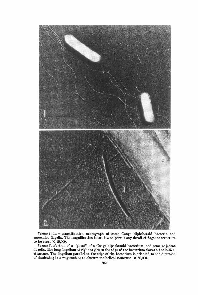
Figure 1. Low magnification micrograph of some Congo diphtheroid bacteria andassociated flagella. The magnification is too low to permit any detail of flagellar structureto be seen. X 10,000.
Figure 2. Portion of a "ghost" of a Congo diphtheroid bacterium, and some adjacentflagella. The long flagellum at right angles to the edge of the bacterium shows a fine helicalstructure. The flagellum parallel to the edge of the bacterium is oriented to the directionof shadowing in a way such as to obscure the helical structure. X 50,000.
702
HELICAL FINE STRUCTURE OF FLAGELLA
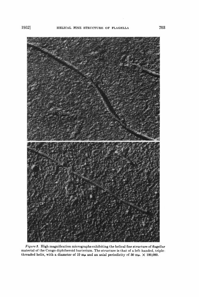
Figure S. High magnification micrographs exhibiting the helical fine structure of flagellarmaterial of the Congo diphtheroid bacterium. The structure is that of a left-handed, triple-threaded helix, with a diameter of 19 miA and an axial periodicity of 50 m/A. X 100,000.
1952] 703

M. P. STARR AND R. C. WILLIAMS
isolated from human hemoculture in the Belgian Congo by Dr. R. de Vignat,and sent to us by Dr. E. V. Morse of the University of Wisconsin.
Cultures of the Congo diphtheroid bacterium were grown on BBL "eugonagar"(trypticase soy agar with cystine and glucose) at 37 C for 18 to 24 hours. Whenexamined with a light microscope in a wet mount, almost all the cells showed ahigh degree of motility. The preparations for electron microscopy were madefrom a water suspension of the bacteria. The suspension was either sprayed(Backus and Williams, 1950) as microdroplets upon the microscope specimenfilms, or it was applied as a gross drop in the usual manner. Reference particlesof polystyrene latex (Backus and Williams, 1949) were added to the preparationin order to establish precisely the angle of shadow-casting and the direction ofthe shadowing beam with respect to the flagellar axis. The shadowing elementwas uranium.
OBSERVATIONS
Figure 1 is a low magnification micrograph of the Congo diphtheroid bacterium,with adjacent flagella. As is the case with most electron micrographs preparedfrom bacteria that have been suspended in distilled water, there is no way ofbeing certain that the flagella seen actually are attached to, or originated from,an adjacent bacterium.
Figure 2 shows a portion of a bacterium and some adjacent flagella at amoderate magnification. Some of the flagella shown here can be seen to have asurface structure of a helical nature. It is found that most of the flagella havethis structure in the several preparations examined on five separate occasionsover a period of eight months.
Figure 3 shows individual flagella at high magnification. The helical structureis plainly seen, and some of its details are these: (1) the diameter of the flagellais 19 m,u; (2) the helix has the form of a triple-threaded screw; (3) the directionof the helical turns is invariably that of a left-handed screw; (4) the distancealong the flagellar axis for one complete period (or turn) is about 50 mg; (5)except for cases of obvious kinking, the length of a full period is approximatelyconstant along one flagellum, and also from one flagellum to another.
DISCUSSION
The first point that arises is the reality of the phenomenon. Do the helicesseen on the micrographs represent helices in the flagella as prepared for microg-raphy? It can be demonstrated easily that a type of "pseudo-helix" appears onmicrographs of nodulated filaments if the electron image has moved during themicrography. In such cases, the direction of the "pseudo-helical" turns (that is,left-handed or right-handed) would then be expected to vary from one photo-graphic exposure to another, depending upon the direction of the random driftof the image. Also, in micrographs showing this "pseudo-helical" structure, thebackground detail would appear to have a linear structure. The invariable left-handed orientation of the helical structure in our micrographs and the absenceof any linear structure in the background, as shown in figure 3, rule out thepossibility of this type of artifact.
704 [VOL. 63
HELICAL FINE STRUCTURE OF FLAGELLA
Some confusion can arise easily in attempting to infer the direction of a helicalstructure in the object from observations of micrographs, owing to possibleleft to right perversion of the image during the photography (Weibull, 1950).For example, a photographic transparency of a left-handed helix will show aright-handed helix if one turns the transparency over and looks through theback.
It appears that the observed helical structure is not likely to be a result ofmechanical intertwisting of smaller fibrils, inasmuch as no partially frayedflagella, nor fibrils of smaller diameter, are ever seen in the micrographs. Webelieve that failure to observe such smaller fibrils is not due to limitations in theresolving power of the microscope.At the present time, the Congo diphtheroid appears to be the only organism
to show this highly organized flagellar fine structure; relatively few bacteria,however, have been photographed with sufficiently high resolution to demon-strate such a structure, if it exists. As far as is known to us, no natural proteinfibrils have ever been reported to exhibit a helical structure of anything like thesmallness of scale of the flagella described here. Even this fineness of structureis not to be confused with the scale of size of the protein helices postulated byPauling et al. (1951), since the diameter of our flagellar helices is about 40 timesthat of their polypeptide configuration. On the other hand, this flagellar finestructure appears, superficially at least, to be a distinctly different propertyfrom the flagellar structure studied by Weibull (1950), which involves a simple"spiral" with a period of about 2 ,u. The helical fine structure found by us for theflagella of the Congo diphtheroid is about one-fiftieth the scale of such grossflagellar forms. For reasons mentioned previously, a comparison with the struc-ture reported in trypsin-treated flagella by de Robertis and Franchi- (1951) isnot possible at this time.
SUMMARY
A helical fine structure is demonstrated in the flagella of a motile diphtheroidbacterium. This structure appears as a left-handed, triple-threaded helix of50 mu axial period. Evidence for the reality of this structure and its relationshipto other reported flagellar structures are discussed.
REFERENCES
BACKUS, R. C., AND WILLIAMS, R. C. 1949 Small spherical particles of exceptionallyuniform size. J. Applied Phys., 20, 224-225.
BACKUS, R. C., AND WILLIAMS, R. C. 1950 The use of spraying methods and of volatilesuspending media in the preparation of specimens for electron microscopy. J. AppliedPhys., 21, 11-15.
CLARK, F. E., AND CARR, P. H. 1951 Motility and flagellation of the soil corynebacteria.J. Bact., 62, 1-6.
DE ROBERTIS, E., AND FRANCHI, C. M. 1951 Electron microscope observation on the finestructure of bacterial flagella. Exptl. Cell Research, 2, 295-298.
HOUWINK, A. L., AND VAN ITERSON, W. 1950 Electron microscopical observations on
bacterial cytology. II. A study on flagellation. Biochim. et Biophys. Acta, 5, 10-44.KNAYSI, G. 1951 Elements of bacterial cytology. Second edition. Comstock Publish-
ing Co., Ithaca, N. Y.
7051952]

706 M. P. STARR AND R. C. WILLIAMS [VOL. 63
PAULING, L., COREY, R. B., AND BRANSON, H. R. 1951 The structure of proteins: Twohydrogen-bonded helical configurations of the polypeptide chain. Proc. Natl. Acad.Sci., 37, 205-211.
PIJPER, A. 1947 Methylcellulose and bacterial motility. J. Bact., 53, 257-269.STARR, M. P., AND PIRONE, P. P. 1942 Phytomonas poinsettiae n. sp., the cause of a
bacterial disease of poinsettia. Phytopathology, 32, 1076-1081.WEIBULL, C. 1950 Investigations on bacterial flagella. Acta Chem. Scand., 4, 268-276.