
Firstfossil mesothele spider, from the Carboniferous ofFrance
12
REVUE SUISSE DE ZOOLOGIE, vol. hors serie: 585-596; aoOt 1996 First fossil mesothele spider, from the Carboniferous of France Paul Antony SELDEN Department of Earth Sciences University of Manchester Manchester M 13 9PL, United Kingdom. First fossil mesothele spider, from the Carboniferous of France. . Eothele montceauensis n. gen., n. sp., is described from two specimens from the Upper Carboniferous (Stephanian) of Montceau-Ies-Mines, France, as the first fossil and oldest known mesothele spider. In addition to the plesiomorphies characteristic of mesotheles, the holotype of Eothele preserves one mesothele synapomorphy (deep, narrow sternum) and at least one autapomorphy of the genus (biserially dentate chelicerae). The fossil is evidence that both mesotheles and opisthotheles were present in the Carboniferous period. Key-words: Araneae - Carboniferous - fossil - France - Mesothelae - palaeontology - phylogeny. INTRODUCTION The south-east Asian spiders Liphistius Schiodte, 1849, and Heptathela Kishida, 1923, constitute the suborder Mesothelae which is sister group to Opistho- thelae to which all other extant spiders belong (PLATNICK & GERTSCH 1976). Meso- theles exhibit the most primitive character states of all living spiders, including segmented opisthosoma, eight spinnerets (seven in Heptathela) in a medioventral position on the opisthosoma, and absence of spinneret tartipores (SELDEN et al. 1991). Autapomorphies of Mesothelae include: modified trichobothria, invaginated fourth coxae, pseudosegmented lateral spinnerets, tibial spurs, and deep and narrow sternum (PLATNICK & GOLOBOFF 1985; RAVEN 1985). As primitive spiders, it would be expected that mesotheles should appear early in the fossil record and, indeed, many Carboniferous spiders were referred to this suborder (as Liphistiina) in the Treatise (PETRUNKEVITCH 1955). However, re-examination of all available types of Manuscript accepted 03.01.1996. Proceedings of the XIIIlh International Congress of Arachnology, Geneva, 3-8.IX.1995.
Transcript of Firstfossil mesothele spider, from the Carboniferous ofFrance

REVUE SUISSE DE ZOOLOGIE, vol. hors serie: 585-596; aoOt 1996
First fossil mesothele spider, from the Carboniferous of France
Paul Antony SELDENDepartment of Earth SciencesUniversity of ManchesterManchester M 13 9PL, United Kingdom.
First fossil mesothele spider, from the Carboniferous of France. .Eothele montceauensis n. gen., n. sp., is described from two specimensfrom the Upper Carboniferous (Stephanian) of Montceau-Ies-Mines,France, as the first fossil and oldest known mesothele spider. In addition tothe plesiomorphies characteristic of mesotheles, the holotype of Eothelepreserves one mesothele synapomorphy (deep, narrow sternum) and atleast one autapomorphy of the genus (biserially dentate chelicerae). Thefossil is evidence that both mesotheles and opisthotheles were present inthe Carboniferous period.
Key-words: Araneae - Carboniferous - fossil - France - Mesothelae palaeontology - phylogeny.
INTRODUCTION
The south-east Asian spiders Liphistius Schiodte, 1849, and HeptathelaKishida, 1923, constitute the suborder Mesothelae which is sister group to Opisthothelae to which all other extant spiders belong (PLATNICK & GERTSCH 1976). Mesotheles exhibit the most primitive character states of all living spiders, includingsegmented opisthosoma, eight spinnerets (seven in Heptathela) in a medioventralposition on the opisthosoma, and absence of spinneret tartipores (SELDEN et al. 1991).Autapomorphies of Mesothelae include: modified trichobothria, invaginated fourthcoxae, pseudosegmented lateral spinnerets, tibial spurs, and deep and narrow sternum(PLATNICK & GOLOBOFF 1985; RAVEN 1985). As primitive spiders, it would beexpected that mesotheles should appear early in the fossil record and, indeed, manyCarboniferous spiders were referred to this suborder (as Liphistiina) in the Treatise(PETRUNKEVITCH 1955). However, re-examination of all available types of
Manuscript accepted 03.01.1996.Proceedings of the XIIIlh International Congress of Arachnology, Geneva, 3-8.IX.1995.

586 PAUL ANTONY SELDEN
Carboniferous spiders for a monograph of the group (in preparation) has revealed thatnone shows autapomorphic characters of Mesothelae. Of the eleven genera includedin Mesothelae in PETRUNKEYITCH (1955), one (Palaeocteniza) has been shown not tobe a spider (SELDEN et al. 1991), and the remainder can be referred to Araneae onlyon the absence of autapomorphies of related orders (they have the general appearanceof spiders, show segmented opisthosomae, but supposed spinnerets are artefacts).
In the last few years, some new specimens of fossil arachnids have beendiscovered at the locality known as Montceau-Ies-Mines in the Massif Central regionof France. Among them, two specimens of Araneae have been identified, one ofwhich is quite remarkable in that spinnerets are clearly preserved. The two specimenswere mentioned briefly by SELDEN (1996) and are described fully here. The fossils arepresumed to belong to the same species of spider, which shares most of theplesiomorphic character states of modern mesotheles, and a single synapomorphy:narrow, deep sternum (RAYEN 1985). At least one autapomorphy (biserial cheliceraIdentition) separates the fossil species from living mesotheles. The fossil is evidencethat both mesotheles and opisthotheles were present in the Carboniferous period.
MATERIAL AND METHODS
A number of fossil arachnids, including opilionids and trigonotarbids, areknown from the Carboniferous (Stephanian) locality of Montceau-Ies-Mines in theMassif Central region of France, most of which are yet to be described. A recentreview of the geology and palaeontology of the locality is given in HEYLER & POPLIN(1994). In that book, two photographs (p. 165) are labelled as spiders; the leftphotograph could be of a spider, but the specimen needs further development(specimen n° 5190/1), the other (specimen n° 6165) is not a spider. The specimendescribed here with the prefix 'In' is housed in the Department of Palaeontology,British Museum (Natural History), London, the other specimen is deposited in theMuseum d'Histoire Naturelle, Autun, France. The fossils are external moulds in clayironstone nodules.
THE MONTCEAU-LES-MINES SPIDER
Order ARANEAE Clerck, 1757Suborder MESOTHELAE Pocock, 1892Eothele n. gen.
Etymology: Greek eos, the dawn, and thele, a nipple; combining reference tothe early age of the spider and the common suffix for mesotheles and mygalomorphs.
Diagnosis: mesothele spider with biserially dentate chelicerae (2 5 teeth onpromargin, 2 3 teeth on retromargin); at least 6 spinnerets in two rows (4, 2) onventral surface of opisthosoma, not bunched together at posterior margin of secondoperculum.
Type and only species: Eothele montceauensis n. sp., described below.

Eothele montceauensis n. sp.
FOSSIL MESOTHELE 587
(Figs 1-4)
Etymology: after Montceau-Ies-Mines, France, the provenance of the fossils.Diagnosis: as for the genus.Horizon & locality: Carboniferous (Stephanian), Montceau-Ies-Mines, Massif
Central, France.Type material: Holotype specimen, numbers 51961 (part) and 51962 (counter
part), housed in Museum d'Histoire Naturelle, Autun, France. Second specimen (notto be considered a paratype), In 62050 (part) and In 62050a (counterpart), samelocality, British Museum (Natural History), London.
DESCRIPTION OF SPECIMEN 51961/2
The specimen consists of ventrolateral (part: specimen n° 51961) and dorsolateral (counterpart: specimen n° 51962) external moulds in a nodule. The part showsmost detail (Figs 1,3), including most of the prosoma and opisthosoma. The counterpart preserves only a few holes where legs disappear into the matrix, and vaguedetails of the carapace. The dorsolateral mould of the opisthosoma, which should beon the counterpart, is missing; it is possible that a sliver of matrix preserving themould was lost on splitting the nodule. It is impossible to tell whether the fossilrepresents a male, female or juvenile specimen.
The morphology of the specimen is essentially clear on the part (Figs 1,3). Theopisthosoma has been compressed, the prosoma less so. The inflated shape of theopisthosoma indicates that this specimen represents a dead animal rather than a moultbecause in spider moults the opisthosoma is shrivelled. The coxae of all prosomalappendages on the left side, both chelicerae, and coxae of the right pedipalp and leg Iare clearly displayed. The prosoma is tilted to the left, so that the coxae of legs 11 toIV are not seen on the part. Between the coxae, the labium and the left two-thirds ofthe sternum can be seen. The edge of the sternum drops steeply then flattens sharplyto the right. The external mould of the labium disappears beneath the internal mouldof the mouth. Labium and sternum are about the same width. Tilting the specimenreveals the external mould of the chelicerae (inset B on Fig. 3). Each of these shows alarge pit distally which represents the external mould of the fang, and a line of holes(5 can be seen on each chelicera) running proximally from the fang represents thepromarginal line of teeth. On the left chelicera, a line of three holes on the left sidemarks a retromarginal tooth row, but this cannot be matched on the right chelicera.Beyond the lateral edges of the coxae is a narrow groove which marks the externalmould of the edge of the carapace. Beyond this there are some holes, the posterior twoof which were developed further. These holes represent the external moulds ofprosomal appendages. The posterior two represent the femora and patella of legs IIIand IV; looking inside the mould of femur IV, two grooves can be seen, which wereridges on the inferior surface of the femur in life. Two elongate holes about 4 mmfrom the edge of the carapace are two distal podomeres of leg I (presumably leg Il isentirely hidden within the matrix). A small hole close to the carapace edge is the

588 PAUl. ANTONY SELlHoN
FIGS I & 2
Eothele montceollensis n. gen., n. sp.. Carboniferous (Stephanian), Montceall~lcs~Mines,
France. Fig. 1 (upper): holotype, specimen 51961 (part); sce Fig. 3 for explanalion; x 13. Fig. 2(I"wpr)' ,,,;ditinmd ,npl';ml'.n In (,?n~() (n~rl): see Fig. 4 for exolanation: x6.

FOSSIL MESOTHELE 585
7105 3 & 4. EOlhele monlceauensis n. gen., n. sp., Carboniferous (Stephanian), Montceau-Iesvlines, France. Fig. 3 (upper): holotype, specimen 51961 (part); A: right dorsolateral view of:xternal mould of left ventrolateral; B: dorsal view of external mould of chelicerae showing fangsholes disappearing into matrix) and tooth rows. Fig. 4 (lower): additional specimen, In 62050part), ventral view of external mould of dorsal. Legend for Figs 3 & 4: I-IO-opisthosomalergite numbers, ALS-anterior lateral spinneret, AMS-anterior median spinneret, anal tubmal tubercle, car-carapace, ch-chelicera, ex-coxa, fe-femur, I-lV-walking legs I-IV,a-labium, operc-book-Iung operculum, op-opisthosoma, pa-patella, pd-pedipalp, PLS-
posterior lateral spinneret, pst-paired structure, st-sternum, te-tergite, tr-trochanter.
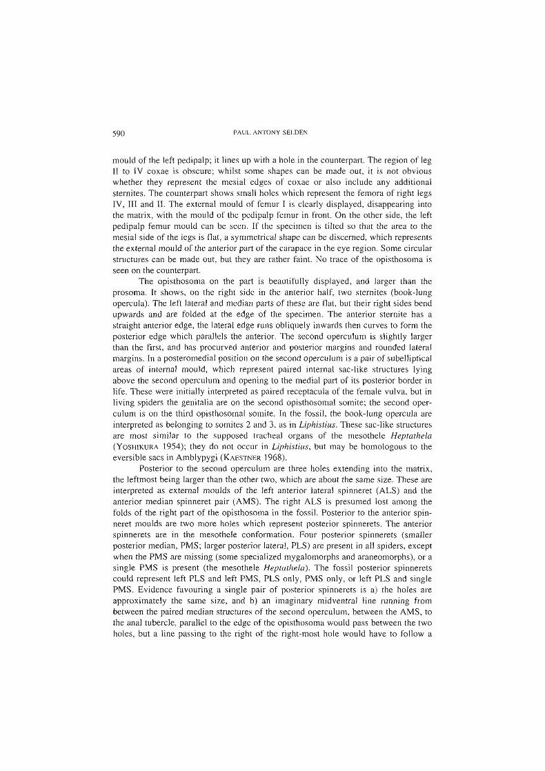
590 PAUL ANTONY SELDEN
mould of the left pedipalp; it lines up with a hole in the counterpart. The region of legII to IV coxae is obscure; whilst some shapes can be made out, it is not obviouswhether they represent the mesial edges of coxae or also include any additionalsternites. The counterpart shows small holes which represent the femora of right legsIV, III and H. The external mould of femur I is clearly displayed, disappearing intothe matrix, with the mould of the pedipalp femur in front. On the other side, the leftpedipalp femur mould can be seen. If the specimen is tilted so that the area to themesial side of the legs is flat, a symmetrical shape can be discerned, which representsthe external mould of the anterior part of the carapace in the eye region. Some circularstructures can be made out, but they are rather faint. No trace of the opisthosoma isseen on the counterpart.
The opisthosoma on the part is beautifully displayed, and larger than theprosoma. It shows, on the right side in the anterior half, two sternites (book-lungopercula). The left lateral and median parts of these are flat, but their right sides bendupwards and are folded at the edge of the specimen. The anterior sternite has astraight anterior edge, the lateral edge runs obliquely inwards then curves to form theposterior edge which parallels the anterior. The second operculum is slightly largerthan the first, and has procurved anterior and posterior margins and rounded lateralmargins. In a posteromedial position on the second operculum is a pair of subellipticalareas of internal mould, which represent paired internal sac-like structures lyingabove the second operculum and opening to the medial part of its posterior border inlife. These were initially interpreted as paired receptacula of the female vulva, but inliving spiders the genitalia are on the second opisthosomal somite; the second operculum is on the third opisthosomai somite. In the fossil, the book-lung opercula areinterpreted as belonging to somites 2 and 3, as in Liphistius. These sac-like structuresare most similar to the supposed tracheal organs of the mesothele Heprathela(YOSHIKURA 1954); they do not occur in Liphistius, but may be homologous to theeversible sacs in Amblypygi (KAESTNER 1968).
Posterior to the second operculum are three holes extending into the matrix,the leftmost being larger than the other two, which are about the same size. These areinterpreted as external moulds of the left anterior lateral spinneret (ALS) and theanterior median spinneret pair (AMS). The right ALS is presumed lost among thefolds of the right part of the opisthosoma in the fossil. Posterior to the anterior spinneret moulds are two more holes which represent posterior spinnerets. The anteriorspinnerets are in the mesothele conformation. Four posterior spinnerets (smallerposterior median, PMS; larger posterior lateral, PLS) are present in all spiders, exceptwhen the PMS are missing (some specialized mygalomorphs and araneomorphs), or asingle PMS is present (the mesothele Heptathela). The fossil posterior spinneretscould represent left PLS and left PMS, PLS only, PMS only, or left PLS and singlePMS. Evidence favouring a single pair of posterior spinnerets is a) the holes areapproximately the same size, and b) an imaginary midventral line running frombetween the paired median structures of the second operculum, between the AMS, tothe anal tubercle, parallel to the edge of the opisthosoma would pass between the twoholes, but a line passing to the right of the right-most hole would have to follow a

FUSSIL MESOTHELE 591
dog-leg course to reach the anal tubercle. Since the PMS are usually smaller than thePLS, or lost (Marples, 1967), the posterior spinnerets present in the fossil, which arerelatively large, are most probably PLS.
The left side of the opisthosoma bears a number of random, irregular nodules.Beyond these are some curved folds which approximately parallel the edge of theopisthosoma. No tergites are visible. This may be because any tergites which werepresent were confined to the dorsal side of the opisthosoma. On the part, only ventraland lateral sides are visible, the counterpart preserves no fragment of the opisthosoma. On the left lateral side of the opisthosoma, which would have been the anteriorpart of the dorsolateral in life, one fold is larger and more robust than the rest (whichsuggests it was composed of tougher cuticle in life) and extends for about 1/3 of thelength of the opisthosoma. In front of this structure is a smaller, yet similarly robustfold. The possibility that the larger structure could represent a dorsal scutum wasconsidered, but further thought and study of some other well-preserved Carboniferousspiders has led to the conclusion that this specimen probably did bear dorsal tergites.These, like the carapace, would have been preserved on the counterpart. However,like the carapace, which is preserved on the part as a thin groove-the external mouldof the edge of the carapace, the opisthosomal tergites have also left their mark on thepart as a narrow external mould. I suggest that during compression of the fossil, thedorsal tergites remained fairly rigid compared to the flexible cuticle of the sides of theopisthosoma. Where there were no adjacent tergites, such as over most of thepreserved left and posterior parts of the opisthosoma, this flexible cuticle is expressedas simple folds on the external mould. On the left anterolateral side, however, thesecond and following tergites above compressed the flexible cuticle onto the sedimentto produce an apparently robust fold. In front of this, the short first opisthosomaltergite has produced a similar fold. Segmentation is not apparent because it is thelateral cuticle which formed the floor of the crevice, not the tergites themselves.
DESCRIPTION OF SPECIMEN IN 62050
The specimen (Figs 2,4) consists of dorsal (part: specimen n° 62050) andventral (counterpart: specimen n° 62050a) external moulds in a nodule. The partshows most detail (Figs 2,4), including much of the prosoma and dorsal opisthosoma.The counterpart preserves only a few holes where legs disappear into the matrix, andsome parts of the book-lung opercula on the ventral opisthosoma. The ventral mouldof the prosoma, which should be on the counterpart, is missing because a sliver ofmatrix preserving the mould was lost on splitting the nodule. In places, details areobscured by a thin veneer of pyrite and calcite (?) crystals; kaolinite is rare, andoccurs mainly as a cast of thin tubes (possibly distal podomeres) towards the edge ofthe nodule. The specimen is strongly three-dimensional, its shape no doubt reflectsthe original shape of the animal. There is slight distortion in that the prosoma isrotated slightly in relation to the opisthosoma, so that in the chelicerae and otherprosomal appendages appear to lie off the midline of the carapace when the specimenis lying with the opisthosoma symmetrical to view. By rotating the specimen, the

592 PAUL ANTONY SELDEN
carapace and appendages can be positioned so that they are almost symmetrical, whenthe opisthosoma is not. The slight displacement of the chelicerae is then explained bya little compaction of the sediment prior to the formation of the nodule. It isimpossible to tell whether the fossil represents a male, female or juvenile specimen.
The carapace is preserved as an external mould, but is obscured, especiallyanteriorly, by mineralisation. It is roughly oval in shape, widest at or behind itsmid length, but the anterior border is not visible in the specimen, being covered bymatrix surrounding the anterior appendages. The posterior margin of the carapace isapproximately straight, with a slightly recurved submarginal line; it is about half thewidth of the carapace at its widest. The chelicerae are preserved as two holes, separated by a mineralised sheet of rock matrix, anterior to the carapace and disappearinginto the nodule anteriorly. No details can be discerned. The pedipalps and walkinglegs are preserved only as external moulds of parts of the proximal podomeres. Holesdescending into the matrix around the carapace represent the upwardly rising femora.A few thin holes well beyond the main body of the fossil may represent distalpodomeres descending again.
The opisthosoma is represented on the part by a sub-hemispherical hole: theexternal mould of its three-dimensional form. The inflated shape of the opisthosomaindicates that this specimen represents a dead animal rather than a moult because inspider moults the opisthosoma is shrivelled. Lateral parts of the opisthosoma,especially anteriorly, are distinctively longitudinally wrinkled. (Similar wrinklingoccurs on the soft parts of the opisthosoma of Liphistius). The dorsal surface of theopisthosoma consists of a series of tergites, at least eight can be counted. The moreanterior tergites are longer (sag.) than the posterior, but telescoping of the opisthosoma posteriorly conceals their true lengths. Another tergite (I) may be presentbetween the most anterior well-defined tergite and the carapace, but wrinkling on theleft side of the specimen (right side of the animal) suggests that this area could consistof soft cuticle. The tergites have fairly straight anterior and posterior margins and,where visible, their lateral margins appear to be rounded. At the posterior end of theopisthosoma, just posterior to a possible microtergite (10), is a hole which representsthe external mould of the anal tubercle. On the counterpart (specimen n° 62050a), theexternal mould of a small part of the left side of the opisthosoma can be seen. Thisregion is characteristically transversely wrinkled. Two transverse grooves mayrepresent the posterior margins of book-lung opercula I and 2.
DISCUSSION
It is tempting to assume that the two Montceau specimens belong to the samespecies, but the morphological evidence neither proves nor disproves this supposition.Features shown by the Autun specimen are not preserved on the London specimen,and vice versa. However, the two fossils are approximately the same size and occur inthe same locality and lithology. At other Upper Carboniferous localities yieldingarachnids in concretions, such as Coseley (UK) and Nyrany (Czech Republic),diversity within orders is low, and similar specimens have usually been assigned te

FOSSIL MESOTHELE S9~
the same species (this is true for the Nyrany 'spiders', but is not yet published). Allten specimens of the earliest mygalomorph, Rosamygale !?rauvogeli Selden & Gall,1992, found at the Triassic locality in the Vosges, France, belonged to one species.For these reasons, the two Montceau-Ies-Mines specimens will be assumed to beconspecific, until further evidence (in the form of more specimens) might proveotherwise.
Since the two specimens reveal essentially different aspects of the morphologyof the Montceau spider, a reasonable reconstruction of the living animal can be builtup. The fossil animal is clearly a spider because of the presence of spinnerets (at least,appendages are present in the position in which spinnerets occur in living spiders, andno other arachnid has such appendages in this position; spinnerets are taken to
indicate the presence of opisthosomal silk glands) combined with a pedicel, an essentially flexible opisthosoma with the tergites 'floating' in softer cuticle, general appearance of the carapace and sternum, and lack of autapomorphies of other arachnidgroups. Some autapomorphies of spiders, naked fang and cheliceral poison gland forexample (SELDEN el a/. 1991), cannot be seen.
There are few described autapomorphies of mesotheles (PLATNICK & GERTSCH1976; PLATNICK & GOLOBOFF 1(85). One of these relates to chromosome numbers socannot be confirmed in fossils; the other characters (trichobothrial bases, invaginations of fourth coxae, multisegmented lateral spinnerets, tibial spurs) are morphological, but none of them can be seen in the Montceau fossils. An additional apomorphy of mesotheles was suggested by RAVEN (1985): a deep and narrow sternum.He contended that the presence of a sternum is an autapomorphy of Araneae (it isabsent or very small in Amblypygi and most other arachnids), and that in Opisthothelae it is wide and low (an autapomorphy of that group). SELDEN el al. (1991)considered the presence of a sternum to be plesiomorphic within the Tetrapulmonata(Trigonotarbida have one also), but this does not alter the apomorphic state of thesternum in the mesotheles and opisthotheles. The characters of dorsal opisthosomaltergites, two ventral opercula (covering the two pairs of book-lungs), orthognathchelicerae, and AMS fully developed, are all indicative of Mesothelae, but are plesiomorphies. The deep, narrow sternum of the Montceau spider is a character sharedwith modern mesotheles. If the paired structures above the second operculum in thefossil are not homologous with amblypygid eversible sacs then this character wouldbe a synapomorphy for Heptathela and Eothele n. gen. (Fig. 5A). The spinneret pairsin Eothele are widely spaced between the second operculum and the anal tubercle, notbunched together just behind the second operculum as in modern mesotheles norclose to the anal tubercle as in Opisthothelae. The position of the spinnerets in Eothelecould reflect the start of their rearward movement towards the opisthothelatecondition, or a more ancestral arrangement prior to the bunching of the spinneretsclose to the second operculum as seen in Liphistius. If the posterior spinnerets inEothele are really PLS, then it would represent a new configuration for Araneae, anadvance over the Heptathela condition (with a single PMS), and an autapomorphy forthe fossil species. Another feature which separates Eothele from modern mesothelesis the presence in the fossil of two rows of teeth on the chelicera (Fig. 3,B); only a

594 PAUL ANTONY StLL>J:::N
promarginal row is present in living forms (ABRAHAM 1929; BRISTOWE 1932). Biserially dentate chelicerae are special adaptations which have evolved in a number ofspider groups. For example, RAYEN (1985) considered the biserial condition to beapomorphic in ctenizoid mygalomorphs and some related idiopids and cyrtaucheniids.Amblypygids and the Devonian spider Attercopus have uniserially dentate chelicerae(SELDEN et al. 1991).
FOSSILS AND PHYLOGENY
Much has been written concerning the value of fossils in piecing together therelationships between animal groups (see a recent review by SMITH, 1994). It hasbecome e1ear in the last twenty years or so that fossils must play a secondary role inestablishing phylogenetic relationships, but an important one in providing raw dataand, in some cases, resolving conflicting e1adograms. Some authors have attempted touse stratigraphic data in cladistic analyses (e.g. FISHER 1992, 1994; WAGNER 1995)but such analyses need to be executed with caution. The fossil record of arachnids isstill so sparse that the discovery of single specimens can make major alterations to theknown fossil record. The new spider described here is a case in point: it is the fjrstknown fossil mesothele, thus extending the record of this group from 0 years to 290million years. The unique contribution fossils can make is in the construction ofevolutionary trees, the next logical step in understanding the phylogenetic history ofan animal group after the construction of cladograms. It has been shown (for exampleby SMITH 1994, chapter 6) that stratigraphic data can be combined with a robuste1adogram to produce an estimate of the evolutionary tree of a group which minimisesstratigraphic gaps. A normal result of this process is the production of range extensions and ghost lineages; these are predictions made by the data. Note that fossil dataprovides minimum estimates for the dates of divergence of e1ades.
Possession of many character states which indicate that the Montceau spider isplesiomorphic with respect to Opisthothelae, and one character state (sternum narrowand deep) which is an autapomorphy of Mesothelae, confirm that the Montceau spiderbelongs in Mesothelae. The fossil species cannot be ancestral to any modern mesothele because of at least one autapomorphy (biserially dentate chelicera). Therefore,the fossil is the oldest record of a mesothele. Furthermore, since Opisthothelae(Mygalomorphae + Araneomorphae) is the sister clade to Mesothelae, it must haveoriginated sometime prior to the age of the fossil. Fig. 5 illustrates these relationships.
ACKNOWLEDGEMENTS
I thank G. Pacaud (Autun) and R. Fortey (London) for the loan of specimens intheir care, P. Schwendinger for specimens of Liphistius, and L. Anderson forbringing the London specimen to my attention; photographs were taken by J. Almond(Fig. l) and L. Anderson (Fig. 2).

rU:>:>IL Ml::SUTHl:Ll: 595
Trigonota rbida
Attercopus 1A"",,,Eothele
Heptathela ]T"""Liphistius
Mygalomorphae] OPtthothelae
Araneomorphae
Amblypygi
B
DevoCarboniferous Permian MESOZOIC CENOZOICnian
• Attercopus .. Eothe/e Mesothelae
.. .. __ ...._-- ....... • Rosamygale _ Mygalomorphae. . _-_ ..... __ . .J
;................ Juraraneus _ Araneomorphae_
408 360 286 248 165 Ma BP
FIG. 5
A: cladogram of relationships among Trigonotarbida, Amblypygi, and selected spider taxa. TheMontceau spider £othele is shown as sister to Heptathela because tracheal sacs are absent inLiphistius and on the assumption that PMS are lost in Eothele. B: Evolutionary tree showingrelationships between spider suborders (emphasising the earliest fossil members) andAttercopus; solid black lines are known ranges (fossil record of Mesothelae consists solely ofEothele; other suborders have better fossil records); horizontal dashed lines are ghost lineagesfor (Mesothelae + Opisthothelae) and (Mygalomorphae + Araneomorphae) and range extension
for Araneomorphae; vertical dashed lines show hypothesized relationships.
REFERENCES
ABRAHAM, H. C. 1929. The male of the spider Liphistius malayunus H. C. Abr., with furthernotes on the female and on its habits. Proceedings of the Zoological Society of London(1929): 671-677; pis I-Ill.
BRISTOWE, W. S. J932. The liphistiid spiders. With an appendix on their internal anatomy by J.Millo!. Proceedings of the Zoological Society ofLondon (1932): 1015-1057; pIs I-VI.
CLERCK, C. 1757. Svenska spindlar (Araneae suecici), L. Salvii, Stockholm. 154 pp.FISHER, D. C. 1992. Stratigraphic parsimony. 124--129. In: MacClade (W. P. MADDISON & D.
R. MADDlSON, eds.), Sinauer Associates, Sunderland, Massachusetts, xi + 398pp.FISHER, D. C. 1994. Stratocladistics: morphological and temporal patterns and their relation to
phylogenetic process. 133-171. In: Interpreting the hierarchy of nature-from systematic patterns to evolutionary theories (L. GRANDE & O. RIEPPEL, eds). AcademicPress, Sun Diego.

596 PAUL ANTUNY SI::LUtN
HEYLER. e. & POPLIN. D. (eds) 1994. Quand le Massif central etait sous I·equateur. CTHS,Paris. 328pp.
KAESTNER. A. 1968. Invertebrate Zoology. Vol. 2. Interscience, New York. 472pp.KISHIDA. K. 1923. Heptathela. a new genus of liphistiid spiders. Nippon dobUlsugaku iho 10:
235-242.MARPLES. B. 1. 1967. The spinnerets and epiandrous glands of spiders. Journal of the Linnean
Society (Zoology) 46.209-222; pI. I.PLATNICK. N. 1. & GERTSCH. W. J. 1976. The suborders of spiders: a cladistic analysis.
American Museum Novitates 2607: I-IS.PLATNICK. N. 1. & GOLOBOFF. P. A. 1985. On the monophyly of the spider suborder Mesothelae
(Arachnida: Araneae). Journal of the New York Entomological Society 93: 1265-1270.PETRUNKEVITCH. A.1995. Arachnida. pp. 42-162. In: Treatise on invertebrate paleontology.
Part P. Arthropoda 2 (R.e. MOORE. ed.). Geological Society of America and Universityof Kansas Press, Boulder, Colorado and Lawrence. Kansas: xvii + 181 pp.
POCOCK. R. 1. 1892. Liphistius and its bearing upon the classification of spiders. Annals andMagazine 0/Natural History. series 6. J0: 306-314.
RAvEN. R. J. 1985. The spider infraorder Mygalomorphae (Araneae): cladistics and systematics. Bulletin a/the American Mu.\'eum o/Natural Histor)' 182: 1-180.
SCHIODTE. J. e. 1849. Om en afvigende Slaegt af Spindlerrnes Orden. Naturhistoriske Tidsskrijl2: 617-624; pI. IV.
SELDEN. P. A. J996. Fossil mesothele spiders. Nature 379: 498-499.SELDEN. P. A. & GALL. 1.-e. 1992. A Triassic mygalomorph spider from the northern Vosges.
France. Palaeontology 35: 211-235.SELDEN. P. A.• SHEAR. W. A. & BONAMO. P. M. 1991. A spider and other arachnids from thE
Devonian of New York. and reinterpretations of Devonian Araneae. Palaeontology 34241-281.
SMITH. A. B. 1994. Systematics and the fossil record. Blackwell Scientific Publications, Oxfordviii + 223pp.
WAGNER. P. J. 1995. Stratigraphic tests of cladistic hypotheses. Paleobiology 21: 153-178.YOSHIKURA. M. 1954. On the tracheae in a liphistiid spider. Heptathela kimurai. Kumamo((
Journal ofScience. series B. 1: 37-40.


![INDEX [assets.cambridge.org]Aachen and Lothar IV ofFrance and Ottonians , , , , , Aachen capitulary Aachen Gospels , , , ,](https://static.fdocuments.net/doc/165x107/60b8d200380fd66cca378262/index-aachen-and-lothar-iv-offrance-and-ottonians-aachen-capitulary.jpg)


