FIRST HISTOLOGICAL AND …rocek.gli.cas.cz/Postdoc/steyer2003a.pdf · TEMNOSPONDYL GROWTH;...
15
FIRST HISTOLOGICAL AND SKELETOCHRONOLOGICAL DATA ON TEMNOSPONDYL GROWTH; PALAEOECOLOGICAL AND PALAEOCLIMATOLOGICAL IMPLICATIONS. J. Sébastien Steyer 1 , Michel Laurin 2 , Jacques Castanet 2 and Armand de Ricqlès 2 . In press Palaeogeography, Palaeoclimatology, Palaeoecology Correspondence should be addressed to J. Sébastien Steyer ; 1. Laboratoire de Paléontologie, UMR 8569 CNRS, Muséum national d’Histoire naturelle, 8 rue Buffon, F-75005 Paris, tel 00 33 1 40 79 30 42, fax 00 33 1 40 79 35 80, [email protected] 2. Equipe “ Formations squelettiques ”, UMR 8570 CNRS, Université Paris 6-7, case 7077, 2 Place Jussieu, F-75251 Paris cedex 05. Abstract – The metoposaurid Dutuitosaurus ouazzoui, from the Carnian (Upper Triassic) of Southern Morocco is one of the best known temnospondyls; more than 75 specimens have been found, including 15 sub-complete skeletons of various sizes from ImiN’Tanoute, a mass mortality locality (Dutuit and Heyler, 1983, p.628) in the Argana Basin, Western High Atlas. Ten femora, sampled from sub-complete skeletons belonging to a growth series, have been sectioned at the mid-diaphyseal level in order to perform histological and skeletochronological analyses. Sections from juveniles and adults show microstructures typical of aquatic stegocephalians (absence of a free medullary cavity, presence of an extensive spongiosa that merges gradually into a cortical compacta). Bone sections from juveniles reveal a relatively high initial growth rate (no secondary osteons, cortex with wide and densely vascularized growth zones, alternating with thin annuli or Lines of Arrested Growth - LAGs-). Bone sections from adults indicate a decreased growth rate (less vascularized external cortex, osteocyte lacunae parallel to the cortical stratification), probably after the acquisition of sexual maturity, and a moderate amount of remodeling (presence of a few secondary osteons, of erosion bays in the cortical compacta). Growth marks have been correlated with environmental changes, likely to be seasonal cycles. Compactness profiles have been quantified using ‘Bone profiler’. Comparison with profiles of extant taxa confirm the hypothesis that Dutuitosaurus was aquatic. Keywords – Temnospondyl, ontogeny, histology, skeletochronology, Triassic, Morocco. 1
Transcript of FIRST HISTOLOGICAL AND …rocek.gli.cas.cz/Postdoc/steyer2003a.pdf · TEMNOSPONDYL GROWTH;...

FIRST HISTOLOGICAL AND SKELETOCHRONOLOGICAL DATA ON
TEMNOSPONDYL GROWTH; PALAEOECOLOGICAL AND PALAEOCLIMATOLOGICAL IMPLICATIONS.
J. Sébastien Steyer1, Michel Laurin2, Jacques Castanet2 and Armand de Ricqlès2.
In press Palaeogeography, Palaeoclimatology, Palaeoecology Correspondence should be addressed to J. Sébastien Steyer ; 1. Laboratoire de Paléontologie, UMR 8569 CNRS, Muséum national d’Histoire naturelle, 8 rue Buffon, F-75005 Paris, tel 00 33 1 40 79 30 42, fax 00 33 1 40 79 35 80, [email protected] 2. Equipe “ Formations squelettiques ”, UMR 8570 CNRS, Université Paris 6-7, case 7077, 2 Place Jussieu, F-75251 Paris cedex 05. Abstract – The metoposaurid Dutuitosaurus ouazzoui, from the Carnian (Upper Triassic) of Southern Morocco is one of the best known temnospondyls; more than 75 specimens have been found, including 15 sub-complete skeletons of various sizes from ImiN’Tanoute, a mass mortality locality (Dutuit and Heyler, 1983, p.628) in the Argana Basin, Western High Atlas. Ten femora, sampled from sub-complete skeletons belonging to a growth series, have been sectioned at the mid-diaphyseal level in order to perform histological and skeletochronological analyses. Sections from juveniles and adults show microstructures typical of aquatic stegocephalians (absence of a free medullary cavity, presence of an extensive spongiosa that merges gradually into a cortical compacta). Bone sections from juveniles reveal a relatively high initial growth rate (no secondary osteons, cortex with wide and densely vascularized growth zones, alternating with thin annuli or Lines of Arrested Growth - LAGs-). Bone sections from adults indicate a decreased growth rate (less vascularized external cortex, osteocyte lacunae parallel to the cortical stratification), probably after the acquisition of sexual maturity, and a moderate amount of remodeling (presence of a few secondary osteons, of erosion bays in the cortical compacta). Growth marks have been correlated with environmental changes, likely to be seasonal cycles. Compactness profiles have been quantified using ‘Bone profiler’. Comparison with profiles of extant taxa confirm the hypothesis that Dutuitosaurus was aquatic. Keywords – Temnospondyl, ontogeny, histology, skeletochronology, Triassic, Morocco.
1

1. INTRODUCTION Bone histology and skeletochronology are increasingly used to gain insights into the biology of extinct amniotes such as Mesozoic reptiles (e.g. Horner et al., 2000) or Cenozoic mammals (e.g. Buffrénil et al., 1990). However, this is not the case for non-amniotic extinct taxa such as temnospondyls. Temnospondyls correspond to a stem-tetrapod clade that includes species of a wide range of sizes (3 to 600 cm in total length) and shapes (brevirostral salamander type to longirostral garhial type). Actualistic comparisons (i.e. comparisons with extant relatives) must be used cautiously to address their physiology, ecology and ethology because their relationships with other vertebrates are rather debated: according to recent phylogenetical analyses, their extant sister-group could be either Tetrapoda (sensu Laurin and Reisz, 1997) or Lissamphibia (Lombard and Sumida, 1992). However, temnospondyls show exceptional preservation, with complete growth series ranging from larval to adult individuals (reviewed in Steyer, 2000, 2001). Their ontogeny has already been studied using anatomical observations, but little information is available on their bone histology (Dutuit, 1974; Ricqlès, 1975, 1977-1979; Damiani, 2000). Extensive histological descriptions of several Metoposaurids (Ricqlès, 1973) remain unpublished.
Bone histology and skeletochronology are rarely used to gain insights into ontogeny of extinct taxa: a few exceptions are well preserved growth series of plesiosaurs (Wiffen et al., 1995), dinosaurs (Horner et al., 2000), Triassic nothosaurs (Sanders, 1990), or Plio-Pleistocene anurans (Esteban et al., 1998).
Dutuitosaurus ouazzoui (Dutuit, 1976) Hunt, 1993 belongs to the Metoposauridae, a temnospondyl family widespread in the Triassic of Europe (Meyer, 1842; Koken, 1913; Steyer, 1999), India (Lydekker, 1885), North Africa (Dutuit, 1967), North America (Leidy, 1856; Baird 1986), and perhaps China (Yang, 1978) and Madagascar (Dutuit, 1978). Dutuitosaurus is considered by Hunt (1993, p. 81) to be ‘the most primitive metoposaurid’ although this author placed Apachesaurus and/or Arganasaurus as stem-group or sister-taxa of the clade that includes Dutuitosaurus and Metoposaurus (Hunt, 1993, p. 88, fig. 18; that contradicts his other statement because Arganasaurus branches off earlier than Dutuitosaurus).
Bone histology of Dutuitosaurus ouazzoui (Dutuit, 1976; Hunt, 1993) has already been studied on the basis of cranial (Dutuit, 1974, 1976; Dutuit and Heyler, 1975), costal, femoral and humeral (de Ricqlès, 1975, 1978, 1989) sections. Here, we analysed femoral sections from various growth stages and using qualitative and quantitative histological techniques, as well as skeletochronology, in order to provide data about growth, ecophysiology and paleoclimatology. This is one of the first growth series of an extinct non-amniotic vertebrate studied with such methods.
Even though Dutuitosaurus is the best known temnospondyl from Africa (with over 75 specimens, including 15 subcomplete skeletons of various sizes; Dutuit, 1976), its paleobiology and palaeoecology remain problematical, as is the case for many others metoposaurids (Hunt, 1993). For instance, it has been suggested that Dutuitosaurus was a bottom-dweller, an ambush predator (the passive ‘death-trap’ model of Ochev, 1966), or a mid/upper-water swimmer (the active predator model of Chernin and Cruickshank, 1978).
2

2. MATERIAL AND METHODS (Figures 1, 2)
The material consists of a growth series of 10 femora of Dutuitosaurus ouazzoui belonging to juvenile and adult specimens from the Middle-Upper Carnian (Upper Triassic, ‘Lower T5’ stratigraphical unit; Jalil, 1996) of ImiN’Tanoute (site nr. XIII; Dutuit, 1976), a mass mortality locality (Dutuit and Heyler, 1983) in the Argana Basin, Western High Atlas, Southern Morocco (Fig. 1).
The studied femora (Fig. 2), sampled from sub-complete skeletons from the MNHN collection and measuring from 79 to 157 mm in length, have been prepared and sectioned at their mid-diaphyseal level. Sections have been mounted on glass slides and observed (i.e. qualitative analysis) under ordinary and polarized transmitted light. Quantitative analyses were made using ‘Bone profiler’ version 1.0.15 (Girondot and Laurin, in press) to quantify and compare the compactness profile of bone sections. 3. HISTOLOGICAL DESCRIPTIONS AND SKELETOCHRONOLOGY
3.1. General features (Figs 3-4)
Sections from juveniles and adults, which range from 9.3 to 24.3 mm in diameter, do not show a free medullary cavity, but an extensive spongiosa which merges gradually into the cortical compacta, a microstructure typical of aquatic stegocephalians (Ricqlès, 1977; Laurin et al., 2000; Steyer et al., 2001). Juvenile and adult stages of these femora have been firstly determined using growth markers (reviewed in Steyer, 2001) observed on associated skeletal elements (Steyer, pers. obs.), and secondarily confirmed according to their various histological structures (see below).
Growth marks such as annuli (thin layers corresponding to temporary decrease of osteogenesis) or Lines of Arrested Growth (LAGs, corresponding to temporary cessation of osteogenesis), have been counted on sections from both juveniles and adults. They are reliable indicators of growth rhythm and are generally laid down annually in extant vertebrates (Castanet et al., 1996).
3.2. Sections from juveniles (Fig. 3) The sections from juveniles have sub-circular outlines and show no secondary
osteons. The cortex contains wide growth zones, alternating with thin annuli and LAGs. The zones are densely vascularized. This fine structure reveals a cyclical growth but a relatively high (for such poïkilo-ectothermic organisms) initial growth rate, perhaps at least until the acquisition of sexual maturity (Castanet, 1975; Steyer et al., 2001).
A maximum of six LAGs were counted in the sections from juveniles. According to their shape and their distance to the center of the bone (i.e. the radius), the last LAGs (i.e. the most peripheral one) in the sections from the late juveniles correspond to the first ones in the sections from the early adults. But in bones of late adults, the first few LAGs could be destroyed by bone remodelling during growth (see above). Both visible and estimated LAGs have therefore been counted (Table 1).
3

3.3. Sections from adults (Fig. 4) The sections from adults have pear-shaped outlines (Fig. 4A), which reflect a positive
allometric development of the adductor femoral crest. They are densely vascularized, yet the cortical vascularization decreases toward the periphery, and the cortex is less vascularized than that of the juveniles.
The external cortex, with osteocyte lacunae parallel to the cortical stratification (Fig. 4B), is mainly composed of a lamellar bone tissue, with primary osteons and vascular canals. The canals are located either in zones (i.e. growth marks laid down during periods of active growth) or annuli, the latter becoming very thin and dark LAGs. The LAGs are more closely spaced in the external cortex than in the inner one. The progressive change from annuli to LAGs is in relationship with a decreased growth rate, probably after the acquisition of sexual maturity. The inner cortex is locally eroded and remodeled. The spongiosa shows a mainly trabeculo-cancellous structure with secondary osteons, revealing a moderate inner remodeling during growth (Francillon-Vieillot et al., 1990; Castanet et al., 1993).
3.4. Individual age and size The maximal diameter of the mid-diaphysis generally increases with femoral length.
The latter also increases with total body length (with a ratio of 1/70, from the snout to the tail extremity) (Table 1), but the number of observed and estimated LAGs do not increase regularly with size. In this work, the number of LAGs has been considered, rather than size, in order to estimate individual age. Individual age and size (i.e. number of LAGs x femoral length) are slightly better correlated by a polynomial curve of second degree (Fig. 5; with a correlation coefficient R2 = 0.922) than by a linear curve (the correlation coefficient of which is R2 = 0.906). The slope of this polynomial curve is slightly stronger during the juvenile than during the adult stage. This suggests a decreasing growth rate during ontogeny. The spacing between the first few visible LAGs is fairly large and even, but starting at the 6th identified LAG, this spacing decreases steadily. This is interpreted as a possible acquisition of sexual maturity. The longevity of Dutuitosaurus is of at least 12 cycles (the maximum number of estimated LAGs). This age corresponds to individuals with a femoral length of 157 mm and a total body length reaching about 2.2 m (the longest known temnospondyl is Mastodonsaurus from the Triassic of Germany, with 6 m of total body length; Schoch and Milner, 2000, and the largest is a Brachyopoid from the Triassic of Lesotho, with 1.5m of skull width; Steyer and Damiani, in press).
3.5. Palaeoecology
Microstructure observed in sections from both juveniles and adults of Dutuitosaurus
(see above) suggests that development of this temnospondyl took place in the aquatic environment. Moreover, densely vascularized growth zones observed in the sections from juveniles (see above) suggest that its development was cyclical but with a relatively high initial growth rate, perhaps at least until the acquisition of sexual maturity. This could confirm previous analyses made on cranial sections of Dutuitosaurus (Dutuit and Heyler, 1975) where large natural cavities, observed in the otic flange and the pterygoidal complex, have been linked with a pneumatization of the auditory complex, and perhaps with relatively high metabolism.
4

The evidence indicates that juveniles grew more rapidly than adults, as in many extant and extinct taxa. A common explanation for this phenomenon is that rapid body length and mass increase limits predation on the juvenile individuals (Wiffen et al., 1995; Horner et al., 2000).
Compactness profiles for each section have been quantified using ‘Bone profiler’ version 1.0.15 (Girondot and Laurin, in press), a new tool to statistically analyse and compare bone section compactness profiles (Fig. 6). These profiles have been statistically compared with those of femora of 24 extant taxa, whose mode of life is well known (Laurin et al., 2000). Both juvenile and adult profiles of Dutuitosaurus resemble those of several predominantly aquatic taxa, such as the chelonian Pelusios subniger. In these taxa, the slope of the profile is gentle; this reflects the gradual transition between the cortical compacta and the medullary spongiosa. Furthermore, the global compactness is fairly high (about 77% in Pelusio subniger, and from 61% to 81% in Dutuitosaurus). Terrestrial taxa are usually quite different. For instance, the profile of the predominantly terrestrial anuran Bufo marinus has an abrupt slope (Fig. 6) that reflects the absence of a spongiosa and a lower global compactness (about 56% to 65%). This strongly suggests that growth of Dutuitosaurus took place in the aquatic environment.
Taphonomical observations of the Dutuitosaurus material from ImiN’Tanoute showed that the adult skeletons fossilized in the center of a basin of several meters in width, just under the juvenile ones; the latter stand at the periphery of this basin (Dutuit, 1976). According to Dutuit and Heyler (1983, p.628), this spatial organisation of the growth series is due to either environmental conditions (a rapid mass mortality in a coastal fluvial environment) or biological conditions (the juvenile individuals were relatively less aquatic and could have lived in relatively shallower water).
3.6. Palaeoclimatology
Growth zones are commonly laid down during favourable environmental conditions, while annuli or LAGs correspond to unfavourable conditions in which growth is slowed or stopped. The presence of both zones and annuli or LAGs in Dutuitosaurus probably results from seasonal changes in the local climate where it lived, even though the zones and annuli may also have had an endogenic component (Buffrénil, 1980; Castanet et al., 1977, 1996). Damiani (2000) similarly inferred local climate seasonality using histological observations on mastodonsaurians from the Triassic of Australia.
The flora and fauna from the stratigraphic level of the Argana Basin (‘Lower T5’, a red siltous and clayed sandstone, Upper-Middle Carnian, Upper Triassic; Jalil, 1996), where our material was found, are composed of Equisetum, Equisetites, Phyllopoda indet., actinopterygians (Colobodontidae, Redfieldiidae, Brookvaliidae), lung-fishes (Arganodontidae, ‘Ceratodus’, Coelacanthidae indet.), other temnospondyls (Almasaurus), dicynodonts (Kannemeyeridae), Rauisuchia indet., phytosaurs, and a prosauropod (review in Jalil, 1996). This association has been suggested to indicate a sub-tropical and seasonal climate (Dutuit, 1976, p.28-31). This corroborates our hypothesis on seasonality based on clear growth mark alternation during bone tissue deposition.
5

4. CONCLUSION
Ontogenetic changes in the bone histology of Dutuitosaurus ouazzoui are revealed by two distinct but nonetheless gradational growth stages: juvenile and adult. These stages are distinguished not only by morphological (i.e. external) markers (reviewed in Steyer, 2001), but also by changes in the histological patterns in femora at each stage, and by the number and relative placement of LAGs.
This histological analysis of femora complements data previously obtained from cranial (Dutuit, 1974, 1976; Dutuit and Heyler, 1975), costal, femoral, and humeral sections (de Ricqlès, 1973, 1975, 1978, 1989). Bone structure of the femoral growth series confirms the aquatic lifestyle of Dutuitosaurus, as already deduced from its morphology (e.g. limb proportions, lateral line system on the adult skull roof), but also reveals modifications in the growth rate, and perhaps seasonal changes in the local environment. De Ricqlès (1973) inferred potential ecological changes during the ontogeny of Dutuitosaurus and Buettneria: the more compact and less vascularized cortex of the smaller individuals suggests an amphibious lifestyle rather than a fully aquatic one, inferred from sections from adults. This is also supported by limb proportion changes along the growth of Buettneria (Olsen, 1951). ACKNOWLEDGEMENTS: This study is a part of the Ph.D. dissertation of one of us (J.S.S.) who sincerely thanks his co-supervisors J.-C. Rage and Ph. Janvier (UMR 8569 CNRS and MNHN) for their encouragement. We are indebted to Ph. Taquet (MNHN) for permission to study (and to cut !) the material from MNHN collection. We sincerely thank Marie-Madeleine Loth (Equipe “ Formations squelettiques ”, Paris 7) for histological preparations, Nour-Eddine Jalil (University of Marrakech, Morocco) for his valuable comments on the Argana stratigraphy as well as for his help in the Moroccan collection stored in Paris, Jean-Michel Pacaud (MNHN) for the casts of the femora, and Denis Serrette and Philippe Loubry (MNHN) for the high-quality photos. A preliminary version of this work was presented at the 61th Meeting of the Society of Vertebrate Paleontology (Bozeman, Montana, 2001). We also sincerely thank R. Reisz, S. Sumida, and B. MacFadden for constructive comments on the draft. This research has been partly supported by the ‘Paul Sangnier’ Prize attributed to one of us (J.S.S.) by the CNFG (‘Comité National Français de Géologie’). REFERENCES Baird, D., 1986. Some Upper Triassic Reptiles, Footprints and an Amphibian from New
Jersey. The Mosasaur 3, 125-153. Buffrénil, V. de., 1980. Mise en évidence de l’incidence des conditions de milieu sur la
croissance de Crocodylus siamensis (Schneider, 1801) et valeur des marques de croissance squelettiques pour l’évaluation de l’âge individuel. Arch. Zool. Exp. Gén. 121, 63-76.
Buffrénil, V. de, Domning, D., Ray, C., Ricqlès, A. de, 1990. Bone histology of the ribs of the Archaeocetes (Mammalia: Cetacea). J. Vert. Paleontol. 10, 455-466.
Castanet, J., 1975. Quelques observations sur la présence et la structure des marques squlettiques de croissance chez les amphibiens. Bull. Soc. Zool. Fr. 100, 603-620.
Castanet, J., Meunier, F., Ricqlès, A. de, 1977. L’enregistrement de la croissance cyclique par le tissu osseux chez les vertébrés poïkilothermes. C. R. Acad. Sci. Paris 270D, 2853-2856.
6

Castanet, J., Francillon-Vieillot, H., Meunier, F. J., Ricqlès, A. de, 1993. Bone and individual aging. In: Hall, B.K. (Ed.), Bone. CRC Press, London, pp. 245-283.
Castanet, J., Grandin, A., Abourachid, A., Ricqlès, A. de, 1996. Expression de la dynamique de croissance dans la structure de l’os périostique chez Anas platyrhynchos. C. R. Acad. Sci. Paris 319, 301-308.
Chernin, S., Cruickshank, R.I., 1978. The myth of the bottom-dwelling capitosaur amphibians. South Afr. J. Sci. 74, 111-112.
Damiani, R.J., 2000. Bone histology of some Australian Triassic temnospondyl amphibians: preliminary data. Mod. Geol. 24, 109-124.
Dutuit, J.M., 1967. Gisements de Vertébrés Triasiques de l'Atlas Marocain. Coll. Intern. CNRS 163, 427-428.
Dutuit, J.M., 1974. Particularités structurales observées dans l’ostéologie de l’arrière-crâne de Metoposaurus ouazzoui. C. R. Acad. Sci. Paris 278D, 221-224.
Dutuit, J.M., 1976. Introduction à l’étude paléontologique du Trias continental marocain. Description des premiers Stégocéphales recueillis dans le couloir d’Argana (Atlas occidental). Mém. Mus. natn. Hist. nat. 36, 1-253.
Dutuit, J.M., 1978. Description de quelques fragments osseux provenant de la région de Folakara (Trias supérieur malgache). Bull. Mus. natn. Hist. nat. 516(3), 79-89.
Dutuit, J.M., Heyler, D., 1975. Présence de cellules d’évidement dans les os de l’arrière-crâne de deux Stégocéphales triasiques. Colloque CNRS, Problèmes actuels de Paléontologie-Evolution des Vertébrés 218, 331-336.
Dutuit, J.M., Heyler, D., 1983. Taphonomie des gisements de Vertébrés triasiques marocains (couloir d’Argana) et paléogéographie. Bull. Soc. géol. France 25(4), 623-633.
Esteban, M., Castanet, J., Sanchiz, B., 1998. Inferring age and growth from remains of fossil and predated recent anurans: a test case using skeletochronology. Can. J. Zool., 76, 1689-1695.
Francillon-Vieillot, H., Buffrénil, V. de, Castanet, J., Géraudie, J., Menier, F., Sire, J.Y., Zylbeberg, L., Ricqlès, A. de, 1990. Microsutructure and mineralization of vertebrate skeletal tissues. In: Carter, J.G. (Ed.), Skeletal Biomineralization: Patterns, Processes and Evolutiony Trends. Van Nostrand Reinhold, New York, pp. 471-530.
Girondot, M., Laurin, M., in press. Bone profiler: a tool to quantify, model and statistically compare bone section compactness profiles. J. Vert. Paleontol., 13 p., 3 figs.
Horner, J.R., Ricqlès, A. de, Padian, K., 2000. Long bone histology of the hadrosaurid Maiasaura peeblesorum: growth dynamics and physiology based on an ontogenetic series of skeletal elements. J. Vert. Paleontol. 20(1), 115-129.
Hunt, A.P., 1993. Revision of the Metoposauridae (Amphibia: Temnospondyli) and description of a new genus from western North America. In: Morales, M. (Ed.), Aspects of Mesozoic Geology and Paleontology of the Colorado Plateau. Museum of Northren Arizona Bulletin 59, 67-97.
Jalil, N.E., 1996. Les Vertébrés permiens et triasiques de la Formation d’Argana (Haut Atlas occidental): liste faunique préliminaire et implications stratigraphiques. In : Medina, F. (Ed.), Le Permien et le Trias du Maroc: état des connaissances. Université Semlalia, Marrakech, pp. 227-250.
Koken, E., 1913. Beitrage zur Kenntnis der Schichten von Heiligenkreuz (Abteital, Sudtirol). Abhandlungen der Kaiserlich-Koniglichen Geologischen Reichsanstalt 15, 1-43.
Laurin, M., Reisz, R. R., 1997. A new perspective on tetrapod phylogeny. In: Sumida, S.S., Martin, K. (Eds.), Amniote origins - completing the transition to land. Academic Press, San Diego, pp. 9-59.
Laurin, M., Steyer, J.S., Girondot, M., Ricqlès, A. de., 2000. Early limb evolution: histological features and their relavance to the problem of the conquest of land by
7

vertebrates. J. Vert. Paleontol. 20(3), 53A. Leidy, J., 1856. Notices of extinct vertebrated animals discovered by Prof. E. Emmons. Proc.
Acad. Nat. Sci. Philadelphia 8, 255-256. Lombard, R.R., Sumida, S.S., 1992. Recent progress in the study of early tetrapods. Am.
Zool., 32: 609-622. Lyddeker, R., 1885. Indian Pre-Tertiary Vertebrata. The Reptilia and Amphibia of the
Maleri and Denwa groups. Palaeont. Indica 1(4-5), 1-38. Meyer, H. von., 1842. Letter on Mesozoic amphibians and reptiles. N. Jb. Min. Geol. Paläont.
1842, 301-304. Ochev, V.G., 1966. Systematics and phylogeny of capitosauroid labyrinthodonts. Saratov
State University Press, Saratov, 184 pp. (In Russian). Olsen, R., 1951. Size relations in the limb bones of Buettneria perfecta. J. Paleont. 25(4), 520-
524. Ricqlès, A. de., 1973. Recherches paléohistologiques sur les os longs de Tétrapodes. Ph-D
thesis dissertation, University Paris 7. Ricqlès, A. de., 1975. Quelques remarques paléohistologiques sur le problème de la néoténie
chez les Stégocéphales. Colloque international du CNRS 218, 351-363. Ricqlès, A. de., 1977. Recherches paléohistologiques sur les os longs de Tétrapodes. VII. Sur
la classification, la signification fonctionnelle et l’histoire des tissus osseux des tétrapodes. Par. 2: Fonctions et bibliographie alphabétique. Ann. Paléontol. 63(1-2), 33-56; 63(2), 133-160.
Ricqlès, A. de., 1978. Recherches paléohistologiques sur les os longs de Tétrapodes. VII. Sur la classification, la signification fonctionnelle et l’histoire des tissus osseux des tétrapodes. Par. 3.: Evolution et bibliographie analytique. Ann. Paléontol. 64(1): 85-111, 64(2): 153-1184.
Ricqlès, A. de., 1979. Relations entre structures histologiques, ontogenèses, stratégies démographiques et modalités évolutives: le cas des Reptiles Captorhinomorphes et Stégocéphales temnospondyles. C.R. Acad. Sci., Paris, 288(D), 1147-1150.
Ricqlès, A. de., 1989. Les mécanismes hétérochroniques dans le retour des tétrapodes au milieu aquatique. Colloque International CNRS: Ontogenèse et Evolution, Dijon, pp. 171-178.
Sanders, P.M., 1990. Skeletochronology in the small Triassic reptile Neusticosaurus. Ann. Sci. nat. Zool., Paris, 13ème série, 11, 213-217.
Schoch, R.R., Milner, A.R., 2000. Handbuch der Paläoherpetologie. Teil 3B. Stereospondyli. Stem-Stereospondyli, Rhinesuchidae, Rhytidostea, Trematosauroidea, Capitosauroidea. Ed. Dr. Friedrich Pfeil, München, 203 pp.
Scotese, C.R., Golonka, J., 1992. Paleogeographic Atlas, PALEOMAP Progress Report 20-0692, Department of Geology, University of Texas, Arlington, 34 pp.
Steyer, J.S., 1999. Comparaison des stégocéphales de France et du Maroc: que peut-on tirer des données actuelles? Deuxième Réunion du Groupe Marocain du Permien et du Trias, Abstracts, Marrakech, pp. 9.
Steyer, J.S., 2000. Ontogeny and phylogeny of temnospondyl amphibians, a new method of analysis. Zool. J. Linn. Soc. 130(3), 449-467.
Steyer, J.S., 2001. Ontogénie et phylogénie des Stégocéphales temnospondyles et seymouriamorphes; implications paléobiologiques et paléoenvironnementales. Unpublished Ph-D dissertation, MNHN, Paris, 237 pp.
Steyer, J.S., Laurin, M., Castanet, J., Ricqlès, A. de., 2001. Histological evidence of rapid growth in the metoposaurian Dutuitosaurus ouazzoui from the Triassic of Morocco. J. Vert. Paleontol. 21(3), 104A-105A.
Steyer, J.S., Damiani, R.J., in press. A giant Brachyopoid temnospondyl from the Upper
8

Triassic of Lesotho. Journal of Vertebrate Paleontology. Wiffen, J., Buffrénil, V. de, Ricqlès, A. de, Mazin, J.-M., 1995. Ontogenetic evolution of
bone structure in Late Cretaceous Plesiosauria from New Zealand. Geobios 28, 625-640.
Yang, C., 1978. A Late Triassic vertebrate fauna from Fukang, Sinkiang. Mem. Inst. Vert. Palaeont. Paleoanthrop. 13, 60-67. (In Chinese).
9

Table 1. Mid-diaphysal diameter, femoral length and LAGs of the studied sections of the metoposaurid temnospondyl Dutuitosaurus ouazzoui (Upper Triassic of Morocco).
___________________________________________________________________________ Nr. max. diaphysal femoral length number of the LAGs MNHN- diameter (mm) (mm) visible estimated ___________________________________________________________________________ AZA 117-1 10.6 79 2 2 AZA 115-2 (Fig. 3) 9.3 63 3 3 AZA 114-2 12 76 4 4 JUVENILES AZA 107-2 13.5 87 2 5 AZA 72-2 12 101 6 6 AZA 78 14 108 7 8 AZA 94-2 16.6 118 7 8 AZA 71-1 18.2 134 3 9 ADULTS AZA 131-1 23 148 3 10 AZA 74-2 (Fig. 4) 23.7 157 6 12 ___________________________________________________________________________
10

Figure 1. Paleogeographic map of the Upper Triassic (after Scotese and Golonka. 1992), showing approximate location of the Argana Basin, Southern Morocco. Abbreviations: NAM, North America; Gon, Gondwana.
Figure 2. Growth series of 10 femora of Dutuitosaurus ouazzoui, from the Carnian (Upper Triassic) of ImiN’Tanoute, Southern Morocco.
11
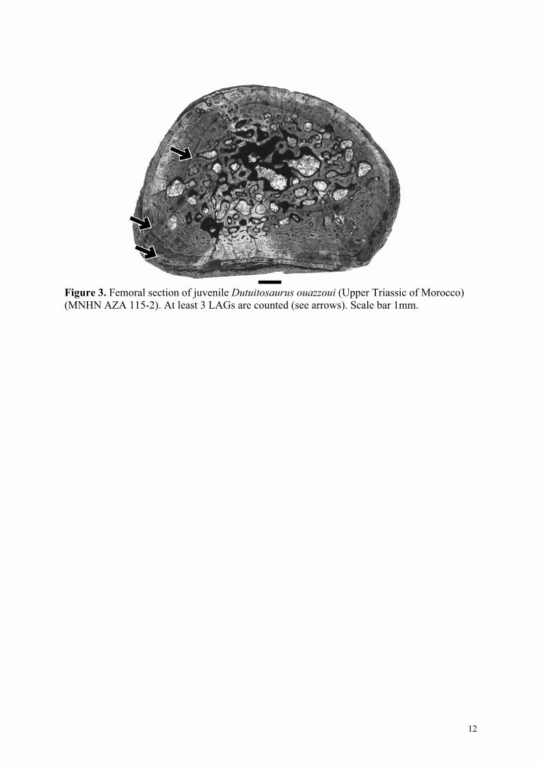
Figure 3. Femoral section of juvenile Dutuitosaurus ouazzoui (Upper Triassic of Morocco) (MNHN AZA 115-2). At least 3 LAGs are counted (see arrows). Scale bar 1mm.
12

Figure 4. Femoral section of adult (A) Dutuitosaurus ouazzoui (Upper Triassic of Morocco) (MNHN AZA 74-2) with its external cortex (B) showing at least 7 closely spaced LAGs (see arrows). Scale bar 1mm.
13
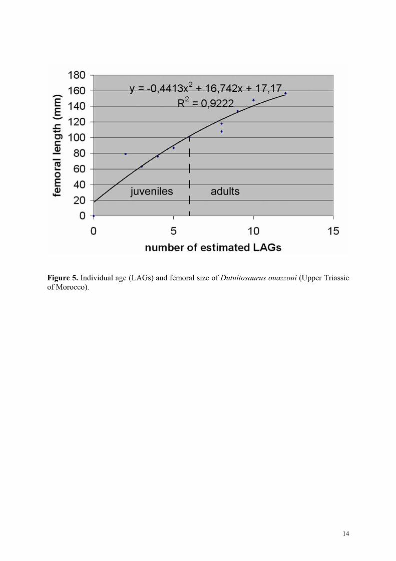
Figure 5. Individual age (LAGs) and femoral size of Dutuitosaurus ouazzoui (Upper Triassic of Morocco).
14
Figure 6. Evolution of the femoral compactness profile of Dutuitosaurus (Upper Triassic of Morocco) during its growth, and comparison with the extant taxa Pelusios subniger and Bufo marinus. Compactness (c) and distance to the center (dc) in % of the section radius.
15