Final Report - ETH Z · 1/14 Final Report . Project reference: A2011-03 . Applicant’s name:...
14
1/14 Final Report Project reference: A2011-03 Applicant’s name: Lagroye Isabelle, Veyret Bernard, Haro Emmanuelle, Hurtier Annabelle, Poulletier de Gannes Florence, Masuda Hiroshi, Jany Marion Project title: NeuroInflammation and Mobile PHone Exposure (NIMPHE) 1. State of Research. 1.1 Research activities performed, milestones and deliverables accomplished Please list against the background of the research proposal. Task 1: Animal exposure All the animals were cared for in accordance with French animal welfare guidelines. The laboratory members have obtained the agreement to experiment on live animals from the French authorities and the protocol was validated by the local ethic committee for animal experiments (reference 00237.01). Four-week male Wistar (Janvier, France) were used. Animals were housed under controlled temperature (22°C) and lighting conditions (monitored light-dark cycles 08:00-20:00), and supplied with water and food ad libitum (Safe, France). Animals were kept for one week in the laboratory before the start of the experiment. Exposure began after a progressive habituation period to confinement in the exposure jigs: 5 min Day 1, 15 min Day 2, 30 min Day 3, 1 hr Day 4, 2 hr Day 5 and 2 hr Day 6. When six-week-old, rats were exposed head-only to the UMTS (1960 MHz) and GSM-900 signals using the loop-antenna setup for 2 hours/day, 5 days/week for 4 weeks (Figure 1). Brain-averaged Specific Absorption Rate (BASAR) tested were 0, 0.5, 5 and 15 W/kg. For each signal, three to six series of exposure were performed for a total of 216 rats, i.e. 24 rats per group. A total of 24 rats were sham-exposed and 16 rats were kept in the animal facility as cage-controls. Figure 1. The loop antenna system for head-only exposure of rats. The loop antenna can be adapted to both GSM-900 and UMTS signals. At 0.5 and 5 W/kg, 8 rats are exposed at a time. At 15 W/Kg, 4 rats are exposed at a time. Sham-exposed rats are placed in the rockets the same way, but without the loop-antenna. Milestone 1: “Completion of RF exposures” was completed. Task 2: Brain slicing Forty-eight hours after the last exposure, rats (10 week-old) were ethically sacrificed using isoflurane inhalation. Using intracardiac perfusion, rats were perfused with phosphate buffer saline (PBS, 8 minutes)
Transcript of Final Report - ETH Z · 1/14 Final Report . Project reference: A2011-03 . Applicant’s name:...

1/14
Final Report Project reference: A2011-03 Applicant’s name: Lagroye Isabelle, Veyret Bernard, Haro Emmanuelle, Hurtier Annabelle, Poulletier de
Gannes Florence, Masuda Hiroshi, Jany Marion Project title: NeuroInflammation and Mobile PHone Exposure (NIMPHE)
1. State of Research.
1.1 Research activities performed, milestones and deliverables accomplished Please list against the background of the research proposal. Task 1: Animal exposure All the animals were cared for in accordance with French animal welfare guidelines. The laboratory members have obtained the agreement to experiment on live animals from the French authorities and the protocol was validated by the local ethic committee for animal experiments (reference 00237.01). Four-week male Wistar (Janvier, France) were used. Animals were housed under controlled temperature (22°C) and lighting conditions (monitored light-dark cycles 08:00-20:00), and supplied with water and food ad libitum (Safe, France). Animals were kept for one week in the laboratory before the start of the experiment. Exposure began after a progressive habituation period to confinement in the exposure jigs: 5 min Day 1, 15 min Day 2, 30 min Day 3, 1 hr Day 4, 2 hr Day 5 and 2 hr Day 6. When six-week-old, rats were exposed head-only to the UMTS (1960 MHz) and GSM-900 signals using the loop-antenna setup for 2 hours/day, 5 days/week for 4 weeks (Figure 1). Brain-averaged Specific Absorption Rate (BASAR) tested were 0, 0.5, 5 and 15 W/kg. For each signal, three to six series of exposure were performed for a total of 216 rats, i.e. 24 rats per group. A total of 24 rats were sham-exposed and 16 rats were kept in the animal facility as cage-controls.
Figure 1. The loop antenna system for head-only exposure of rats. The loop antenna can be adapted to both GSM-900 and UMTS signals. At 0.5 and 5 W/kg, 8 rats are exposed at a time. At 15 W/Kg, 4 rats are exposed at a time. Sham-exposed rats are placed in the rockets the same way, but without the
loop-antenna. Milestone 1: “Completion of RF exposures” was completed. Task 2: Brain slicing Forty-eight hours after the last exposure, rats (10 week-old) were ethically sacrificed using isoflurane inhalation. Using intracardiac perfusion, rats were perfused with phosphate buffer saline (PBS, 8 minutes)
2/14
and fixed for 8 minutes with 4% Paraformaldehyde in 0.1 M phosphate buffer. The brains were removed and kept in the fixative solution overnight at 4°C. They were dehydrated using 20 % sucrose in phosphate buffer for 48 hours at 4°C and then cryo-preserved using isopentane at -80°C. To ensure blinding of the experiments, brains were coded at this step, before slicing and analysis. From the brains, prefrontal cortex and hippocampal CA1, CA2, CA3 and dentate gyrus (DG) serial floating cryosections (40 µm thick) were prepared and stored at –20°C in a cryoconservative medium. Task 3: Slice staining and analysis The following stainings were performed CD68/ED1, Iba1, and GFAP. Microglia expressing CD68 markers are known as phagocytic cells and were found, in the Nimphe project, as a rare event after microscopic observations. Unbiased stereology is considered the best method to estimate this sparse labelling. Therefore, for CD68, four to six slide-mounted serial sections were analyzed with a macro developped in Imaje J software. Results are expressed as the averaged sum of cell count/total area. These values were multiply by 100 to draw the figures and are expressed as arbitrary unit (A.U.). The levels of expression of GFAP and Iba1 proteins (numerous stellate cells) were obtained after conventional image analysis using ImageJ software. The use of conventional method image analysis with Image J allows us to compare our results with published findings looking at GFAP and Iba1 immunostaining after RF exposure. Results are expressed in percentage of labelled area. Task 4: Decoding and statistical analysis Partial decoding and statistical analysis were performed for Iba1, CD68 and GFAP in brains of 8 rats exposed to the UMTS signal for reporting. This process involved persons not involved in the analysis. All other codes were broken once the whole analysis was completed. Statistical analysis used the Kruskall-Wallis test (for small samples) on all experimental groups followed, when significant difference were detected, by the Mann-Whitney test to compare each exposed group to the sham-exposed group. To test for a possible strees induced by the contention (although the habituation protocol), sham-exposed rats were compared to cage control rats using the Mann-Whitney test. Since UMTS exposure and GSM exposure were performed in the different buildings (see section 1.3), we considered that the experimental groups had to be compared to their respective sham-exposed group. Task 5: Reporting Reporting includes the different reports to the Foundation, including the present report. Preliminary data were presented as a poster at the BioEM2013 meeting (June 2013) and a platform presentation at the BioEM2014 meeting (June 2014).
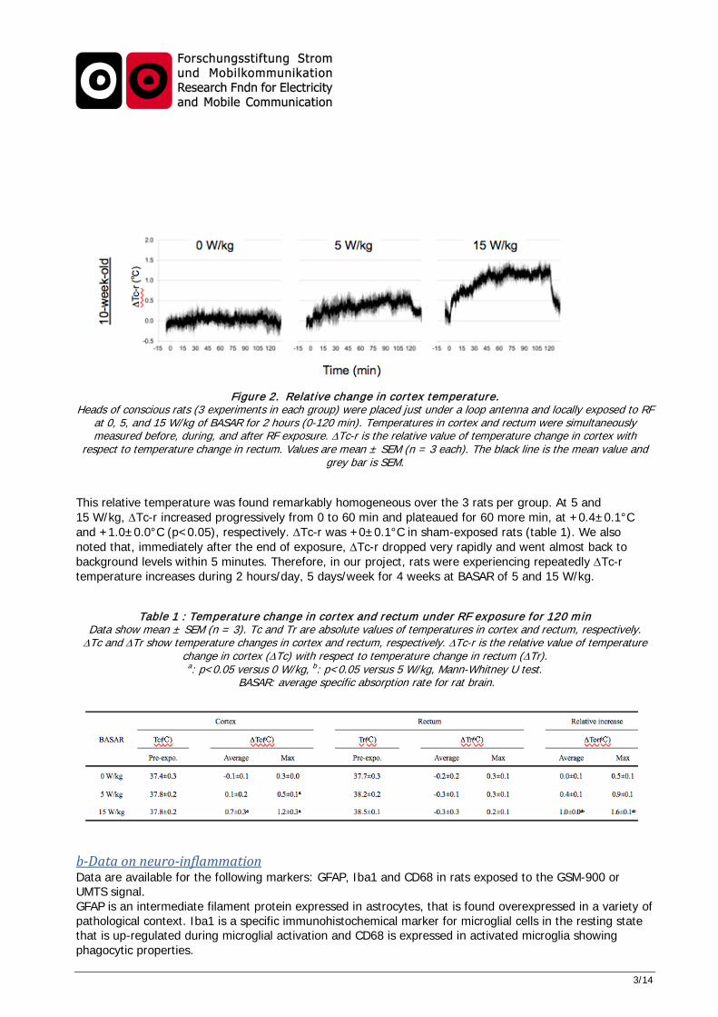
1.2 Findings Comment on achieved scientific insights. a-Temperature measurements In a preliminary experiment on a single rat exposed at 15 W/kg, a 0.5 °C increase in brain temperature was observed over a 30 minute-exposure, while core temperature did not change. In further experiments, temperature was measured in the rat brain during a 2-hour UMTS exposure at 0, 5, and 15 W/kg BASAR. Groups of 3 rats of 10 weeks of age were used. For that purpose, temperature probes were implanted in the rectum and dura mater under anesthesia. After a one-day recovery, rats were placed in a jig and exposed. Figure 2 shows the relative value of temperature change in cortex with respect to temperature change in the rectum (∆Tc-r in °C).
3/14
Figure 2. Relative change in cortex temperature. Heads of conscious rats (3 experiments in each group) were placed just under a loop antenna and locally exposed to RF
at 0, 5, and 15 W/kg of BASAR for 2 hours (0-120 min). Temperatures in cortex and rectum were simultaneously measured before, during, and after RF exposure. ∆Tc-r is the relative value of temperature change in cortex with
respect to temperature change in rectum. Values are mean ± SEM (n = 3 each). The black line is the mean value and grey bar is SEM.
This relative temperature was found remarkably homogeneous over the 3 rats per group. At 5 and 15 W/kg, ∆Tc-r increased progressively from 0 to 60 min and plateaued for 60 more min, at +0.4±0.1°C and +1.0±0.0°C (p<0.05), respectively. ∆Tc-r was +0±0.1°C in sham-exposed rats (table 1). We also noted that, immediately after the end of exposure, ∆Tc-r dropped very rapidly and went almost back to background levels within 5 minutes. Therefore, in our project, rats were experiencing repeatedly ∆Tc-r temperature increases during 2 hours/day, 5 days/week for 4 weeks at BASAR of 5 and 15 W/kg.
Table 1 : Temperature change in cortex and rectum under RF exposure for 120 min Data show mean ± SEM (n = 3). Tc and Tr are absolute values of temperatures in cortex and rectum, respectively. ∆Tc and ∆Tr show temperature changes in cortex and rectum, respectively. ∆Tc-r is the relative value of temperature
change in cortex (∆Tc) with respect to temperature change in rectum (∆Tr). a: p<0.05 versus 0 W/kg, b: p<0.05 versus 5 W/kg, Mann-Whitney U test.
BASAR: average specific absorption rate for rat brain.
b-Data on neuro-inflammation Data are available for the following markers: GFAP, Iba1 and CD68 in rats exposed to the GSM-900 or UMTS signal. GFAP is an intermediate filament protein expressed in astrocytes, that is found overexpressed in a variety of pathological context. Iba1 is a specific immunohistochemical marker for microglial cells in the resting state that is up-regulated during microglial activation and CD68 is expressed in activated microglia showing phagocytic properties.

4/14
Effects of GSM-900 exposure The percentage of labelled area corresponding to GFAP positive cells in cage control rats (n=9) was 27.1±3.4% in the prefrontal cortex and 35.1±5.9% in the hippocampus. No significant difference was found in the Sham-exposed rats (n=11), with a GFAP-positive percent area of 38.1±3.1% in the prefrontal cortex and 50.7±4.7% in the hippocampus. In the experimental groups, GFAP expression was not found significantly affected in the prefrontal cortex and the hippocampus (Figure 3). Figure 3: Expression of GFAP in the prefrontal cortex and the hippocampus of rats exposed to GSM-900. Rats were repeatedly exposed to the GSM-900 signal at different SAR levels. Sham: n=11; 0.5 W/kg: n=24; 5 W/kg:
n=20; 15 W/kg: n=16. The percentage of labelled area corresponding to Iba-1 positive cells in cage control rats (n=16) was 10.3±1.3% in the prefrontal cortex and 8.1±1.1% in the hippocampus. No significant difference was found in the Sham-exposed rats (n= 11), with a Iba-1-positive percent area of 11.2±0.5% in the prefrontal cortex and 9.8±0.9% in the hippocampus. By contrast, Iba1 expression was significantly decreased after exposure at 5 and 15 W/kg in the prefrontal cortex with a Iba-1 positive labelled area of 8.2±0.4% and 6.3±1.4%, respectively. At 15 W/kg, the Iba-1 positive labelled area dropt to 4.5±1% in the hippocampus (Figure 4).
Figure 4: Expression of Iba1 in the prefrontal cortex and the hippocampus of rats exposed to GSM-900.
Rats were repeatedly exposed to the GSM-900 signal at different SAR levels. Sham: n=11; 0.5 W/kg: n=24; 5 W/kg: n=24; 15 W/kg: n=16. *: p<0.05 as compared to sham

5/14
The CD68-positive cell population (averaged sum of cell count/total area) was 0.049±0.006 in the prefrontal cortex and 0.113±0.006 in the hippocampus of the cage control rats (n=16). No significant difference was found in the Sham-exposed rats (n= 11), with a CD68-positive cell population of 0.051±0.005 in the prefrontal cortex and 0.103±0.018 in the hippocampus. In the experimental groups, CD68 expression was not found significantly affected in the prefrontal cortex and the hippocampus (Figure 5).
Figure 5: Expression of CD68 in the prefrontal cortex and the hippocampus of rats exposed to GSM-900. Rats were repeatedly exposed to the GSM-900 signal at different SAR levels. Sham: n=10; 0.5 W/kg: n=24;
5 W/kg: n=23; 15 W/kg: n=24. Effects of UMTS exposure
Figure 6: Expression of GFAP in the prefrontal cortex and the hippocampus of rats exposed to UMTS. Rats were repeatedly exposed to the UMTS signal at different SAR levels. Sham: n=14; 0.5 W/kg: n=19;
5 W/kg: n=22; 15 W/kg: n=20. No significant difference was found within the experimental groups. The percentage of labelled area corresponding to GFAP positive cells in cage control rats (n=13) was 28.8±3.2% in the prefrontal cortex and 49.3±4.1% in the hippocampus. No significant difference was found in the Sham-exposed rats (n=14), with a GFAP-positive percent area of 32.1±3.6% in the prefrontal cortex and 45.2±3.5% in the hippocampus.
6/14
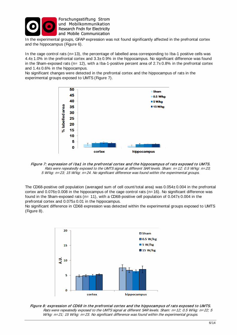
In the experimental groups, GFAP expression was not found significantly affected in the prefrontal cortex and the hippocampus (Figure 6). In the cage control rats (n=13), the percentage of labelled area corresponding to Iba-1 positive cells was 4.4±1.0% in the prefrontal cortex and 3.3±0.9% in the hippocampus. No significant difference was found in the Sham-exposed rats (n= 12), with a Iba-1-positive percent area of 2.7±0.8% in the prefrontal cortex and 1.4±0.6% in the hippocampus. No significant changes were detected in the prefrontal cortex and the hippocampus of rats in the experimental groups exposed to UMTS (Figure 7).
Figure 7: expression of Iba1 in the prefrontal cortex and the hippocampus of rats exposed to UMTS. Rats were repeatedly exposed to the UMTS signal at different SAR levels. Sham: n=12; 0.5 W/kg: n=23;
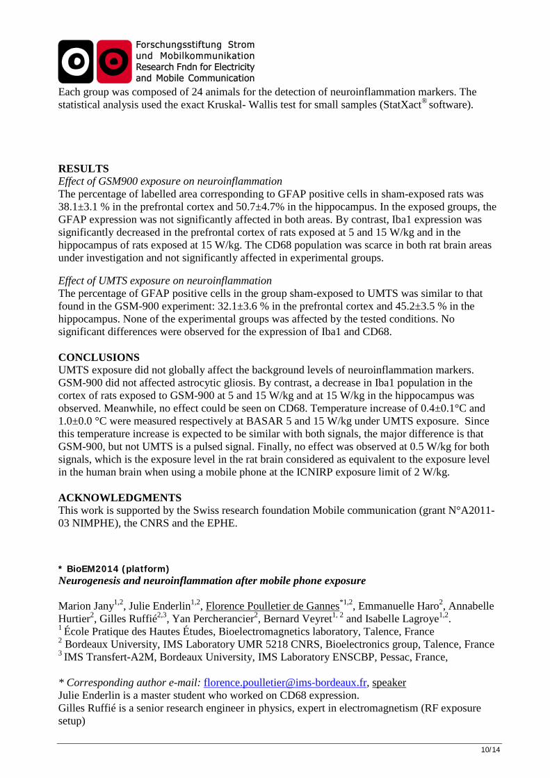
5 W/kg: n=23; 15 W/kg: n=24. No significant difference was found within the experimental groups. The CD68-positive cell population (averaged sum of cell count/total area) was 0.054±0.004 in the prefrontal cortex and 0.076±0.008 in the hippocampus of the cage control rats (n=16). No significant difference was found in the Sham-exposed rats (n= 11), with a CD68-positive cell population of 0.047±0.004 in the prefrontal cortex and 0.075±0.01 in the hippocampus. No significant difference in CD68 expression was detected within the experimental groups exposed to UMTS (Figure 8).
Figure 8: expression of CD68 in the prefrontal cortex and the hippocampus of rats exposed to UMTS. Rats were repeatedly exposed to the UMTS signal at different SAR levels. Sham: n=12; 0.5 W/kg: n=22; 5
W/kg: n=21; 15 W/kg: n=23. No significant difference was found within the experimental groups.

7/14
Conclusion A set of markers was investigated to get a global view of the influence of exposure to RF signals on neuroinflammation in rats. Head-only exposures lasted 2 hours per day, 5 days per week per 4 weeks, at SAR levels of 0; 0.5, 5 and 15 W/kg. The preliminary results obtained with the UMTS signal (a drop in the CD68 positive cell population in the cortex and the hippocampus) could not be confirmed, and globally UMTS exposures did not affect the background level of inflammation in the brains of rats. GSM-900 exposures did not induce reactive astrocytic gliosis. By contrast, a decrease in the Iba-1 positive cell population was shown in the cortex of rats exposed at 5 and 15 W/kg and at 15 W/kg in their hippocampus when exposed to the GSM-900 signal. The amplitude of the effect ranged from 27% at 5 W/kg in the prefrontal cortex to 44 and 54% at 15 W/kg in the prefrontal cortex and the hippocampus, respectively. Meanwhile, no effect could be seen on the CD68 positive cell population. Overall, the results obtained suggest no pro-inflammatory effects of the GSM-900 and UMTS signals in the rat brain repeatedly exposed for 4 weeks, head-only, and up to 15 W/kg. This is in agreement with previous work testing for chronic exposures of mice (Kim et al. 2008, Finnie et al. 2010, Court-Kowalski et al. 2015) or rats (Aït-Aïssa et al. 2010) to radiofrequency fields ranging from 849 to 2450 MHz. However, they are in contradiction with a set of data testing the effects of chronic (8 and 24 weeks) GSM-900 head-only exposures (loop antennas) on gliosis (Ammari et al., 2008; 2010). In these studies, reactive astrocytic gliosis was mainly seen at 6 W/kg (15 min/day, 5 days/week), and 3 and 10 days after the least exposure. In the NIMPHE project, the 5 W/kg level tested was similar to the 6 W/kg exposure level of Ammari et al. (2008, 2010), but exposure schedule was different (2 hr/day, 4 weeks). Exposures of more than 4 weeks may then be needed to induce reactive gliosis. In the NIMPHE project, experimental groups have 15 to 24 animals, while there were 3 animals per group in the Ammari 2008 paper and 6 animals per group in the Ammari 2010 paper. It thus cannot be ruled out that part of the findings in the smallest groups could be chance findings. We, however, found a differential effect of the GSM-900 signal as compared to the UMTS signal. The effect of repeated exposures to GSM-900 was observed at 5 and/or 15 W/kg in the brain regions tested, and was a decrease in the activated microglial cell population on one out the two microglial activation markers tested. This effect may be related to a defect in early microglial activation. An increase in activated microglial death could also be hypothesized. Based on the brain slices counterstaining, we noted that the total number of astrocytes and microglial cells seemed to not be affected by repeated RF exposures in both cortex and hippocampus. However, a specific assay for cell viability would be needed to exclude this possibility (TUNEL assay for instance). We showed that during UMTS exposures for 2 hours/day, 5 days/week for 4 weeks rats were repeatedly experiencing a mean ∆Tc-r1 temperature increase of 0.4±0.1°C and 1.0±0.0°C at the BASAR of 5 and 15 W/kg, respectively. Since this temperature increases are expected to be similar with both signals, the major difference is that GSM-900, but not UMTS, is a pulsed signal. The peak power level is thus higher in the brains of rats exposed to the GSM-900 signal, and could explain the differential effect. The possibility for a differential effect of the modulation (pulsed versus non-pulsed) of radiofrequency fields signals was suggested possible in the central nervous system only by contrast to any other organs (Juutilainen et al., 2011). Recently, evidence has grown that a pulsed signal could affect the electroencephalogramme spectrum while a non-pulsed signal could not (Regel et al. 2007, Kwon et al. 2011; Schmid et al. 2012; Suhhova et al., 2013; SCENIHR 2015).
1 ∆Tc-r is the relative value of temperature change in cortex with respect to temperature change in rectum

8/14
Finally, in the NIMPHE project, no effects were observed at the 0.5 W/kg BASAR level for both GSM and UMTS signals, which is considered as the exposure level in the rat brain equivalent to the exposure level in the brain of humans when using a mobile phone at the ICNIRP local exposure limit of 2 W/kg. Bibliography Aït Aïssa S et al. (2010) In situ detection of gliosis and apoptosis in the brains of young rats exposed in utero to a Wi-Fi signal. C. R. Physique. 11 : 592–601 Ammari M et al. (2008) Effect of a chronic GSM 900 MHz exposure on glia in the rat brain. Biomedicine & Pharmacotherapy 62 : 273-281. Ammari M et al. (2010) GFAP expression in the rat brain following sub-chronic exposure to a 900 MHz electromagnetic field signal. Int J Radiat Biol. 86(5):367-375. Court-Kowalski S et al. (2015) Effect of long-term (2 years) exposure of mouse brains to global system for mobile communication (GSM) radiofrequency fields on astrocytic immunoreactivity. Bioelectromagnetics. 36(3):245-250. Finnie JW et al. (2010) Microglial activation as a measure of stress in mouse brains exposed acutely (60 minutes) and long-term (2 years) to mobile telephone radiofrequency fields. Pathology. 42(2):151-154. Juutilainen J et al. (2011) Review of Possible Modulation-Dependent Biological Effects of Radiofrequency Fields. Bioelectromagnetics 32: 511-534. Kim et al. (2008) Local exposure of 849 MHz and 1763 MHz radiofrequency radiation to mouse heads does not induce cell death or cell proliferation in brain. Exp Mol Med. 40(3):294-303. Kwon MS & Hämäläinen H (2011) Effects of mobile phone electromagnetic fields: critical evaluation of behavioral and neurophysiological studies. Bioelectromagnetics 32: 253-272. Regel SJ et al. (2007) Pulsed radio frequency radiation affects cognitive performance and the waking electro-encephalogram. Neuroreport 18: 803-807. SCENIHR (2105) Health effects of EMF – Scientific Committee on Emerging and Newly Identified Health Risks. http://ec.europa.eu/health/scientific_committees/emerging/index_en.htm Schmid MR et al. (2012) Sleep EEG alterations: effects of different pulse-modulated radio frequency electromagnetic fields. J Sleep Res 21: 50-58. Suhhova A et al. (2013) Effect of microwave radiation on human EEG at two different levels of exposure. Bioelectromagnetics. 34: 264-74.
1.3 Problems Expand on research, financial or schedule problems, if any. For the intermediate report: please include problems that might occur in the upcoming period. This part of the report must not exceed one page (intermediate report) and two pages (final report). Our laboratory moved into a new building in December 2012. Although we tried to optimize our work plan, delay was experienced in exposure to the GSM-900 signal. Moreover, Murielle Taxile, our engineer in biology since 1999, got a permanent position in a private company at the end of 2012. Marion Jany was hired to replace her but had to deal with repeated health problems, which impeded the work progression. Using stereology also multiplied the number of stainings and increased the time for analysis as compared to the classical methods, and we obviously under-estimated this point when adding stereological analyses in our project was discussed. Therefore, it was not possible to provide data on other markers as previously planned. We acknowledge the 15-month extension of the contract duration given by the Foundation.
2. Annex
2.1 Publications The data analysis was completed very recently. Our plan is to write one paper to be submitted to Plos One.
2.2 Documents In case publications are not yet available or cover only part of the funded research, please include: - submitted papers (confidentiality is secured), or

9/14
- concise internal documents that inform about your research work, or - a short progress (2-3 pages) or final report that summarises the state of research The project and preliminary data were presented at the BioEM 2013 and BioEM2014 meetings and the support of the Foundation was mentioned. Final results will be presented at BioEM2016 (platform). * BioEM2016 (platform) Neuroinflammation after mobile phone exposure Florence Poulletier de Gannes*1, Emmanuelle Haro1
, Rémy Renom1, Annabelle Hurtier1, Marion Jany1, Julie Enderlin1, Gilles Ruffié 1,3, Yan Percherancier1, Bernard Veyret1, 2
and Isabelle Lagroye1,2. 1 Bordeaux University, IMS Laboratory UMR 5218 CNRS, Bioelectronics group, Talence, France 2 École Pratique des Hautes Études, Bioelectromagnetics laboratory, Talence, France 3 IMS Transfert-A2M, Bordeaux University, IMS Laboratory ENSCBP, Pessac, France, * Corresponding author e-mail: [email protected] Julie Enderlin and Rémy Renom are master students who worked on CD68 expression. Gilles Ruffié is a senior research engineer in physics, expert in electromagnetism (RF exposure setup) INTRODUCTION After partial results presented in BioEM2014 in Cape Town, we have completed the NIMPHE project to provide answers to controversial findings related to the effect of RF exposure on neuroinflammation. A set of markers was investigated to get a global view of neuroinflammation after GSM-900 or UMTS exposures. MATERIALS AND METHODS Animal exposure Following a one-week acclimatization to the animal facility and another week of progressive habituation to confinement in exposure jigs, male Wistar rats (6 week old) were exposed head-only, using a loop antenna, to GSM-900 or UMTS-1960 signals. Exposure lasted 2 hrs/day, 5 days/week, for 4 weeks at brain-averaged specific absorption rates (BASAR) of 0 (sham-exposed group), 0.5, 5, and 15 W/kg. We used a control group of non-exposed animals that remained in the animal facility during the whole experiment period as negative control (cage controls). Three series of exposure for each RF signal were done. Rat brain preparation Forty-eight hours after the last exposure, rats were ethically sacrificed and their brains fixed with paraformaldehyde, removed, coded, frozen and stored at –80°C. Prefrontal cortex, hippocampal CA1, CA2, CA3 and dentate gyrus serial floating cryosections (40 µm thick) were prepared and stored at –20°C in a cryoconservative medium. Immunohistochemistry Neuroinflammation was investigated using specific antibodies directed against GFAP, CD68/ ED1, and Iba-1 (Wako, Germany). Staining was performed using the avidin-biotin-peroxydase amplificating method and/or diaminobenzidine as chromagen (Vector Laboratories, France). When necessary, a pH 6 citric-acid retrieval was used. Cell nuclei were counterstained using blue counterstaining medium and ammonium revelation. Brain slices were coverslipped with DPX mounting medium (Sigma Aldrich, France), and viewed using an Olympus BX51WIF microscope (Olympus, Japan). For CD68, due to the round shape of super-activated microglia, a stereological method of cell counting could be used. Statistical analysis
10/14
Each group was composed of 24 animals for the detection of neuroinflammation markers. The statistical analysis used the exact Kruskal- Wallis test for small samples (StatXact®
software). RESULTS Effect of GSM900 exposure on neuroinflammation The percentage of labelled area corresponding to GFAP positive cells in sham-exposed rats was 38.1±3.1 % in the prefrontal cortex and 50.7±4.7% in the hippocampus. In the exposed groups, the GFAP expression was not significantly affected in both areas. By contrast, Iba1 expression was significantly decreased in the prefrontal cortex of rats exposed at 5 and 15 W/kg and in the hippocampus of rats exposed at 15 W/kg. The CD68 population was scarce in both rat brain areas under investigation and not significantly affected in experimental groups. Effect of UMTS exposure on neuroinflammation The percentage of GFAP positive cells in the group sham-exposed to UMTS was similar to that found in the GSM-900 experiment: 32.1±3.6 % in the prefrontal cortex and 45.2±3.5 % in the hippocampus. None of the experimental groups was affected by the tested conditions. No significant differences were observed for the expression of Iba1 and CD68. CONCLUSIONS UMTS exposure did not globally affect the background levels of neuroinflammation markers. GSM-900 did not affected astrocytic gliosis. By contrast, a decrease in Iba1 population in the cortex of rats exposed to GSM-900 at 5 and 15 W/kg and at 15 W/kg in the hippocampus was observed. Meanwhile, no effect could be seen on CD68. Temperature increase of 0.4±0.1°C and 1.0±0.0 °C were measured respectively at BASAR 5 and 15 W/kg under UMTS exposure. Since this temperature increase is expected to be similar with both signals, the major difference is that GSM-900, but not UMTS is a pulsed signal. Finally, no effect was observed at 0.5 W/kg for both signals, which is the exposure level in the rat brain considered as equivalent to the exposure level in the human brain when using a mobile phone at the ICNIRP exposure limit of 2 W/kg. ACKNOWLEDGMENTS This work is supported by the Swiss research foundation Mobile communication (grant N°A2011-03 NIMPHE), the CNRS and the EPHE. * BioEM2014 (platform) Neurogenesis and neuroinflammation after mobile phone exposure Marion Jany1,2, Julie Enderlin1,2, Florence Poulletier de Gannes*1,2, Emmanuelle Haro2, Annabelle Hurtier2, Gilles Ruffié2,3, Yan Percherancier2, Bernard Veyret1, 2 and Isabelle Lagroye1,2. 1 École Pratique des Hautes Études, Bioelectromagnetics laboratory, Talence, France 2 Bordeaux University, IMS Laboratory UMR 5218 CNRS, Bioelectronics group, Talence, France 3 IMS Transfert-A2M, Bordeaux University, IMS Laboratory ENSCBP, Pessac, France,
* Corresponding author e-mail: [email protected], speaker Julie Enderlin is a master student who worked on CD68 expression. Gilles Ruffié is a senior research engineer in physics, expert in electromagnetism (RF exposure setup)

11/14
Yann Percherancier is a senior researcher who worked on neurogenesis-related gene expression (RT-PCR). SHORT SUMMARY There are some doubts related to neuroinflammation and cognitive functions following exposure to wireless communication signals. Our work aims at providing answers to these open questions. Rats were repeatedly exposed to two types of mobile phone signals (GSM-900 and UMTS), 2 hrs/day, 5 days/week, for 4 weeks at Brain Averaged SAR of 0, 0.5, 5, and 15 W/kg. Neurogenesis and neuroinflammation were investigated. INTRODUCTION The effects of wireless communication exposure on the brain are still a matter of debate. Two projects are in progress in our laboratory to provide answers to controversial findings related to the effects of RF fields on neuroinflammation1 and cognitive functions2. Regarding the first research topic, both astroglial and microglial cell populations were assessed along with some other neuroinflammation markers. In the second project, adult hippocampal neurogenesis was monitored using proliferation and neuronal markers, and signalling pathways regulating neurogenesis in the hippocampal dentate gyrus area. All processes were investigated using repeated exposures to two types of mobile phone signals (GSM-900 and UMTS). MATERIALS AND METHODS Animal exposure Following a one-week acclimatization to the animal facility and another week of progressive habituation to confinement in exposure jigs, male Wistar rats (6 week old) were exposed head-only, using a loop antenna, to GSM-900 or UMTS-1960 signals. Exposure lasted 2 hrs/day, 5 days/week, for 4 weeks at brain-averaged specific absorption rates (BASAR) of 0 (sham-exposed group), 0.5, 5, and 15 W/kg. We used a control group of non-exposed animals that remained in the animal facility during the whole experiment period as negative control (cage controls). Three series of exposure for each RF signal were done. An extra exposure was added for an RT-PCR study of the signalling pathways. Rat brain preparation Forty-eight hours after the last exposure, rats were ethically sacrificed and their brains fixed with paraformaldehyde, removed, coded, frozen and stored at –80°C. Prefrontal cortex and hippocampal CA1, CA2, CA3 and dentate gyrus serial floating cryosections (40 µm thick) were prepared and stored at –20°C in a cryoconservative medium. In the RT-PCR study, hippocampi were dissected immediately after decapitation and rapidly frozen in liquid nitrogen and store at –80°C. Immunohistochemistry Neurogenesis was investigated using the thymidine analogue 5-bromo-2′ - deoxyuridine (BrdU) (Abdserotec, UK) and the doublecortin (DCX) labelling (Millipore, France). Neuroinflammation was investigated using specific antibodies directed against GFAP, CD68/ ED1, COX2 (Santa Cruz Biotechnology, Germany), S100B (Abcam, France), Iba-1 (Wako, Germany), isolectin GS1-ß4 (Abcys SA, France) or iNOS (Millipore, France). Staining was performed using the avidin-biotin-peroxydase amplificating method and/or diaminobenzidine as chromagen (Vector Laboratories, France). When necessary, a pH 6 citric-acid retrieval was used. Cell nuclei were counterstained using blue counterstaining medium and ammonium revelation.

12/14
Brain slices were coverslipped with DPX mounting medium (Sigma Aldrich, France), and viewed using an Olympus BX51WIF microscope (Olympus, Japan). For a selected marker, CD68, an unbiased stereological method of cell counting was used. RT-PCR Total RNA was extracted from samples of the hippocampus (Rneasy plus mini kit, Qiagen, France) then reverse transcripted by Quantitect reverse transcription (1g of RNA), and amplified by RT real-time PCR (Rotor-Gene, Qiagen, France). Primers for the following genes of interest: Notch1, Hes1 and Hes5, JAG1 and internal controls ß-actin and HPRT (Hypoxanthine phosphoribosyltransferase) were tested (Realtimeprimer.com). Amplification was obtained using the Rotorgene SYBR green kit (Qiagen, France) containing SYBR Green and Taq DNA polymerase. Statistical analysis Each group was composed of 24 animals for the detection of neuroinflammation markers, 8 for neurogenesis markers, and 3 for RT-PCR. The statistical analysis used the exact Kruskal-Wallis test for small samples (StatXact® software). RESULTS Immunohistochemistry analysis is in progress. Results for at least 8 rats per group will be presented at the meeting for CD68, Iba1 and GFAP. Exposures are in progress for the RT-PCR assay and results will be analysed for the meeting report. CONCLUSION Our projects will provide extensive information on neuroinflammation and neurogenesis after RF exposure in adult rats. Astroglial and microglial activation is being explored as well as the presence of inflammatory markers. Such a comprehensive approach will help reach a conclusion on pro-inflammatory effects of RF wireless signals on the rat brain. To our knowledge, it is the first time that a neurogenesis study is performed after RF exposure in order to support or not the data reported in the literature on rodent cognitive functions. REFERENCES 1 Mausset-Bonnefont et al., 2004, Neurobiol. Dis., 17:445–54 ; Brillaud et al., 2007, Toxicology, 238:23–33; Carballo-Quintas et al., 2011, NeuroToxicology, 32 : 478–494; Ammari et al., 2008, Biomed Pharmacother., 62:273–81; Ammari et al., 2010, Int. J. Radiat. Biol. 86:367–75; Maskey et al., 2010, Brain Res., 1346: 237–46; Fritze et al., 1997, Neuroscience., 81(3):627–39, Watilliaux et al., 2011, Neurotox. Res., 20:109–119; Finnie et al., 2010, Pathology, 42:151–154; Hirose et al., 2010, Bioelectromagnetics, 31:104-12; Thorlin et al., 2006, Radiat. Res., 166:409–421. 2 Nittby et al. Bioelectromagnetics, 2008, 29:219-32 ; Fragopoulou et al. 2010, Pathophysiology 17:179-87 ; Ntzouni et al., 2013, Electromagnetic Biology and Medicine, 32: 95-120; Kumlin et al., 2007, Radiation Research, 168: 471–479 ACKNOWLEDGMENTS This work is supported by the Swiss research foundation Mobile communication (grant N°A2011-03 NIMPHE), Bouygues Telecom (grant TEMCER), the CNRS and the EPHE. * BioEM2013 (poster)

13/14
The abstract “Neuroinflammation and mobile phone exposure: the NIMPHE project” was accepted for a poster presentation at the BioEM2013 meeting in Thessaloniki, Greece. Authors: Marion Jany1,2, Florence Poulletier de Gannes1,2, Murielle Taxile1,2, Annabelle Hurtier2, Emmanuelle Haro2, Gilles Ruffié2,3, Bernard Veyret1, 2 and Isabelle Lagroye*1,2. 1 Ecole Pratique des Hautes Études, Bioelectromagnetics laboratory, Talence, France 2 Bordeaux University, IMS Laboratory UMR 5218 CNRS, Bioelectronics group, Talence, France 3 IMS Transfert-A2M, Bordeaux University, IMS Laboratory ENSCBP, Pessac, France, INTRODUCTION The NIMPHE project aims at providing extensive information on neuroinflammation under repeated exposures to two types of mobile phone signals (GSM-900 and UMTS), through the investigation of a panel of neuroinflammation markers in rat brains. Both astroglial and microglial cell populations, total and activated, are being tested along with some other neuroinflammation markers. MATERIALS AND METHODS Animal exposure Following a one-week acclimatization to the animal facility and a one-week habituation period to confinement in exposure jigs, rats were exposed head-only using a loop antenna to a GSM-900 or a UMTS-1960 signal. Exposure lasted 2 hrs/day, 5 days/week, for 4 weeks at brain-averaged specific absorption rates (BASAR) of 0 (sham-exposed group), 0.5, 5, and 15 W/kg. We used a group of LPS intraventricular injected rats as inflammation positive control and a group of non-exposed animals staying in the animal facility during the whole experiment period as exposure negative control (cage controls). Rat brain preparation Forty-eight hours after the last exposure, rats were ethically sacrificed and their brains removed, coded, frozen and stored at –80°C. Prefrontal cortex and hippocampal CA1, CA2, CA3 and dentate gyrus serial floating cryosections (40 µm thick) were prepared and stored at –20°C in a cryoconservative medium. Immunohistochemistry Neuroinflammation was investigated using specific antibodies directed against GFAP, CD68/ ED1, COX2 (Santa Cruz Biotechnology, Germany), S100B (Abcam, France), Iba-1 (Wako, Germany), isolectin GS1-ß4 (Abcys SA, France) or iNOS (Millipore, France). Staining was performed using the avidin-biotin-peroxydase amplificating method and/or diaminobenzidine as chromagen (Vector Laboratories, France). When necessary, a pH 6 citric acid retrieval was used. Cell nuclei were counterstained with blue counterstaining medium and ammonium revelation. Brain slices were coverslipped with DPX mounting medium (Sigma Aldrich, France), and viewed using an Olympus BX51WIF microscope (Olympus, Japan). For a selected marker, an unbiased stereological method of cell counting was carried out using the Mercator® software (Explora Nova, France). Statistical analysis Each group was composed of 24 animals. The statistical analysis used exact Kruskal-Wallis statistics for small samples (StatXact® software). RESULTS The work began early 2012 and will be completed in 2014. Three series of exposure for each RF signal were planned. For UMTS, the last exposure series is ongoing. In order to determine the

14/14
temperature in a rat brain under exposure to UMTS at the highest BASAR tested (15 W/kg), temperature probes were implanted in the anus and the dura matter. Over a 30 minute-exposure, a 0.5°C increase in brain temperature was observed while core temperature did not change. Immuno-histochemistry analysis is in progress. A minimum of 8 rats per group will be analysed for this meeting report. CONCLUSION There are some controversial findings related to neuroinflammation following wireless communication signals exposure. While a trend for the induction of gliosis in the brains of rats has been described1,2,3,4,5, there was no microglial activation reported in two papers6,7. The NIMPHE project will provide extensive information on neuroinflammation with such a wide panel of markers. Evidence for the presence of several converging endpoints will strengthen the conclusion about neuroinflammatory effects of mobile phone exposure of rats. REFERENCES 1Mausset-Bonnefont et al., 2004, Neurobiol. Dis., 17:445–54 2Brillaud et al., 2007, Toxicology, 238:23–33 3Carballo-Quintas et al., 2011, NeuroToxicology, 32 : 478–494 4Ammari et al., 2008, Biomed Pharmacother., 62:273–81 5Ammari et al., 2010, Int. J. Radiat. Biol. 86:367–75. 6Watilliaux et al., 2011, Neurotox. Res., 20:109–119. 7Finnie et al., 2010, Pathology, 42:151–154. ACKNOWLEDGMENTS This work is supported by the Swiss research foundation Mobile communication, the CNRS and the EPHE.
Date and Signature : 10 May 2016 Isabelle Lagroye