Field chest radiography
12
Chest Radiography in the field Narayan PENDSE, MD 25, Rue Daubin 1203 Geneva Switzerland 1. Background and purpose 2. Scope of this document 3. List of abbreviations 4. Introduction to field radiography 5. Pre requisites 6. Recommended Specifications 7. Manpower 8. Consumables 9. Criteria for selection 10. Details of radiograph 11. X‐ray interpretation 12. Radiation protection 13. Practical tips 14. Illustrations 15. References for further reading 1. BACKGROUND AND PURPOSE Chest radiography is an important tool to detect and monitor respiratory infections (including TB) and is the single most performed medical diagnostic imaging investigation. Chest radiography was extensively used for population screening for TB few decades back, after which it fell out of favour. Riding on technological improvements, chest radiography is reasserting itself in this role, and is increasingly finding mention in prevalence surveys and active case detection of TB. This is particularly true in high prevalence settings. However, use of radiography for this purpose is significantly different and more complex than it’s use in hospitals or clinics. Apart from type and cost of technology, factors like availability & expertise of manpower, status of allied infrastructure, number of x‐rays planned, general civic conditions, and many others play an important role for successful use of Chest radiography for prevalence surveys. Unfortunately, no ready reference document or simple‐to‐use tool is available for providing into Chest radiography in field conditions. This document is a concise guidance document for using Chest radiography for TB prevalence surveys. Recommended specifications for using radiography equipment are also provided, as is a check list for equipment.
Transcript of Field chest radiography

Chest Radiography in the field Narayan PENDSE, MD 25, Rue Daubin 1203 Geneva Switzerland
1. Background and purpose 2. Scope of this document 3. List of abbreviations 4. Introduction to field radiography 5. Pre requisites 6. Recommended Specifications 7. Manpower 8. Consumables 9. Criteria for selection 10. Details of radiograph 11. X‐ray interpretation 12. Radiation protection 13. Practical tips 14. Illustrations 15. References for further reading
1. BACKGROUND AND PURPOSE
Chest radiography is an important tool to detect and monitor respiratory infections (including TB) and is the single most performed medical diagnostic imaging investigation. Chest radiography was extensively used for population screening for TB few decades back, after which it fell out of favour. Riding on technological improvements, chest radiography is reasserting itself in this role, and is increasingly finding mention in prevalence surveys and active case detection of TB. This is particularly true in high prevalence settings.
However, use of radiography for this purpose is significantly different and
more complex than it’s use in hospitals or clinics. Apart from type and cost of technology, factors like availability & expertise of manpower, status of allied infrastructure, number of x‐rays planned, general civic conditions, and many others play an important role for successful use of Chest radiography for prevalence surveys. Unfortunately, no ready reference document or simple‐to‐use tool is available for providing into Chest radiography in field conditions.
This document is a concise guidance document for using Chest radiography for TB prevalence surveys. Recommended specifications for using radiography equipment are also provided, as is a check list for equipment.

The various technologies and equipment currently available for this purpose and their respective advantages, disadvantages and suitability, are dealt with in a separate document (Chest radiography ‐ technologies and their use in prevalence surveys). 2. SCOPE This document is a working document for use of chest radiography in the field, particularly for TB prevalence surveys and active case detection, by planners. 3. ABBREVIATIONS AC : Alternating Current AEC : Automatic Exposure Control CE : Conformite Europienne (standard) DC : Direct Current FDA : Food and Drug Agency (USA; standard) FS : Focal Spot FSR : Film Screen Radiography HF : High Frequency (X‐ray generator) HU : Heat Unit (Anode ) Kg : Kilogram KV : Kilo Volt KW : Kilo Watt mA : milli Ampere (filament current) mAs : milli Ampere seconds mmAl : millimetre equivalent Aluminium (filtration of x‐ray beam) Pb : Plumbum (Lead) V : Volt VAC : Volt AC (Input power supply) 4. INTRODUCTION TO FIELD RADIOGRAPHY The most commonly employed form of radiography is when x‐rays are obtained in the hospital or healthcare facility – indoor or departmental radiography. Almost always, such settings have a dedicated space specifically designed for this purpose ‐ with Pb reinforced walls and doors for radiation protection. A fixed (non mobile) x‐ray machine is used, and a x‐ray table and stand are used for positioning of the patient. A dark room is provided for developing the x‐ray films subsequent to exposure, though automatic (day light) film processors are increasingly being used. Larger hospitals also have provision for mobile radiography – like in Intensive Care or Post Operative wards where mobilizing the patient is unsuitable – and a mobile x‐ray machine is wheeled to the respective ward, rest of the steps being the same as for departmental radiography.

A less common use of radiography is when general population needs to be screened for certain diseases. Hospitals or healthcare facilities are unsuitable for this purpose, since majority of the population may not avail such facility due to a lack of motivation and also logistic problems (like transport). In such instances, it is more feasible to make radiography available on‐the‐spot or ‘field radiography’. Field radiography poses certain difficulties since the situation is far from ideal and very different from the more organized hospital (departmental) radiography. Factors taken for granted for imaging in a hospital set up, like availability of space, planning for various aspects – like waiting area, equipment room, film processing unit or dark room, availability of routine maintenance etc. are almost always missing in field conditions. Only limited planning for field radiography is possible and different work sites have different issues and complexities. It should be noted that radiography involves the use of electromagnetic radiation (x‐rays) with a fundamental property of producing ionization (which can lead to biological damage), thus carrying certain risk. This merits judicious and safe use of this technology. In this regard, compliance with the regional / national laws pertaining to use of ionizing radiation, and use of ‘safe radiation’ practices like ALARA (As Low As Reasonably Achievable). Details on radiation protection practice are mentioned in section 12. 5. PRE‐REQUISITES ‐ Needs assessment: A comprehensive local needs assessment is critical for
deriving maximum benefit from use of field radiography. ‐ Teamwork: Radiography requires teamwork of a medical expert (radiologist, or
chest physician with experience in radiography and interpretation), radiographer or radiological technologist, biomedical engineer or maintenance staff or service engineer, civil/electrical engineer familiar with local conditions and sometimes an IT expert.
‐ Awareness of local factors: o Power supply: in terms of importance, this is the single most important
factor. Depending on availability of power the choice may be to use a self propelled or battery operated x‐ray generator. Configuration of the capacitor or electric generator also depends on this
o Clear communication with the supplier about existing conditions like details of input power supply is essential. The supplier may need to modify the supply configurations accordingly or may need to provide add‐ons for compatibility
o Conditions of roads and transport are also factors to be considered since equipment durability may be of overwhelming importance. Equipment weight may be an issue since conditions of roads may not allow transport of heavy equipment. Also, more the paraphernalia, more are the chances of breakdown

o General civic conditions also have a bearing on equipment function. For example, clean water is required for film processing.
o Compliance with legal & regulatory issues is a must. For example, open radiography may not be possible or practical in some regions, since each site will have to be inspected and okayed by the radiation regulatory authority. In such instances, closed radiography using a radioprotective chamber/truck may have to be used; further increasing the overall cost
‐ Costing: Awareness with the Life Cycle Concept helps in better planning for
purchase and maintenance of equipment. Though prevalence surveys are short as compared to the equipment life (usually considered as 8 years), continues use of equipment elsewhere should be planned and included in costing. Cost of manpower and consumables (like x‐ray films, chemical solutions for film processing, computers, printers if used, etc.) should also be included in the overall cost
6. RECOMMENDED SPECIFICATIONS S. No. Parameter Recommendation
1. X‐ray Generator ‐ Type (available types are single‐
phase, triple‐phase, high frequency and constant potential)
High Frequency type ideal. For any generator ‐ lower the ripple, better the quality of x‐rays produced
‐ Power Rating* 1.6 kW or higher 2. X‐ray Tube
Anode type Rotating (better) OR Stationary Heat capacity Minimum 20 kHU Cooling (HU/minute) Minimum 10000 X‐ray output range Up to 100 kVp (minimum) mA 20 mA (minimum) or higher Focal spot 1.2 mm ideal (upto 2mm) mAs range & increments 0.4 mAs to 50 mAs (or higher)
3. Filteration (mm AL) 4. Exposure control Digital (better) OR manual 5. Collimator Adjustable multileaf collimator 6. Input Power Supply 7. Weight ( If mounted on truck, weight
may not be a major issue) 50 Kg or less
8. Handheld control Minimum cord length 2 meters, Cordless if available is better
9. FDA/CE clearance Preferable

10. Service availability Preferably global. Minimal continent specific
11. Radiographic Grid (Type, Ratio) Focussed, 8:1 12. X‐ray protective screen Accordion type, 1800 x 1500 mm
(Height x Width). 0.5 mm Pb equivalence (Minimum). With Lead glass window
13. Protective lead apron Shoulder to knee length, 0.25 mm Pb equivalence
14. Lead markers / Number set (laminated in plastic)
Numbers 0‐9, Side R&L, +/‐, Letters A‐Z
15. X‐ray Cabinet (unexposed film storage, film size up to 14” x 17”)
1.0 mm Pb equivalence
16. Chest Stand (for film size up to 14” x 17”)
Height adjustable type
17. X‐ray cassette with intensifying screens
Green type, Minimum speed 200, (cassettes should be marked with speed of the screens)
18. X‐ray film (14” x 14” or 14” x 17”) Green type 19. Dark Room** Compartment type with thick
(double layered) curtains and safe lamp. Minimum dimension ‐ 1200 x 1200 x 1800 (W x D x H)
20. Processing chemicals (Developer and Fixer)
21. X‐ray view box Lit with warm‐white fluorescent tubes, 15W
22. Electric generator *** 23. Mobile stand (for x‐ray unit)**** Height adjustable up to 180 cms *Power rating of x‐ray generator and tube‐ is given in kilowatt and indicates the maximum kV and mA that the x‐ray generator can produce and the x‐ray tube can receive. Power rating is not constant over the entire kV range; this must be borne in mind while interpreting the information provided by the manufacturer. A common practice is to specify power rating at 100 kVp. ** Can be a regular (brick walled) room customized to act as a dark room. Alternatively, a compartment type can be used *** Electric Generator – specifications would depend on power rating of the x‐ray generator and on the total power requirement (x‐ray production, autoprocessor, view box, computer, paper printer etc.). **** Depending on the type of X‐ray unit used, a mobile stand may be required. This item is sometimes part of the specifications provided and sometimes listed separately.
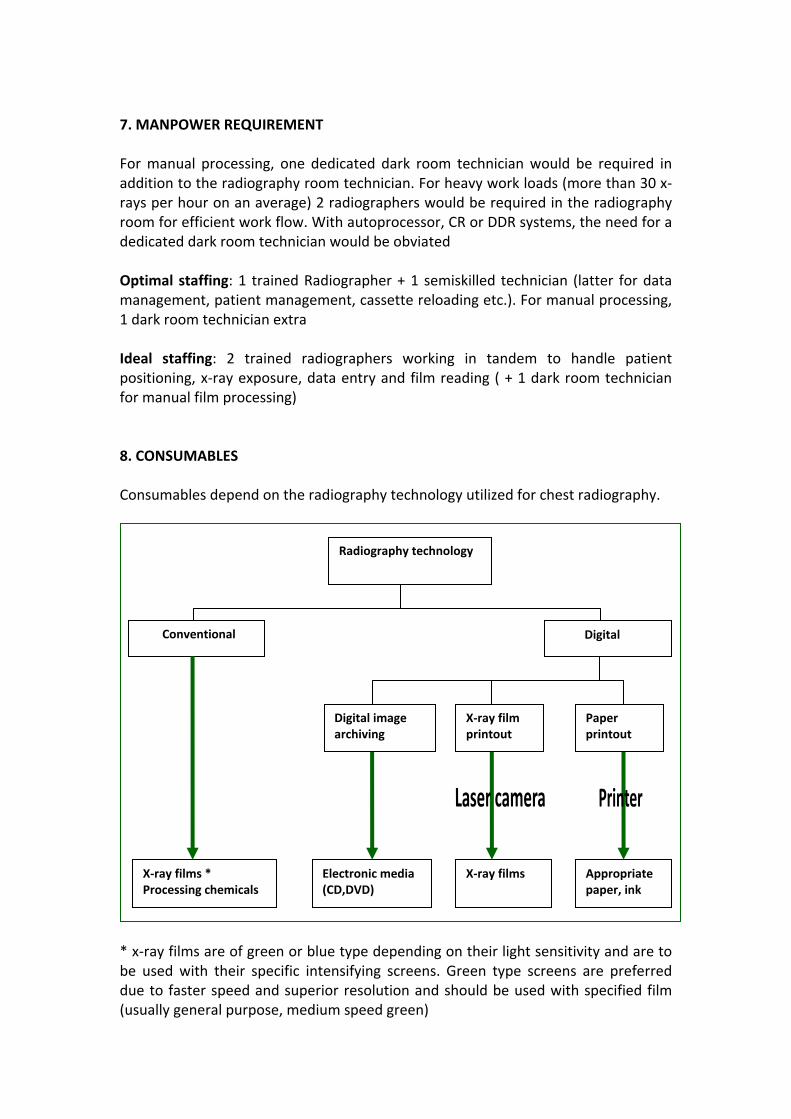
7. MANPOWER REQUIREMENT For manual processing, one dedicated dark room technician would be required in addition to the radiography room technician. For heavy work loads (more than 30 x‐rays per hour on an average) 2 radiographers would be required in the radiography room for efficient work flow. With autoprocessor, CR or DDR systems, the need for a dedicated dark room technician would be obviated Optimal staffing: 1 trained Radiographer + 1 semiskilled technician (latter for data management, patient management, cassette reloading etc.). For manual processing, 1 dark room technician extra Ideal staffing: 2 trained radiographers working in tandem to handle patient positioning, x‐ray exposure, data entry and film reading ( + 1 dark room technician for manual film processing) 8. CONSUMABLES Consumables depend on the radiography technology utilized for chest radiography.
Radiography technology
Conventional Digital
Appropriate paper, ink
X‐ray films * Processing chemicals
Electronic media (CD,DVD)
X‐ray films
Digital image archiving
X‐ray film printout
Paper printout
* x‐ray films are of green or blue type depending on their light sensitivity and are to be used with their specific intensifying screens. Green type screens are preferred due to faster speed and superior resolution and should be used with specified film (usually general purpose, medium speed green)
9. CHECK LIST FOR EQUIPMENT FOR FIELD CHEST RADIOGRAPHY This section provides a check list for the equipment to be used. Specific points to be taken into account are also mentioned. ‐ X‐ray machine including x‐ray generator, x‐ray tube, carry case and mobile
stand: check specifications and power rating, input power supply requirements ‐ Input power supply: Mains or battery/generator driven. Compatible with
required load ‐ Chest stand: for positioning ‐ Radiographic grid: as recommended ‐ X‐ray cassettes with intensifying screens; cassettes should be marked with
speed of screens, type of film used and date of initial use. Ideally 3 per team, with provision of replacement if required
‐ X‐ray films (film size as decided, number as per sample size); expiry date checked
‐ Lead marker set and numbers ‐ Dark room: with sections for film developing, washing and fixing ‐ Water supply source for running water ‐ Film processing chemicals ‐ Developer and Fixer; amount to be calculated as
per requirement ‐ Autoprocessor: If used, include in total power requirement calculation ‐ [Dark room] Film hangers: for film size used (14”x 14”) ‐ Film cabinet: for storage of unexposed x‐ray films. Lead lined ‐ X‐ray viewing box; 1 each for radiographer and film‐reader; of similar
illumination ‐ Radiation protection screen ‐ Radiation protection apron ‐ Equipment maintenance manuals, log books for data entry ‐ Equipment service contract documentation ‐ Computer and printer(s) – if required ‐ Acknowledgement / No objection Certificate from regulatory authority on
Ionizing Radiation* – specially if open radiography is considered ‐ Adequate backup equipment (including spares) and manpower
*X‐ray being ionizing radiation, its use is regulated in most countries. Consultation with the ionizing radiation authority prior to finalization of the fieldwork is essential. This also has bearing on the logistics involved. For example, open radiography (without any protective walls/screens) may not be legally acceptable in many countries and options like mobile x‐ray truck or chambers will have to considered, adding to the cost and to the burden of transportation and handling. 10. DETAILS OF RADIOGRAPH A good quality, full size posteroanterior (PA) view of the chest should be obtained in inspiration. The radiograph should be appropriately labelled.

11. INTERPRETATION OF RADIOGRAPHS At least 2 independent readings, one each by trained and experienced film readers (preferably 1 Radiologist and 1 Chest Physician) should be done for each radiograph. A third ‘expert’ opinion should be obtained in case of difference of opinion. Inter observer variation can be minimized by use of standardized reporting methodology adopted by consensus before start of survey. A few training sessions for film readers should be planned before start of survey to homogenize the interpretation results. Concept of ‘intentional over‐reading’ may be useful, provided it is followed by all survey teams. A good practice is to run a ‘screening’ test at the end of each session/day, to help ensure quality control and to help facilitate identification of cases for sputum/culture examination. 12. RADIATION PROTECTION This section provides some tips for good radiation safety. It is implied that prior to commencing fieldwork, all necessary clearances from the Ionizing Radiation Board/Authority have been obtained (see footnote, section 9). The guiding principle of radiation protection is ALARA – As Low As Reasonably Achievable. It is recorded that good radiation practice can minimize the average radiation dose by about 40% .To achieve ALARA, the main component are:
• TIME: shorter exposure time implies lower radiation dose to the patient as well surroundings
• DISTANCE: Longer the distance of patient from the tube, lower is the
radiation dose. A good practice is to stick to the suggested protocols for various procedures. If open radiography is practiced, all persons (except the patient and radiographer) should be at a distance of 2 meters or more from the x‐ray tube. A good practice is to cordon off the area at 3 to 4 meters.
• SHIELDING: X‐ray tubes are housed within lead lining to block leakage
radiation. Proper calibration and maintenance ensures safety from leakage radiation. Further, protection devices like lead aprons, gonad shields etc., should be used as appropriate
• Use of intensifying screens and appropriate film improves quality and
reduces dose
• Use of high kV technique reduces radiation dose
• During exposure, all other staff and persons (except patient) should be outside the x‐ray room
• Collimation should be used to restrict the x‐ray beam to the chest
• Appropriate radiation protective devices should be used
• Radiation/X‐ray warning sign should be present at appropriate locations
• It is increasingly being reported that the X‐ray dose produced by single exposure for chest radiography does not carry significant risk for pregnant women, however, it is prudent to inform them about possible risk to the unborn child and if needed, written consent should be obtained
13. PRACTICAL TIPS Appointments for clustering – survey teams can give hour wise appointments for undergoing radiography. This ensures better work efficiency. The number of x‐rays per hour depends on the sample size and the radiography technology utilized. It should be kept in mind that some extra time needs to be provisioned for repeat x‐rays. Open radiography – does not require much preparation. Ensure compliance with national radiation governing laws and make sure that adequate distance from x‐ray source (recommended 2 m, extend as further as practically possible) is maintained. Closed radiography – ensure that the radiography room meets standard requirements and that adequate space is provided. Other options are to have the X‐ray machine and room mounted on a truck. Another option available with some of the technologies is a 20 feet container cabin which houses the x‐ray unit. Type of transport – ensure that the transport is appropriate for all areas to be covered in the survey. 4 wheel drive vehicles are better suited for heavy equipment and for rugged terrain. Costly and delicate equipment requires air suspension vehicles, but these are costly Importance of weight – Include weight of x‐ray equipment, consumables and electric generator (if required) while calculating the total weight. While weight of the x‐ray machine is almost always mentioned in the specifications provided, a critical factor is the choice of the electric generator depending on the total power requirement and availability. High power generators are heavy and may need separate transportation.

14. ILLUSTRATIONS
Fig 1 – X-ray positioning stand and carrying case
Fig 2 : An example of portable x-ray unit

Fig 3 - Mobile x-ray unit Fig 4 - Chest stand for positioning
Fig 5 – Tank for manual processing with chemicals (developer, fixer)
Fig 6 – Automatic film processor

15. REFERENCES A list of reference documents and education material related to chest radiography, useful for training/self‐study by staff is provided as a separate document. Some of the relevant references for this document are listed here: ‐ Basics of radiation protection. How to achieve ALARA: Working tips and
guidelines. WHO 2004. ‐ Consumer Guide for the purchase of X‐ray equipment. WHO 2000. ‐ Handbook for District Hospitals in Resource Constrained Settings on Quality
Assurance of Chest Radiography: for better TB control and health system strengthening. The Tuberculosis Coalition for Technical Assistance, 2008.
*************************************