February 22, 2011, neoplasia 1 lecture - Duke University · Note microscopic similarity to normal...
79
Introduction to NEOPLASIA (Part 1) Rex Bentley, M.D. Department of Pathology DUMC
Transcript of February 22, 2011, neoplasia 1 lecture - Duke University · Note microscopic similarity to normal...

Introduction to NEOPLASIA
(Part 1)
Rex Bentley, M.D.Department of Pathology
DUMC
ts135
Text Box
Today: Vocab Later: Epidemiology and Molecular Biology of Cancer followed by immunology and treatment of cancers
hulet001
Approved

NEOPLASIAToday’s Goals and Objectives
1. Define neoplasm
2. Define benign and malignant
3. Differentiate benign from malignant neoplasmsbased on histologic appearance
4. Explain how neoplasms are named and infer properties of a neoplasm from its name
5. Explain what grade is, and how it impacts prognosis

1. What is a Neoplasm?• NEOPLASM = “New growth”
• Synonym: TUMOR = “swelling”– Originally used for inflammation, but
now used as synonym for neoplasm
• Oncology = the study of tumors (Greek “oncos” = tumor)
ts135
Text Box
Tumor and neoplasm are used interchangeably nowadays.

NEOPLASMDefinition
“A neoplasm is an abnormal mass of tissue which exceeds and is uncoordinated with that of the normal tissues, and persists in the same excessive manner after the cessation of the stimuli which evoked the change.”
Sir Rupert Willis, 1952
ts135
Highlight
ts135
Callout
mass lesion in tissue that outgrows what it normally should be
ts135
Highlight
ts135
Callout
becomes autonomous

Two Fundamental Features of Neoplasms
1. Unregulated growth2. Clonal genetic defects
Subject of later lectureNeoplasia III (Dr. Yan)
ts135
Text Box
Derived from single cells, and all the cells within the neoplasm are clonally related
ts135
Line

Mount Sacagawea, Montana

2. What Do “Benign” and “Malignant” Mean?
ts135
Text Box
The fundamental difference for tumors arising from most tissues is the ability to metastasize - see next slide. Brain tumors are the notable exception to this rule. Glioblastomas are malignant but they do not metastasize.

Malignant Neoplasm“CANCER”
• Metastasis = Malignant.• Metastasis: spread to distant, non-
contiguous site– Lymphatic metastases (nodes)– Hematogenous metastases (lung, liver,
bone, brain)– Implantation in body cavities
• Fatal if untreated
ts135
Text Box
If the tumor gains access to body cavities (pleural space, etc) it can implant in these cavities
ts135
Line

Lymph Node Metastasis
Cancer
Normal
ts135
Text Box
Dark blue is the normal small lymphocytes

Hematogenous Metastases
Breast cancer metastases in liver
Courtesy PEIR digital library
ts135
Callout
white nodules are foci of metastatic cancer
ts135
Text Box
Green because of the bile backup due to tumor blocking the bile excretion

Hematogenous Metastases
Breast cancer metastases in vertebra Courtesy PEIR digital library
ts135
Callout
pale areas are cancer in bone marrow

Peritoneal Metastases
Ovarian Cancer
ts135
Callout
omentum covered with thousands of nodules - common way ovarian cancer likes to spread
ts135
Text Box
Loop of colon

Benign Neoplasms• Do not metastasize• In general, do not result in
death of the patient–Location, location, location!–Secretory products can be
lethal (e.g. endocrine tumors)
ts135
Highlight
ts135
Highlight
ts135
Highlight
ts135
Callout
unless in a bad location

From a practical standpoint, benign neoplasms often can be cured by simple surgical
excision while malignant neoplasms often cannot be
cured by surgery alone
ts135
Callout
risk of spreading to distant sites

Benign vs. Malignant
Benign Malignant
Distant Metastases?
No Yes
Life-threatening
No (usually)
Yes
Malignant neoplasms have the potential for metastasis
Benign vs. MalignantBenign Malignant
Distant Metastases?
No Yes
Definition correct but clinically not helpful…do you want to wait for your patient to develop metastatic disease before you start treatment for cancer?
ts135
Text Box
The answer here is no

Cham Museum, Danang, Viet Nam

3. How can we tell if a neoplasm is malignant
BEFORE it metastasizes?
Histopathology!

Histologic Features Distinguishing Benign vs. Malignant
a) Bordersb) Growth
ratec) Anaplasia
Is this cancer or not?
Courtesy PEIR digital library
ts135
Text Box
Anaplasia - lack of differentiation
ts135
Text Box
Three big things to look at

Benign Neoplasms• Encapsulated (pushing borders)
–Do not invade locally• Slow growth• Mild anaplasia (well differentiated)
ts135
Text Box
Can push local structures aside / put pressure on them but don't invade
ts135
Text Box
Can be evaluated both clinically and histopathologically - malignant tumors grow much faster

Breast, fibroadenoma
Pushing Borders
ts135
Callout
tumor with sharp circumscribed borders

Pushing Borders
Breast, fibroadenoma
ts135
Callout
sharp interface - pushing duct to the side
ts135
Callout
not invading the duct

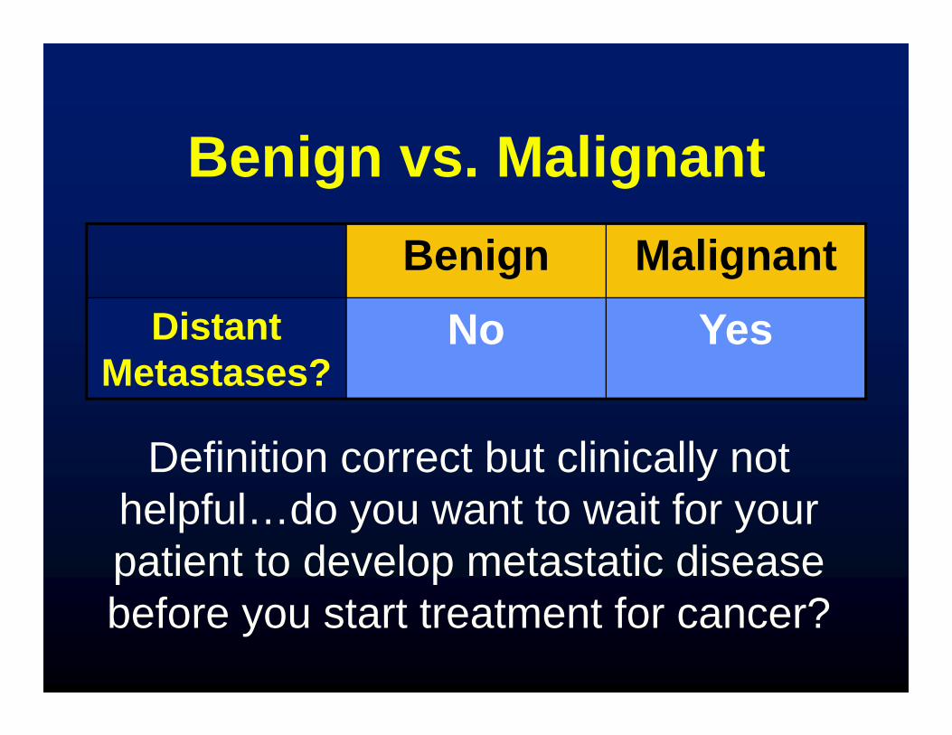
Malignant Neoplasms
• Local Invasion–Infiltrative borders–“Stellate” or “spiculated”
ts135
Text Box
Can be seen on x-rays and pathologically. Malignant tumors are fixed to adjacent structures, not mobile. This can be palpated on physical exam if the tumor is large.

Local Invasion
Lung Cancer
ts135
Callout
Pale tumor sending fingers throughout the vessels and into the pleural space of the lung - infiltrative growth pattern
ts135
Highlight
Local Invasion
Breast cancer
ts135
Callout
spiculated/stellate architecture
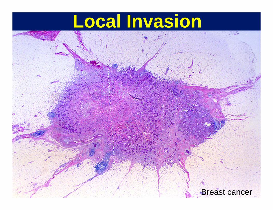
Malignant Neoplasms• Local Invasion• Rapid growth rate
– Histology: Mitotic figures numerous
– Not unique to malignancies, many normal tissues grow rapidly (GI mucosa, endometrium, bone marrow)
ts135
Text Box
Can be seen clinically too - something that has grown over several weeks or months
ts135
Highlight
ts135
Line
ts135
Highlight

Mitotic Figures in Cancer
Breast, malignant phyllodes tumor
ts135
Text Box
Arrows pointing to mitoses Frequently talked about how many mitoses are seen on a high power field (40x field microscope). This allows the pathologist to tell the clinician how badly the tumor is likely to behave.

Malignant Neoplasms
• Local Invasion• Rapid growth rate• Anaplasia
ts135
Text Box
Lack of differentiation

ANAPLASIA“Lack of Differentiation”
• “Differentiation” is the extent to which neoplastic cells resemble normal tissues, both morphologically and functionally– Well-differentiated: closely resembles
tissue of origin– Poorly-differentiated: unspecialized, little
resemblance to tissue of origin
Anaplastic cells are poorly differentiated
ts135
Text Box
Earlier thinking was that tumor cells were reverting to an earlier embryologic state. However, we now realize they go to more of a stem cell state.
ts135
Highlight
ts135
Highlight

ANAPLASIA“Lack of Differentiation”
–Anaplastic skeletal muscle cells make little actin and myosin (lose cross striations)
–Anaplastic colonic epithelial cells make little or no mucin
–Anaplastic glandular cells make only few glands
ts135
Text Box
Some examples of Anaplasia:

Benign: No Anaplasia
Note microscopic similarity to normal smooth muscle
Uterus, leiomyoma
ts135
Text Box
uterus - the white nodules are fibroids (benign common tumors)
ts135
Text Box
Tumor that has no anaplasia - histologically looks almost exactly the same as normal smooth muscle cells
ts135
Callout
cervix

Normal
Benign: Mild Anaplasia
Neoplastic glands still resemble normal endometrial gland
ts135
Text Box
pinker glands are neoplastic ones - still look similar to normal though

Cancer: Moderate Anaplasia
Neoplastic squamous cells still make abundant keratin (arrows)
Normal Skin Squamous cell carcinoma
ts135
Text Box
The cancer side is more disorganized than the left side, but still making keratin (pink whirls).

Normal
Breast Cancer: No gland formation
Severe Anaplasia
ts135
Text Box
Can't even recognize the breast histologically - no glands, only solid cord of cells

Severe Anaplasia
Normal
Colon Cancer
No resemblance to normal
ts135
Text Box
Sheets of unrecognizable cells - doesn't even look like an epithelium

– High ratio of nucleus to cytoplasm– Nuclear hyperchromasia.– Clumped chromatin.– Prominent nucleoli.
ANAPLASIA: Abnormal Nuclei
“Blue is BAD”
ts135
Text Box
Known as the N/C ratio
ts135
Highlight
ts135
Callout
first impression of looking at a neoplasm under the microscope is blueness.
ts135
Text Box
Does this correlate with the rate of growth? A: Cells that are dividing rapidly have less cytoplasm, nuclear hyperchromasia is from replicating DNA so it's all related. Anaplastic cells may have multiple copies of chromosomes and therefore more DNA
ts135
Stamp

– Pleomorphism• Variation in size and shape • Nuclear and cytoplasmic• Tumor giant cells
– Frequent and sometimes abnormal mitoses
ANAPLASIA:
Other Nuclear Features
ts135
Text Box
In normal tissues, the cells are relatively uniform

Mild Anaplasia: Nuclei
Normal Colon Adenoma
Remember--blue is bad!
ts135
Text Box
Benign neoplasm of the colon
ts135
Text Box
Still making glands, but a little bluer, lost some mucin, a little darker and a little larger than normal

Severe Anaplasia: Nuclei
Nuclear pleomorphism, tumor giant cells, tripolar mitosis
ts135
Callout
Mercedes Benz sign - cell dividing three ways (almost always a sign of malignancy)

Histologic Diagnosis Of Malignancy
There is no single parameter (other than metastasis) which always allows recognition of a malignant neoplasm microscopically. However, the presence of severe anaplasia and a pattern of invasiveness are the criteria which are most generally useful.
ts135
Highlight
ts135
Highlight

NEOPLASMS
BENIGN INTERMEDIATE MALIGNANT
SPECTRUM
ts135
Text Box
There is gray area with neoplasms. Can have tumors that metastasize 1/10 or 1/1000 times. You have to recognize lesions that are intermediate in their biology.
ts135
Highlight
ts135
Highlight
ts135
Highlight

Quick Review: Which of these is malignant?
ts135
Text Box
see next slide

Quick Review: Which of these is malignant?
Benign (pushing borders)
Malignant (infiltrative borders)
ts135
Text Box
Breast cancer: infiltrative stellate borders
ts135
Callout
smooth, pushing rounded borders

Quick Review: Which of these thyroid tumors is malignant?

Quick Review: Which of these thyroid tumors is malignant?
Malignant (severe anaplasia!)
Benign (no anaplasia!)
ts135
Text Box
Upper left is super anaplastic. Big giant nuclei, very pleomorphic.

Duke University, North Carolina
ts135
Text Box
malignant

4. How do we name neoplasms?

Nomenclature
Neoplasms are composed of proliferating neoplastic cells but also contain non-neoplastic supportive stroma of connective tissue and blood vessels.
ts135
Text Box
Ignore the supporting stroma - only look at the clonally neoplastic cells for nomenclature
mff4
Highlight

NomenclatureTumors are named
according to the neoplastic component
(Cell type) + (modifier to indicate benign/malignant)
+ (site of origin)

Benign Neoplasms: Nomenclature
• Benign tumors are often designated by the suffix -“oma”.
• Prefix designates the cell of origin

Benign Mesenchymal Neoplasms
CELL TYPE• Fat• Smooth muscle• Skeletal muscle• Fibrous tissue• Blood vessel • Cartilage
BENIGN TUMORLipomaLeiomyomaRhabdomyomaFibromaHemangiomaChondroma

Benign Epithelial Neoplasms
• ADENOMA: benign neoplasm derived from glandular epithelium
• CYSTADENOMA: benign epithelial neoplasm with cystic or fluid-filled cavity
• PAPILLOMA: benign epithelial neoplasm producing finger-like or papillary projections (think sea anemone)
ts135
Text Box
epithelial cells are more complicated:
ts135
Callout
cystic + glandular
ts135
Callout
Papillary growth pattern typically within a cyst but not always (bladder tumors are a common example)

Interior of tumor
Papillary growth inside cyst
ts135
Text Box
papilloma - can see little fingers growing in the cyst

…Then add site of origin:
Examples of benign neoplasms• Leiomyoma of the uterus• Chondroma of the femur• Adenoma of the colon• Cystadenoma of the ovary• Papilloma of the larynx

Malignant Neoplasms:Nomenclature
CARCINOMA: arising from epithelial tissue
ADENOCARCINOMA: arising from glandular epithelium
SARCOMA: arising from mesenchymal tissue

Malignant NeoplasmsNomenclature
LYMPHOMA = arising from lymphoid tissue
LEUKEMIA = arising from blood or bone marrow elements
ts135
Text Box
hematopoietic tissue

Examples of malignant neoplasms• Leiomyosarcoma of the uterus• Chondrosarcoma of the femur• Adenocarcinoma of the colon• Squamous cell carcinoma of the
larynx
…Then add site of origin:
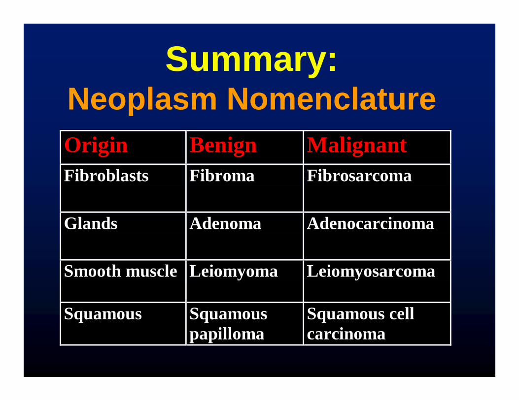
Summary:Neoplasm NomenclatureOrigin Benign Malignant Fibroblasts Fibroma Fibrosarcoma
Glands Adenoma Adenocarcinoma
Smooth muscle Leiomyoma Leiomyosarcoma
Squamous Squamous papilloma
Squamous cell carcinoma

Tissue Benign MalignantLymphocytes (?) Lymphoma
Granulocytes (?) Leukemia
3 germ celllayers
Teratoma Teratocarcinoma
GI wall GI stromal tumor GI stromal tumor
Summary:Neoplasm Nomenclature
ts135
Text Box
No real benign tumor because once lymphs proliferate in bloodstream, they go everywhere
ts135
Text Box
Occurs in the wall of the GI tract and they have the same name with benign or malignant added in front (i.e. malignant GI stromal tumor)

Exceptions• Many “-omas” are malignant
–Lymphoma–Hepatoma–Seminoma–Melanoma
ts135
Text Box
Seminifierous Tubules
ts135
Text Box
Should be called "melanosarcoma" but it's not. Tumor of melanocytes
ts135
Text Box
lymphocytes
ts135
Text Box
liver cells

Exceptions• Some “carcinomas” or
“sarcomas” are benign–Basal cell carcinoma of skin–Cystosarcoma phyllodes of
breast–Well differentiated
liposarcoma of skin
ts135
Text Box
Most common cancer, but it almost never metastasizes
ts135
Line
ts135
Text Box
Point: If you're not sure about whether something is malignant or benign - look it up.

Name that tumor!

Tumor #1 –Liver
ts135
Text Box
Answer in two slides
ts135
Text Box
How would you describe the liver? Irregular borders, nodules everywhere. Could be necrotic in the center.

Tumor #1Tumor #1 –Liver
Mitoses
ts135
Text Box
this is bad
ts135
Line
ts135
Text Box
Where is it coming from? Bile ducts.

Tumor #1• Dx: Adenocarcinoma of the
bile duct • Malignant features
– Infiltrative borders, many mitoses
– Gland forming neoplasm• aka “Cholangiocarcinoma”

Tumor #2-Adrenal
Courtesy Healthcentral.org
ts135
Text Box
How would you describe the border of this tumor?Pushing border
ts135
Text Box
answer in two slides

Tumor #2-Adrenal
Minimal ana plasia-resembles normal adrenal
ts135
Text Box
No mitoses or pleomorphisms

Tumor #2• Dx: Adenoma of the Adrenal
Cortex• Benign features
– Pushing, circumscribed borders, no mitoses or anaplasia
ts135
Text Box
Is the adrenal gland an epithelial structure? No. Historically called this though


5. What is Grade?

Grading Of Cancer
Grade: A histologic parameter quantitating the degree of differentiation of the cancer cells.
ts135
Text Box
capture the anaplasticity of the tumor. Grade is a way to describe (quantify) how anaplastic the tumor cells are.

Differentiation• Well-differentiated (“low grade”)
tumors resemble mature normal cells of the tissue of origin.
• Poorly differentiated (“high grade”) tumors show little resemblance to the tissue of origin.
ts135
Text Box
highly anaplastic
ts135
Text Box
little anaplasia

Grading of Cancer• Many tumors graded according
to a three-tiered scheme: well, moderately, and poorly differentiated (grade 1, 2, 3).
• Grading systems vary by different tumor type.
ts135
Text Box
1 are the well differentiated tumors (looks like normal) 3 is poorly differentiated tumors (look very little like normal)
ts135
Highlight

Importance of Grade
Many tumors show a range of differentiation from low grade to high grade. For those that do…
Grade predicts behavior(for many common malignancies)
ts135
Text Box
Predicts response to chemotherapy, how they will metastasize, etc.

Grade and PrognosisBreast Cancer
Grade 5 yr survival
1 95
2 75
3 50
ts135
Text Box
Can base your treatment strategy based on the grade

Grading Of Cancer• Limitations:
– Many tumors are of intermediate differentiation
– There is sampling error with small biopsies
– Grading is based on subjective light microscopic interpretation
ts135
Text Box
Such as colon cancer
ts135
Text Box
observer variation and error based on the person looking at it

Factors that would influence whether a surgical resection would be curative include:
A. Whether it is benign or malignant
B. Location of the neoplasm
C. Cell type of the neoplasm
D. Degree of anaplasia of the neoplasm
E. All of the above
Quick Review

Factors that might influence whether surgery for a neoplasm will be curative include:
A. Whether the neoplasm is benign or malignant
B. Location of the neoplasm
C. Cell type of the neoplasm
D. Degree of anaplasia of the neoplasm
E. All of the above
Quick Review

The EndIntroduction to Neoplasia
(Part I)
Rex Bentley684-6423
[email protected] Duke South
Green Zone
ts135
Text Box
Q: Some tumor types are class associated? i.e. GBM (glioblastoma multiforme - which is a common, aggressive brain tumor) have a grade and a class GBM is always Grade 4. Staging is not applied to GBM because they do not metastasize (More detail after Spring Break) A: Every tumor has its own grading scheme, but what we have learned is how most tumors are graded.
ts135
Text Box
Very important Addendum: Staging a tumor - a different concept from grade. Grade is a histological parameter based on anaplasia in the tumor Stage is a clinical parameter that tells how far advanced a tumor is (How big it is, what critically local structures it has invaded, if it has any metastasis). Stage can put patients into prognostic categories. For example: Colon cancer invasiveness in the wall defines stage. Minimally invasive in the wall is a low stage and a good prognosis. If it goes deep into the wall, it will be a high stage. More details about staging will come in subsequent lectures.
ts135
Text Box
Q: Is there a range of differentiation in the same tumor? A: Yes, within one tumor if there is a range, you use the worst looking area. It is assumed that this area will be the most aggressive area.
ts135
Text Box
Can a benign tumor transfer into a malignant one? Sometimes - depends on the benign tumor. Fibroadenoma in breast never becomes. However, tubular adenomas of the colon, frequently lead to colon cancer. We think that the majority of colon cancers may come from colon adenomas (hence the colonoscopy screening after 50)
ts135
Text Box
Q: If you perform a surgery on a benign tumor, will it convert it to a malignant one? A:Usually no. The only potential problem is that some benign neoplasms don't spread distantly but spread locally so if you do a bad job taking it out, you can spread benign neoplasms locally but you don't convert it to a malignancy. Sometimes a pathologist (horrors) may make a mistake an call a malignant tumor benign. There are many reasons for this which include sampling error and communication difficulties. Take home lesson - develop a good working relationship with your pathologist