FDG-PET/CT in Head & Neck Cancer - up.ac.za · Parotis Base of tongue Soft palate Submandibular...
46
FDG-PET/CT in Head & Neck Cancer IAEA PET/CT Workshop: Improving Patient Care Midrand, November 2010 Ora Israel, MD Department of Nuclear Medicine Rambam Health Care Campus Haifa, Israel
Transcript of FDG-PET/CT in Head & Neck Cancer - up.ac.za · Parotis Base of tongue Soft palate Submandibular...

FDG-PET/CT in Head & Neck Cancer
IAEA PET/CT Workshop: Improving Patient Care
Midrand, November 2010
Ora Israel, MD
Department of Nuclear Medicine
Rambam Health Care Campus
Haifa, Israel

Head & Neck Malignancies Overview
• 5% of all malignant tumors • ~550,000 new cases/year, >300,000 deaths/year • Greatest burden: low- and medium-income countries • Western world: >90% squamous cell (larynx,
oropharynx, oral) • Survival: poor, little improvement over last 3 decades • Etiology: tobacco & alcohol account for >75% • Open issues:
• Genetic susceptibility • Tumors in young patients • Relationship to HPV

Head & Neck Malignancies Overview
Staging – early and accurate is critical • selection of appropriate treatment strategy • prognostic significance – high 5-yr DFS from 55% to 35% with LN involvement Treatment – challenging multidisciplinary approach After therapy • ~ 1/3 of cases: late/inadequate dg. of recurrence • early dg. recurrence - critical for better outcome

H & N Tumors Tools for Diagnosis
• Histology
• Extension to bone and vessels – MRI & CT
• FDG imaging: benign vs. malignant (old studies 1994)
• PET/CT: small, highly metabolic tumor
– prognostic value of high FDG uptake
– correlates with high proliferation index

Specific Role of Imaging in H&N Tumors
• Depth of primary tumor invasion
• Lymph node status
• Synchronous 2nd primary lesions
Surgical interventions • following concomitant chemo/radiotherapy • repeated direct biopsy for susp. local failure • planned neck dissection for advanced nodal disease • possible tracheostomy - for compromised
edematous airways post-laryngeal biopsy

FDG-PET/CT in H&N Tumors Patient Preparation & Imaging Protocol
• Fast 4 - 6 hrs; Good hydration; low glucose levels <150 • FDG – injected dose: 15 mCi • Uptake phase: 60-90 min • No talk, drink & chew • Imaging:
– Head fixation – Head (top-of-the-ear) to mid-thigh – Both PET & CT are Head-to-Thigh or 2 separate
acquisitions • i.v. contrast
– easier definition of vessels & separation from nodes – care for PET attenuation correction artifacts

Head & Neck Malignancies Anatomic Localization

FDG – PET : Normal biodistribution

Normal FDG Anatomy of the Head & Neck Areas of Physiologic FDG Uptake
• Neural tissue: brain, cerebellum, spinal cord • Active striate muscles: ocular, genio-glossus,
cricoarytenoid, vocal cords • Normal lymphoid tissue: Waldeyer’s ring, tonsils,
base of tongue • Activated brown fat (neck & shoulder girdle) • Low uptake: salivary glands [submandibular &
sublingual] - due to physiologic secretion • Minimal uptake: normal thyroid

Physiologic FDG Uptake in Head & Neck
Parotis Base of tongue Soft palate
Submandibular glands
Longus Capitis
Vocal cords

FDG Imaging of H&N Tumors Limitations, False Negative
• Lesion size <6 mm
• Metabolic rate
• Not tumor specific
(quantitation attempts – SUV measurements)

FDG Imaging of the Head & Neck Pitfalls & Artifacts
• Movement between PET and CT
• Metal artefacts (dental)
• Asymetric physiologic uptake
– Paralysis of one vocal cord & relative increased uptake in the other cord
– Focal uptake in mastication & sternocleidomastoid muscles (strain or excessive use)
• FDG-avid benign lesions
e.g. Warthin’s tumor with FDG-avidity 78%

FDG-PET/CT – Pitfalls in H&N Region NSC Lung Ca – Staging, Equiv. upper neck uptake
? Primary Mets in colon
Physiologic asymmetric uptake in rt. vocal cord (due to paralysis of lt. vocal cord)

Patterns of Physiologic FDG Uptake in Neck
In Muscles In Brown Fat

FDG Imaging of the Head & Neck Pitfalls & Artifacts after Treatment
• Assessing response - facilitated if pre- and post-treatment FDG-PET/CT studies are available for comparison
• Timing of post-treatment study
– After radiotherapy: delay of at least 8-12 weeks to decrease the potential for false positive inflammatory radiation-related reactions.
– After chemotherapy: delay of at least 2 weeks to avoid false negative study results

M, 67, advanced parotis ca, s/a total parotidectomy & post-op radiotherapy (1y). Focal FDG uptake in the left maxilla localized by PET/CT to a further diagnosed dental abscess

FDG-PET/CT in Newly Diagnosed H&N Tumors
Accurate staging - essential for Rx planning
T: Limited PET/CT use (less anatomical details than MRI – mainly for planning of surgery & radiotherapy)
N: LN+ important prognostic factor, cure rate declines by ~ 50% in regional LN+ tumors
• FDG-PET/CT: improved nodal staging
• Challenge: clinical negative neck (N0) 10-45% LN+ at surgery FDG-PET/CT: sensitivity 67%, specificity 95% for LN+
Mainly in squamous cell tumors (pharynx & larynx)
> CT/MR sparing neck dissection (not for anatomic delineation)
M: advanced H&N tumors benefit from preRx PET/CT
25% distant mets & 10% synchronous malignancy

Staging of H&N Tumors
• T: size & subsite involvement
T1-3: increasing size
T4: invasion of surounding structures
• N: size & number of LN, & relationship to primary (ipsi- or contralateral)
• M: distant mets (25%)
• Attention: 10% synchronous mets.

SCC of base of tongue & cervical LN mets

Advanced Ca of the Mandible Loco-regional & Distant Involvement
LN, liver & bone metastases
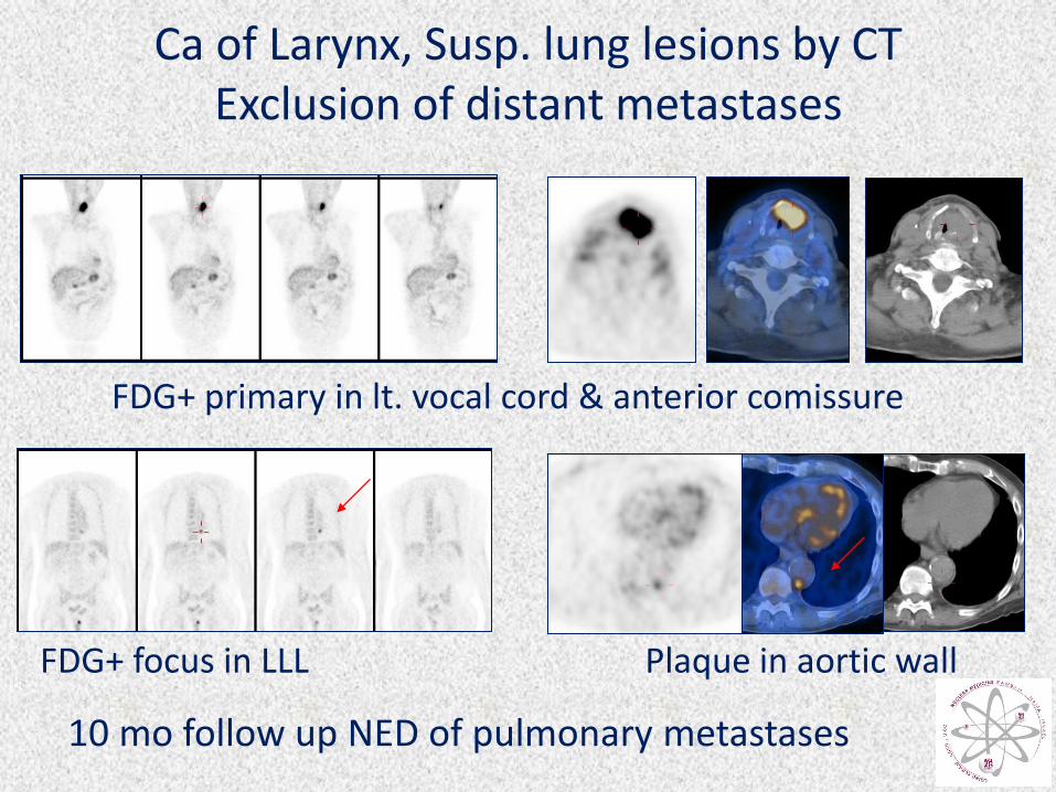
Ca of Larynx, Susp. lung lesions by CT Exclusion of distant metastases
FDG+ primary in lt. vocal cord & anterior comissure
FDG+ focus in LLL Plaque in aortic wall
10 mo follow up NED of pulmonary metastases

FDG Imaging Improves Staging & Management in H&N Squamous Cell Ca
Lonneux et al, JCO 2010, Multicenter prospective, 233 pts
• Discordant FDG & conventional imaging: 43% pts
– FDG accurate stage change: 20%
– FDG error rate: 6% (FDG+ inflammatory LN & pneumonia)
• Accuracy: conventional +FDG > conventional only
• FDG impact on management:
– Low: 81%
– Medium:5% (intramodality changes)
– High: 9% (intermodality; curative to palliation; pallation to cure)

FDG-PET/CT for treatment planning
Multimodality treatment strategies
Induction of :
• More aggressive chemotherapeutic regimens
• Radiation treatment planning
• Planning of the surgical procedure
Radiation Tx planning based on metabolic & biologic features
• Increase in gross tumor volume >25% in 17% patients
• Decreased risk of geographic misses
• Decrease in gross tumor volume in 33% patients
• Minimize dose to non-target organs

SCC of Sinuses - Staging
Whole extent of primary tumor

Head & Neck Malignancies Lymph Node Regions Levels I-VI
Courtesy, EORTC Task Force

FDG-PET/CT in H&N Malignancies
Monitoring Response to Treatment • Rx options: surgery, radiotherapy, chemo-radiation
• Early assessment of response to chemo- radiotherapy: salvage surgery with improved local disease control.
• FDG PET/CT (& ∆SUV changes) : sens 90%, spec 83%
– 4 mo post-Rx > 1 mo post-Rx
– > CT/MRI for detecting residual tumor after chemoradiation
– Negative FDG-PET/CT: highly reliable
– Positive FDG-PET/CT: residual disease vs. inflammation
• Main Indications for FDG-PET/CT after treatment: – Detection of residual tumor – Guiding invasive biopsy at edematous /fibrotic site

Nasopharynx Ca, End of treatment Equivocal MRI
FDG-PET/CT Residual Tumor

Advanced supraglottic tumor, end of chemo-radiation FDG- PET/CT Residual Mass - no Residual Tumor
CT - diffuse supraglottic edema PET/CT - no uptake in the edematous region.
Negative clinical & radiological follow-up: 24 mo

FDG-PET/CT in H&N Tumors Diagnosis of Recurrence & Restaging
Early dg: salvage surgery - improved outcome & prognosis
Biopsy of irradiated tissue: high morbidity, necrosis, failure to heal
CT & MRI: impaired by post-surgery/radiation distorted anatomy, loss of landmarks and symmetry
FDG-PET/CT • High sensitivity 78-96%, vs. CT/MRI: 38-80%
• High accuracy (scar vs. recurrence): 81% vs. CT/MRI: 45%
• Higher specificity for dg. of loco-regional recurrence
Potential 1st study for early dg. of recurrence in larynx Ca

Advanced Nasopharynx CA, s/a chemo-radiation (2 y) Normal size (8 mm) right jugulo-digastric lymph node on CT with increased FDG uptake FNA from node – negative Neck dissection: Metastatic Nasopharynx Ca

FDG-PET/CT in H&N Tumors Diagnosis of Recurrence & Restaging
DD: scar vs. recurrent tumor [in distorted anatomy]
• Accuracy PET – 81%; CT/MRI – 45%
• Recurrent tumor in primary site:
• FDG-PET/CT: sens: 88-100; spec: 75-100
• CT/MRI: sens: 70-92; spec: 50-57
• Planning of total salvage laryngectomy:
accuracy: CT - 42%; PET - 85%
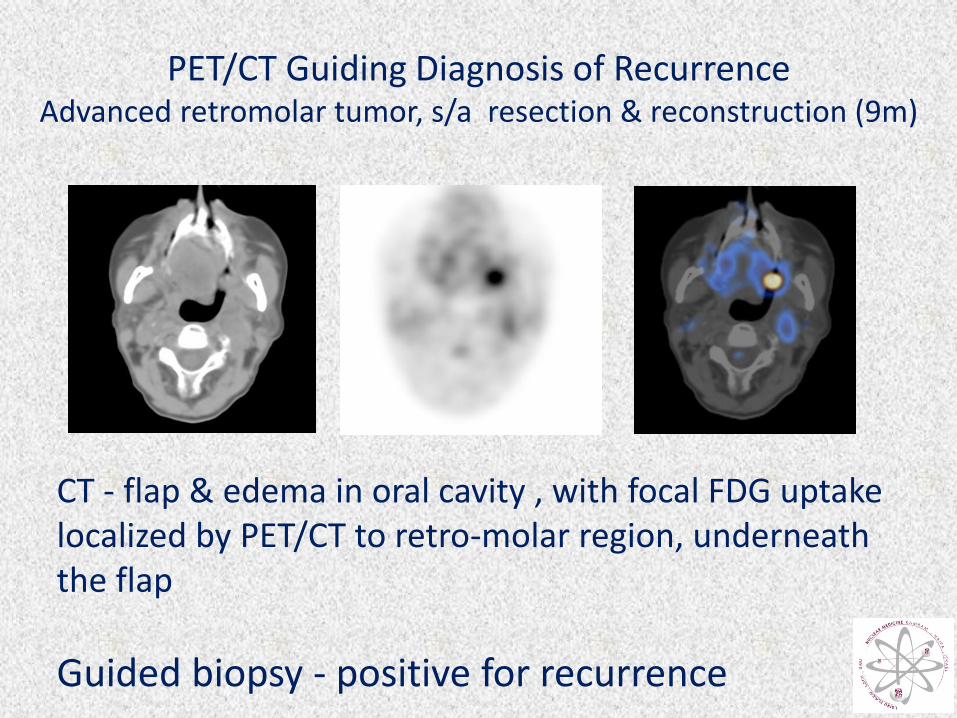
PET/CT Guiding Diagnosis of Recurrence Advanced retromolar tumor, s/a resection & reconstruction (9m)
CT - flap & edema in oral cavity , with focal FDG uptake localized by PET/CT to retro-molar region, underneath the flap
Guided biopsy - positive for recurrence

PET/CT Guide for Biopsy Larynx Ca, new edema, 3 mo s/p radiotherapy
FDG+ focus in neck: SUVmax 4.4 CT: laryngeal edema (rt. vocal cord & anterior comissure) FDG uptake only in edematous changes at anterior comissure PET/CT guided biopsy: Squamous Cell Carcinoma

FDG-PET/CT Diagnosis & Extent of Recurrence Nasopharynx Ca, equivocal MRI
Local recurrence & LN involvement

2nd Primary Tumors (Synchronous or Metachronous)
• Risk: 4%/year >20% within 5 years • Location:
–40% larynx or pharynx –31% lung –9% esophagus

FDG-PET/CT Dg. of 2nd Primary Tumor Larynx Ca, NED 18 mo, New hoarseness & swelling of rt. vocal
cord (CT) Susp. recurrence
FDG+ focus anterior neck PET/CT: •no FDG uptake in vocal cord •FDG+ lesion in mass in proximal esophagus
Biopsy: Carcinoma of esophagus

0
20
40
60
80
100
Sen Spec PPV NPV Acc
Staging
Treatment response
Distant meta
Loco-regional
Performance of FDG-PET/CT in H&N Tumors Gordin et al, Otolaryngol Head Neck Surg. 2007
0
20
40
60
80
100
Sen Spec PPV NPV Acc
Nasopharynx
Larynx
Salivary Gland
Oral Cavity

Larynx Ca FDG-PET/CT Impact on Patient Care
PET/CT altered management in 30% patients
• Cancelled planned biopsy in FDG-negative lesions
• Guide for tissue sampling biopsy from metabolically active area in edematous larynx
• Modified treatment planning:
– from chemotherapy to surgery
– surgery cancelled
– radiotherapy cancelled
Gordin et al, Laryngoscope, 2006

Recurrent/residual Nasopharynx Ca Impact of FDG-PET/CT
• Radiology 2001, 36 pts - best dg. tool
FDG: sens 100, spec 96, acc 97
CT: 73, 88, 83
• Cancer 2003, 67 pts
FDG: sens 100, spec 93, acc 96, PPV 88, NPV 100
MRI: 62, 44, 49, 33, 70

Metastatic Cancer of Unknown Origin
• <10% of squamous cell tumors present with neck mets and no primary
• Diagnostic and therapeutic challenge
• Debilitating blind treatment
• FDG-PET/CT sensitivity for detection of primary
40-65% vs. CT/MRI & random biopsy: 10-20%.
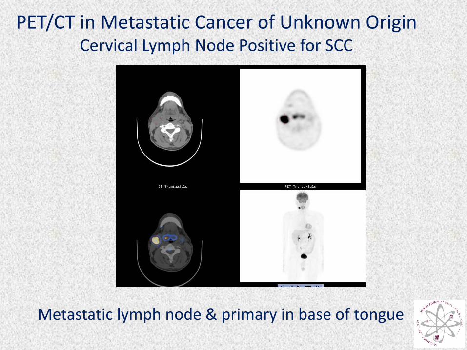
PET/CT in Metastatic Cancer of Unknown Origin Cervical Lymph Node Positive for SCC
Metastatic lymph node & primary in base of tongue

Value of FDG PET/CT in Management of H&N Malignancies
• Guide and facilitates targeted biopsy
(less sampling error)
• Optimized definition of extent of disease
• Exclusion of disease in sites of physiologic FDG uptake
• From “watch-only” expectative policy to therapy (determining the need and type of treatment)
• Intra-modality and inter-modality treatment changes
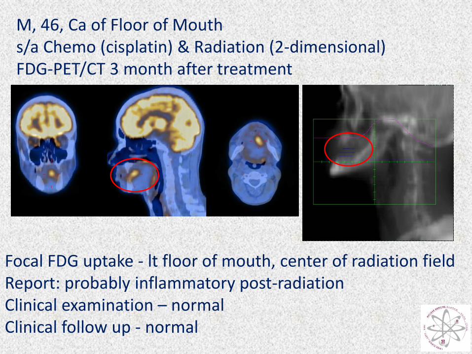
M, 46, Ca of Floor of Mouth s/a Chemo (cisplatin) & Radiation (2-dimensional) FDG-PET/CT 3 month after treatment
Focal FDG uptake - lt floor of mouth, center of radiation field Report: probably inflammatory post-radiation Clinical examination – normal Clinical follow up - normal

F, 61, SCC Base of Tongue & Cervical LN Mets s/a Chemo & Radiation (IMRT 70 Gy Primary & 50Gy Neck FDG-PET/CT 4 month after treatment
Focal FDG uptake – lt. hard palate (border of radiation field) PET/CT guided biopsy: Recurrent SCC Additional chemo-radiotherapy FDG-PET/CT 10 weeks after treatment – Negative

FDG-PET/CT in H&N Tumors Guidelines & Recommendations
(NCCN 2007, multidisciplinary panel – JNM 2008)
Recommended for routine:
• Search for occult primary malignancies not identified by other tests
• For nodal and distant staging
• In suspected recurrence
Not recommended:
• Diagnostic work-up of primary tumor

Thank You